Introduction
Articular hyaline cartilage has poor regenerative capacity, and the loss of its function is, in the long term, often painful and debilitating. Th erefore, attempts have been made to intervene in cartilage defects with the objective of supporting biological repair of tissue. Alongside cell-based strategies for in situ regeneration [1], autologous chondrocyte transplantation was initiated as the fi rst cell therapy for cartilage [2]. Th e requirement for biopsies from a healthy area of the cartilage cap and the necessity of surgical intervention prior to transplantation are evident disadvantages of this therapy, and multipotent mesenchymal stromal cells (MSCs) represent an appeal-ing alternative cell source for cartilage repair.
Th e therapeutic potential of MSCs for cartilage repair is clear; however, the requirements and conditions for eff ective induction of chondrogenesis in MSCs and for the production of a stable cartilaginous tissue by these cells are far from being understood. Diff erent sources of MSCs have been considered for cartilage tissue engineer-ing, mainly based on criteria of availability, as for adipose tissue (AT), or of proximity to cartilage and the joint
environment in vivo, as for bone marrow (BM) and
synovial tissues. Focussing on human MSCs, this review will provide an overview of studies featuring comparative analysis of the chondrogenic diff erentiation of MSCs from diff erent sources.
Defi nition of multipotent mesenchymal stromal cells
Th e presence of cells with osteochondral diff erentiation potential in BM was shown in the late 1960s [3]. Th eir isolation as the adherent mononuclear cell fraction of BM and ex vivo cultivation allowed their further charac-terization as colony-forming unit fi broblasts (CFU-Fs) [4]. Th is pioneering work in guinea-pig was followed by the identifi cation of human BM CFU-Fs [5] and the demonstration of their osteogenic potential in diff usion chambers [6]. Th e in vitro diff erentiation of cloned MSC populations along the osteogenic, chondrogenic and adipogenic lineages demonstrated the multilineage potential of these cells [7].
Th e diff erentiation potential of MSCs was the feature that fostered their discovery and characterization. In vivo, MSCs function to support the homeostasis of mesenchymal tissues, and this mesengenic activity bears high therapeutic potential. However, it has been recog-nized in recent years that the potential therapeutic benefi ts of MSCs do not reside solely in their ability to diff erentiate towards multiple lineages but also in paracrine mechanisms [8]. In particular, the cardio-vascular reparative eff ects attributed to MSCs appear to be mediated predominantly through the secretion of factors targeting cells at the site of repair [9]. Indeed, MSCs secrete a variety of bioactive molecules with trophic, immunomodulatory, anti-scarring and chemo-attractant activities [10]. New therapeutic strategies thus
Abstract
Multipotent mesenchymal stromal cells (MSCs) are an attractive cell source for cell therapy in cartilage. Although their therapeutic potential is clear, the requirements and conditions for eff ective induction of chondrogenesis in MSCs and for the production of a stable cartilaginous tissue by these cells are far from being understood. Diff erent sources of MSCs have been considered for cartilage tissue engineering, mainly based on criteria of availability, as for adipose tissue, or of proximity to cartilage and the joint environment in vivo, as for bone marrow and synovial tissues. Focussing on human MSCs, this review will provide an overview of studies featuring comparative analysis of the chondrogenic diff erentiation of MSCs from diff erent sources. In particular, it will examine the infl uence of the cells’ origin on the requirements for the induction of chondrogenesis and on the phenotype achieved by the cells after diff erentiation.
© 2010 BioMed Central Ltd
Chondrogenesis of mesenchymal stem cells:
role of tissue source and inducing factors
Stephane Boeuf and Wiltrud Richter*
R E V I E W
*Correspondence: wiltrud.richter@med.uni-heidelberg.de
Research Centre for Experimental Orthopaedics, Orthopaedic University Hospital Heidelberg, Schlierbacher Landstrasse 200a, 69118 Heidelberg, Germany
include transplantation of MSCs for the promotion of haematopoietic engraftment or for immunosuppression in graft-versus-host disease [11]. Whether benefi cial eff ects can be expected from the immunomodulatory activities of MSCs for the treatment of rheumatic arthritis is still under debate [12].
Th e term ‘mesenchymal stem cells’ was proposed by Caplan [13] on the basis of the ability of these cell populations to diff erentiate towards tissues of mesen-chymal origin. Based on diff erent isolation methods to obtain MSCs or subpopulations of MSCs, investigators have given diff erent names, such as bone marrow stromal cells (BMSCs) [14], marrow-isolated adult multipotent inducible (MIAMIs) cells [15] or multipotent adult progenitor cells (MAPCs) [16] to these cell populations. Th e International Society for Cellular Th erapy proposed the term ‘multipotent mesenchymal stromal cell’ for the plastic-adherent cell population isolated from BM or other tissues, thus avoiding use of the term ‘stem cells’ to designate a population that does not consist entirely of such cells [17].
Indeed, early evidence for heterogeneity of MSC popu-lations in terms of morphology, growth characteristics and diff erentiation potential has been reported [18]. MSC populations are heterogeneous cell populations whose composition depends on isolation methods and expansion conditions that diff er largely among investi-gators. A recent publication on cloned populations of MSCs showed that nearly 50% of CFU-Fs from BM were tripotent MSCs while the remaining population of cells showed varied phenotypes [19].
So far, no clear marker for MSCs has been identifi ed. Th e criteria for the defi nition of MSCs, as set by the International Society for Cellular Th erapy, are the ability of MSCs to adhere to plastic in standard culture conditions, their phenotypical characterization based on the expression of a set of surface antigens and their in vitro diff erentiation along the osteogenic, the adipogenic and the chondrogenic lineages [20]. Th e ability to form CFU-Fs is another commonly accepted criterion. How-ever, none of these criteria is unequivocal and only their combination can be used to defi ne MSC populations.
Sources of MSCs in diff erent organs
While it was the characterization of BM stromal cells that introduced the concept of MSCs, analysis of progenitor cell populations isolated from other tissues showed that they shared the properties ascribed to BM MSCs. Th is was not restricted to mesodermal tissues, on which this review will focus, as MSCs have also been isolated from ectodermal tissues, such as skin or hair follicles, as well as from perinatal tissue and umbilical cord blood [21].
Th e multilineage diff erentiation potential of a cell population can be due to a mixture of diff erent
committed progenitor cells. Th erefore, to demonstrate the presence of multipotent cells in a cell population, it is essential to show the multilineage potential of cloned cell populations. A tissue other than BM whose osteogenic properties were described at an early stage was the periosteum [22], in which the presence of clonogenic multipotent cells has been demonstrated [23]. Th e presence of MSCs was also demonstrated in stromal cells isolated from AT [24]. Th e synovial membrane (SM) appeared a particularly interesting source of cells for cartilage tissue engineering owing to its proximity to articular cartilage. Th e presence of MSCs with multi-lineage potential was shown in the SM of healthy and osteoarthritic patients [25]. A more extended analysis of clonal populations of SM MSCs distinguished two popu-lations: 30% of cells were tripotent while the remainder displayed only osteo-chondral diff erentiation potential [26]. Th is heterogeneity could be linked to the presence of synovial fi broblasts among SM MSC populations. Th e presence of MSCs has also been demonstrated in the synovial fl uid of healthy and arthritic patients [27]. Dediff erentiated chondrocytes from articular cartilage of healthy and osteoarthritic donors have been shown to exhibit MSC characteristics. Th e reported rates of tripotent cloned cell populations arising from them varies from 10% to 30% [28,29]. In trabecular bone, cell populations with chondrogenic, osteogenic and adipo-genic diff erentiation potential have been isolated [30], but the multidiff erentiation potential of cloned popula-tions has been shown only along the adipogenic and osteogenic lineages [31].
Putative MSC populations have also been isolated from other tissues, for which, to our knowledge, the demonstration of clonal multilineage potential has not been provided. Th ese tissues include muscle [32] as well as joint-related tissues such as meniscus, intra-articular ligament [33] and infrapatellar fat pad [34].
Origin of MSCs in vivo
Th e possible identity of MSCs and pericytes is not restricted to BM. Based on the expression of CD146 and other markers, pericytes have been identifi ed in multiple human organs, including skeletal muscle, pancreas, adipose tissue and placenta, and clonogenic populations from these cell populations have been found to display osteogenic, chondrogenic and adipogenic diff erentiation potential [38]. Possibly, MSCs derived from all vascu lar-ized tissues, such as BM, AT, trabecular bone, periosteum and SM, could share a common origin as perivascular cells. Evidence for this common origin of MSCs is so far based on similarities between MSCs and pericytes and on the shared expression of marker genes. Further investigations are needed to test this hypothesis.
However, cells with MSC characteristics have also been isolated from articular cartilage, which is an avascular tissue [28,29]. Provided the tissue of origin was not contaminated, a perivascular origin of these cells can be excluded. It is likely, therefore, that cells other than perivascular cells can contribute to multipotent MSC populations.
Numbers of MSCs in diff erent tissues
In relation to tissue mass, yields of adherent stromal cells from BM and AT have been described as similar, with an average 2 × 10⁵ cells per gram of tissue [39]. As AT MSCs are most frequently isolated from lipoaspirates, no correlation to initial tissue mass is possible. Th e total number of nucleated cells is much higher in BM than in AT and, accordingly, investigators have reported higher amounts of CFU-Fs per total cell number in AT [33,40]. Identical yields of CFU-Fs per total cell number were shown from periosteum and from AT [41], while more CFU-Fs could be isolated from SM than from subcutaneous fat [41,42] or infrapatellar fat [43].
Th e analysis of diff erences in the growth kinetics of MSCs from diff erent sources would require precise monitoring of initial cell numbers. It is not clear whether longer growth potential of AT MSCs than of BM MSCs before senescence, as suggested by some studies [40,44,45], can be convincingly demonstrated. Th e better growth characteristics sometimes reported for AT MSCs may, instead, be linked to higher initial cell numbers. In terms of accessibility and yield of adherent cells, AT indeed appears one of the most attractive sources of MSCs for therapeutic use.
Molecular characterization of MSCs
No specifi c marker combination for MSCs has been identifi ed so far. However, immunophenotypical profi les of expanded MSCs from diff erent sources have generally been found to be very similar. Th e analysis of MSCs from BM, AT, SM and periosteum showed that these cells can be characterized by the absence of expression of surface
markers for the haematopoietic lineage, such as the cluster of diff erentiation (CD) molecules CD14, CD31 or CD45, and by positivity for a panel of markers, including CD13, CD29, CD44, CD73, CD90, CD105, CD147 or CD166 [33,40,41,44,46-53]. Results from fl ow cytometric analysis of MSCs showing signifi cant diff erences between diff erent sources are summarized in Table 1.
While absence of CD34 is generally considered as a criterion for the defi nition of MSCs [20], some investi-gators have reported low expression in AT MSCs [46,51,53] and one group described selected cell populations with multidiff erentiation potential from AT using CD34 [54]. Th e presence of a pericytic CD34-positive subpopulation in AT has been shown, but it has not yet been determined by cloning studies whether these cells indeed bear stem cell charac teristics [55,56]. While the stem cell marker CD133 is not expressed in expanded MSC populations obtained by adhesion to plastic [40,47], the isolation of CD133-positive cell populations from blood and BM with high proliferation potential and multilineage potential, including mesodermal lineages, has been described [57,58]. CD271, a marker that is highly expressed in BM and AT MSCs and allows the isolation of MSC populations from primary tissues [54,59], has been reported not to be expressed in SM MSCs [26,60]. However, to our knowledge, no direct comparison of stem cell populations from diff erent sources has been performed for this marker. Several studies have pointed to higher expression of CD106 in BM than in AT [40,46,53,61]. Th e vascular cell adhesion molecule CD106/VCAM1 has been shown to be involved in homing of haematopoietic stem cells (HSCs) [62]. Th is diff erence may, therefore, be related to the specifi c micro environment in BM and has indeed been correlated with a functional diff erence between AT and BM MSCs, the latter showing a higher capacity to maintain long-term cultures of primary HSCs [63]. Another marker with potential functional relevance is the platelet-derived growth factor receptor CD140a/ PDGFRα, which is involved in proliferation and migration of MSCs and osteoblasts and has been
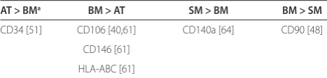
Table 1. Surface markers for which diff erent expression profi les in human MSCs from adipose tissue (AT), bone marrow (BM) and synovial membrane (SM) have been reported
AT > BMa BM > AT SM > BM BM > SM
CD34 [51] CD106 [40,61] CD140a [64] CD90 [48]
CD146 [61]
HLA-ABC [61]
aHigher expression in multipotent mesenchymal stromal cells from adipose
described in one study to be expressed more highly in SM than in BM [64].
Comparative array analysis of expanded MSCs from diff erent sources was published by several groups and provided, overall, very similar expression profi les of MSC populations [44,47,48,65-67]. Interestingly, intra-articular MSCs from SM and MSC-like cells from anterior cruciate ligament and meniscus were found to cluster separately from AT, BM and muscle MSCs [33]. Similar results were found by two-dimensional gel electrophoresis analysis of the proteome of MSCs, where the expression profi les of AT and BM MSCs were closer to each other than either was to SM MSCs [53]. While the functional heterogeneity of SM MSCs has been characterized [26], it remains unknown for other intra-articular sources of cells. It is not clear, therefore, whether the separate clustering of SM MSCs is due to particular characteristics of MSCs from the joint environment or to a higher heterogeneity of these populations. Altogether, the comparative trans-criptome analyses of MSCs from diff erent sources have revealed few diff erences, suggesting that these cell popu-lations contain a common population of similar cells.
Epigenetic characterization of MSCs
Large-scale analysis of DNA methylation in embryonic and adult stem cells has shown that embryonic stem (ES) cells can clearly be discriminated from MSCs by specifi c hypermethylation of numerous genes. In contrast, the comparison of AT and BM MSCs revealed few diff erences [66]. A comparison of DNA methylation profi les in MSCs from AT, BM and muscle and in HSCs also revealed specifi c hypermethylation of numerous genes in HSCs while the methylation patterns of MSCs from diff erent sources were very similar. Most promoters specifying mesodermal, endodermal and ectodermal diff erentiation were hypomethylated in all MSC populations [68]. Th is suggests that promoter hypomethylation is not predictive for the diff erentiation potential of cells, while hyper-methy lation sets restrictions that defi ne frames for diff erentiation potentials, distinguishing MSCs from ES cells or HSCs.
Accordingly, genes related to the adipogenic and myogenic lineage were found to be equally hypo-methylated in MSCs from AT, BM or muscle [69], and in an analysis of the methylation patterns in the promoters
of COL2A1 (collagen type II gene) and COL10A1
(collagen type X gene) in MSCs we found no diff erences between BM- and AT-derived MSCs [70]. However, two cytosines in the COL10A1 promoter were consistently hypomethylated in MSCs in comparison with articular chondrocytes, correlating to the inducibility of COL10A1 expression and hypertrophy during in vitro chondro-genesis of MSCs [70]. Diff erences between the DNA methylation patterns of diff erentiated cells originating
from embryonic precursors and MSCs could thus be of functional relevance for tissue engineering.
Post-translational histone modifi cations have been mapped in ES cells and in MSCs and have been recog-nized to play an important role in transcriptional regulation in stem cells [71]. To date, no comparative analysis of MSCs from diff erent sources has been published. Histone modifi cations and histone-modifying molecules are regulated, while MSCs enter senescence in vitro and could be involved in the ensuing loss of diff erentiation potential [72]. Th ey are also actively involved in diff erentiation. Several studies have indicated that histone deacetylases, in particular HDAC4, may represent important regulators of chondrogenesis [73,74].
MicroRNAs represent a further epigenetic regulation mechanism relevant for stem cell biology [75]. Th e comparison of the microRNA expression profi les of MSCs from BM and AT revealed that only one microRNA was diff erentially expressed while the diff er-ences with ES cells were high [66]. Studies have shown regulation of the expression of microRNAs in MSC senescence [76] and chondrogenic diff erentiation [77],
but the functional mechanisms are unknown. Th e
epigenetic characterization of MSCs is a relatively new fi eld of investigation that has so far revealed only minor diff erences between MSCs from diff erent sources at all levels. Refi nement of the analysis of profi les may, however, lead to an epigenetic defi nition of MSCs, which could have the advantage of correlating with the functional potential of the cells.
Induction of chondrogenic diff erentiation of MSCs in vitro
chondrogenic diff erentiation of human MSCs in a three-dimensional structure.
One of the most widely applied culture systems for chondrogenesis is pellet culture, alternatively termed aggregate or spheroid culture [82,83]. Pellets comprising between 200,000 and 500,000 cells, depending on the investigators, are submitted to chondrogenic induction with a basal medium containing, conventionally, dexamethasone, ascorbate, insulin, transferrin and
selenous acid [82,83]. Th e classic growth factor
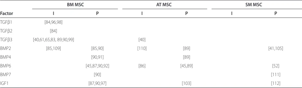
supplementation for this medium is 10 ng/ml of transforming growth factor (TGF)β. TGFβ1, 2 and 3 are the only well-established full inducers of chondrogenesis that lead to deposition of proteoglycan and collagen type II when added as single factors [83,84]. Although other inducers of chondrogenesis, such as the bone morpho-genic proteins BMP2 for BM MSCs and BMP6 for AT MSCs, have been described [85,86], this has not been confi rmed by other investigators [65,87-90] and may apply only to MSCs from selected donors. Rather, BMP2 [85], BMP4 [91], BMP6 [92] and the insulin-lik e growth factor IGF1 [87] may be regarded as promoters of chondrogenesis in MSCs when used together with the inducer TGFβ. Table 2 gives an overview of studies characterizing growth factors as full inducers or promoters of chondrogenesis in MSCs. Besides soluble factors, environmental factors such as mechanical stimu-lation [93] and hypoxia [94,95] have also been reported to modulate chondrogenesis of MSCs in vitro.
With some exceptions [40], most studies undertaking a direct comparison of BM and AT MSCs have described a lower chondrogenic diff erentiation potential of AT MSCs in pellet culture under induction with TGFβ1 or 3 alone [61,65,96] or with TGFβ2 and IGF1 [97], including studies using cells isolated from the same patients [98,99]. Cultures in alginate beads [50,100], hyaluronic
acid scaff olds [101] and cartilage-derived matrix [100] also showed a lower response of AT MSCs to TGFβ-driven chondrogenic induction.
Looking for factors that might explain the reduced inducibility of AT MSCs with TGFβ, we analysed the expression of relevant growth factors in expanded MSCs from BM and AT and found reduced expression of BMP2, 4, 6 and the TGFβ receptor 1 (TGFBR1) in AT MSCs [89] and enhanced levels of the integral membrane protein 2A (ITM2A) gene [102]. Th e high expression of ITM2A during the early phase of the induction of chondro genesis correlated to inhibition of chondro-genesis, and forced overexpression of ITM2A was indeed able to inhibit chondrogenesis in a mouse cell line [102].
One strategy explored for the enhancement of chondro genesis in AT MSCs was to increase concen-trations of TGFβ. While we found that concen concen-trations up to 50 ng/ml did not enhance the chondrogenesis of AT MSCs [89], another group found that a combination of 25 ng/ml TGFβ2 and 500 ng/ml IGF1 induced a chondrogenic phenotype in AT MSCs similar to that induced by 5 ng/ml TGFβ2 in BM MSCs [103]. A second strategy arising from the diff erences in the growth factor repertoire of AT and BM MSCs was to add BMPs for the induction of chondrogenesis. Th e addition of BMPs at a concentration of 10 ng/ml indeed enhanced the chondro-genic diff erentiation of AT MSCs. Among the BMPs the most potent inducer was BMP6, which eliminated the diff erences in diff erentiation potential between AT and BM MSCs [45,89,100].
Th e third source of MSCs often considered for
applications in cartilage tissue engineering is the SM. While the chondrogenic potential of SM MSCs was initially described with TGFβ1 as inducing factor [25,104], another laboratory found no induction of chondro genesis in pellet cultures with TGFβ3 alone, but
Table 2. Growth factors reported as full inducers (I) or promoters (P) of chondrogenic diff erentiation of human MSCs in pellet culture in vitro
BM MSC AT MSC SM MSC
Factor I P I P I P
TGFβ1 [84,96,98]
TGFβ2 [84]
TGFβ3 [40,61,65,83, 89,90,99] [40]
BMP2 [85,109] [85,90] [110] [89] [41,105]
BMP4 [90,91] [89]
BMP6 [45,87,90,92] [86] [45,89] [52]
BMP7 [90] [111]
IGF1 [87,90,97] [103] [112]
only with a supplementary high-dose BMP2 treatment [105]. Under these conditions with TGFβ3 and high-dose BMP2, chondrogenic diff erentiation was higher in MSCs from BM, SM and periosteum than in those from muscle and subcutaneous AT [41,42]. We found the response of SM MSCs to chondrogenic induction with TGFβ3 alone to be higher than that of AT MSCs, but lower than that of BM MSCs. While TGFβ3 was able to induce chondro-genesis in only 50% of SM MSC populations from distinct donors, 100% of SM MSCs responded when TGFβ3 was combined with 10 ng/ml BMP6 [52].
Th e requirements for the induction of chondrogenesis in MSCs from diff erent sources thus appear to diff er in terms of growth factors. Th e comparative analysis of AT and BM suggests these diff erent requirements may be related to diff erences in the growth factor repertoires expressed by the cells or to active pathways at the time point of the initiation of chondrogenesis, which may depend on their microenvironment in vivo.
Hypertrophic diff erentiation of MSCs in vitro
Despite diff erences in the conditions necessary for eff ective induction of chondrogenesis, the chondrogenic phenotype and molecular profi le achieved by AT and BM MSCs under appropriate conditions were found to be similar [65,89]. Th e chondrogenic induction of MSCs in pellet culture with TGFβ3 is accompanied by an undesired up-regulation of hypertrophy-associated marker molecules, such as collagen type X and the matrix metallo-proteinase MMP13, and by an activation of alkaline phosphatase (ALP) activity in vitro [65,96,100,106]. After ectopic transplantation into subcutaneous pouches of
severe combined immuno defi cient (SCID) mice, the
hyper trophic phenotype of diff erentiated pellets of both AT and BM MSCs leads to pronounced matrix calcifi cation accompanied by vascular invasion and even micro-ossicle formation [89,106]. Common in vitro protocols of chondrogenesis thus produce MSC-derived chondrocytes that undergo premature hypertrophy and develop into transient endochondral cartilage, instead of stable articular cartilage-like tissue. As AT MSCs, in spite of their origin, mineralized their surrounding matrix in vivo to an extent similar to BM MSCs, the predisposition for osteogenesis and matrix calcifi cation does not appear to be due to an origin of MSCs from bone.
In vitro, SM MSCs showed a tendency identical to those of MSCs from AT and BM to induce expression of osteogenic genes [105] and collagen type X after chondrogenic diff erentiation [52]. Under TGFβ3 and BMP6, the mean up-regulation of ALP activity was lower in SM MSCs than in AT and BM MSCs, but ALP activity in SM MSCs cell populations displayed extremely high donor variability compared with other MSCs, ranging from negative to very strong signals [52]. In vivo, cell
populations that showed low ALP activity in vitro displayed low calcifi cation, and this was surprisingly accompanied by a loss of already deposited collagen type II protein, possibly due to high MMP2, 3 and 13 activity. Th e MSCs in these transplants thus lost their diff er-entiated phenotype, while SM MSCs from other donors, which displayed high ALP activity in vitro, showed calcifi cation in a similar way as AT and BM MSCs [52]. Th e cause of this variability of phenotypes in SM MSCs after chondrogenic induction is unknown. Although SM MSCs show a diff erent phenotype than AT and BM MSCs after chondrogenesis in vitro, their origin from the joint environment appears not to be suffi cient to program them towards a stable chondrogenic phenotype.
Undesired hypertrophic development of expanded MSCs seems no concern in vivo in a cartilage micro-environment where expanded animal MSCs spontan-eously mature into collagen type II-positive and collagen type X-negative chondrocytes [107]. In vitro co-culture experiments with articular chondrocytes demonstrate that the hypertrophic diff erentiation of MSCs can be inhibited by soluble factors secreted from chondrocytes, and parathyroid hormone-related protein (PTHrP) may be a candidate molecule for involvement in this inhibition [108]. PTHrP1-34 displays an inhibitory action on the TGFβ-induced hypertrophic diff erentiation of MSCs in vitro [90]. As a counteractor of Indian hedgehog, which is up-regulated during chondrogenesis of MSCs, it could represent an important factor for the stabilization of an articular phenotype in MSCs. Further in vivo and co-culture experiments may enable identifi cation of factors that are active in the microenvironment of cartilage and have the ability to lock cells in a hyaline chondrogenic stage.
Conclusion
Despite the growth of knowledge on the origin and composition of MSC populations from diff erent tissues, their heterogeneity is poorly understood. Transcriptional and epigenetic analyses of diff erent MSC populations reveal very similar profi les. However, diff erences in expressed growth factors or active pathways between MSCs from diff erent sources could explain the diff erent requirements for the induction of chondrogenesis. Conditions allowing effi cient chondrogenic in vitro diff erentiation of MSCs from AT, BM and SM have been described. For long-lasting cell therapy in cartilage it is, however, essential to be able to achieve a stable
chondrogenic phenotype. Th e endochondral pathway
Abbreviations
ALP = alkaline phosphatase; AT = adipose tissue; BM = bone marrow; BMP = bone morphogenic protein; CD = cluster of diff erentiation; CFU-F = colony-forming unit fi broblast; ES = embryonic stem; HSC = haematopoietic stem cell; IGF = insulin-like growth factor; MMP = matrix metalloproteinase; MSC = multipotent mesenchymal stromal cell; PTHrP = parathyroid hormone-related protein; SM = synovial membrane; TGF = transforming growth factor.
Competing interests
The authors declare that they have no competing interests.
Authors’ contributions
Both authors contributed to the writing of the manuscript and read and approved the fi nal version.
Acknowledgments
This work was fi nancially supported by the Deutsche Forschungsgemeinschaft (grant number RI707/7-1) and by the EuroBoNeT consortium, a European Commission FP-6 granted Network of Excellence for studying the pathology and genetics of bone tumours.
Published: 13 October 2010
References
1. Richter W: Mesenchymal stem c ells and cartilage in situ regeneration. J Intern Med 2009, 266:390-405.
2. Brittberg M, Lindahl A, Nilsson A, Ohlsson C, Isaksson O, Peterson L:
Treatment of deep cartilage defects in the knee with autologous chondrocyte transplantation.N Engl J Med 1994, 331:889-895. 3. Friedenstein AJ, Piatetzky-Shapiro II, Petrakova KV: Osteogenesis in
transplants of bone marrow cells.J Embryol Exp Morphol 1966, 16:381-390. 4. Friedenstein AJ, Chailakhjan RK, Lalykina KS: The development of fi broblast
colonies in monolayer cultures of guinea-pig bone marrow and spleen cells.Cell Tissue Kinet 1970, 3:393-403.
5. Castro-Malaspina H, Gay RE, Resnick G, Kapoor N, Meyers P, Chiarieri D, McKenzie S, Broxmeyer HE, Moore MA: Characterization of human bone marrow fi broblast colony-forming cells (CFU-F) and their progeny.Blood
1980, 56:289-301.
6. Bab I, Passi-Even L, Gazit D, Sekeles E, Ashton BA, Peylan-Ramu N, Ziv I, Ulmansky M: Osteogenesis in in vivo diff usion chamber cultures of human marrow cells.Bone Miner 1988, 4:373-386.
7. Pittenger MF, Mackay AM, Beck SC, Jaiswal RK, Douglas R, Mosca JD, Moorman MA, Simonetti DW, Craig S, Marshak DR: Multilineage potential of adult human mesenchymal stem cells.Science 1999, 284:143-147. 8. Caplan AI, Dennis JE: Mesenchymal stem cells as trophic mediators.J Cell
Biochem 2006, 98:1076-1084.
9. Psaltis PJ, Zannettino AC, Worthley SG, Gronthos S: Concise review: mesenchymal stromal cells: potential for cardiovascular repair.Stem Cells
2008, 26:2201-2210.
10. Meirelles LS, Fontes AM, Covas DT, Caplan AI: Mechanisms involved in the therapeutic properties of mesenchymal stem cells.Cytokine Growth Factor Rev 2009, 20:419-427.
11. Tolar J, Le Blanc K, Keating A, Blazar BR: Hitting the right spot with mesenchymal stromal cells (MSCs).Stem Cells 2010, 28:1446-1455. 12. Ichim TE, Harman RJ, Min WP, Minev B, Solano F, Rodriguez JP, Alexandrescu
DT, Necochea-Campion R, Hu X, Marleau AM, Riordan NH: Autologous stromal vascular fraction cells: A tool for facilitating tolerance in rheumatic disease.Cell Immunol 2010, 264:7-17.
13. Caplan AI: Mesenchymal stem cells.J Orthop Res 1991, 9:641-650. 14. Gronthos S, Zannettino AC, Hay SJ, Shi S, Graves SE, Kortesidis A, Simmons PJ:
Molecular and cellular characterisation of highly purifi ed stromal stem cells derived from human bone marrow.J Cell Sci 2003, 116:1827-1835. 15. D’Ippolito G, Diabira S, Howard GA, Menei P, Roos BA, Schiller PC:
Marrow-isolated adult multilineage inducible (MIAMI) cells, a unique population of postnatal young and old human cells with extensive expansion and diff erentiation potential.J Cell Sci 2004, 117:2971-2981.
16. Belema-Bedada F, Uchida S, Martire A, Kostin S, Braun T: Effi cient homing of multipotent adult mesenchymal stem cells depends on FROUNT-mediated clustering of CCR2.Cell Stem Cell 2008, 2:566-575.
17. Horwitz EM, Le Blanc K, Dominici M, Mueller I, Slaper-Cortenbach I, Marini FC, Deans RJ, Krause DS, Keating A: Clarifi cation of the nomenclature for MSC:
The International Society for Cellular Therapy position statement. Cytotherapy 2005, 7:393-395.
18. Phinney DG: Building a consensus regarding the nature and origin of mesenchymal stem cells.J Cell Biochem Suppl 2002, 38:7-12.
19. Russell KC, Phinney DG, Lacey MR, Barrilleaux BL, Meyertholen KE, O’Connor KC: In vitro high-capacity assay to quantify the clonal heterogeneity in trilineage potential of mesenchymal stem cells reveals a complex hierarchy of lineage commitment.Stem Cells 2010, 28:788-798. 20. Dominici M, Le Blanc K, Mueller I, Slaper-Cortenbach I, Marini F, Krause D,
Deans R, Keating A, Prockop D, Horwitz E: Minimal criteria for defi ning multipotent mesenchymal stromal cells. The International Society for Cellular Therapy position statement.Cytotherapy 2006, 8:315-317. 21. Kuhn NZ, Tuan RS: Regulation of stemness and stem cell niche of
mesenchymal stem cells: implications in tumorigenesis and metastasis. J Cell Physiol 2010, 222:268-277.
22. Ollier L: Recherches experimentales sur la Production artifi cielle des Os au Moyen de la Transplantation du Perioste et sur la Regeneration des Os apres les Resections et les Ablations completes.J de Physiol de l’Homme et des Animaux 1859, 2:1-27.
23. De Bari C, Dell’Accio F, Vanlauwe J, Eyckmans J, Khan IM, Archer CW, Jones EA, McGonagle D, Mitsiadis TA, Pitzalis C, Luyten FP: Mesenchymal multipotency of adult human periosteal cells demonstrated by single-cell lineage analysis.Arthritis Rheum 2006, 54:1209-1221.
24. Zuk PA, Zhu M, Ashjian P, De Ugarte DA, Huang JI, Mizuno H, Alfonso ZC, Fraser JK, Benhaim P, Hedrick MH: Human adipose tissue is a source of multipotent stem cells.Mol Biol Cell 2002, 13:4279-4295.
25. De Bari C, Dell’Accio F, Tylzanowski P, Luyten FP: Multipotent mesenchymal stem cells from adult human synovial membrane.Arthritis Rheum 2001,
44:1928-1942.
26. Karystinou A, Dell’Accio F, Kurth TB, Wackerhage H, Khan IM, Archer CW, Jones EA, Mitsiadis TA, De Bari C: Distinct mesenchymal progenitor cell subsets in the adult human synovium.Rheumatology (Oxford) 2009, 48:1057-1064. 27. Jones EA, English A, Henshaw K, Kinsey SE, Markham AF, Emery P, McGonagle
D: Enumeration and phenotypic characterization of synovial fl uid multipotential mesenchymal progenitor cells in infl ammatory and degenerative arthritis.Arthritis Rheum 2004, 50:817-827.
28. Barbero A, Ploegert S, Heberer M, Martin I: Plasticity of clonal populations of dediff erentiated adult human articular chondrocytes.Arthritis Rheum 2003,
48:1315-1325.
29. de la Fuente R, Abad JL, Garcia-Castro J, Fernandez-Miguel G, Petriz J, Rubio D, Vicario-Abejon C, Guillen P, Gonzalez MA, Bernad A: Dediff erentiated adult articular chondrocytes: a population of human multipotent primitive cells. Exp Cell Res 2004, 297:313-328.
30. Noth U, Osyczka AM, Tuli R, Hickok NJ, Danielson KG, Tuan RS: Multilineage mesenchymal diff erentiation potential of human trabecular bone-derived cells.J Orthop Res 2002, 20:1060-1069.
31. Nuttall ME, Patton AJ, Olivera DL, Nadeau DP, Gowen M: Human trabecular bone cells are able to express both osteoblastic and adipocytic phenotype: implications for osteopenic disorders.J Bone Miner Res 1998,
13:371-382.
32. Grande DA, Southerland SS, Manji R, Pate DW, Schwartz RE, Lucas PA: Repair of articular cartilage defects using mesenchymal stem cells.Tissue Eng
1995, 1:345-353.
33. Segawa Y, Muneta T, Makino H, Nimura A, Mochizuki T, Ju YJ, Ezura Y, Umezawa A, Sekiya I: Mesenchymal stem cells derived from synovium, meniscus, anterior cruciate ligament, and articular chondrocytes share similar gene expression profi les.J Orthop Res 2009, 27:435-441.
34. Wickham MQ, Erickson GR, Gimble JM, Vail TP, Guilak F: Multipotent stromal cells derived from the infrapatellar fat pad of the knee.Clin Orthop Relat Res
2003, 412:196-212.
35. Doherty MJ, Ashton BA, Walsh S, Beresford JN, Grant ME, Canfi eld AE:
Vascular pericytes express osteogenic potential in vitro and in vivo.J Bone Miner Res 1998, 13:828-838.
36. Farrington-Rock C, Crofts NJ, Doherty MJ, Ashton BA, Griffi n-Jones C, Canfi eld AE: Chondrogenic and adipogenic potential of microvascular pericytes. Circulation 2004, 110:2226-2232.
37. Sacchetti B, Funari A, Michienzi S, Di Cesare S, Piersanti S, Saggio I, Tagliafi co E, Ferrari S, Robey PG, Riminucci M, Bianco P: Self-renewing osteoprogenitors in bone marrow sinusoids can organize a hematopoietic
microenvironment.Cell 2007, 131:324-336.
Zheng B, Zhang L, Norotte C, Teng PN, Traas J, Schugar R, Deasy BM, Badylak S, Buhring HJ, Giacobino JP, Lazzari L, Huard J, Peault B: A perivascular origin for mesenchymal stem cells in multiple human organs.Cell Stem Cell 2008,
3:301-313.
39. De Ugarte DA, Morizono K, Elbarbary A, Alfonso Z, Zuk PA, Zhu M, Dragoo JL, Ashjian P, Thomas B, Benhaim P, Chen I, Fraser J, Hedrick MH: Comparison of multi-lineage cells from human adipose tissue and bone marrow.Cells Tissues Organs 2003, 174:101-109.
40. Kern S, Eichler H, Stoeve J, Kluter H, Bieback K: Comparative analysis of mesenchymal stem cells from bone marrow, umbilical cord blood, or adipose tissue.Stem Cells 2006, 24:1294-1301.
41. Sakaguchi Y, Sekiya I, Yagishita K, Muneta T: Comparison of human stem cells derived from various mesenchymal tissues: superiority of synovium as a cell source.Arthritis Rheum 2005, 52:2521-2529.
42. Mochizuki T, Muneta T, Sakaguchi Y, Nimura A, Yokoyama A, Koga H, Sekiya I:
Higher chondrogenic potential of fi brous synovium- and adipose synovium-derived cells compared with subcutaneous fat-derived cells: distinguishing properties of mesenchymal stem cells in humans.Arthritis Rheum 2006, 54:843-853.
43. Lee SY, Nakagawa T, Reddi AH: Mesenchymal progenitor cells derived from synovium and infrapatellar fat pad as a source for superfi cial zone cartilage tissue engineering: analysis of superfi cial zone protein/lubricin expression.Tissue Eng Part A 2010, 16:317-325.
44. Lee RH, Kim B, Choi I, Kim H, Choi HS, Suh K, Bae YC, Jung JS: Characterization and expression analysis of mesenchymal stem cells from human bone marrow and adipose tissue.Cell Physiol Biochem 2004, 14:311-324. 45. Izadpanah R, Trygg C, Patel B, Kriedt C, Dufour J, Gimble JM, Bunnell BA:
Biologic properties of mesenchymal stem cells derived from bone marrow and adipose tissue.J Cell Biochem 2006, 99:1285-1297.
46. De Ugarte DA, Alfonso Z, Zuk PA, Elbarbary A, Zhu M, Ashjian P, Benhaim P, Hedrick MH, Fraser JK: Diff erential expression of stem cell mobilization-associated molecules on multi-lineage cells from adipose tissue and bone marrow.Immunol Lett 2003, 89:267-270.
47. Wagner W, Wein F, Seckinger A, Frankhauser M, Wirkner U, Krause U, Blake J, Schwager C, Eckstein V, Ansorge W, Ho AD: Comparative characteristics of mesenchymal stem cells from human bone marrow, adipose tissue, and umbilical cord blood.Exp Hematol 2005, 33:1402-1416.
48. Djouad F, Bony C, Haupl T, Uze G, Lahlou N, Louis-Plence P, Apparailly F, Canovas F, Reme T, Sany J, Jorgensen C, Noel D: Transcriptional profi les discriminate bone marrow-derived and synovium-derived mesenchymal stem cells.Arthritis Res Ther 2005, 7:R1304-R1315.
49. Musina RA, Bekchanova ES, Sukhikh GT: Comparison of mesenchymal stem cells obtained from diff erent human tissues.Bull Exp Biol Med 2005,
139:504-509.
50. Mehlhorn AT, Niemeyer P, Kaiser S, Finkenzeller G, Stark GB, Sudkamp NP, Schmal H: Diff erential expression pattern of extracellular matrix molecules during chondrogenesis of mesenchymal stem cells from bone marrow and adipose tissue.Tissue Eng 2006, 12:2853-2862.
51. Rebelatto CK, Aguiar AM, Moretao MP, Senegaglia AC, Hansen P, Barchiki F, Oliveira J, Martins J, Kuligovski C, Mansur F, Christofi s A, Amaral VF, Brofman PS, Goldenberg S, Nakao LS, Correa A: Dissimilar diff erentiation of mesenchymal stem cells from bone marrow, umbilical cord blood, and adipose tissue.Exp Biol Med (Maywood) 2008, 233:901-913.
52. Dickhut A, Pelttari K, Janicki P, Wagner W, Eckstein V, Egermann M, Richter W:
Calcifi cation or dediff erentiation: requirement to lock mesenchymal stem cells in a desired diff erentiation stage.J Cell Physiol 2009, 219:219-226. 53. Roche S, Delorme B, Oostendorp RA, Barbet R, Caton D, Noel D, Boumediene
K, Papadaki HA, Cousin B, Crozet C, Milhavet O, Casteilla L, Hatzfeld J, Jorgensen C, Charbord P, Lehmann S: Comparative proteomic analysis of human mesenchymal and embryonic stem cells: towards the defi nition of a mesenchymal stem cell proteomic signature.Proteomics 2009, 9:223-232. 54. Quirici N, Scavullo C, de Girolamo L, Lopa S, Arrigoni E, Deliliers GL, Brini AT:
Anti-L-NGFR and -CD34 monoclonal antibodies identify multipotent mesenchymal stem cells in human adipose tissue.Stem Cells Dev 2010,
19:915-925.
55. Traktuev DO, Merfeld-Clauss S, Li J, Kolonin M, Arap W, Pasqualini R, Johnstone BH, March KL: A population of multipotent CD34-positive adipose stromal cells share pericyte and mesenchymal surface markers, reside in a periendothelial location, and stabilize endothelial networks. Circ Res 2008, 102:77-85.
56. Zimmerlin L, Donnenberg VS, Pfeifer ME, Meyer EM, Peault B, Rubin JP,
Donnenberg AD: Stromal vascular progenitors in adult human adipose tissue.Cytometry A 2010, 77:22-30.
57. Tondreau T, Meuleman N, Delforge A, Dejeneff e M, Leroy R, Massy M, Mortier C, Bron D, Lagneaux L: Mesenchymal stem cells derived from CD133-positive cells in mobilized peripheral blood and cord blood: proliferation, Oct4 expression, and plasticity.Stem Cells 2005, 23:1105-1112.
58. Pozzobon M, Piccoli M, Ditadi A, Bollini S, Destro R, Andre-Schmutz I, Masiero L, Lenzini E, Zanesco L, Petrelli L, Cavazzana-Calvo M, Gazzola MV, De Coppi P:
Mesenchymal stromal cells can be derived from bone marrow CD133+ cells: implications for therapy.Stem Cells Dev 2009, 18:497-510. 59. Jones EA, Kinsey SE, English A, Jones RA, Straszynski L, Meredith DM,
Markham AF, Jack A, Emery P, McGonagle D: Isolation and characterization of bone marrow multipotential mesenchymal progenitor cells.Arthritis Rheum 2002, 46:3349-3360.
60. Van Landuyt KB, Jones EA, McGonagle D, Luyten FP, Lories RJ: Flow cytometric characterization of freshly isolated and culture expanded human synovial cell populations in patients with chronic arthritis.Arthritis Res Ther 2010, 12:R15.
61. Rider DA, Dombrowski C, Sawyer AA, Ng GH, Leong D, Hutmacher DW, Nurcombe V, Cool SM: Autocrine fi broblast growth factor 2 increases the multipotentiality of human adipose-derived mesenchymal stem cells. Stem Cells 2008, 26:1598-1608.
62. Simmons PJ, Masinovsky B, Longenecker BM, Berenson R, Torok-Storb B, Gallatin WM: Vascular cell adhesion molecule-1 expressed by bone marrow stromal cells mediates the binding of hematopoietic progenitor cells. Blood 1992, 80:388-395.
63. Wagner W, Roderburg C, Wein F, Diehlmann A, Frankhauser M, Schubert R, Eckstein V, Ho AD: Molecular and secretory profi les of human mesenchymal stromal cells and their abilities to maintain primitive hematopoietic progenitors.Stem Cells 2007, 25:2638-2647.
64. Nimura A, Muneta T, Koga H, Mochizuki T, Suzuki K, Makino H, Umezawa A, Sekiya I: Increased proliferation of human synovial mesenchymal stem cells with autologous human serum: comparisons with bone marrow mesenchymal stem cells and with fetal bovine serum.Arthritis Rheum 2008,
58:501-510.
65. Winter A, Breit S, Parsch D, Benz K, Steck E, Hauner H, Weber RM, Ewerbeck V, Richter W: Cartilage-like gene expression in diff erentiated human stem cell spheroids: a comparison of bone marrow-derived and adipose tissue-derived stromal cells.Arthritis Rheum 2003, 48:418-429.
66. Aranda P, Agirre X, Ballestar E, Andreu EJ, Roman-Gomez J, Prieto I, Martin-Subero JI, Cigudosa JC, Siebert R, Esteller M, Prosper F: Epigenetic signatures associated with diff erent levels of diff erentiation potential in human stem cells.PLoS One 2009, 4:e7809.
67. Jansen BJ, Gilissen C, Roelofs H, Schaap-Oziemlak A, Veltman JA, Raymakers RA, Jansen JH, Kogler G, Figdor CG, Torensma R, Adema GJ: Functional diff erences between mesenchymal stem cell populations are refl ected by their transcriptome.Stem Cells Dev 2010, 19:481-490.
68. Sorensen AL, Jacobsen BM, Reiner AH, Andersen IS, Collas P: Promoter DNA methylation patterns of diff erentiated cells are largely programmed at the progenitor stage.Mol Biol Cell 2010, 21:2066-2077.
69. Sorensen AL, Timoskainen S, West FD, Vekterud K, Boquest AC, Ahrlund-Richter L, Stice SL, Collas P: Lineage-specifi c promoter DNA methylation patterns segregate adult progenitor cell types.Stem Cells Dev 2010,
19:1257-1266.
70. Zimmermann P, Boeuf S, Dickhut A, Boehmer S, Olek S, Richter W: Correlation of COL10A1 induction during chondrogenesis of mesenchymal stem cells with demethylation of two CpG sites in the COL10A1 promoter.Arthritis Rheum 2008, 58:2743-2753.
71. Collas P: Epigenetic states in stem cells.Biochim Biophys Acta 2009,
1790:900-905.
72. Noer A, Lindeman LC, Collas P: Histone H3 modifi cations associated with diff erentiation and long-term culture of mesenchymal adipose stem cells. Stem Cells Dev 2009, 18:725-736.
73. Bobick BE, Chen FH, Le AM, Tuan RS: Regulation of the chondrogenic phenotype in culture.Birth Defects Res C Embryo Today 2009, 87:351-371. 74. Haberland M, Montgomery RL, Olson EN: The many roles of histone
deacetylases in development and physiology: implications for disease and therapy.Nat Rev Genet 2009, 10:32-42.
75. Stadler BM, Ruohola-Baker H: Small RNAs: keeping stem cells in line.Cell
2008, 132:563-566.
J, Pfi ster S, Eckstein V, Ho AD: Replicative senescence of mesenchymal stem cells: a continuous and organized process.PLoS One 2008, 3:e2213. 77. Sorrentino A, Ferracin M, Castelli G, Biff oni M, Tomaselli G, Baiocchi M, Fatica
A, Negrini M, Peschle C, Valtieri M: Isolation and characterization of CD146+ multipotent mesenchymal stromal cells.Exp Hematol 2008, 36:1035-1046. 78. Woods A, Wang G, Beier F: Regulation of chondrocyte diff erentiation by the
actin cytoskeleton and adhesive interactions.J Cell Physiol 2007, 213:1-8. 79. Oberlender SA, Tuan RS: Expression and functional involvement of
N-cadherin in embryonic limb chondrogenesis.Development 1994,
120:177-187.
80. Tuli R, Tuli S, Nandi S, Huang X, Manner PA, Hozack WJ, Danielson KG, Hall DJ, Tuan RS: Transforming growth factor-beta-mediated chondrogenesis of human mesenchymal progenitor cells involves N-cadherin and mitogen-activated protein kinase and Wnt signaling cross-talk.J Biol Chem 2003,
278:41227-41236.
81. Dickhut A, Gottwald E, Steck E, Heisel C, Richter W: Chondrogenesis of mesenchymal stem cells in gel-like biomaterials in vitro und in vivo.Front Biosci 2008, 13:4517-4528.
82. Johnstone B, Hering TM, Caplan AI, Goldberg VM, Yoo JU: In vitro chondrogenesis of bone marrow-derived mesenchymal progenitor cells. Exp Cell Res 1998, 238:265-272.
83. Mackay AM, Beck SC, Murphy JM, Barry FP, Chichester CO, Pittenger MF:
Chondrogenic diff erentiation of cultured human mesenchymal stem cells from marrow.Tissue Eng 1998, 4:415-428.
84. Barry F, Boynton RE, Liu B, Murphy JM: Chondrogenic diff erentiation of mesenchymal stem cells from bone marrow: diff erentiation-dependent gene expression of matrix components.Exp Cell Res 2001, 268:189-200. 85. Schmitt B, Ringe J, Haupl T, Notter M, Manz R, Burmester GR, Sittinger M, Kaps
C: BMP2 initiates chondrogenic lineage development of adult human mesenchymal stem cells in high-density culture.Diff erentiation 2003,
71:567-577.
86. Estes BT, Wu AW, Guilak F: Potent induction of chondrocytic diff erentiation of human adipose-derived adult stem cells by bone morphogenetic protein 6.Arthritis Rheum 2006, 54:1222-1232.
87. Indrawattana N, Chen G, Tadokoro M, Shann LH, Ohgushi H, Tateishi T, Tanaka J, Bunyaratvej A: Growth factor combination for chondrogenic induction from human mesenchymal stem cell.Biochem Biophys Res Commun 2004,
320:914-919.
88. Xu D, Gechtman Z, Hughes A, Collins A, Dodds R, Cui X, Jolliff e L, Higgins L, Murphy A, Farrell F: Potential involvement of BMP receptor type IB activation in a synergistic eff ect of chondrogenic promotion between rhTGFbeta3 and rhGDF5 or rhBMP7 in human mesenchymal stem cells. Growth Factors 2006, 24:268-278.
89. Hennig T, Lorenz H, Thiel A, Goetzke K, Dickhut A, Geiger F, Richter W:
Reduced chondrogenic potential of adipose tissue derived stromal cells correlates with an altered TGFbeta receptor and BMP profi le and is overcome by BMP-6.J Cell Physiol 2007, 211:682-691.
90. Weiss S, Hennig T, Bock R, Steck E, Richter W: Impact of growth factors and PTHrP on early and late chondrogenic diff erentiation of human mesenchymal stem cells.J Cell Physiol 2010, 223:84-93.
91. Sekiya I, Larson BL, Vuoristo JT, Reger RL, Prockop DJ: Comparison of eff ect of BMP-2, -4, and -6 on in vitro cartilage formation of human adult stem cells from bone marrow stroma.Cell Tissue Res 2005, 320:269-276.
92. Sekiya I, Colter DC, Prockop DJ: BMP-6 enhances chondrogenesis in a subpopulation of human marrow stromal cells.Biochem Biophys Res Commun 2001, 284:411-418.
93. Potier E, Noailly J, Ito K: Directing bone marrow-derived stromal cell function with mechanics.J Biomech 2010, 43:807-817.
94. Araldi E, Schipani E: Hypoxia, HIFs and bone development.Bone 2010,
47:190-196.
95. Khan WS, Adesida AB, Tew SR, Lowe ET, Hardingham TE: Bone marrow-derived mesenchymal stem cells express the pericyte marker 3G5 in culture and show enhanced chondrogenesis in hypoxic conditions. J Orthop Res 2010, 28:834-840.
96. Liu TM, Martina M, Hutmacher DW, Hui JH, Lee EH, Lim B: Identifi cation of
common pathways mediating diff erentiation of bone marrow- and adipose tissue-derived human mesenchymal stem cells into three mesenchymal lineages.Stem Cells 2007, 25:750-760.
97. Im GI, Shin YW, Lee KB: Do adipose tissue-derived mesenchymal stem cells have the same osteogenic and chondrogenic potential as bone marrow-derived cells?Osteoarthritis Cartilage 2005, 13:845-853.
98. Huang JI, Kazmi N, Durbhakula MM, Hering TM, Yoo JU, Johnstone B:
Chondrogenic potential of progenitor cells derived from human bone marrow and adipose tissue: a patient-matched comparison.J Orthop Res
2005, 23:1383-1389.
99. Afi zah H, Yang Z, Hui JH, Ouyang HW, Lee EH: A comparison between the chondrogenic potential of human bone marrow stem cells (BMSCs) and adipose-derived stem cells (ADSCs) taken from the same donors.Tissue Eng 2007, 13:659-666.
100. Diekman BO, Rowland CR, Caplan AI, Lennon D, Guilak F: Chondrogenesis of adult stem cells from adipose tissue and bone marrow: Induction by growth factors and cartilage derived matrix.Tissue Eng Part A 2010,
16:523-533.
101. Jakobsen RB, Shahdadfar A, Reinholt FP, Brinchmann JE: Chondrogenesis in a hyaluronic acid scaff old: comparison between chondrocytes and MSC from bone marrow and adipose tissue.Knee Surg Sports Traumatol Arthrosc
2010, doi10.1007/s00167-009-1017-4.
102. Boeuf S, Borger M, Hennig T, Winter A, Kasten P, Richter W: Enhanced ITM2A expression inhibits chondrogenic diff erentiation of mesenchymal stem cells.Diff erentiation 2009, 78:108-115.
103. Kim HJ, Im GI: Chondrogenic diff erentiation of adipose tissue-derived mesenchymal stem cells: greater doses of growth factor are necessary. J Orthop Res 2009, 27:612-619.
104. Nishimura K, Solchaga LA, Caplan AI, Yoo JU, Goldberg VM, Johnstone B:
Chondroprogenitor cells of synovial tissue.Arthritis Rheum 1999,
42:2631-2637.
105. Shirasawa S, Sekiya I, Sakaguchi Y, Yagishita K, Ichinose S, Muneta T: In vitro chondrogenesis of human synovium-derived mesenchymal stem cells: optimal condition and comparison with bone marrow-derived cells.J Cell Biochem 2006, 97:84-97.
106. Pelttari K, Winter A, Steck E, Goetzke K, Hennig T, Ochs BG, Aigner T, Richter W:
Premature induction of hypertrophy during in vitro chondrogenesis of human mesenchymal stem cells correlates with calcifi cation and vascular invasion after ectopic transplantation in SCID mice.Arthritis Rheum 2006,
54:3254-3266.
107. Steck E, Fischer J, Lorenz H, Gotterbarm T, Jung M, Richter W: Mesenchymal stem cell diff erentiation in an experimental cartilage defect: restriction of hypertrophy to bone-close neocartilage.Stem Cells Dev 2009, 18:969-978. 108. Fischer J, Dickhut A, Rickert M, Richter W: Articular chondrocytes secrete
PTHrP and inhibit hypertrophy of mesenchymal stem cells in coculture during chondrogenesis.Arthritis Rheum 2010, doi10.1002/art.27565. 109. Noel D, Caton D, Roche S, Bony C, Lehmann S, Casteilla L, Jorgensen C, Cousin
B: Cell specifi c diff erences between human adipose-derived and mesenchymal-stromal cells despite similar diff erentiation potentials.Exp Cell Res 2008, 314:1575-1584.
110. Wei Y, Hu Y, Lv R, Li D: Regulation of adipose-derived adult stem cells diff erentiating into chondrocytes with the use of rhBMP-2.Cytotherapy
2006, 8:570-579.
111. Miyamoto C, Matsumoto T, Sakimura K, Shindo H: Osteogenic protein-1 with transforming growth factor-beta1: potent inducer of chondrogenesis of synovial mesenchymal stem cells in vitro.J Orthop Sci 2007, 12:555-561. 112. Pei M, He F, Vunjak-Novakovic G: Synovium-derived stem cell-based
chondrogenesis.Diff erentiation 2008, 76:1044-1056.
doi:10.1186/scrt31