PRIMARY RESEARCH
Peripheral memory and naïve T cells
in non-small cell lung cancer patients with lung
metastases undergoing stereotactic body
radiotherapy: predictors of early tumor
response
Chao Liu
1,2,3†, Qinyong Hu
1†, Bin Xu
1, Xiaoyu Hu
2, Huichao Su
2, Qian Li
1, Xiaoling Zhang
4*, Jinbo Yue
2*and Jinming Yu
1,2*Abstract
Background: Further analysis of phase I trial of the KEYNOTE-001 has shown that previous radiotherapy improves the outcomes of patients with advanced non-small cell lung cancer (NSCLC) who received pembrolizumab treatment, possibly explained by the radiation-induced specific anti-cancer immunity with a memory effect. In this study, we aimed to investigate the peripheral memory and naïve T cells as predictors of early response in lung metastases post-stereotactic body radiotherapy (SBRT).
Methods: Sixty-six lung metastases patients with NSCLC who received SBRT were enrolled in this study. Analyses of peripheral memory CD4+ T, memory CD8+ T, naive CD4+ T, and naive CD8+ T in NSCLC patients were performed by flow cytometry. Evaluations of the link between immune cells and early radiation response a month after SBRT were carried out via logistic regression analyses.
Results: Higher levels of memory CD4+ T, memory CD8+ T, and lower levels of naïve CD4+ T, CD4+ naïve/memory ratio, and CD8+ naïve/memory ratio were shown in responders compared with non-responders (all P < 0.05). Logis-tic regression analyses of univariate and multivariate revealed that peripheral memory CD4+ T (OR: 0.14, 95% CI 0.04–0.50, P= 0.003; OR: 0.17, 95% CI 0.05–0.66, P= 0.010), memory CD8+ T (OR: 0.11, 95% CI 0.01–0.87, P= 0.037; OR: 0.11, 95% CI 0.01–0.97, P= 0.047), naïve CD4+ T (OR: 16.25, 95% CI 3.17–83.13, P= 0.001; OR: 12.67, 95% CI 2.26–71.18,
P= 0.004) and CD4+ naïve/memory ratio (OR: 11.27, 95% CI 2.67–47.58, P= 0.001; OR: 8.50, 95% CI 1.90–38.14,
P= 0.005) were independent predictors for tumor response to SBRT in the lung metastases of NSCLC patients.
© The Author(s) 2019. This article is distributed under the terms of the Creative Commons Attribution 4.0 International License (http://creat iveco mmons .org/licen ses/by/4.0/), which permits unrestricted use, distribution, and reproduction in any medium, provided you give appropriate credit to the original author(s) and the source, provide a link to the Creative Commons license, and indicate if changes were made. The Creative Commons Public Domain Dedication waiver (http://creat iveco mmons .org/ publi cdoma in/zero/1.0/) applies to the data made available in this article, unless otherwise stated.
Open Access
*Correspondence: [email protected]; [email protected]; [email protected]
†Chao Liu and Qinyong Hu contributed equally to this work 1 Department of Oncology, Renmin Hospital of Wuhan University, Wuhan 430060, China
2 Department of Radiation Oncology, Shandong Cancer Hospital and Institute, Shandong Cancer Hospital Affiliated to Shandong University, Shandong Academy of Medical Sciences, Jinan 250117, Shandong, China
4 Department of Gynecologic Oncology, Shandong Cancer Hospital and Institute, Shandong Cancer Hospital Affiliated to Shandong University, Shandong Academy of Medical Sciences, Jinan 250117, Shandong, China
Page 2 of 9 Liu et al. Cancer Cell Int (2019) 19:121
Background
To date, radiotherapy, alone and combined with other therapies, such as chemotherapy, targeted therapies, and immunotherapy, is given as a frontline therapy to nearly 60% of all patients newly diagnosed with cancer [1–4]. Stereotactic body radiotherapy (SBRT), known for its high local control rate and insignificant toxicity, has become the primary technique for treating non-small cell lung cancer (NSCLC), especially in the early stage and the oligometastatic types [5–8]. Radiation’s clinical effectiveness has previously been credited to its DNA damage-inducing capacity, which sometimes causes direct tumor-related cell death [9]. Antitumor immunity mobilization has subsequently become a significant con-tributing factor to the whole clinical efficiency of tumor radiotherapy [10–13].
The “abscopal effect” is a fascinating but sporadic occurrence prominent with SBRT that describes the extra tumor burden regression in non-irradiated spots post-local radiotherapy [14–16]. This special phenom-enon has been explained by SBRT’s activation of the anti-tumor immune response [11, 17–19]. Specifically, SBRT induces immunogenic cell stress or death of cancer cells and eases DCs’ recruitment into the tumor bed, DCs’ washout of tumor antigens, and a peak antigen perfor-mance in lymph nodes’ T cells. Eventually, primed and activated T cells exit the lymph nodes, home to irradiated and non-irradiated tumors, and kill tumor cells [9, 20].
Despite its ability, clinical tests with SBRT have rarely yielded abscopal effects in patients with advanced cancer, probably due to the immunosuppressive feature of these patients [20]. Researchers have found that CD8+ T cells are required to trigger SBRT’s healing effects on local tumors, suggesting that host immune status is important for SBRT to take effect in cancer patients [10]. However, to date, we have not found a study that has examined peripheral immune cells for predictive roles in tumor response to SBRT in lung cancer patients. Further analy-sis of the phase I trial of the KEYNOTE-001 has shown that preceding radiotherapies in patients with advanced NSCLC undergoing pembrolizumab treatment results in more favorable outcomes when compared with pem-brolizumab treated patients who underwent no earlier radiotherapy, which could be explained by the radiation-induced specific anti-cancer immunity with a memory effect [21, 22]. Our hypothesis was that memory T and
naïve T cells could influence the anti-tumor effect of SBRT. We sought, therefore, to study the predictive val-ues of memory CD8+ T, memory CD4+ T, naïve CD8+ T, and naïve CD4+ T for tumor response to SBRT in patients with NSCLC lung metastases.
Methods Patients
The Ethical Committee of the Affiliated Hospital of the Academy of Military Medical Sciences approved our investigation. All patients presented written informed consents before enrolment. We selected 66 patients with NSCLC lung metastases undergoing SBRT between December 2014 and January 2018. To be selected, patients had to meet the following criteria: a histol-ogy examination confirming lung metastases, lung metastases treated with SBRT, performance status ≤ 1, age > 18 years, and definitive treatment for prior NSCLC. Conclusions: The tumor response of lung metastases a month after SBRT independently correlated with peripheral memory CD4+ T, memory CD8+ T, naïve CD4+ T, and CD4+ naïve/memory ratio. These findings could be helpful in incorporating additional treatments to improve clinical outcomes in the case of poor responders.
Keywords: Memory T, Naïve T, Lung metastases, SBRT, Predictive value
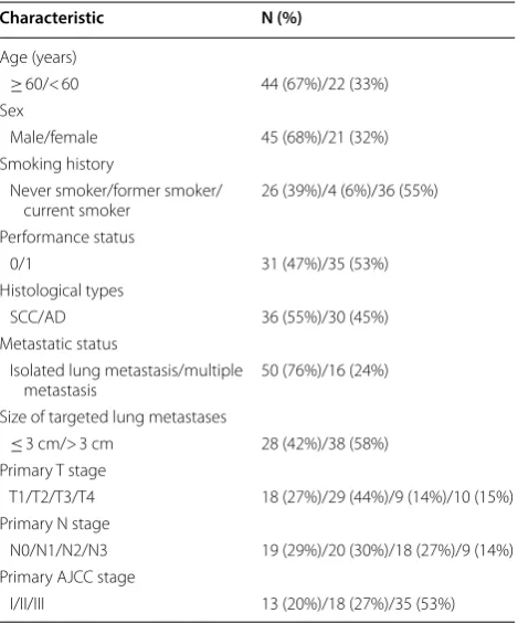
Table 1 Baseline characteristics of 66 NSCLC patients with lung metastases
Characteristic N (%)
Age (years)
≥ 60/< 60 44 (67%)/22 (33%) Sex
Male/female 45 (68%)/21 (32%) Smoking history
Never smoker/former smoker/
current smoker 26 (39%)/4 (6%)/36 (55%) Performance status
0/1 31 (47%)/35 (53%)
Histological types
SCC/AD 36 (55%)/30 (45%) Metastatic status
Isolated lung metastasis/multiple
metastasis 50 (76%)/16 (24%) Size of targeted lung metastases
≤ 3 cm/> 3 cm 28 (42%)/38 (58%) Primary T stage
T1/T2/T3/T4 18 (27%)/29 (44%)/9 (14%)/10 (15%) Primary N stage
N0/N1/N2/N3 19 (29%)/20 (30%)/18 (27%)/9 (14%) Primary AJCC stage
The exclusion criteria were as follows: patients concur-rently receiving other anti-tumor treatments (chemother-apy, immunother(chemother-apy, targeted therapy) within 1 month of SBRT; patients who had received anti-tumor treatment or steroids 3 months prior to enrollment; hematonosis, systemic lupus erythematosus, ulcerative colitis, hyper-thyroidism, scleroderma, rheumatoid arthritis, chronic liver disease, renal diseases, and other malignant tumors. The baseline characteristics of patients considered were age, sex, smoking history, performance status, histologi-cal types, size of lung metastases, primary tumor (T) stage, node (N) stage, and American Joint Committee on Cancer (AJCC) stage according to AJCC-7 criteria [23].
Flow cytometry
Additional file 1: Figure S1 shows representative flow cytometry plots and gating. We collected 4 mL of fresh blood from patients 7 days before SBRT. The flow cytom-etry protocol, the same as described in our previous study [24], was used to detect memory CD8+ T (CD3+ CD8+ CD45RA–CD45RO+), naive CD8+ T (CD3+ CD8+ CD45RA+ CCR7+), memory CD4+ T (CD3+ CD4+ CD45RA–CD45RO+), and naïve CD4+ T (CD3+ CD4+ CD45RA+ CCR7+).
SBRT and tumor response
SBRT was employed to treat lung metastases by CyberKnife. BED10 was calculated using the formula D × [1 + d/(α/β)]; D represents total dose, d stands for dose per fraction, and α/β = 10 [25]. The SBRT dose pre-scribed for radiation therapy was the responsibility of the oncologist carrying out the radiation treatment and was
prescribed in consideration of normal tissue tolerances; our patients were dosed with 5 fractions of 10 Gy or 10 fractions of 7 Gy.
Tumor response was evaluated 1 month after SBRT using a computed tomography (CT) or positron emission tomography-computed tomography (PET-CT) accord-ing to the RECIST 1.1 guideline [26]. On the one hand, a minimum 20% increase in the diameter of targeted lung metastases was used to mark a progressive disease (PD), while a minimum 30% reduction in the diameter of targeted lung metastases represented a partial response (PR). On the other hand, the absence of an adequate shrinkage to qualify for PR or an acceptable increase to qualify for PD marked a stable disease (SD), whereas, the diminishing targeted lung metastases represented a com-plete response (CR).
Statistical analysis
The link between predictors and early radiation response was assessed by analyses of logistic regression. Univari-ate analytical predictive findings with P < 0.1 were fur-ther subjected to multivariate analyses. Comparisons of immune cells differences between responsive and non-responsive patients were carried out using the independ-ent Studindepend-ent t-test. Cut-off values represindepend-enting immune cells’ ability to discriminate between responsive and non-responsive patients were determined using the receiver operating characteristic (ROC) curve and were used to determine high and low immune cells. Cut-off values for memory CD8+ T, naive CD8+ T, memory CD4+ T, naive CD4+ T, CD4+ naïve/memory ratio, and CD8+ naïve/memory ratio were 43.5, 33.1, 58.9, 20.9, 0.32, and 0.99, respectively. Statistical significance was marked by
Fig. 1 Numbers and proportions of responders and non-responders
Page 4 of 9 Liu et al. Cancer Cell Int (2019) 19:121
P-value < 0.05. Data were analyzed on the SPSS 23.0 soft-ware (SPSS Inc., Chicago, IL).
Results
Baseline characteristics
Baseline characteristics of all 66 patients with lung metas-tases of NSCLC are presented in Table 1. There were 45 (68%) males and 21 (32%) females; 36 (55%) squamous cell carcinomas (SCCs) and 30 (45%) adenocarcinomas (ADs); 28 (42%) patients with lung metastases ≤ 3 cm and 38 (58%) patients with lung metastases > 3 cm. As shown in Fig. 1, 49 (74.2%) of the 66 patients with lung metastases undergoing SBRT experienced PR (responders), while 17 (25.8%) expe-rienced SD (non-responders) 1 month after SBRT. The indi-vidual tumor changes in target lesion size from baseline are shown in Fig. 2. The absolute numbers of memory CD8+ T, naive CD8+ T, memory CD4+ T and naïve CD4+ T cells were (0.18 ± 0.15) × 109/L, (0.19 ± 0.13) × 109/L, (0.41 ± 0.25) × 109/L, and (0.13 ± 0.10) × 109/L.
Differences of immune factors between responders and non‑responders
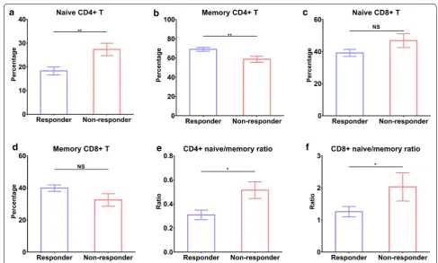
As shown in Fig. 3, there were lower levels of naïve CD4+ T (18.29 ± 1.66 vs. 27.33 ± 2.623, P < 0.01), CD4+
naïve/memory ratio (0.31 ± 0.04 vs. 0.51 ± 0.07, P < 0.01), CD8+ naïve/memory ratio (1.26 ± 0.16 vs. 2.03 ± 0.44, P < 0.05), and higher memory CD4+ T (68.99 ± 1.99 vs. 58.71 ± 3.21, P < 0.05) and memory CD8+ T (39.86 ± 2.02 vs. 32.48 ± 3.90, P= 0.07) in responders compared to non-responders.
ROC curves for immune factors’ ability to discrimi-nate between responders and non-responders are shown in Fig. 4. The most sensitive and specific marker was naïve CD4+ T with an area under curve (AUC) of 0.769, whereas, memory CD4+ T with an AUC of 0.740, naïve CD8+ T with an AUC of 0.648, memory CD8+ T with an AUC of 0.674, CD4+ naïve/memory ratio with an AUC of 0.753, and CD8+ naïve/memory ratio with an AUC of 0.669 were somewhat less sensitive and specific.
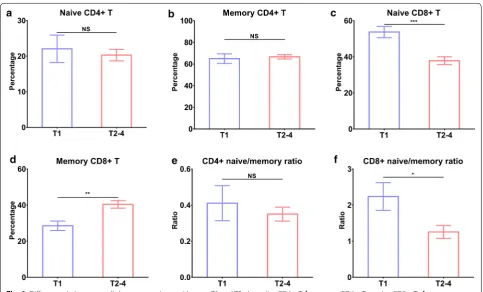
We also compared immune cells between 36 SCCs and 30 ADs and found no significant difference between them (all P > 0.05, Fig. 5). Patients with stage T1 had higher lev-els of naïve CD8+ T (P < 0.001), CD8+ naïve/memory ratio (P < 0.05), and lower memory CD8+ T (P < 0.01) when compared to those with stage T2–4 (Fig. 6).
Fig. 4 ROC curves of immune cells’ ability to discriminate between responders and non-responders for SBRT. a naïve CD4+ T, b memory CD4+ T, c
naive CD8+ T, d memory CD8+ T, e CD4+ naïve/memory ratio, and f CD8+ naïve/memory ratio
Page 6 of 9 Liu et al. Cancer Cell Int (2019) 19:121
Univariate logistic regression analysis
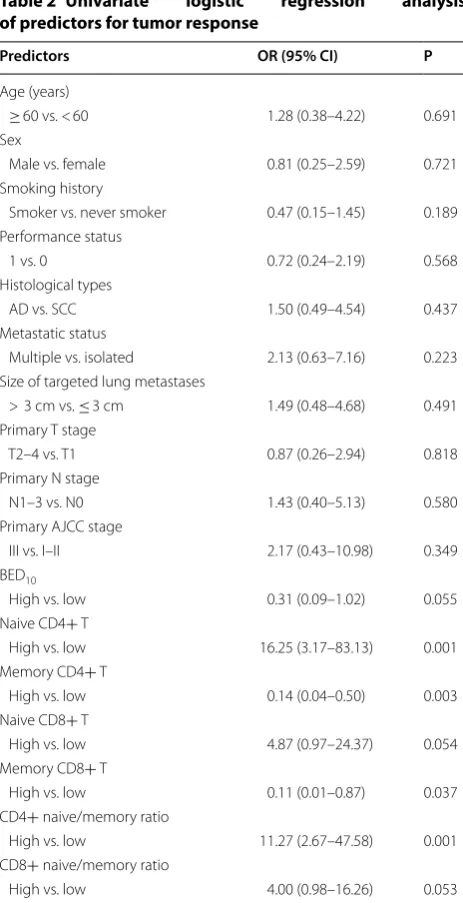
As shown in Table 2, low levels of naïve CD4+ T (OR: 16.25, 95% CI 3.17–83.13, P= 0.001), CD4+ naive/mem-ory ratio (OR: 11.27, 95% CI 2.67–47.58, P= 0.001), and high levels of memory CD4+ T (OR: 0.14, 95% CI 0.04– 0.50, P= 0.003) and memory CD8+ T (OR: 0.11, 95% CI 0.01–0.87, P= 0.037) predicted better tumor response to SBRT. The correlation between levels of naive CD8+ T, CD8+ naive/memory ratio, and BED10 and tumor response demonstrated strong trends (P= 0.054, 0.053, and 0.055, respectively).
Multivariate logistic regression analysis
Univariate analytical findings with P < 0.1, including naive CD4+ T, memory CD4+ T, naive CD8+ T, mem-ory CD8+ T, CD4+ naïve/memory ratio, CD8+ naïve/ memory ratio, and BED10, were enrolled in multivari-ate analysis. Naïve CD4+ T (OR: 12.67, 95% CI 2.26– 71.18, P= 0.004), memory CD4+ T (OR: 0.17, 95% CI 0.05–0.66, P= 0.010), memory CD8+ T (OR: 0.11, 95% CI 0.01–0.97, P= 0.047), and CD4+ naïve/memory ratio (OR: 8.50, 95% CI 1.90–38.14, P= 0.005) were independ-ent predictors of tumor response to SBRT (Table 3).
Discussion
Few studies have investigated tumor response predic-tors after SBRT. One of those, a recent study, revealed that the mean and maximum values of pre-SBRT stand-ard uptake value could predict a complete response in lung metastases from various primary tumors 6 months after SBRT [27]. In addition, a minimum 20% shrink-age in lung lesion during the final SBRT was revealed to correlate positively with a complete response, 6 months after SBRT [27, 28]. Here, we have provided additional information, that peripheral memory CD4+ T, memory CD8+ T, naïve CD4+ T, and CD4+ naïve/memory ratio were independent tumor response predictors to SBRT in NSCLC lung metastases.
CD45RO has been identified as a common marker of all subdivisions of memory T-cells, such as subdivisions of the bone marrow and secondary lymphoid organs, and subdivisions of circulating and tissue-resident nature, but it is not known to mark T memory-stem cells [29]. The elimination of an antigen necessitates the generation of Memory T cells during cell-mediated immune responses, and these generated cells last months and years after the antigens are gone, causing quicker and bigger responses to secondary and ensuing antigen exposures [30]. Upon tumor antigen stimulation, activated memory CD4+ T
cells respond very early to impede extensive replication or any significant impairment, either directly attack-ing the invadattack-ing organism or providattack-ing assistance to B or cytotoxic T cells [31]. Memory CD8+ T cells have the ability to persist for years and kill tumor and virally infected cells [32].
The prognostic value of tumor-infiltrating memory T cells has been assessed by many researchers in lung cancer. Memory T cells that infiltrate tumors in lymph-node metastases reportedly are positive independent factors of prognosis for survival in patients with NSCLC [33]. A positive correlation between tumor-associated
memory T cells and survival of SCLC patients has been shown to exist [34]. Interestingly, the correlation between memory T cells that infiltrate renal cell carcinoma and survival was negative, possibly due to impaired infiltrat-ing lymphocytes within the renal cell carcinoma [35]. In our study, we found more memory CD4+ T and CD8+ T cells in responders than non-responders in lung metasta-ses undergoing SBRT. In multivariate logistic regression analysis, memory CD4+ T and CD8+ T were independ-ent predictors of tumor response to SBRT, consistindepend-ent with the function of memory cells.
Also, we found fewer naïve CD4+ T cells, CD4+ naïve/memory ratio, and CD8+ naïve/memory ratio in responders than non-responders and an unfavorable pre-dictive value of naive CD4+ T and CD4+ naïve/memory ratio for tumor response after SBRT. Recently, Su et al. [36] reported that the chemotaxis of circulating naive CD4+ T cells differentiating into Tregs in situ and caus-ing immunosuppression of tumors trigger the infiltration of breast tumors by Tregs, which may explain our find-ings on naïve CD4+ T.
Several limitations exist in our study. First, different histological types were enrolled, including adenocarci-noma and squamous cell carciadenocarci-noma. Second, the sample size of 66 patients was limited and the study contained an unavoidable selection bias. Third, different doses of SBRT were used in our study. Finally, although the tumor response was evaluated 1 month after SBRT, we were unable to evaluate it after 6 months, since most patients went back to their local hospitals 1 month after SBRT. Nevertheless, our study could potentially be a step towards providing additional biomarkers for predicting tumor response after SBRT.
Table 2 Univariate logistic regression analysis
of predictors for tumor response
Predictors OR (95% CI) P
Age (years)
≥ 60 vs. < 60 1.28 (0.38–4.22) 0.691 Sex
Male vs. female 0.81 (0.25–2.59) 0.721 Smoking history
Smoker vs. never smoker 0.47 (0.15–1.45) 0.189 Performance status
1 vs. 0 0.72 (0.24–2.19) 0.568 Histological types
AD vs. SCC 1.50 (0.49–4.54) 0.437 Metastatic status
Multiple vs. isolated 2.13 (0.63–7.16) 0.223 Size of targeted lung metastases
> 3 cm vs. ≤ 3 cm 1.49 (0.48–4.68) 0.491 Primary T stage
T2–4 vs. T1 0.87 (0.26–2.94) 0.818 Primary N stage
N1–3 vs. N0 1.43 (0.40–5.13) 0.580 Primary AJCC stage
III vs. I–II 2.17 (0.43–10.98) 0.349 BED10
High vs. low 0.31 (0.09–1.02) 0.055 Naive CD4+ T
High vs. low 16.25 (3.17–83.13) 0.001 Memory CD4+ T
High vs. low 0.14 (0.04–0.50) 0.003 Naive CD8+ T
High vs. low 4.87 (0.97–24.37) 0.054 Memory CD8+ T
High vs. low 0.11 (0.01–0.87) 0.037 CD4+ naive/memory ratio
High vs. low 11.27 (2.67–47.58) 0.001 CD8+ naive/memory ratio
High vs. low 4.00 (0.98–16.26) 0.053
Table 3 Multivariate logistic regression analysis of predictors for tumor response
Predictors OR (95% CI) P
Naive CD4+ T
High vs. low 12.67 (2.26–71.18) 0.004 Memory CD4+ T
High vs. low 0.17 (0.05–0.66) 0.010 Naive CD8+ T
High vs. low 4.00 (0.76–20.91) 0.101 Memory CD8+ T
High vs. low 0.11 (0.01–0.97) 0.047 CD4+ naive/memory ratio
High vs. low 8.50 (1.90–8.14) 0.005 CD8+ naive/memory ratio
Page 8 of 9 Liu et al. Cancer Cell Int (2019) 19:121
Conclusions
We revealed that peripheral memory CD4+ T, memory CD8+ T, naïve CD4+ T, and CD4+ naïve/memory ratio were independent predictors for tumor response to SBRT in NSCLC lung metastases. Larger, in-depth studies are necessary to verify our findings.
Additional file
Additional file 1: Figure S1. Representative flow cytometry plots and gating for (A) memory CD4+ T and naïve CD4+ T cells, (B) memory CD8+ T and naive CD8+ T cells.
Abbreviations
SBRT: stereotactic body radiotherapy; NSCLC: non-small cell lung cancer; AJCC: American Joint Committee on Cancer; PD: progressive disease; PR: partial response; SD: stable disease; CR: complete response; AD: adenocarcinoma; SCC: squamous cell carcinomas.
Acknowledgements Not applicable.
Authors’ contributions
JMY, JBY and XLZ conceived and designed the study. CL, QYH, BX, XYH, HCS and QL enrolled patients and collected samples. CL performed the experi-ments and wrote the manuscript. All authors read and approved the final manuscript.
Funding
This work was supported by National Key Research and Development Pro-gram of China (No. 2018YFC1313200), Shandong Key Research and Develop-ment Program (No. 2016CYJS01A03), and National Natural Science Foundation of China (Nos. 81472813 and 81871895).
Availability of data and materials
All data included in our study are shown in our manuscript.
Ethics approval and consent to participate
This investigation received approval from the Ethics Committee of the Affili-ated Hospitals of Academy of Military Medical Sciences.
Consent for publication
All patients and healthy volunteers provided written informed consents.
Competing interests
The authors declare that they have no competing interests.
Author details
1 Department of Oncology, Renmin Hospital of Wuhan University, Wuhan 430060, China. 2 Department of Radiation Oncology, Shandong Can-cer Hospital and Institute, Shandong CanCan-cer Hospital Affiliated to Shandong University, Shandong Academy of Medical Sciences, Jinan 250117, Shandong, China. 3 Department of Radiation Oncology, Affiliated Hospital of Academy of Military Medical Sciences, Beijing 100071, China. 4 Department of Gyneco-logic Oncology, Shandong Cancer Hospital and Institute, Shandong Cancer Hospital Affiliated to Shandong University, Shandong Academy of Medical Sciences, Jinan 250117, Shandong, China.
Received: 26 February 2019 Accepted: 25 April 2019
References
1. Jaffray DA. Image-guided radiotherapy: from current concept to future perspectives. Nat Rev Clin Oncol. 2012;9:688–99.
2. Timmerman RD, Paulus R, Pass HI, Gore EM, Edelman MJ, Galvin J, et al. Stereotactic body radiation therapy for operable early-stage lung cancer: findings from the NRG Oncology RTOG 0618 Trial. JAMA Oncol. 2018;4:1263–6.
3. Gomez DR, Blumenschein GR Jr, Lee JJ, Hernandez M, Ye R, Camidge DR, et al. Local consolidative therapy versus maintenance therapy or observa-tion for patients with oligometastatic non-small-cell lung cancer without progression after first-line systemic therapy: a multicentre, randomised, controlled, phase 2 study. Lancet Oncol. 2016;17:1672–82.
4. Kwint M, Walraven I, Burgers S, Hartemink K, Klomp H, Knegjens J, et al. Outcome of radical local treatment of non-small cell lung cancer patients with synchronous oligometastases. Lung Cancer. 2017;112:134–9. 5. Lodeweges JE, Klinkenberg TJ, Ubbels JF, Groen HJM, Langendijk JA,
Wid-der J. Long-term outcome of surgery or stereotactic radiotherapy for lung oligometastases. J Thorac Oncol. 2017;12:1442–5.
6. Xu Q, Zhou F, Liu H, Jiang T, Li X, Xu Y, et al. Consolidative local ablative therapy improves the survival of patients with synchronous oligometa-static NSCLC Harboring EGFR activating mutation treated with first-line EGFR-TKIs. J Thorac Oncol. 2018;13:1383–92.
7. Sun B, Brooks ED, Komaki R, Liao Z, Jeter M, McAleer M, et al. Long-term outcomes of salvage stereotactic ablative radiotherapy for isolated lung recurrence of non-small cell lung cancer: a phase ii clinical trial. J Thorac Oncol. 2017;12:983–92.
8. Sutera P, Clump DA, Kalash R, D’Ambrosio D, Mihai A, Wang H, et al. Initial results of a multicenter phase II trial of stereotactic ablative radia-tion therapy for oligometastatic cancer. Int J Radiat Oncol Biol Phys. 2018;103:116–22.
9. Herrera FG, Bourhis J, Coukos G. Radiotherapy combination opportunities leveraging immunity for the next oncology practice. CA Cancer J Clin. 2017;67:65–85.
10. Lee Y, Auh SL, Wang Y, Burnette B, Wang Y, Meng Y, et al. Therapeutic effects of ablative radiation on local tumor require CD8+ T cells: chang-ing strategies for cancer treatment. Blood. 2009;114:589–95.
11. Filatenkov A, Baker J, Mueller AM, Kenkel J, Ahn GO, Dutt S, et al. Ablative tumor radiation can change the tumor immune cell micro-environment to induce durable complete remissions. Clin Cancer Res. 2015;21:3727–39.
12. Weichselbaum RR, Liang H, Deng L, Fu YX. Radiotherapy and immuno-therapy: a beneficial liaison? Nat Rev Clin Oncol. 2017;14:365–79. 13. Bernstein MB, Krishnan S, Hodge JW, Chang JY. Immunotherapy and
stereotactic ablative radiotherapy (ISABR): a curative approach? Nat Rev Clin Oncol. 2016;13:516.
14. Mole RH. Whole body irradiation; radiobiology or medicine? Br J Radiol. 1953;26:234–41.
15. Tang C, Welsh JW, de Groot P, Massarelli E, Chang JY, Hess KR, et al. Ipilimumab with stereotactic ablative radiation therapy: phase I results and immunologic correlates from peripheral T cells. Clin Cancer Res. 2017;23:1388–96.
16. Golden EB, Chhabra A, Chachoua A, Adams S, Donach M, Fenton-Kerimian M, et al. Local radiotherapy and granulocyte-macrophage colony-stimulating factor to generate abscopal responses in patients with metastatic solid tumours: a proof-of-principle trial. Lancet Oncol. 2015;16:795–803.
17. Postow MA, Callahan MK, Barker CA, Yamada Y, Yuan J, Kitano S, et al. Immunologic correlates of the abscopal effect in a patient with mela-noma. N Engl J Med. 2012;366:925–31.
18. Rodriguez-Ruiz ME, Rodriguez I, Garasa S, Barbes B, Solorzano JL, Perez-Gracia JL, et al. Abscopal effects of radiotherapy are enhanced by combined immunostimulatory mAbs and are dependent on CD8 T cells and crosspriming. Cancer Res. 2016;76:5994–6005.
•fast, convenient online submission •
thorough peer review by experienced researchers in your field • rapid publication on acceptance
• support for research data, including large and complex data types •
gold Open Access which fosters wider collaboration and increased citations maximum visibility for your research: over 100M website views per year •
At BMC, research is always in progress.
Learn more biomedcentral.com/submissions
Ready to submit your research? Choose BMC and benefit from:
20. Popp I, Grosu AL, Niedermann G, Duda DG. Immune modulation by hypofractionated stereotactic radiation therapy: therapeutic implications. Radiother Oncol. 2016;120:185–94.
21. Shaverdian N, Lisberg AE, Bornazyan K, Veruttipong D, Goldman JW, Formenti SC, et al. Previous radiotherapy and the clinical activity and tox-icity of pembrolizumab in the treatment of non-small-cell lung cancer: a secondary analysis of the KEYNOTE-001 phase 1 trial. Lancet Oncol. 2017;18:895–903.
22. De Ruysscher D. Radiotherapy and PD-L1 inhibition in metastatic NSCLC. Lancet Oncol. 2017;18:840–2.
23. Edge SB, Compton CC. The American Joint Committee on Cancer: the 7th edition of the AJCC cancer staging manual and the future of TNM. Ann Surg Oncol. 2010;17:1471–4.
24. Liu C, Wu S, Meng X, Liu G, Chen D, Cong Y, et al. Predictive value of peripheral regulatory T cells in non-small cell lung cancer patients under-going radiotherapy. Oncotarget. 2017;8:43427–38.
25. Fowler JF. 21 years of biologically effective dose. Br J Radiol. 2010;83:554–68.
26. Eisenhauer EA, Therasse P, Bogaerts J, Schwartz LH, Sargent D, Ford R, et al. New response evaluation criteria in solid tumours: revised RECIST guideline (version 1.1). Eur J Cancer. 2009;45:228–47.
27. Mazzola R, Fiorentino A, Di Paola G, Giaj Levra N, Ricchetti F, Fersino S, et al. Stereotactic ablative radiation therapy for lung oligometastases: predictive parameters of early response by (18)FDG-PET/CT. J Thorac Oncol. 2017;12:547–55.
28. Mazzola R, Fiorentino A, Ricchetti F, Giaj Levra N, Fersino S, Di Paola G, et al. Cone-beam computed tomography in lung stereotactic ablative radiation therapy: predictive parameters of early response. Br J Radiol. 2016;89:20160146.
29. Paulsen EE, Kilvaer T, Khanehkenari MR, Maurseth RJ, Al-Saad S, Hald SM, et al. CD45RO(+) Memory T lymphocytes—a candidate marker for
TNM-immunoscore in squamous non-small cell lung cancer. Neoplasia. 2015;17:839–48.
30. Farber DL, Yudanin NA, Restifo NP. Human memory T cells: gen-eration, compartmentalization and homeostasis. Nat Rev Immunol. 2014;14:24–35.
31. MacLeod MK, Kappler JW, Marrack P. Memory CD4 T cells: generation, reactivation and re-assignment. Immunology. 2010;130:10–5. 32. Henning AN, Roychoudhuri R, Restifo NP. Epigenetic control of CD8(+) T
cell differentiation. Nat Rev Immunol. 2018;18:340–56.
33. Kilvaer TK, Paulsen EE, Khanehkenari MR, Saad S, Johansen RM, Al-Shibli K, et al. The presence of intraepithelial CD45RO+ cells in resected lymph nodes with metastases from NSCLC patients is an independent predictor of disease-specific survival. Br J Cancer. 2016;114:1145–51. 34. Wang W, Hodkinson P, McLaren F, Mackean MJ, Williams L, Howie SE,
et al. Histologic assessment of tumor-associated CD45(+) cell numbers is an independent predictor of prognosis in small cell lung cancer. Chest. 2013;143:146–51.
35. Hotta K, Sho M, Fujimoto K, Shimada K, Yamato I, Anai S, et al. Prognostic significance of CD45RO+ memory T cells in renal cell carcinoma. Br J Cancer. 2011;105:1191–6.
36. Su S, Liao J, Liu J, Huang D, He C, Chen F, et al. Blocking the recruitment of naive CD4+ T cells reverses immunosuppression in breast cancer. Cell Res. 2017;27:461–82.
Publisher’s Note