E D U C A T I O N A L R E V I E W
Open Access
Gallstones top to toe: what the radiologist
needs to know
M. C. Murphy
*, B. Gibney, C. Gillespie, J. Hynes and F. Bolster
Abstract
Gallstone-related disease can have significant associated morbidity and mortality worldwide. The incidence of gallstone-related disease in the Western world is on the increase. There are multiple different
pathological manifestations of gallstone disease: the presentation, diagnosis and associated complications of which vary significantly depending on anatomical location. The role of imaging in gallstone-related disease is broad with radiology playing an essential role in the diagnosis, management and follow-up of gallstone-related pathologies. This paper distills the broad range of gallstone-gallstone-related pathologies into an anatomical map, discussing the disease processes involved at each point along the biliary tree and reviewing the strengths and weaknesses of different imaging modalities for each distinct disease process.
Keywords: Gallstones, Gallbladder, Biliary, Calculus, Pathology
Key points
Gallstone-related pathology is on the increase in the
Western world.
Gallstones can be located within the gallbladder,
migrate into the biliary tree or outside the
pancreaticobiliary system altogether with associated pathology.
Imaging of gallstones and associated pathology
requires a multimodality approach.
Introduction
Gallstones are solid rounded particles composed of a combination of cholesterol and bilirubin that form within the gallbladder and within the biliary system. The size and number of gallstones is variable with some patients forming multiple small gallstones and others forming single or few large stones.
The incidence of gallstones is increasing in Western populations as obesity levels rise. In the USA, 8.6% of Caucasian men and 16.6% of women have gallstones [1]. The vast majority of gallstones
are asymptomatic and require no follow-up; however, approximately 10–15% of gallstones will become symptomatic over a period of 10–15 years of follow-up [2, 3]. The symptomatic manifestations of gall-stones are variable and range from mild symptoms such as biliary colic to severe acute presentations such as pancreatitis, which can be associated with significant morbidity and mortality. In a patient that has suffered a symptomatic manifestation of gall-stones, the incidence of a further manifestation over their lifetime is approximately 3% per year [2]. The risk for gallstone-related pathology is also related to the number and size of stones with numerous larger stones more likely to cause symptoms. For this reason, it is suggested that once symptoms present, the patient should be offered a cholecystectomy [3–5].
The main risk factors for gallstone formation are outlined in Table 1, and the factors protective against gallstones are outlined in Table2.
There are three main types of gallstones: choles-terol, mixed and pigmented. Cholesterol gallstones account for 10% of gallstones and are composed of at least 50% cholesterol and form with supersatur-ation of bile. Patients with high-fat diets and high serum lipids are more likely to have a higher choles-terol composition. Pigmented gallstones, which are darker in colour and are composed mainly of
© The Author(s). 2020Open AccessThis article is distributed under the terms of the Creative Commons Attribution 4.0 International License (http://creativecommons.org/licenses/by/4.0/), which permits unrestricted use, distribution, and reproduction in any medium, provided you give appropriate credit to the original author(s) and the source, provide a link to the Creative Commons license, and indicate if changes were made.
* Correspondence:[email protected]
bilirubin with < 20% cholesterol, form with super-saturation of unconjugated bilirubin. There are two subtypes of pigmented gallstones: black pigmented gallstones which are formed due to chronic haemoly-sis, cirrhosis or intestinal malabsorption, and brown pigmented gallstones which are formed secondary to bacterial and parasitic infections such as Clonorchis
sinensis or due to stasis of bile. Finally, mixed gall-stones account for 80% of gallgall-stones and have a cholesterol content of 20–50% [6, 7].
Imaging in gallstone-related disease
Multiple imaging modalities play a role in the diagno-sis of the broad spectrum of gallstone-related disease including plain radiography, ultrasonography, com-puted tomography (CT), scintigraphy and magnetic resonance imaging (MRI).
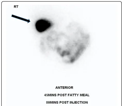
Ultrasound is by far the most common and useful imaging modality in assessing gallstones within the gallbladder (cholelithiasis) and associated gallbladder pathology and has the added benefit of no radiation dose to the patient. With ultrasound, gallstones are characteristically echogenic and demonstrate poster-ior acoustic shadowing regardless of the gallstone composition (Fig. 1). As ultrasound is a bedside dynamic study, stone mobility can be demonstrated with patient manoeuvering, and this can help in differentiating gallstones from gallbladder polyps, which can mimic cholelithiasis sonographically.
Another principle advantage of ultrasound over other imaging techniques in the investigation of acute cholecystitis is the ability to evaluate for a sonographic Murphy’s sign, which can be a reliable indicator of acute cholecystitis with a high sensitivity [8]. Sonographic Murphy’s sign is where the patient reports maximal pain as the sonographer presses over the fundus of the distended gallbladder with the ultrasound probe (and differs to clinical Murphy’s sign).
The major limitation of ultrasound in the imaging of gallstone-related disease is the frequent inability to assess the distal common bile duct (CBD) due to overlying bowel gas. Occasionally, a stone can be seen within the biliary system; however, often the presence of a CBD stone can be inferred from visua-lised proximal biliary dilatation and clinical presenta-tion with painful jaundice or an obstructive pattern on liver function tests. This requires further work up with alternative imaging.
Plain radiography is limited in the diagnosis of gall-stones as only 15–20% of gallstones are radio-opaque on X-ray [6]. The classical radiographic appearance is the “Mercedes Benz sign” which is an outer radio-opaque rim with a radiolucent centre which is caused by calcification of the gallstone rim and gas fissuring within the gallstone. Calcification of gallstones occurs with increased calcium in bile [9] (Fig. 2).
On CT, a high percentage of cholesterol stones are hypoattenuating relative to bile (Fig. 3) and calcified Table 1Main risk factors for gallstone formation
Risk factors for gallstone formation
Comment
Age The incidence increases with age but
symptomatic presentation is most common in middle age.
Gender More common in females by a ratio
of 2:1.
Race More common in Western, Caucasian,
Hispanic and Native American populations.
Family history A first degree relative with a history of gallstones doubles the risk.
Obesity Increased risk of cholesterol stone formation.
Rapid weight loss Bile stasis due to reduced calorie intake and increased cholesterol mobilisation.
Haemolysis There is an increased incidence of associated with haemolytic disorders such as sickle cell disease and the thalassemias.
Oral contraceptives and oestrogen replacement therapy
Pregnancy
Raised serum lipids Increased risk of cholesterol stone formation.
Raised serum bilirubin Increased risk of pigmented stone formation.
Cirrhosis
Gallbladder stasis Stasis of flow allows stones time to form.
Diabetes mellitus Insulin resistance increases circulating cholesterol.
Crohn’s disease
Certain medications
Table 2Factors protective against gallstones
Factors protective against gallstone formation
Comment
Statins Reduce bile cholesterol
concentration
Ascorbic acid Alters cholesterol catabolism
Unsaturated fats Alters bile acid composition
Coffee Alters cholesterol catabolism
Vegetable proteins and nuts Increase ascorbic acid levels
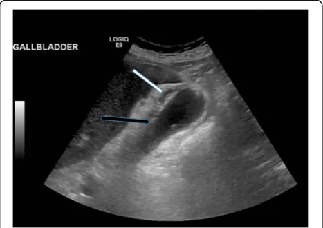
stones are hyperattenuating relative to bile (Fig. 4); however, a significant proportion of stones are iso-attenuating relative to surrounding bile and may be radiologically occult on CT. Dual-energy CT has been shown to improve detection of gallstones with low KV imaging and base substance imaging, such as calcium-based and lipid-based imaging, which is more sensitive at detecting gallstones than traditional higher KV imaging. Despite these imaging advances, CT remains inferior to ultrasound at assessing the
gallbladder and results in a significant radiation dose to the patient. As such, ultrasound is the imaging modality of choice for initial assessment of suspected gallbladder pathology. CT however can be very effective at assessing extra-biliary gallstone pathology and complications arising from gallstone pancreatitis and cholecystitis [8, 10].
MRI is another very effective tool for diagnosing gallstones and associated pathological processes. Mag-netic resonance cholangiopancreatography (MRCP) is typically performed using heavily T2-weighted se-quences, supplemented by fat-saturated T1- and T2-weighted MRI, and with steady-state gradient-echo acquisitions. Heavily T2-weighted image sequences are particularly helpful in delineating ductal anatomy.
Fig. 1Sagittal ultrasound image of the gallbladder which demonstrates the typical appearance of a gallstone that is hyperechoic with posterior acoustic shadowing (arrow)
Fig. 2Abdominal radiograph revealing a large rounded calcified density with a central lucency in the right upper quadrant consistent with a peripherally calcified gallstone
Gallstones typically appear as low signal or signal void on T2-weighted imaging surrounded by T2 hyperintense bile [11] (Fig. 5). MRCP has largely re-placed endoscopic retrograde cholangiopancreatogra-phy (ERCP) as the gold standard for diagnosing choledocholithiasis due to its high sensitivity of 90– 94% and specificity of 95–99% without the use of ionising radiation or ERCP-related complications such as pancreatitis which can result in significant morbi-dity and even mortality [12, 13].
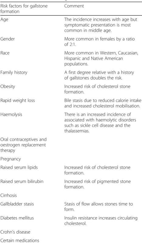
Nuclear medicine imaging with scintigraphy and with SPECT/CT can be used to dynamically assess
the gallbladder. Technicium-99 labeled mebrofenin is administered and taken up by bile producing cells and subsequently excreted into the biliary system. The patient is typically imaged at 1 h and at 4 h post-administration of radioisotope. A normal
Fig. 4Axial contrast-enhanced CT of the abdomen. There are several calcified gallstones (arrow) within the gallbladder which are hyper attenuating relative to surrounding bile
Fig. 5Axial T2-weighted MRI of the abdomen. There is a large gallstone within the gallbladder which is markedly hypointense relative to the surrounding hyperintense bile. The gallbladder wall is thickened with surrounding pericholecystic fluid consistent with acute cholecystitis
Fig. 6Normal cholescintogram 80 min post-injection of mebrofenin labeled with technetium-99. The radio-isotope is taken up by the liver and excreted into bile. Bile will pool in a non-obstructed normal gallbladder (arrow). In a gallbladder obstructed due to gallstones, the radioactive bile is unable to pool
Fig. 7Cholangiogram via a cholecystostomy tube demonstrating a distended gallbladder with a large filling defect consistent with a gallstone. The cystic duct and common bile duct are patent. Contrast is seen in the duodenum
gallbladder will be well delineated as it fills with radioactive bile. In cases of cholecystitis or gallblad-der obstruction due to an impacted stone in the cys-tic duct, the gallbladder will not be visualised as the radioactive will not accumulate within the gallblad-der. If the gallbladder is not visualised, morphine analogues can be given to induce sphincter of Oddi
contraction and aid gallbladder filling. Cholescinti-graphy for acute cholecystitis has a sensitivity of 97% and a specificity of 94% [14]. This is actually superior to ultrasound; however, this technique is more expensive and time consuming and also confers a radiation dose to the patient and staff and thus is generally not a first-line investigation (Fig. 6).
There is an increasing role for interventional ra-diology (IR) in gallstone-related diseases. Percuta-neous cholecystostomy can be used as a temporizing measure in critically ill patients who are too sick to proceed to immediate surgery and cholecystectomy. It is also occasionally being used as a therapeutic measure in the setting of an ageing population with multiple co-morbidities, many of whom may have contraindications to general anaesthesia and surgical intervention. In patients who have had a cholecys-tostomy placed, cholangiograms can be performed to demonstrate resolution of or ongoing obstruction of the biliary tree by assessing for filling defects (gall-stones) within the biliary system (Fig. 7). Percutan-eous transhepatic cholangiography (PTC) with biliary stenting is a very effective treatment to de-compress the biliary system in the case of obstruct-ing choledocholithiasis, most often in cases not amenable to ERCP. IR also play an active role in the
Fig. 8Illustration outlines the multitude of locations within the digestive tract where gallstones can manifest and lists the pathological processes that occur in these locations
management of the complications of gallstone dis-ease through drain insertion for hepatic or peripan-creatic abscesses, in the case of pancreatitis, and for post-operative intra-abdominal collections post-cholecystectomy.
Gallstone pathology
The broad spectrum of gallstone-related disease can be broken down based on the anatomical locations in which they occur (Fig.8).
Gallbladder
Unsurprisingly, the most common location for gall-stones and thus gallstone-related disease is the gallbladder.
Biliary colic is caused when a stone temporarily obstructs the drainage from the cystic duct, resulting in severe cramping abdominal and right upper quad-rant pain that can radiate to the back and right shoulder tip as the gallbladder contracts (typically, these symptoms are temporary and subside with resolution of the cystic duct obstruction). Ultra-sonography will often demonstrate cholelithiasis without associated complications in patients with simple biliary colic.
Calculous cholecystitis refers to infection and in-flammation of the gallbladder wall caused by irrita-tion from gallstones, and this can be an acute or chronic process. The typical clinical presentation of acute cholecystitis is of right upper quadrant pain, with or without radiation to the right shoulder, which is more constant compared to the intermit-tent pain seen in biliary colic. There is usually asso-ciated pyrexia and other infective symptoms such as nausea and vomiting. The typical imaging features
of acute cholecystitis are as follows: gallbladder wall thickening, pericholecystic fluid and a distended gallbladder [8] (Figs. 9 and 10). Chronic cholecystitis is caused by repeated episodes of biliary colic and acute cholecystitis over time, and in contrast to
Fig. 10Coronal (a) and axial (b) contrast-enhanced CT of the abdomen. There is gallbladder wall thickening (black arrow) and pericholecystic fluid (white arrows) consistent with acute cholecystitis
Fig. 11Coronal contrast-enhanced CT of the abdomen demonstrating a markedly distended gallbladder. There are multiple locules of gas within the anti-dependent gallbladder wall (arrow). Findings are consistent with emphysematous cholecystitis
acute cholecystitis, the gallbladder is usually shrunken down and the wall is thickened and scarred.
Emphysematous cholecystitis (EC) is a particular entity in which the gallbladder wall becomes nec-rotic, and this typically occurs with bacterial organ-isms such as the gas forming organorgan-isms such as clostridium, or with E. coli infections. Although rare, EC is associated with high mortality secondary to gallbladder perforation and gangrene, and EC is seen
more commonly in patients with diabetes mellitus, coronary artery disease and SIRS (systemic inflam-matory response syndrome) [8]. On plain films and CT, gas can be seen within the gallbladder wall (Fig. 11).
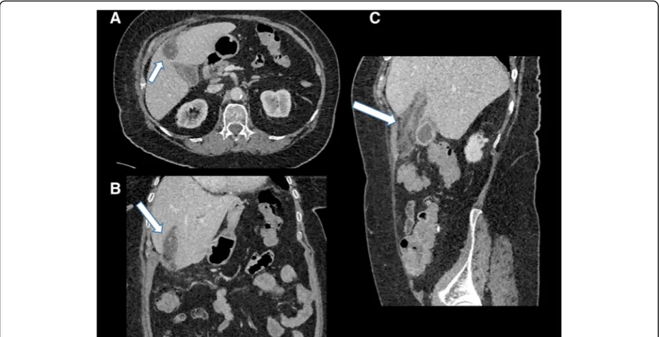
If an episode of acute cholecystitis is particularly severe or left untreated, it can progress to a gall-bladder perforation. This can be appreciated on US or on CT and MRI with pericholecystic abscesses or a defect in the gallbladder wall and a rim of bilious fluid outside of the gallbladder (Fig. 12). There may be an associated intrahepatic abscess which may require radiological or surgical drainage. Other rare complications of severe cholecystitis include cholecysto-cutaneous fistula and thrombophlebitis of a recanalised umbilical vein (Figs. 13 and 14).
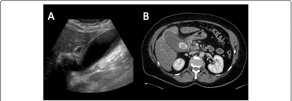
Along the spectrum of chronic cholecystitis is the porcelain gallbladder, where calcification of the gall-bladder wall is caused by repeated episodes of cholecystitis (Fig. 15). There is evidence of a causal relationship between gallstones, chronic cholecystitis and gallbladder carcinoma (Fig. 16), and malignancy often presents at an advancedstage; however, definitive proof is lacking [15].
A gallbladder mucocoele results when a stone obstructs the cystic duct causing the gallbladder to become distended with bile. When the bile within the mucocoele becomes infected, this is known as a gallbladder empyema (Fig.17).
Mirizzi syndrome refers to a gallstone that is impacted in the cystic duct or neck of gallbladder which causes extrinsic compression on the common bile duct resulting in obstructive jaundice (Fig. 18).
Both biliary dilatation and the offending gallstone can be seen on ultrasound; however, cross-sectional
Fig. 12Axial contrast-enhanced CT of the abdomen. There is gallbladder wall thickening and pericholecystic fluid consistent with acute cholecystitis. There is a defect in the medial gallbladder wall with a hypoattenuating collection within segment 5 of the liver (arrow). Appearances are consistent with gallbladder perforation and hepatic abscess
imaging with MRI or CT, or ERCP, may be needed to confirm that biliary dilatation is secondary to compression from a gallbladder/cystic duct stone rather than secondary to a CBD calculus.
Pancreaticobiliary system
The next anatomical location where gallstones can be found is outside the gallbladder but within the
pancreaticobiliary system. When gallstones exit the gallbladder into the common bile duct (choledocholi-thiasis), they can often obstruct the normal drainage of bile which can lead to jaundice. This is typically associated with pain, unlike malignant biliary ob-struction which is characteristically painless (Fig. 19). The obstruction of biliary drainage and stasis of bile may result in infection in the form of ascending
Fig. 14Axial (a), coronal (b) and sagittal (c) contrast-enhanced CT of the abdomen. There is gallbladder wall thickening and pericholecystic fluid consistent with acute cholecystitis. There is marked expansion and oedema surrounding the falciform ligament (arrows). Appearances are due to thrombophlebitis of a recanalised umbilical vein
Fig. 15Axial (a) and sagittal (b) contrast-enhanced CT of the abdomen demonstrating peripheral calcification of the gallbladder wall (arrows) consistent with porcelain gallbladder
cholangitis and associated sepsis. The clinical picture associated with this is described in Reynolds’ pentad consisting of fever, right upper quadrant pain, jaun-dice, hypotension and altered mental status. These patients may require urgent decompression of the biliary system.
In rare cases, there can be retrograde passage of gallstones into the common hepatic duct or the right or left main hepatic ducts, or stones can form in intrahepatic ducts due to biliary stasis.
If a gallstone passes down the common bile duct and comes to rest at the ampulla of Vater, it may block the drainage of the pancreatic duct causing back pressure on the pancreatic cells and resulting in gallstone pancreatitis. These patients present with
epigastric pain radiating to the back and the severity ranges from mild to severe. There is a significant mortality associated with severe pancreatitis, and critically ill patients should be managed in a high dependency or intensive care monitored environ-ment. While imaging is not usually required or indi-cated to confirm the diagnosis of acute pancreatitis, an ultrasound of the gallbladder can confirm or rule out the presence of gallstones. CT abdomen/pelvis is best performed 48 h after the onset of symptoms to assess for complications of pancreatitis such as peripancreatic collections or pancreatic necrosis (Fig. 20).
In general, larger gallstones are more likely to obstruct higher in the common bile duct, and as such are more likely to cause obstructive jaundice or cholangitis. Smaller gallstones are more likely to cause pancreatitis as they more freely pass down to the level of the ampulla of Vater [4, 5].
Extra-biliary complications
Gallstones can also cause pathology outside of the bil-iary system. The most common cause, although rare, is a cholecystoenteric fistula. Chronic irritation from a large gallstone can erode through the gallbladder wall with fistulisation into small bowel. This can be seen on imaging with air seen within the gallbladder or biliary tree (pneumobilia).
When a gallstone passes through the fistula into the small bowel, this can result in intestinal obstruc-tion, either proximal or more commonly distal. The most common place for distal small bowel obstruc-tion and gallstone ileum is at the level of ileocecal
Fig. 16Sagittal ultrasound of the gallbladder demonstrating a soft tissue mass within the gallbladder with internal vascularity consistent with a gallbladder carcinoma
valve as this is the narrowest point; however, gall-stone ileus can occur anywhere in the gastrointes-tinal tract. The diagnosis is suggested on abdominal X-ray by the presence of pneumobilia in the right upper quadrant with dilated loops of bowel consist-ent with bowel obstruction. Gallstone ileus is more accurately diagnosed with CT which may show pneumobilia or may directly demonstrate the pres-ence of a cholecystoenteric fistula and associated bowel obstruction (Fig. 21).
Bouveret’s syndrome is a particular eponymous syndrome in which a stone obstructs the upper GI tract proximally at the level of duodenum or gastric outlet. Patients typically present with copious vomit-ing owvomit-ing to the proximal level of obstruction. There may be little or no small bowel dilatation; in particu-lar, the X-ray abdomen may be completely normal which can falsely reassure. Imaging will demonstrate evidence of gastric outlet or duodenal obstruction related to a gallstone in the upper GI tract (Fig. 22).
Fig. 18aMaximum intensity projection (MIP).bAxial T2-weighted MRCP image of the biliary tree. The gallbladder is distended with extensive intrahepatic biliary duct dilatation. The common bile duct is normal in calibre. Appearances are consistent with Mirizzi
syndrome, with a stone in Hartmann’s pouch of the gallbladder causing extrinsic compression of the common hepatic duct
Fig. 19(a) Sagittal ultrasound, (b) fluoroscopic ERCP cholangiogram, (c) T2-weighted axial MRI and (d) coronal MIP MRI of the biliary tree. These images demonstrate multiple filling defects (arrows) within the common bile duct with associated biliary duct dilatation consistent with obstructing choledocholithiasis
Post-surgery/cholecystectomy complications
Finally, there are a number of imaging features post-cholecystectomy that the radiologist should be aware of. Immediate complications can include post-operative bleeding or an injury to the common bile duct resulting in a bile leak and subsequent biloma. CT is the optimal imaging modality for the initial imaging of post-operative complications, where these complications and fluid collections are well appre-ciated. It can be difficult to differentiate between blood and bile on CT, and measuring a region of interest to obtain the Hounsfield attenuation value of the fluid can help differentiate between the two. The typical Hounsfield unit of blood is 25–75 and that of bile is usually < 20; however, there can be some overlap. Other factors should be considered to ascertain the aetiology of any visualised collection, for example, a layering haemotocritl level with altered attenuation values can be a feature seen with haemorrhagic collections where the inferior denser (haemorrhagic) component is seen dependently [16] (Fig. 23).
Dropped gallstones at time of laparoscopy can have a delayed presentation with post-operative complications such as intrabdominal abscess forma-tion and CT demonstrating a radio-opaque gallstone surrounded by abscess (Fig. 24). Gallstone abscesses without radiopaque gallstones can pose a particular diagnostic challenge as the nidus for infection is not definitely confirmed on imaging. Abscesses related to dropped gallstones can be complex and may
Fig. 20Axial contrast-enhanced CT of the abdomen (a) and axial fat-suppressed T2-weighted MRI of the abdomen (b) in the same patient demonstrating extensive inflammation and oedema of the pancreas secondary to gallstone pancreatitis with a peripancreatic collection (arrow)
extend through abdominal planes and extend extra-peritoneally into adjacent subcutaneous and soft tissue plains. The clinical history will often include a history of prior or difficult cholecystectomy. Gallbladder clips or an absent gallbladder can be seen on cross-sectional imaging as clues.
Patients with occult choledocholithiasis that proceed to cholecystectomy can present with obstructive
jaundice and cholangitis in the post-operative period. It is important that any patient in whom choledocholi-thiasis is suspected undergo MRCP prior to surgery. Alternatively, an intra-operative cholangiogram or choledochoscope can be performed intra-operatively to ensure the common bile duct is clear of stones. Late post-cholecystectomy complications can include stump cholecystitis or a retained cystic duct stump or
Fig. 22Ultrasound (a), CT (b,c), MRI (d) and endoscopic images (e). Demonstrating a large calcified gallstone in the proximal duodenum with a massively dilated stomach. Findings are consistent with a proximal bowel obstruction consistent with Bouveret’s syndrome
Fig. 23Axial and coronal contrast enhanced CT of the abdomen in a patient several hours post-cholecystectomy. There is large volume perihepatic fluid with an average Hounsfield unit of 55 consistent with post-cholecystectomy bleeding
common bile duct stone. These findings result from incomplete cholecystectomy and can be identified on imaging [17] (Fig.25).
Conclusion
Radiologists should be familiar with the wide range of pathological processes that can be seen secondary to gallstones in order to aid prompt diagnosis, treatment and intervention
It is hoped that through understanding the role of multimodality imaging and understanding the anatomic locations of the manifestations of gallstone-related disease, this review paper will assist the radiologist in diagnosing common and less common manifestations of gallstone-related pathology.
Abbreviations
CBD:Common bile duct; CT: Computed tomography; EC: Emphysematous cholecystitis; ERCP: Endoscopic retrograde cholangiopancreatography; IR: Interventional radiology; MRCP: Magnetic resonance
cholangiopancreatography; MRI: Magnetic resonance imaging; SIRS: Systemic inflammatory response syndrome
Authors’contributions
MM contributed to the drafting of manuscript, image provision and final approval of manuscript. BG contributed to the manuscript edits and image provision. CG contributed to the manuscript edits and image provision. JH contributed to the manuscript edits and image provision. FB contributed to the manuscript edits, image provision and final approval of manuscript. All authors read and approved the final manuscript.
Funding None.
Availability of data and materials Not applicable.
Ethics approval and consent to participate Not required.
Consent for publication
All imaging is completely anonymised with no patient identifiers.
Competing interests
The authors declare that they have no competing interests.
Fig. 24Axial and sagittal images of a contrast-enhanced CT abdomen in a patient several days post-laparoscopic cholecystectomy. There is a rim-enhancing fluid collection compatible with an abscess which contains multiple (dropped) gallstones
Received: 17 September 2019 Accepted: 5 December 2019
References
1. Everhart JE, Khare M, Hill M, Maurer KR (1999) Prevalence and ethnic differences in gallbladder disease in the United States. Gastroenterology 117(3):632-9.https://www.ncbi.nlm.nih.gov/m/pubmed/10464139/
2. Friedman GD, Raviola CA, Fireman B (1989) Prognosis of gallstones with mild or no symptoms; 25 years of follow up in a health maintenance organisation. J Clin Epidemiolhttps://doi.org/10.1016/0895-4356(89)90086-3
3. Thistle JL, Cleary PA, Lachin JM, Tyor MP, Hersh T (1984) The natural history of cholelithiasis: the national cooperative gallstone study. Ann Intern Med
https://doi.org/10.7326/0003-4819-101-2-171
4. Shabanzadeh DM, Sørensen LT, Jørgensen T (2016) A prediction rule for risk stratification of incidentally discovered gallstones: results from a large cohort study. Gastroenterology.https://doi.org/10.1053/j.gastro.2015.09.002
5. Friedman G (1993) Natural history of asymptomatic and symptomatic gallstones. Am J Surg 165:399–404
6. Bortoff GA, Chen MY, Ott DJ, Wolfman NT, Routh WD (2000) Gallbladder stones: imaging and intervention. Radiographics 20(3):751–66
7. Clavien PA, Baillie J (Eds) (2006) Diseases of the Gallbladder and Bile Ducts: Diagnosis and Treatment, Second Edition. Wiley-Blackwell Press.https://doi. org/10.1002/9780470986981
8. O’Connor OJ, Maher MM (2011) Imaging of cholecystitis. AJR Am J Roentgenol 196:W367–W374https://doi.org/10.2214/AJR.10.4340
9. Meyers MA, O'Donohue N (1973) The Mercedes-Benz sign: insight into the dynamics of formation and disappearance of gallstones. Am J Roentgenol Radium Ther Nucl Medhttps://doi.org/10.2214/ajr.119.1.63
10. Chen AL, Liu AL, Wang S et al (2015) Detection of gallbladder stones by dual-energy spectral computed tomography imaging. World J Gastroenterol
https://doi.org/10.3748/wjg.v21.i34.9993
11. O’Connor OJ, O’Neill S, Maher MM (2011) Imaging of biliary tract disease. AJR Am J Roentgenolhttps://doi.org/10.2214/AJR.10.4341
12. Chen W, Mo JJ, Lin L, Li CQ, Zhang JF (2015) Diagnostic value of magnetic resonance cholangiopancreatography in choledocholithiasis. World J Gastroenterol 21(11):3351–3360https://doi.org/10.3748/wjg.v21.i11.3351
13. Maher M, Dixon AK (2015) Grainger & Allison’s diagnostic radiology: abdominal imaging. Elsevier, ISBN: 9780702069383ISBN: 9780702069383
14. Shea JA, Berlin JA, Escarce JJ et al (1994) Revised estimates of diagnostic test sensitivity and specificity in suspected biliary tract disease. Arch Intern Med 154(22):2573–81
15. Shrikhande SV, Barreto SG, Singh S, Udwadia TE, Agarwal AK (2010) Cholelithiasis in gallbladder cancer: coincidence, co-factor or cause? Eur J Surg Oncolhttps://doi.org/10.1016/j.ejso.2010.05.002
16. Vazquez JL, Thorsen MK, Dodds WJ et al (1985) Evaluation and treatment of intraabdominal bilomas. AJR Am J Roentgenol.https://doi.org/10.2214/ ajr.144.5.933
17. Chowbey P, Sharma A, Goswami A et al (2015) Residual gallbladder stones after cholecystectomy: a literature review. J Minim Access Surghttps://doi. org/10.4103/0972-9941.158156
Publisher’s Note
Springer Nature remains neutral with regard to jurisdictional claims in published maps and institutional affiliations.