Original Article
Comparative Evaluation of Penetration Ability of Three Pit and Fissure Sealants
and Their Relationship with Fissure Patterns
Nikita Garg 1, KR Indushekar 1, Bhavna Gupta Saraf 1, Neha Sheoran 1, Divesh Sardana 1
1 Dept. of Paediatric Dentistry, Sudha Rustagi Dental College, Faridabad, India.
KEY WORDS
Pit and fissure sealants;
Dental caries;
Glass ionomer cements;
Resins;
Received August 2017;
Received in Revised form October 2017; Accepted November 2017;
ABSTRACT
Statement of the Problem: Pit and fissure sealant placement is considered as an
effective modality for prevention of caries on occlusal surfaces. Penetration,
reten-tion and lateral wall adaptareten-tion are the key factors in success of pit and fissure
sealant restorations.
Purpose: The purpose of this paper was to compare penetration ability and lateral
wall adaptation of three commercially available pit and fissure sealants.
Materials and Method: The present in-vitro study was done on 45 extracted sound
human molars to evaluate the fissure pattern and assess the penetration ability of
three commercially available sealants [Delton® FS Sealant (Dentsply DeTrey
GmbH, Konstanz, Germany), Clinpro™ Sealant (3M™ ESPE™, Minnesota, USA)
and GC Fuji VII Glass Ionomer Cement (GC Asia Dental Pte Ltd, Singapore)] on
molars divided into 3 equal groups of 15 each, with further sectioning of each
sam-ple into 2 parts giving 30 samsam-ples per group. Following thermocycling and
embed-ding of teeth in clear auto-polymerizing acrylic resin, sections were evaluated for
fissure morphology, sealant penetration, unfilled space, lateral wall adaptation, and
for presence of voids.
Results: Penetrability of all the sealants studied was found to be significantly more
in U-type fissure pattern (93.89%) followed by V-type (78.62%), IK-type (74.34%)
and then in I-type (65.91). The depth of penetration of the GC Fuji VII Glass
Iono-mer sealant (85.82%) was found to be superior followed by unfilled resin sealant
(Clinpro™ Sealant- 78.26%) and then by filled resin sealant (Delton® FS Sealant-
74.89%).
Conclusion: U- type fissure pattern was more common than other fissure patterns
and showed significantly higher penetrability of different type of sealants evaluated
in the present study. GIC based sealant, due to significantly higher penetration
depth than unfilled and filled sealants used in the present study, can be preferred
over filled or unfilled resin sealants.
Corresponding Author: Divesh Sardana, Paediatric Dentistry Unit, Faculty of Dentistry, University of Hong Kong, Hong Kong. Email: [email protected]
Cite this article as: Garg N., Indushekar KR., Saraf BG., Sheoran N., Sardana D. Comparative Evaluation of Penetration Ability of Three Pit and Fissure Sealants and Their Relationship with Fissure Patterns. J Dent Shiraz Univ Med Sci., 2018 June; 19(2): 92-99.
Introduction
Caries involving occlusal surfaces of molars comprises
52.7% to 66.3% of all carious lesions while occlusal
surface of molars makes only 13% of total surfaces. [1]
Molars may have more risk for caries due to the
com-plex shape of their occlusal fissure morphology, which
is considered an ideal site for the retention of bacteria
and food remnants, and is inaccessible to mechanical
cleaning/debridement. The occlusal fissure pattern has
(Y1), IK (Y2) and inverted Y types (Nagano, 1961). [2]
Early attempts to protect pits and fissures from
caries attack by prophylactic odontotomy (Hyatt, 1921)
and fissure eradication were tried, but with little success.
[3] Similar results were met with chemical agents like
ammonical silver nitrate, [4] zinc chloride & potassium
ferrocyanide. [5] Placement of pit and fissure sealants is
considered an effective modality for prevention of caries
onset on occlusal surfaces of posterior teeth. Cost-
ef-fectiveness and decreased caries risk up to nine times
are some of the potential advantages of placing pit and
fissure sealants, which justify their use in prevention
against dental caries. [6] Resin based materials are the
materials of choice today because of their high retention
rates and superior wear resistance; however, these
mate-rials are clinically limited because of their inherent
hy-drophobic nature and inability to be used in moist
envi-ronment. [7] In contrast, glass ionomer pit and fissure
sealant has the advantage of very high fluoride release
along with antibacterial property, free flowing
con-sistency, and improved adherence to enamel. [8-9]
Penetration of the sealant into the complete depths
of pits and fissures, its lateral wall adaptation and
sub-sequent retention are the key factors in the longevity of
these restorations. [10] Apart from the penetration depth
of the sealant, extension of the sealant material over the
enamel of cuspal inclines is another factor considered in
successful bonding of the pit and fissure sealant to the
tooth. [11] The penetration of sealant into pit and fissure
depends on its geometric configuration, the presence of
material deposits within it, the physical and chemical
properties of the enamel, and good clinical technique.
Hence, the purpose of this study was to compare
pene-tration ability and lateral wall adaptation of three
com-mercially available pit and fissure sealants.
Materials and Method
The present in-vitro study was conducted on 45
ex-tracted third molars, which had been exex-tracted due to
orthodontic or periodontal reasons. Third molars with
macroscopic fractures, hypoplasia, restorations, attrition,
or carious occlusal surface were excluded from the
study. Forty-five third molars were divided into three
groups of 15 each to study the fissure morphology and
penetration ability of three commercially available
sealants. The sealants studied in the present
investiga-tion were:
1. Group A: Filled fluoride releasing (Delton® FS
sealant, (Dentsply DeTrey GmbH, Konstanz,
Ger-many) sealant was used which is white, light cure
resin sealant supplied in syringe with disposable tip
2. Group B: Unfilled fluoride releasing (ClinproTM,
3M™ ESPE™, Minnesota, USA) sealant was used
which is pink, light cure resin sealant supplied in
sy-ringe with disposable tip
3. Group C: Fuji VII Glass ionomer (GC Asia Dental
Pte Ltd, Singapore) sealant was used which is pink
and dual cure sealant supplied in bottle-liquid
sys-tem
The extracted molars were cleaned of debris, soft
tissues and calculus and were then stored in distilled
water until further use. The occlusal surface of the
ex-tracted teeth was cleaned with oil-free aqueous pumice
slurry using a prophylactic nylon brush in a slow-speed,
contra-angle handpiece. The pits and fissures were then
rinsed with an air-water spray and air-dried. The teeth
were mounted in clay so that the crown was exposed.
Sharp explorer was then run through the pits and
fis-sures to remove any remaining debris/slurry followed
by air-drying with a three-way syringe.
In Group I and II (Delton® FS Sealant and
Clinpro™ Sealant), acid etching was done using 37%
phosphoric acid gel (3M™ Scotchbond™ Universal
Etchant gel, Minnesota, USA) which was carefully
ap-plied to the occlusal surface covering all the pits and
fissures for 60 seconds with a brush tipped applicator.
Then, thorough rinsing was done with air-water spray
for 10 seconds to remove all etchant completely.
Air-drying was then done for 5 seconds with a
three-way syringe and observed for the frosty
appear-ance of the enamel.
Then, Delton® FS Sealant and Clinpro™ Sealant
were carefully dispensed in the appropriate amount onto
the etched occlusal surface from mesial (in maxillary
molars) or distal pit (in mandibular molars) and guided
to flow to the other pits and fissures with the help of a
brush tipped applicator. The sealant was allowed to flow
for 10 seconds, and then it was polymerised using a
light cure unit for 20-40 seconds, and checked with an
Figure 1: Ground sections of fissures (4X magnification) a. Well-adapted sealant (resin-based) b. Partially adapted (resin based) c. Pres-ence of void in sealant d. Sealant not adapted (Glass ionomer sealant)
The resin sealant Clinpro™, which was initially
pink on application turned light yellow after
polymeri-zation.
In Group III (GC Fuji VII Glass Ionomer Cement),
occlusal surface was gently cleaned with GC
condition-er for 20 seconds, and rinsed for 20 seconds. The
sur-faces were then dried by blotting with cotton pellet. The
GC Fuji VII Glass Ionomer Cement was mixed as per the manufacturer’s instruction and was applied to the occlusal surface using a plastic filling instrument. A
disposable fine brush was used to spread it on pits and
fissures and sealant was then allowed to flow for 10
seconds. After that, sealant was light cured for 20
se-conds followed by application of petroleum jelly on the
occlusal surfaces.
Subsequent to the pit and fissure sealants
applica-tion, teeth were then demounted from clay and were
subjected to thermocycling for 200 cycles in 5°C and
55°C with a dwell time of 60 seconds in each bath and a
transfer time of 3 seconds. These specimens were
com-pletely embedded in clear autopolymerising acrylic
res-in and were sectioned longitudres-inally bucco-lres-ingually
from the central fossa, yielding two sections of 150µ per
tooth using hard tissue microtome (Leica Biosystems,
Germany). Sections were then viewed using a binocular
light microscope at a minimum magnification of 4X for
fissure morphology, sealant penetration, unfilled space,
lateral wall adaptation and for any voids (Figure 1). The
Figure 2: Different Fissure patterns (Longitudinal ground sections 4X magnification):
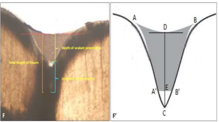
Figure 3: Calculations for depth of fissure sealants
parameters that were studied were:
1. Fissure morphology (classified as U, V, I and IK) [2]
(Figure 2)
2. Sealant penetration depth (µ) calculated as length
measured (µ) from the deepest point on concavity of
the upper margin of the occlusal sealant (D) to the
base of the sealant (E). (Figure 3)
3. Length of Unfilled space (µ) calculated as length
measured (µ) from base of the sealant (E) to the base
of the fissure (C). (Figure 3)
4. Total length of fissure (µ) calculated as length
measured (µ) from deepest point on the upper
mar-gin of the sealant (D) to the base of the fissure (C).
(Figure 3)
5. Penetrability (%) = Sealant Penetration Depth × 100
(Figure 3) Total Length of Fissure
All the linear measurements were made with the
help of software (ImageJ, National Institutes of Health,
Bethesda, USA) and calculated in microns (µ). Three
measurements of each section were taken and the mean
value of these three readings per section was adopted as
the representative values. The values were then
tabulat-ed and subjecttabulat-ed to statistical analysis.
Results
Results of the study are summarized in Table 1. U- type
fissure pattern (N=30; 33.3%) was found to be more
common followed by V-type and I-type (N=26; 28.9%)
and least for IK-type (N=8; 8.9%). However, no
statis-tical significant difference (p> .05) was found between
the prevalence of four types of fissure patterns studied
Table 1: Summary of penetrability and lateral wall adaptation of three different sealants and their distribution according to fissure patterns
Mean depth of penetration (%) Lateral wall adaptation
U (N=30) V (N=26) I (N=26) IK (N=8) Total
(N=90) Adapted
Partially Adapted
Not adapted Group A Filled
fluo-ride releasing (Delton® FS sealant, Dentsply)
93.45±7.13 (N=12)
66.58±28.08 (N=8)
61.52±9.11 (N=8)
50.25±0.59 (N=2)
74.89±21.96
(N=30) 18(60%) 12(40%) 0(0%)
Group B Unfilled fluoride releasing (ClinproTM, 3M ESPE)
91.11±12.65 (N=10)
83.67±15.19 (N=6)
64.04±13.34 (N=10)
73.55±11.25 (N=4)
78.26±17.13
(N=30) 20(66.67%)
10(33.33%)
0(0%)
Group C Fuji VII Glass ionomer (GC) sealant
98.01±3.69 (N=8)
84.12±12.64 (N=12)
72.65±23.96 (N=8)
100.00±0.00 (N=2)
85.82±17.50
(N=30) 20(66.67%) 8(26.7%) 2(6.63%)
on using ANOVA. The depth of penetration of the GC
Fuji VII Glass Ionomer sealant (85.82%) was found to
be superior followed by unfilled resin sealant (Clinpro™
Sealant- 78.26%) and then by filled resin sealant
(Del-ton® FS Sealant- 74.89%). However, the result was
sta-tistically non-significant (p> .05) between the three
groups of sealants on using ANOVA. The overall
pene-trability of all the pit and fissure sealants studied was
found more in U-type fissure pattern (93.89%) followed
by V-type (78.62%), IK-type (74.34%) and then in
I-type (65.91). However, the result was statistically non-
significant (p= 0.138) between the four types of fissures
on using ANOVA. Overall 64.4% of the sections in all
the three sealant groups showed good lateral wall
adap-tation except 2.2% of the sections in group III (GC Fuji
VII Glass Ionomer Cement) where sealant did not adapt
to both the lateral walls of the fissure. However, the
result was statistically non-significant (p= 0.138) when
the mean values of lateral wall adaptation was compared
between different sealant groups.
Discussion
In the present study, higher prevalence of U-type fissure
pattern (33.3%) was observed followed by V-type
(28.9%), I-type (28.9%) and least for IK- type (8.9%).
These findings also correlate with the studies conducted
by Duangthip et al. [12], Selectman et al. [13], and
Marks et al. [14]
Two large groups of materials were used in the
present study for sealing pits and fissures: resin based
sealant (Delton® FS Sealant and Clinpro™ Sealant) and
a glass ionomer cement (GC Fuji VII Glass Ionomer
Cement). The resin-based sealants were further
catego-rized as filled (Delton® FS Sealant) and unfilled
(Clinpro™ Sealant) based on the presence or absence of
the fillers respectively. Fillers are added to the pit and
fissure sealants in order to increase their wear and
abra-sion resistance. Resin-based materials possess high
re-tention rates and superior wear resistance but are
clini-cally limited by the difficulties inherent in their use due
to the technique sensitivity, as these materials are
pri-marily hydrophobic in natureand require a dry field. [6]
Whereas, glass ionomer pit and fissure sealant has the
advantage of very high fluoride release along with
anti-bacterial property, better handling property, free flowing
consistency and improved adherence to enamel. [8-9] In
addition, Antonson et al. found that glass ionomer
seal-ant performed better under wet contamination
condi-tions compared to resin-based sealant hence, material
type could be given more consideration while treating
pediatric patients. [15]
Properties of the pit and fissure sealants such as
surface tension and viscosity are the most important
factors that influence penetration of the sealants. [16]
Addition of the filler particles alters the viscosity and lowers the sealant’s ability to penetrate into fissures and microporosities of etched enamel. In the present study,
depth of penetration was found to be superior for GC
Fuji VII glass Ionomer Cement (85.82%) followed by
Clinpro™ Sealant (78.26%) and Delton® FS Sealant
(74.89%). The lower penetrability noticed in case of
filled sealant (Delton® FS Sealant), when compared
with the unfilled sealant (Clinpro™ Sealant), could be
because of added filler particles. This increases the
vis-cosity of the material and lead to reduced flow and
in-complete depth of penetration of the sealant to the
bot-tom of the occlusal fissures especially in case of deep
and narrow fissures like I-type and IK- type. Clinically,
lesser depth of penetration can also affect the retention
of the sealant, which might be one of the factors in the
reduction of retention rate. However, with the unfilled
sealant (Clinpro™ Sealant) which is less viscous, there is
greater potential for the sealant to flow, spread more
rapidly over surface, and penetrate.
At times, the size of the filler particles may be
larger than the porosities of the enamel. Therefore,
fast-er penetration rates are found with largfast-er holes, densfast-er
liquids and those with high surface tension, but slower
rates are found with fluids that are more viscous. Thus,
it is not only the addition of filler particles that alter the
flow of the sealant but the size of the filler particles used
can also influence the depth of penetration of the pit and
fissure sealant. Moreover, it has been studied that the
sealants containing fluoride tend to be thicker than those
without fluoride [17] but it did not affect the result of
our study as all the three sealants chosen were fluoride
containing.
In addition, penetrability of a pit and fissure
seal-ant varies depending on the occlusal fissure morphology
of the pit and fissure sealants was found to be
signifi-cantly more in U-type fissure pattern (93.89%) followed
by V-type (78.62%), IK-type (74.34%) and then in
I-type (65.91%). The results of the present study
corre-lated with the studies done by Gwinnett et al., [18]
Powell et al. [19], Symons et al. [11], Durmusoglu et al.
[20], Petrovic et al. [21], Selectman et al. [13], Grewal
N et al. [10], and Zhao L et al. [22]
The depth of penetration was greater in case of
U-type and V-type fissure pattern as they are wide and
shallow when compared to I-type and IK-type fissures.
The I-shaped fissure is quite constricted and may
re-semble a bottleneck in that the fissures may have an
extremely narrow slit appearance with a larger base as it
extends towards the dentino-enamel junction. Complete
penetration of sealant into complex fissure systems,
especially deep and narrow fissures is difficult
com-pared to wide and shallow fissures due to the
phenom-enon of closed end capillaries or isolated capillaries.
These fissures may also have a number of different
branches fail to be filled with sealants. Hence, this in
vitro study also proves that occlusal morphology is a
limiting factor for penetration of fissure sealants.
The results in our study suggest that the overall
depth of penetration of all the three pit and fissure
seal-ants is more than 70% of entire fissure depth which can
be correlated to the study done by Covey et al. [23]
Further, 31.11% of the fissures showed the complete
depth of penetration by the sealants. The complete
pen-etrability of the sealants was more common in both
U-type and V-type of fissure pattern due to their wide
and shallow anatomy, which favored greater flow of the
sealant when compared to I-type and IK-type that are
deep and narrow. However, Symons states that fissure
sealants show very good adaptability against vertical
fissure walls, but lack the ability to penetrate entirely to
the fissure bottom. [11] Study done by Bottenberg et al.
and Duangthip and Lussi also suggest that it is difficult
for etchant and consequently sealant to reach the bottom
of the fissure especially in ampular or constrictive
fis-sures. [12, 24]
No material is able to penetrate down to the
bot-tom of deep and narrow fissures; it is understandable
that some clinicians suspect that there are
microorgan-isms in unfilled space or that the sealant is often placed
over an incipient caries lesion. However, there are
evi-dences that bacteria cannot remain vital and that caries
lesion stops if the sealant is placed over an incipient
lesion. Sealing material eliminates nourishment sources
for S. Mutans and converts an active lesion into the
pas-sive caries lesion. [25]Hence, authors feel that clinically
maximum depth of penetration and good adaptation is
more important than the complete penetration of the
sealant to the base of the fissure.
Lateral wall adaptation of the resin sealants
(Del-ton® FS Sealant and Clinpro™ Sealant) was found to be
superior when compared to Glass Ionomer Cement (GC
Fuji VII). The presence of minute voids were noticed as
tiny black dots under the light microscopy in all the
three pit and fissure sealants (Delton® FS Sealant,
Clinpro™ Sealant and GC Fuji VII Glass Ionomer
Ce-ment). This might in turn affect the integrity and
strength of the pit and fissure sealants. These voids
could have been resulted because of the air entrapment
within the pit and fissure sealant material.
Another contrasting feature noticed in the ground
sections of molars with resin-based sealant (Delton® FS
Sealant and Clinpro™ Sealant) and GC Fuji VII Glass
Ionomer Cement was the presence of a smooth surface
in the former and rough surface in the latter as finishing
and polishing was not done in the present study.
There-fore, the placement of pit and fissure sealants in the
oc-clusal fissures is incomplete without finishing and
pol-ishing the surface of the sealant as well as removal of
the occlusal interferences. Hence, glass ionomer sealant
requires adequate finishing and polishing following its
placement in the occlusal pits and fissures.
One of the practical difficulty encountered in the
present in-vitro study while viewing the ground sections
of molars under a light microscope at 4X was that the
base of the occlusal fissure could not be clearly
appreci-ated. However, other modalities like stereomicroscope
and scanning electron microscope can be used to
over-come such problem.
Thus, pit and fissure sealant placement can be
considered as an effective treatment modality in
preven-tive dentistry. Though all the three sealants have shown
the depth of penetration of more than 70% but due to
Del-ton® FS Sealant (74.89%), former may be preferable
over resin based sealants for sealing the occlusal pits
and fissures in order to prevent dental caries.
Conclusion
1. Four fissure patterns were studied (U, V, I, IK) and
U- type fissure pattern (33.3%) was found to be more
prevalent followed by V-type (28.9%) and I-type
(28.9%) and least prevalent for IK-type (8.9%).
2. Penetrability of all the pit and fissure sealants studied
was found to be significantly more in U-type fissure
pattern (93.89%) followed by V-type (78.62%),
IK-type (74.34%) and then in I-type (65.91%).
3. The depth of penetration of the GC Fuji VII Glass
Ionomer sealant (85.82%) was found to be superior
followed by unfilled resin sealant (Clinpro™ Sealant-
78.26%) and then by filled resin sealant (Delton® FS
Sealant- 74.89%).
4. Lateral wall adaptation of the resin sealants (Delton®
FS Sealant and Clinpro™ Sealant) was found to be
superior when compared to Glass Ionomer Cement
(GC Fuji VII) and presence of voids were noticed in
all the three pit and fissure sealants (Delton® FS
Sealant, Clinpro™ Sealant and GC Fuji VII Glass
Ionomer Cement).
Conflict of Interest
The authors disclose no potential conflicts of interest.
References
[1] Demirci M, Tuncer S, Yuceokur AA. Prevalence of caries on individual tooth surfaces and its distribution by age and gender in university clinic patients. Eur J Dent. 2010; 4: 270-279.
[2] Nagano T. Relation between the Form of Pit and Fissure and the Primary Lesion of Caries. Dent Abstr. 1961; 6: 4265.
[3] Hyatt TP. Prophylactic odontotomy: the cutting into the tooth for the prevention of disease. Dental Cosmos. 1923; 65: 234-241.
[4] Klein H, Knutson JW. Studies on Dental Caries. XIII. Effect of Ammoniacal Silver Nitrate on Caries in the First Permanent Molar. J Am Dent Assoc. 1942; 29:1420-1426. [5] Ast DB, Bushel A, Chase HC. A clinical study of caries prophylaxis with zinc chloride and potassium
ferrocya-nide. J Am Dent Assoc. 1950; 41: 437-442.
[6] Droz D, Schiele MJ, Panighi MM. Penetration and micro-leakage of dental sealants in artificial fissures. J Dent Child (Chic). 2004; 71: 41-44.
[7] Hoffman I. A moisture tolerant, resin-based pit and fissure sealant. Dental Tribune. 2009; 4: 17A-18A.
[8] Khoroushi M, Keshani F. A review of glass-ionomers: From conventional ionomer to bioactive glass-ionomer. Dent Res J (Isfahan). 2013; 10: 411-420. [9] Hafshejani TM, Zamanian A, Venugopal JR, Rezvani Z,
Sefat F, Saeb MR, et al. Antibacterial glass-ionomer ce-ment restorative materials: A critical review on the current status of extended release formulations. J Control Re-lease. 2017; 262: 317-328.
[10]Grewal N, Chopra R. The effect of fissure morphology and eruption time on penetration and adaptation of pit and fissure sealants: An SEM study. J Indian Soc Pedod Prev Dent. 2008; 26: 59-63.
[11]Symons AL, Chu CY, Meyers IA. The effect of fissure morphology and pretreatment of the enamel surface on penetration and adhesion of fissure sealants. J Oral Re-habil. 1996; 23: 791-798.
[12]Duangthip D, Lussi A. Effects of fissure cleaning meth-ods, drying agents, and fissure morphology on microleak-age and penetration ability of sealants in vitro. Pediatr Dent. 2003; 25: 527-533.
[13]Selecman JB, Owens BM, Johnson WW. Effect of prepa-ration technique, fissure morphology, and material char-acteristics on the in vitro margin permeability and pene-trability of pit and fissure sealants. Pediatr Dent. 2007; 29: 308-314.
[14]Marks D, Owens BM, Johnson WW. Effect of adhesive agent and fissure morphology on the in vitro microleak-age and penetrability of pit and fissure sealants. Quintes-sence Int. 2009; 40: 763-772.
[15]Antonson SA, Antonson DE, Brener S, Crutchfield J, Larumbe J, Michaud C, et al. Twenty-four month clinical evaluation of fissure sealants on partially eruptedperma-nent first molars: glass ionomer versus resin-based seal-ant. J Am Dent Assoc. 2012; 143: 115-122.
[16]Burrow MF, Burrow JF, Makinson OF. Pits and fissures: etch resistance in prismless enamel walls. Aust Dent J. 2001; 46: 258-262.
Oper Dent. 2000; 25: 306-310.
[18]Gwinnett AJ, Ripa LW. Penetration of pit and fissure sealants into conditioned human enamel in vivo. Arch Oral Biol. 1973; 18: 435-439.
[19]Powell KR, Craig GG. An in vitro investigation of the penetrating efficacy of BIS-GMA resin pit and fissure-coatings. J Dent Res. 1978; 57: 691-695.
[20]Durmuglu Ö, Ta Tekin DA, Özyöney G, Bozkurt FÖ, Yaniko Lu FC. Light microscopy and SEM evaluation of three different fissure sealants. OHDMBSC. 2005; 4: 26-32.
[21]Markovic D, Petrovic B, Peric T, Miletic I, Andjelkovic S. The Impact of Fissure Depth and Enamel Conditioning Protocols on Glass-ionomer and Resin-based Fissure Sealant Penetration. J Adhes Dent. 2011; 13: 171-178.
[22]Zhao L, Shi Q. Microleakage and penetration ability of different sealants. Int J Ped Dent. 2009; 19: 71-75. [23]Covey DA, Johnson WW, Hopper LR. Penetration of
various pit and fissure sealants into occlusal grooves. Available at: https://www.researchgate.net/publication/ 47393422_The_impact_of_occlusal_morphology_on_fiss ure_sealant_penetration
[24]Bottenberg P, Gräber HG, Lampert F. Penetration of etch-ing agents and its influence on sealer penetration into fis-sures in vitro. Dent Mater. 1996; 12: 96-102.