REVI E W
Open Access
Atypical I
κ
B proteins
–
nuclear modulators of
NF-
κ
B signaling
Marc Schuster
1,2, Michaela Annemann
1,2, Carlos Plaza-Sirvent
1,2and Ingo Schmitz
1,2*Abstract
Nuclear factorκB (NF-κB) controls a multitude of physiological processes such as cell differentiation, cytokine expression, survival and proliferation. Since NF-κB governs embryogenesis, tissue homeostasis and the functions of innate and adaptive immune cells it represents one of the most important and versatile signaling networks known. Its activity is regulated via the inhibitors of NF-κB signaling, the IκB proteins. Classical IκBs, like the prototypical protein IκBα, sequester NF-κB transcription factors in the cytoplasm by masking of their nuclear localization signals (NLS). Thus, binding of NF-κB to the DNA is inhibited. The accessibility of the NLS is controlled via the degradation of IκBα. Phosphorylation of the conserved serine residues 32 and 36 leads to polyubiquitination and subsequent proteasomal degradation. This process marks the central event of canonical NF-κB activation. Once their NLS is accessible, NF-κB transcription factors translocate into the nucleus, bind to the DNA and regulate the transcription of their respective target genes. Several studies described a distinct group of atypical IκB proteins, referred to as the BCL-3 subfamily. Those atypical IκBs show entirely different sub-cellular localizations, activation kinetics and an unexpected functional diversity. First of all, their interaction with NF-κB transcription factors takes place in the nucleus in contrast to classical IκBs, whose binding to NF-κB predominantly occurs in the cytoplasm. Secondly, atypical IκBs are strongly induced after NF-κB activation, for example by LPS and IL-1βstimulation or triggering of B cell and T cell antigen receptors, but are not degraded in the first place like their conventional relatives. Finally, the interaction of atypical IκBs with DNA-associated NF-κB transcription factors can further enhance or diminish their transcriptional activity. Thus, they do not exclusively act as inhibitors of NF-κB activity. The capacity to modulate NF-κB transcription either positively or negatively, represents their most important and unique mechanistic difference to classical IκBs. Several reports revealed the importance of atypical IκB proteins for immune homeostasis and the severe consequences following their loss of function. This review summarizes insights into the physiological processes regulated by this protein class and the relevance of atypical IκB functioning.
Keywords:NF-kappaB, Atypical IkappaB proteins, BCL-3, IkappaBNS, IkappaBzeta, IkappaBL, Nuclear NF-kappaB modulation, IkappaB eta, MAIL, NFkBID
Review NF-κB signaling
NF-κB transcription factors are homo- or hetero dimers composed of two REL proteins, such as p50 and p65 [1]. This family consists of p65/RelA, p50, p52, RelB and c-Rel. Their common structural motif is the Rel homology domain (RHD) [2]. It contains a dimerisation sequence for the inter-action with other REL proteins, a nuclear localization signal (NLS) regulating its subcellular localization and a DNA
binding motif for the interaction withκB sites in regulatory sequences of their respective target genes. Transactivation domains, TAD, are found in p65/RelA, c-Rel and RelB, whereby NF-κB dimers containing at least one of these sub-units can induce transcription [2]. On the other hand, NF-κB homodimers of p50 and p52 function as transcriptional repressors due to the lack of such a sequence [2]. They ei-ther compete for activating NF-κB transcription factors by occupation of DNA binding sites, or recruit gene-silencing proteins such as histone deacetylases (HDACs) [3], or inhibit transcription by use of both mechanisms. Each REL-protein subunit, with its individual and slightly different DNA-binding domain, contributes to the total DNA-affinity of the * Correspondence:[email protected]
1Systems-oriented Immunology and Inflammation Research, Helmholtz
Center for Infection Research, 38124, Braunschweig, Germany
2Institute for Molecular and Clinical Immunology, Otto-von-Guericke
University, 39120, Magdeburg, Germany
dimeric transcription factor [4-6]. Thus, the optimal se-quence for NF-κB binding is not identical among the differ-ent dimer combinations. This results in a magnitude of optimal regulatory sequences. The diversity of ideal binding sites, the multitude ofκB-sites in the DNA and the existence of suppressive and inducive NF-κB dimers are the reasons of the complexity and versatility of the downstream signal-ing network.
NF-κB can be activated in two different fashions, called canonical and non-canonical NF-κB activation. Both pathways use a complex formed by IκB kinase pro-teins, however, in slightly different compositions [2,7]. Regulation of the upstream signaling events and detailed differences between the canonical and non-canonical NF-κB activation were previously illustrated and are not part of this review [2,7].
Canonical NF-κB activation
The canonical signaling is initiated by a variety of recep-tors, like members of the TNF receptor super-family, Toll-like receptors, interleukin receptors and antigen re-ceptors of B and T cells [2]. Their common downstream signaling complex is a trimeric IκB kinase complex consisting of the catalytic subunits IKKα, IKKβand the regulatory subunit IKKγ/NEMO [8,9]. The sequestration of NF-κB in the cytoplasm is mediated by the association of classical IκBs such as the prototypical protein IκBαto inhibit NF-κB binding to the DNA [10-13]. The charac-teristic structural motif of IκB proteins is a repetitive se-quence of 6 to 10 ankyrin domains [2]. Binding of these ankyrin repeats to the REL homology domain of NF-κB results in masking of the NLS [14,15]. Crystallography demonstrated that the ankyrin domain of IκBα localizes between the carboxy-terminal Ig-like sequences of the REL homology domains of two NF-κB subunits [16]. When the NLS is accessible the NF-κB transcription fac-tor can localize in the nucleus and bind to the DNA, which depends on IκBα degradation [17,18]. In case of IκBα this process is initiated by phosphorylation of the serine residues 32 and 36 by activated IKKβ[18-20]. The phosphorylated serines within the so-called“destruction box”of IκBαare subsequently recognized by the E3 lig-ase βTRCP leading to polyubiquitination and eventually causing proteasomal degradation of IκBα [17,21-23]. As the NLS of the NF-κB dimer is accessible the transcrip-tion factor localizes into the nucleus and modulates transcription via binding to the DNA.
Non-canonical NF-κB activation
The non-canonical NF-κB activation depends on an IKKαhomodimer, activated for example by triggering of the BAFF receptor, CD40 or the lymphotoxin-βreceptor [7,24,25]. The NF-κB dimers activated in the non-canonical signaling cascade are composed of p52 and
RelB [26]. Their NLS sequences are masked intra-molecularly by the precursor protein of p52, p100, which displays carboxy-terminal ankyrin repeats to interact with the REL domains and hide the NLS [27]. Phosphor-ylation of p100 causes cleavage of the protein into p52 leading to the nuclear translocation of NF-κB [26,28]. As the ankyrin repeats are part of the sequences of p100 as well as p105, the precursor of p50, the existence of a common evolutionary ancestor for both, IκB and REL proteins is reasonable. Alternatively, p100 and p105 could be the result of a gene fusion.
Nuclear modulation of NF-κB activity
NF-κB activity is fine-regulated in the nucleus by a variety of mechanisms, including post translational modifications of REL proteins for example sumoylation, phosphoryl-ation, acetylation and ubiquitination [3,29]. Besides, the nuclear IκB proteins of the BCL-3 class BCL-3, IκBNS,
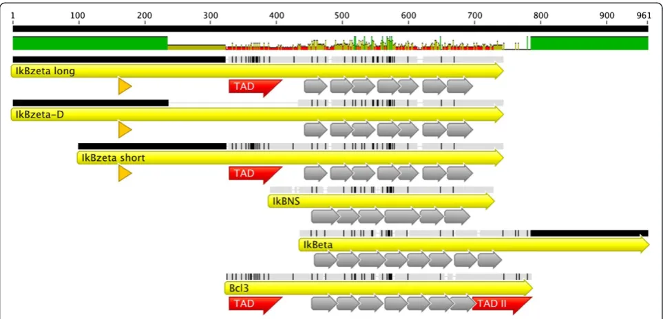
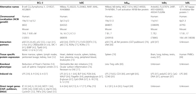
IκBζand IκBηcan dramatically alter NF-κB-mediated ef-fects via the regulation of dimer exchange, the recruitment of histone modifying enzymes or the stabilization of NF-κB dimers on the DNA. Although, these proteins formally belong to the IκBs due to the presence of ankyrin repeats in their structure (Figure 1), they do not functionally act exclusively as repressors of NF-κB-mediated transcription, but more as NF-κB modulators (Table 1).
BCL-3
Initial description and structure
BCL-3 was the first identified atypical IκB protein. It con-sists of an amino-terminal TAD followed by 7 Ankyrin re-peats and a second carboxy terminal TAD, displaying an overall length of 448 amino acids (Figure 1). It was first described as a proto-oncogene expressed in patients, which suffered from B-cell chronic lymphocytic leukemia displaying the translocation t (14:19)(q32;q13.1) [30].
Function
The oncogenic potential of BCL-3 is illustrated by its capacity to dampen the tumor suppressor p53 and to force Cyclin D1 expression in order to enhance prolifer-ation [31,32]. As the protein is expressed by a variety of different non-Hodgkin and Hodgkin lymphomas it could represent a suitable pharmacological target for the treat-ment of cancer [33,34]. Electrophoretic mobility shift as-says initially revealed that the protein interacts with p50 and p52 and can inhibit DNA binding [32,35]. In con-trast to IκBα, which sequesters p50 in the cytoplasm, BCL-3 is localized in the nucleus and alters the sub-nuclear localization of p50. In COS cells p50 was rela-tively equally distributed when overexpressed alone, however cotransfection with BCL-3 resulted in its accu-mulation in nuclear spots [36]. From these analyses it was thought that BCL-3 might act as an anti-repressor
Schusteret al. Cell Communication and Signaling2013,11:23 Page 2 of 11
by removing suppressive p50/p50 homodimers from the promoters of its target genes, which allows binding of activating p50/p65 or comparable heterodimers and in-directly forces transcriptional activation. Transcriptional repression of BCL-3 is also directly regulated via its binding to HDAC-1, -3 and -6 [37]. In macrophages, LPS is a potent inducer of BCL-3 [38,39], which inter-acts with p50 to reduce NF-κB-mediated TNF-α produc-tion. In agreement with the formation of nuclear suppressor complexes, it was suggested that this effect is mediated via chromatin remodeling. This is based on the fact that HDAC-1 overexpression further enhanced BCL-3-mediated suppression and trichostatin A treatment abrogated the BCL-3-mediated effects [39]. Alternatively, BCL-3 can suppress transcription via block of the ubiquitination of p50 to stabilize a suppressive NF-κB com-plex within the nucleus [38]. Thus, BCL-3-deficient macro-phages display enhanced expression of pro-inflammatory cytokines upon LPS treatment, as degradation of inhibitory p50 homodimers is not blocked. Surprisingly, an early study reported that BCL-3 had the potential to induce transcrip-tion directly via an amino terminal proline rich region and a second carboxy-terminal serine/proline rich region [40]. It was later shown that its carboxy-terminal tail enhances tran-scription via interaction with c-Jun, c-Fos, CREB-binding protein/p300 and the steroid receptor coactivator-1 (SRC-1), with CBP/p300 and SRC-1 having acetyltransferase activity [41]. Remarkably, both the amino and carboxy terminal
sequences are needed for the full transcriptional activity of BCL-3, suggesting, that both sequences function coopera-tively [40]. An additional part of the nuclear BCL-3 complex is the BCL-3 binding protein B3BP thought to be involved in DNA repair, which forms a complex with p300 and BCL-3 [42]. In addition to p300, the acetyltransferase Tip60 is another interaction partner of BCL-3/p50 complexes, which can enhance transcriptional activity [43]. A recent re-port nicely demonstrated that p52/BCL-3 complexes bind to A/T and G/C centric NF-κB binding sites sequences, however, with a dramatically altered transcriptional outcome [44]. Via G/C centric elements these complexes induce tran-scription through Tip60 recruitment, but suppress via bind-ing to A/T centric elements and recruitment of HDAC3.
BCL-3 itself is critically regulated via post-translational modifications, especially via phosphorylation and ubiqui-tination. It was shown that phosphorylation of BCL-3 via GSK3 regulated BCL-3 degradation and oncogenicity [37,45]. However, its proteasomal degradation in the cyto-plasm is regulated by an E3-ligase complex containing TBLR1, which appears to be independently of GSK3 [46]. In all known pathways, NF-κB activity is regulated by sev-eral upstream ubiquitination events through the bal-ance between ubiquitin ligases and deubiquitinases [2,47]. CYLD, a K63-deubiquitinase inhibits NF-κB ac-tivation in TRAF2-mediated NF-κB signaling pathways [48]. Remarkably, BCL-3 also becomes deubiquitinated by CYLD in the nucleus, upon UV-irradiation. This Figure 1Alignment of atypical IκB proteins.Alignment of murine atypical IκB proteins IκBζlong, IκBζD, IκBζshort, IκBNS, IκBηand BCL-3 is
Table 1 Properties of atypical IκB proteins
BCL-3 IκBζ IκBNS IκBη IκBL
Alternative names B-cell CLL/lymphoma 3 , D19S37, AI528691
Nfkbiz, FLJ30225, FLJ34463, INAP, MAIL, AA408868
Nfkbid, IkB-delta, MGC11314, MGC149503, TA-NFKBH, T-cell activation NFKB-like protein
Ankrd42, FLJ37874, SARP, 4931426M20,
4933417L02Rik
LST1, NF-kappa -B inhibitor-like protein 1
Chromosomal Localisation (NCBI geneID)
Human: Human: Human: Human: Human:
19q13.1-q13.2 3p12-q12 19q13.12 11q14.1 6p21.3
(602) (64332) (84807) (338699) (4795)
Mouse: Mouse: Mouse: Mouse: Mouse:
7A3; 7 9.95 cM 16; 16 C1.2-C1.3 7 B1; 7 7; 7E2 17 B1; 17
(12051) (80859) (243910) (73845) 18.6 cM (18038)
Interaction partners
p50 [35,36,40], p52 [32], c-Jun [41], c-Fos [41], CREB/p300 [41,43], SRC-1 [41], B3BP [42], Tip60 [43], HDAC-1/-3/-6 [37,45]
p50 [64], STAT3 [71], FUSS-DDIT3 [72], p65 [73], RORγ[77], RORα[77]
p50 [79], all Rel proteins (GST-pulldown) [78], cRel [80]
p50 [87] Unknown
Tissue specific protein expression
Bone marrow, spleen, lymph nodes, peritoneal lavage, kidney, liver [52]
Heart, skeletal muscle, spleen, kidney, liver, placenta, lung, peripheral blood, leukocytes [73]
Spleen [78] Brain, lung, kidney, testis, ovary [87]
Human PMBCs [88]
Knockout phenotype
Reduction of Peyer’s Patches [53]. Lack of splenic marginal centers [51].
Dermatitis-like skin irritations [74]. Ocular surface inflammation [76]. Resistant to EAE [77].
Less Treg cells [80]. Unknown Unknown
Induced via LPS [39], IL-9 [54], IL-4 [57] LPS [61], IL-1 [64], BLP [64], PGN [64], MALP [64], Flagellin [64], peptidoglycan [67],
β-glucan [67], CpG-DNA [67], IL-18 [70], IL-12 [70]
LPS [79,82], CD3 [80], anti-IgM [83], CD40 [83]
LPS [87], poly(I:C) [87], CpG-DNA [87], zymosan [87]
LPS [88]
Direct target genes (ChIP, pulldown, EMSA)
IP-10 [44], IL-10 [44], MCP-1 [44], CD95 [44], CD40 [44], IL-23p19 [44], CyclinD1 [32], TNFα[39], Gata3 [55]
IL-6 [64], IL8 [72], IL-17 [77], IFNγ[70] IL-2 [81], IL-6 [82], Foxp3 [80] Unknown Unknown
Schuster
et
al.
Cell
Communicati
on
and
Signaling
2013,
11
:23
Page
4
o
f
1
1
http://ww
w.biosignali
ng.com/cont
causes the rapid export of BCL-3 from the nucleus and its inactivation [48].
Transgenic mouse models
BCL-3 function was examined using a variety of different transgenic mouse models. Eμ-BCL-3 transgenic mice display splenomegaly, lymphadenopathy and elevated levels of ma-ture B cells in the secondary lymphoid organs, the peritoneal cavity and the bone marrow, suggesting that BCL-3 overexpression renders B cells into a state of hyperactivation [49]. In agreement with this observation, BCL-3-deficient mice display a variety of defects in their humoral immune response. They lack germinal centers in the spleen and show impaired clearance ofListeria, StreptococciandToxoplasma infections since they cannot mount a pathogen-specific antibody response [50-52]. Upon Listeriainfection, re-duced IL-12p70 and IFNγ levels were detected, which is presumably the result of increased levels of anti-inflammatory IL-10 produced by macrophages [50]. In addition, like p50-deficient mice, BCL-3-deficient mice display reduced Peyer´s Patches but not a complete ab-sence of them as seen in p52/p100-deficient mice [53]. Besides the role of BCL-3 in B cells, the protein has several properties important for T cells survival and differentiation. In T cells and mast cells, BCL-3 is upregulated by IL-9 and IL-4 via the Jak/STAT path-way [54]. When BCL-3 is absent, induction of GATA-3 by IL-4 is dramatically impaired and, thus, TH2 devel-opment [55]. In contrast to this, the generation of IFNγ-producing TH1 cells is not altered in BCL-3 compromised mice [55,56]. However, the protein en-hances IFNγ expression in CD8 cells upon second antigen exposure [56]. In addition, IL-4 protects cells from apoptosis via BCL-3. One report demonstrated that BCL-3 expression is lost upon IL-4 deprivation, leading to apoptosis [57]. In agreement, ectopic overexpression of BCL-3 effectively protected cells from IL-4 deprivation-induced death [57]. Consequently, it was suggested that BCL-3 could have anti-apoptotic poten-tial. Indeed, another report demonstrated that BCL-3-defi-cient T cells are highly sensitive towards activation-induced cell death due to over-activated pro-apoptotic Bim [58]. In line, transgenic overexpression of BCL-3 prolonged T cell survival. In the context of T cells it was further shown, that BCL-3 in cooperation with p52 is important in regulating central tolerance [59]. However, this effect is not intrinsically mediated by T cells, but controlled by medullary thymic epi-thelial cells, which are required for selection of T cells. These cells display impaired maturation in BCL-3/p100 double-deficient mice, leading to severe autoimmunity [59]. In terms of autoimmune diseases it should be noted, that BCL-3 is also a suppressor of autoimmune diabetes, as BCL-3-deficient NOD mice are more susceptible to auto-immune diabetes and display higher levels of IL-17 [60].
Conclusive remarks
The protooncogene BCL-3 displays remarkable versatility in the regulation of NF-κB, for example via NF-κB stabilization in the nucleus or removal of the transription factor from the DNA. Via the recruitment of HAT and HDAC proteins BCL-3 can mediate opposing effects on transcription.
IκBζ
Initial description and structure
IκBζwas first identified by a differential display analysis in a variety of tissues upon i.p. injection of LPS in wildtype mice [61]. It was initially termed “molecule possessing ankyrin repeats induced by LPS” (MAIL), which is still a frequently used name for its murine isoforms [61]. A second study found IκBζ upon IL-1β treatment of OP9 stroma cells leading to its alternative name “interleukin-1 inducible nuclear ankyrin-repeat protein” (INAP) [62]. Up to now, its most common name is IκBζ for both the human and murine proteins [63]. IκBζ consists of a NLS (amino acids 163–178), a transactivation domain (amino acids 329–429), and seven ankyrin repeats (amino acids 450–700) (Figure 1). Early studies demonstrated the nuclear localization of IκBζin 3T3 and OP9 cells, strong sequence homology of its Ankyrin-repeat containing C-terminal tail to BCL-3 and interaction with p50/p50 homodimers [61,62,64,65]. So far, three murine isoforms of the protein were de-scribed. Initially reported were MAILL(728 amino acids)
and the N-terminal truncated isoform MAILS (629
amino acids) (Figure 1) [61]. Of these two isoforms, the long protein seems to be more prominently expressed [66]. The third isoform, IκBζ-D, is a splicing variant lack-ing amino acids 236–429 [65]. This deletion results in loss of the suggested transactivation domain (Figure 1). Consequently, IκBζ-D fails to augment NF-κB activity in contrast to the full length protein [65].
Function
[62]. Remarkably, the promoter activity of the Nfkbiz gene (encoding for IκBζ) upon TNFαtreatment is not markedly different compared to stimulation with IL-1βor LPS [68]. IκBζmRNA is not detectable upon TNFαtreatment alone, because it requires stabilization via IL-1β, LPS or IL-17 [68]. TNFαand IL-17 treatment in combination, however, is suf-ficient to induce IκBζ. Analyses of the murine IκBζ locus also revealed the presence of κB binding sites in its pro-moter, which suggests its regulation by NF-κB [69]. In agree-ment with this report, overexpression of dominant negative IκBα can prevent the induction of IκBζ by LPS treatment [67]. Interestingly, ectopic overexpression of the upstream kinases NIK and IKKβ was also sufficient to cause IκBζ-induction in contrast to overexpression of the downstream protein p65 [67].
A recent investigation addressed the function of IκBζ in NK cells. It was shown that IκBζ is induced and recruited to proximal promoter regions of the ifnggene upon IL-12 or IL-18 stimulation [70]. As a result of im-paired NK cell activation IκBζ-deficient mice were more susceptible to MCMV infections. The effect could be pinpointed to impaired binding of STAT4 to the ifng locus. Remarkably, in IκBζ-deficient NK cells STAT4 phosphorylation remained unaffected [70]. Next to the regulation of STAT4, IκBζ was also reported to interact directly with STAT3 via its coiled-coiled domain [71]. Binding of IκBζ results in a dramatic reduction of the transcriptional activity of STAT3. Thereby, transcription of an anti-apoptotic target gene of STAT3, MCL-1, is impaired leading to enhanced apoptosis [71]. Another study revealed its co-localization and interaction with the nuclear fusion oncoprotein FUSS-DDIT3, originating from t(12;16)(q13;p11), which forces the development of myxoid liposarcomas [72]. It was shown that this com-plex binds to the IL8locus and thereby enhances its ex-pression [72]. The modulation of chromatin remodeling through IκBζwas also suggested by the observation that the human protein co-localizes with HDAC-4 and HDAC-5 in nuclear spots [73]. In contrast to the murine protein, human IκBζ presumably interacts with p65 and suppresses its transcriptional activity through HDAC re-cruitment [73]. However, interaction studies and reporter assays were performed using ectopically overexpressed proteins in HEK 293 cells. As a consequence it still re-mains uncertain whether murine and human IκBζ show differences regarding the interaction with Rel proteins. It was further shown that the human protein is inducible by IL-1βand TNFαin MCF-7 and Hela cancer cells [73]. In contrast, stimulation of murine macrophages with TNFα alone was not sufficient to induce IκBζmRNA [62,64,68], indicating that either tumor development alters IκBζ lation or that the murine and human proteins are regu-lated in a different fashion. In addition, RNAi-mediated knockdown of IκBζ rendered Hela cells more resistant
towards TNFα and CD95-mediated apoptosis [73]. Al-though certain data indicate differences in the regulation and interaction of murine and human IκBζ, further inves-tigations need to be done to verify functional differences between the proteins of the two species.
Transgenic mouse models
IκBζ-deficient mice develop several signs of autoimmune syndromes. These comprise severe skin irritations in the face, neck and periocular regions appearing between weeks 4 and 8 after birth [74]. Further analyses revealed constitutive expression of IκBζin keratinocytes [75]. Re-markably, its expression was not altered upon LPS treat-mentin vivo orin vitro, in contrast to IL-1β treatment, which enhanced IκBζ transcription. This indicates the specific repression of LPS-induced IκBζ expression in keratinocytes. Thus, IκBζ appears to be a mediator of skin homeostasis, whereby its deficiency causes a dermatitis-like phenotype. Remarkably, IκBζis expressed in a variety of mucosal tissues, such as the ocular surface epithelium [76]. Its deficiency causes chronic inflamma-tion of the ocular surface, leading to infiltrainflamma-tion of B220+ and CD4+ cells in the submucosa. This proposes a role as negative regulator of pathologic progression of ocular surface inflammations [76]. The importance of IκBζfor adaptive immune cells was impressively demonstrated for TH17 cells. IκBζ binds, together with RORγ or RORα, to the IL-17a locus [77]. Their combined overexpression could enhance TH17 development from naïve T cells, even without TGFβ and IL-6 treatment. Moreover, IκBζ deficiency impairs TH17 development and results in complete resistance to experimental in-duced autoimmune encephalomyelitis (EAE) [77]. Thus, IκBζcould be a pharmaceutical target for the treatment of multiple sclerosis (MS).
Conclusive remarks
In summary, IκBζcan be considered a pro-inflammatory IκB protein, as it is necessary for the generation of TH17 cells and the production of IL-6 upon LPS expos-ure. However, as constitutive protein expression in keratinocytes prevent immune cell infiltration in the skin and IκBζ-deficient mice display signs of dermatitis, loss of the protein can also cause inflammatory syndromes.
IκBNS
Initial description and structure
IκBNS, also known as TA-NFKBH and Nfkbid, consists
of 327 amino acids and, therefore, is the smallest member of the BCL-3 subfamily [78]. IκBNS was
ini-tially identified by RDA analysis, investigating genes in-duced upon negative selection of T cells in the thymus [78]. It consists almost entirely of six ankyrin repeats and short C- and N-terminal tails, but no transactivation
Schusteret al. Cell Communication and Signaling2013,11:23 Page 6 of 11
domains were reported yet (Figure 1). The interaction of IκBNS with other NF-κB family members is not entirely
clear. It was shown that overexpressed IκBNS
predomin-antly interacts with p50 but not p65 in RAW264.7 macro-phages [79]. However, pulldown experiments using GST-IκBNS and protein extracts from stimulated N15 TCR
transgenic thymocytes demonstrated binding to cytoplas-mic and nuclear p50 as well as nuclear p52, p65, RelB and c-Rel [78]. Therefore, it is conceivable that IκBNS can
interact with several different NF-κB dimers in the nu-cleus. One study reported mild interaction of endogenous IκBNSand c-Rel in stimulated T cells [80]. The presence of
a specific interaction might depend on posttranslational modifications and on the analyzed cell type.
Transgenic mouse models and function
The generation of IκBNS-deficient mice revealed that the
protein is dispensable for negative selection, since CD4 and CD8 T cell numbers and Vβexpression are identical between IκBNS-deficient and wildtype mice [81].
Moover, analyses of TCR specificities indicated unaltered re-activity to antigens compared to wildtype mice. However, it was shown that IκBNSis inducible in mature CD4 T cells
upon TCR stimulation [80]. Its deficiency causes reduced expression of IL-2 and IFNγ upon stimulation by anti-CD3 and anti-CD28 and mildly impaired proliferation, which could be overcome by treatment with PMA and ionomycin [81]. In IκBNS-deficient macrophages and DCs,
however, LPS triggering resulted in prolonged and en-hanced expression of IL-6 and IL-12p40 [79,82]. To this end it is thought that a complex containing p50 and IκBNS
is required to terminate IL-6 expression. The reductions of IL-6 and IL-12p40 on the one hand and the inductions of IL-2 and IFNγ on the other hand underline the dual function of atypical IκB proteins as repressors or inducers of transcription also for IκBNS. It is also interesting, that
IκBNSacts antagonistic to IκBζin the regulation of IL-6 in
macrophages [64,79]. Next to macrophages and T cells, a recent report suggested a role for IκBNSin B cell
develop-ment, as it is induced by LPS, anti-IgM and CD40 trigger-ing [83]. Notably, IκBNS-deficient mice lack the entire B1
B cell compartment and display reduced B cell numbers in the marginal zone [83,84]. Corresponding to impaired T cell proliferation upon TCR triggering, proliferation was reduced upon LPS and anti-CD40 triggering in IκBNS
-deficient B cells [83]. In agreement with the impaired generation of plasma cells in vitro, serum IgM and IgG3 levels were dramatically reduced and less antigen-specific antibodies were produced upon influ-enza infection of IκBNS-deficient mice. Remarkably,
one report demonstrated, that IκBNS expression is
suppressed by an AP-1/Foxp3 complex [85]. Foxp3 governs the generation and function of immunosup-pressive regulatory T cells. Of note, IL-2 secretion in
Treg cells is prevented. Thus, it is conceivable that IκBNS repression might ensure silencing of IL-2
tran-scription in Treg cells, as it is needed for IL-2 induc-tion upon activainduc-tion of CD4 and CD8 cells [81]. Although the protein is repressed in Foxp3+ Tregs, IκBNS is important for the maturation of Foxp3-Treg
precursors [80]. Thus, Treg cells are reduced in IκBNS
-deficient mice. Whether or not human IκBNSfunctions
in a similar fashion remains unknown. The sole report on human IκBNS demonstrated that its mRNA is
in-duced upon IL-1β treatment of immortalized human gingival fibroblasts, along with the other NF-κB pro-teins p50, p52, p65, RelB IκBα, IκBε, and IκBζ[86].
Conclusive remarks
IκBNSis necessary for the generation of
immunosuppres-sive Treg cells and the termination of pro-inflammatory cytokines like IL-6 and IL12p40. On the other hand it pro-motes germinal center reactions and IL-2 induction. Thus, the protein mediates immune activation as well as sup-pression. Therefore, it is an important regulator of im-mune homeostasis, although it cannot simply be classified as a pro- nor anti-inflammatory signaling protein.
IκBη
IκBηis the most recently identified member of the BCL-3 subfamily, found by microarray analyses of bone marrow derived DCs [87]. It was shown that the protein made up of 516 amino acids is induced upon LPS, polyI:C, CpG DNA and zymosan treatment in RAW264.7 macrophages [87]. In contrast to the other BCL-3 proteins, it consists of 8 ankyrin domains and a prolonged carboxy terminal tail (Figure 1). Co-Immunoprecipitation experiments demon-strated its interaction with p50, but not with p65. Its siRNA-mediated knockdown led to the loss of the expres-sion of several pro-inflammatory genes, such as the clas-sical NF-κB target genes Il6, Il1b and ifnb [87]. In agreement with the reduced expression of cytokines upon IκBηloss, its overexpression mediated increased luciferase activity of NF-κB consensus constructs [87]. The obvious functional similarity to IκBζ suggests redundancy of the two proteins, but the prolonged carboxy terminal tail is unique to IκBη (Figure 1). Generation of IκBη-deficient mice is essential to further determine functional differ-ences or redundancies between the two proteins.
IκBL
dot-like structures [89], a property of BCL-3 [36], IκBζ [73], IκBη [87] as well as IκBNS (unpublished
data). Although both reports strongly indicate the identification of an additional nuclear IκB protein, interaction with an NF-κB subunit is a prerequisite to consider IκBL part of this class.
Conclusions
The BCL-3 subfamily of IκB proteins alters NF-κB activ-ity in a positive or negative fashion. BCL-3, IκBζ, IκBNS
and IκBηexhibit their function in the nucleus, via asso-ciation with NF-κB subunits on the DNA. Their main interaction partners are p50 and p52 within the NF-κB pathway [32,36,62,78,87]. The observed interaction of overexpressed human IκBζ with p65 and interaction studies using GST-IκBNS and in vitro translated REL
proteins suggest, that atypical IκB proteins can also bind to the other NF-κB subunits [73,78]. However, these in-teractions might be cell type specific and could depend on specific stimuli or posttranslational modifications of IκBs and REL proteins as well. Atypical IκBs exert their transcriptional function by a magnitude of mechanisms, whose regulations and interplay are not completely understood. BCL-3 stabilizes p50 homodimers on the DNA to silence specific genes because their capacity to compete with activating p50/p65 heterodimers is in-creased. On the other hand, it is also possible that BCL-3 removes p50 homodimers and relocalizes them to the nucleus in dot-like structures that are associated with HDAC proteins in order to repress transcription [37]. In both examples, BCL-3 acts as a factor, which regulates the maintenance of NF-κB binding to the DNA. Remark-ably, atypical IκBs can also recruit proteins, which alter transcription via changes of the chromatin structure.
BCL-3 interaction with the histone acetyl transferases p300 and Tip60 [42,43], as well as co-localization of IκBζ with HDAC4 and HDAC5 was observed using confocal microscopy [73]. Apparently the interaction with chro-matin remodeling enzymes is a dynamic process, as BCL-3 does not exclusively bind to acetyl transferases, but can also co-localize with HDAC proteins in the nu-cleus. Further analyses of IκBNS and IκBηare needed to
determine, whether recruitment of histone modifying enzymes is a mechanism common to all atypical IκB proteins.
Remarkably, all atypical IκBs are induced via LPS stimulation (Table 1) [39,61,79,87]. Although the se-quence similarities of the proteins is high (Figure 2), it remains unknown, how and if atypical IκB proteins co-operate or compete with each other during the regula-tion of common target genes and common interacregula-tion partners like p50. As an example, IκBη and IκBζ were both shown to force IL-6 production in macrophages, as loss of these proteins shortened the expression period and the level of the secreted cytokines [64,87]. These data suggest a cooperative function. Nevertheless, it is unknown, whether this depends on direct protein inter-action between the two IκBs or whether it is mediated via two differentκB binding sites, or via two different di-mers, which are sequentially exchanged. On the other hand, IL-6 expression is repressed by IκBNS, since its
loss prolongs the period of cytokine secretion and in-creases their expression level [79,82]. Thus, IκBNS acts
in an opposite fashion to IκBζ and IκBη. It is highly likely, that these proteins are sequentially recruited to the IL-6 locus, to regulate the induction and termination of cytokine expression. However, comprehensive studies are needed, to verify this hypothesis.
0.6493
1.063
1.411 0.4198
0.4593 0.0139
0
Figure 2Homology of atypical IκB proteins.Homology tree between the atypical IκB proteins IκBζlong, IκBζshort, IκBNS, IκBηand BCL-3 is
shown. Numbers indicate evolutionary distance between the different proteins. Tree was generated by Geneious v5.3 software (Drummond et al., 2010; available at www.geneious.com/).
Schusteret al. Cell Communication and Signaling2013,11:23 Page 8 of 11
Several reports demonstrated the oncogenic potential of BCL-3, as the protein is highly upregulated in a var-iety of cancer cells, suppresses the activity of p53 and acts in an anti-apoptotic fashion [31-33]. Thus, the ana-lyses of the BCL-3 expression status might be suitable for determining the prognosis of tumor progression and the disease course. It might also represent a suitable pharmacological target for cancer treatment. Atypical IκBs are of particular importance for the development of distinct T helper cell subsets. BCL-3 is an essential me-diator of TH2 development via GATA-3 upregulation and IL-4 secretion, without affecting the TH1 subset [55]. IκBζ-deficient mice are completely protected from EAE as IκBζ-deficient T cells fail to develop into IL-17 producing TH17 cells [77]. At last, IκBNS drives the
de-velopment of regulatory T cells by binding to regulatory elements of the Foxp3 locus, whereby IκBNS-deficient
mice display reduced Treg numbers [80]. Thus, there ex-ists compelling evidence that atypical IκBs are specific regulators of T helper cell subsets. Therefore, pharmaco-logical targeting of atypical IκBs might help to develop therapies to treat diseases, which depend on a certain T cell subset. As atypical IκBs are involved in a variety of cellular processes, but the understanding of their mo-lecular regulation and relationship remains incomplete, further studies are necessary to uncover their pharma-cological potential in the future.
Abbreviations
AP-1:Activator protein 1; BCL-3: B-cell lymphoma 3-encoded protein; CD: Cluster of differentiation; ChIP: Chromatin Immunoprecipitation; CREB: cAMP response element-binding protein; CtBP: C-terminal-binding protein 1; CYLD: Cylindromatosis; DNA: Deoxyribonucleic acid;
EAE: Experimantal induced autoimmune encephalomyelitis; Foxp3: Forkhead box protein 3; GSK3: Glycogen synthase kinase 3; GST: Glutathione S-transferase; HAT: Histone acetyl transferase; HDAC: Histone deacetylase; IFNγ: Interferon gamma; Ig: Immunoglobuline; IκB: Inhibtior of NF-κB; IKK: IκB Kinase; IL: Interleukin; INAP: Interleukin-1 inducible nuclear ankyrin-repeat protein; JAK: Janus kinase; LPS: Lipopolysaccharide; LSD1: Lysine-specific demethylase 1; MAIL: Molecule possessing ankyrin repeats induced by LPS; MCMV: Murine cytomegaly virus; MS: Multiple Sclerosis; MyD88: Myeloid differentiation primary response gene (88); NCOR: Nuclear receptor co-repressor; NEMO: NF-κB essential modulator; NF-κB: Nuclear factor kappa B; NIK: NF-κB inducing kinase; NK: Natural killer; NLS: Nuclear localization signal; NOD: Non-obese diabetic;
RDA: Representational difference analysis; RHD: REL homology domain; RNA: Ribonucleic acid; RAR: Retinoic acid receptor; ROR: RAR-related orphan receptor; SRC-1: Steroid receptor coactivator 1; STAT: Signal transducer and activator of transcription; TAD: Transactivation domain; TNFα: Tumor necrosis factor alpha; TRAF2: TNF receptor associated factor.
Competing interests
The authors declare that they have no competing interests.
Authors’contributions
All authors contributed in the conception and writing of the manuscript. All authors edited and approved the final version.
Acknowledgements
This work is supported by Fritz-Thyssen foundation and by the President’s Initiative and Networking Fund of the Helmholtz Association of German Research Centers (HGF) under contract number VH- GS-202.
Received: 13 December 2012 Accepted: 28 March 2013 Published: 11 April 2013
References
1. Baeuerle PA, Baltimore D:A 65-kappaD subunit of active NF-kappaB is required for inhibition of NF-kappaB by I kappaB.Genes Dev1989, 3(11):1689–1698.
2. Hayden MS, Ghosh S:NF-kappaB, the first quarter-century: remarkable progress and outstanding questions.Genes Dev2012,26(3):203–234. 3. Chen LF, Greene WC:Shaping the nuclear action of NF-kappaB.Nat Rev
Mol Cell Biol2004,5(5):392–401.
4. Siggers T,et al:Principles of dimer-specific gene regulation revealed by a comprehensive characterization of NF-kappaB family DNA binding.Nat Immunol2012,13(1):95–102.
5. Wang JK,et al:Evaluating the binding affinities of NF-kappaB p50 homodimer to the wild-type and single-nucleotide mutant Ig-kappaB sites by the unimolecular dsDNA microarray.Anal Biochem2003,316(2):192–201. 6. Wong D,et al:Extensive characterization of NF-kappaB binding uncovers
non-canonical motifs and advances the interpretation of genetic functional traits.Genome Biol2011,12(7):R70.
7. Razani B, Reichardt AD, Cheng G:Non-canonical NF-kappaB signaling activation and regulation: principles and perspectives.Immunol Rev2011, 244(1):44–54.
8. Rothwarf DM,et al:IKK-gamma is an essential regulatory subunit of the IkappaB kinase complex.Nature1998,395(6699):297–300.
9. Zandi E,et al:The IkappaB kinase complex (IKK) contains two kinase subunits, IKKalpha and IKKbeta, necessary for IkappaB phosphorylation and NF-kappaB activation.Cell1997,91(2):243–252.
10. Baeuerle PA, Baltimore D:I kappa B: a specific inhibitor of the NF-kappa B transcription factor.Science1988,242(4878):540–546.
11. Baeuerle PA, Baltimore D:Activation of DNA-binding activity in an apparently cytoplasmic precursor of the NF-kappa B transcription factor.
Cell1988,53(2):211–217.
12. Davis N,et al:Rel-associated pp 40: an inhibitor of the rel family of transcription factors.Science1991,253(5025):1268–1271.
13. Haskill S,et al:Characterization of an immediate-early gene induced in adherent monocytes that encodes I kappa B-like activity.Cell1991, 65(7):1281–1289.
14. Huxford T,et al:The crystal structure of the IkappaBalpha/NF-kappaB complex reveals mechanisms of NF-kappaB inactivation.Cell1998, 95(6):759–770.
15. Malek S,et al:X-ray crystal structure of an IkappaBbeta x NF-kappaB p65 homodimer complex.J Biol Chem2003,278(25):23094–23100.
16. Baeuerle PA:IkappaB-NF-kappaB structures: at the interface of inflammation control.Cell1998,95(6):729–731.
17. Henkel T,et al:Rapid proteolysis of I kappa B-alpha is necessary for activation of transcription factor NF-kappa B.Nature1993,365(6442):182–185. 18. Mellits KH, Hay RT, Goodbourn S:Proteolytic degradation of MAD3 (I
kappa B alpha) and enhanced processing of the NF-kappa B precursor p105 are obligatory steps in the activation of NF-kappa B.Nucleic Acids Res1993,21(22):5059–5066.
19. Regnier CH,et al:Identification and characterization of an IkappaB kinase.
Cell1997,90(2):373–383.
20. DiDonato JA,et al:A cytokine-responsive IkappaB kinase that activates the transcription factor NF-kappaB.Nature1997,388(6642):548–554. 21. Chen Z,et al:Signal-induced site-specific phosphorylation targets I kappa
B alpha to the ubiquitin-proteasome pathway.Genes Dev1995, 9(13):1586–1597.
22. Yaron A,et al:Inhibition of NF-kappa-B cellular function via specific targeting of the I-kappa-B-ubiquitin ligase.EMBO J1997,16(21):6486–6494. 23. Winston JT,et al:The SCFbeta-TRCP-ubiquitin ligase complex associates specifically with phosphorylated destruction motifs in IkappaBalpha and beta-catenin and stimulates IkappaBalpha ubiquitination in vitro.Genes Dev1999,13(3):270–283.
24. Coope HJ,et al:CD40 regulates the processing of NF-kappaB2 p100 to p52.EMBO J2002,21(20):5375–5385.
25. Senftleben U,et al:Activation by IKKalpha of a second, evolutionary conserved, NF-kappa B signaling pathway.Science2001,293(5534):1495–1499. 26. Solan NJ,et al:RelB cellular regulation and transcriptional activity are
27. Neri A,et al:B cell lymphoma-associated chromosomal translocation involves candidate oncogene lyt-10, homologous to NF-kappa B p50.
Cell1991,67(6):1075–1087.
28. Fong A, Sun SC:Genetic evidence for the essential role of beta-transducin repeat-containing protein in the inducible processing of NF-kappa B2/p100.J Biol Chem2002,277(25):22111–22114.
29. Mankan AK,et al:NF-kappaB regulation: the nuclear response.J Cell Mol Med2009,13(4):631–643.
30. Ohno H, Takimoto G, McKeithan TW:The candidate proto-oncogene bcl-3 is related to genes implicated in cell lineage determination and cell cycle control.Cell1990,60(6):991–997.
31. Kashatus D, Cogswell P, Baldwin AS:Expression of the Bcl-3 proto-oncogene suppresses p53 activation.Genes Dev2006,20(2):225–235. 32. Park SG,et al:Up-regulation of cyclin D1 by HBx is mediated by NF-kappaB2/BCL3 complex through kappaB site of cyclin D1 promoter.
J Biol Chem2006,281(42):31770–31777.
33. Canoz O,et al:Immunohistochemical detection of BCL-3 in lymphoid neoplasms: a survey of 353 cases.Mod Pathol2004,17(8):911–917. 34. Mathas S,et al:Elevated NF-kappaB p50 complex formation and Bcl-3
expression in classical Hodgkin, anaplastic large-cell, and other peripheral T-cell lymphomas.Blood2005,106(13):4287–4293. 35. Hatada EN,et al:The ankyrin repeat domains of the NF-kappa B
precursor p105 and the protooncogene bcl-3 act as specific inhibitors of NF-kappa B DNA binding.Proc Natl Acad Sci USA1992,89(6):2489–2493. 36. Zhang Q,et al:BCL3 encodes a nuclear protein which can alter the
subcellular location of NF-kappa B proteins.Mol Cell Biol1994,14(6):3915–3926. 37. Viatour P,et al:GSK3-mediated BCL-3 phosphorylation modulates its
degradation and its oncogenicity.Mol Cell2004,16(1):35–45. 38. Carmody RJ,et al:Negative regulation of toll-like receptor signaling by
NF-kappaB p50 ubiquitination blockade.Science2007,317(5838):675–678. 39. Wessells J,et al:BCL-3 and NF-kappaB p50 attenuate
lipopolysaccharide-induced inflammatory responses in macrophages.J Biol Chem2004, 279(48):49995–50003.
40. Bours V,et al:The oncoprotein Bcl-3 directly transactivates through kappa B motifs via association with DNA-binding p50B homodimers.Cell
1993,72(5):729–739.
41. Na SY,et al:Bcl3, an IkappaB protein, stimulates activating protein-1 transactivation and cellular proliferation.J Biol Chem1999,274(40):28491–28496. 42. Watanabe N, Wachi S, Fujita T:Identification and characterization of BCL-3
-binding protein: implications for transcription and DNA repair or recombination.J Biol Chem2003,278(28):26102–26110.
43. Dechend R,et al:The Bcl-3 oncoprotein acts as a bridging factor between NF-kappaB/Rel and nuclear co-regulators.Oncogene1999, 18(22):3316–3323.
44. Wang VY,et al:The transcriptional specificity of NF-kappaB dimers is coded within the kappaB DNA response elements.Cell Rep2012,2(4):824–839. 45. Viatour P,et al:Protein phosphorylation as a key mechanism for the
regulation of BCL-3 activity.Cell Cycle2004,3(12):1498–1501. 46. Keutgens A,et al:The repressing function of the oncoprotein BCL-3
requires CtBP, while its polyubiquitination and degradation involve the E3 ligase TBLR1.Mol Cell Biol2010,30(16):4006–4021.
47. Wei N, Serino G, Deng XW:The COP9 signalosome: more than a protease.
Trends Biochem Sci2008,33(12):592–600.
48. Massoumi R,et al:Cyld inhibits tumor cell proliferation by blocking Bcl-3-dependent NF-kappaB signaling.Cell2006,125(4):665–677.
49. Ong ST,et al:Lymphadenopathy, splenomegaly, and altered immunoglobulin production in BCL3 transgenic mice.Oncogene1998, 16(18):2333–2343.
50. Riemann M,et al:The IkappaB protein Bcl-3 negatively regulates transcription of the IL-10 gene in macrophages.J Immunol2005, 175(6):3560–3568.
51. Schwarz EM,et al:Immunological defects in mice with a targeted disruption in Bcl-3.Genes Dev1997,11(2):187–197.
52. Franzoso G,et al:Critical roles for the Bcl-3 oncoprotein in T cell-mediated immunity, splenic microarchitecture, and germinal center reactions.Immunity1997,6(4):479–490.
53. Paxian S,et al:Abnormal organogenesis of Peyer's patches in mice deficient for NF-kappaB1, NF-kappaB2, and Bcl-3.Gastroenterology2002, 122(7):1853–1868.
54. Richard M,et al:Interleukin-9 regulates NF-kappaB activity through BCL3 gene induction.Blood1999,93(12):4318–4327.
55. Corn RA,et al:Opposing roles for RelB and Bcl-3 in regulation of T-box expressed in T cells, GATA-3, and Th effector differentiation.J Immunol
2005,175(4):2102–2110.
56. Chilton PM, Mitchell TC:CD8 T cells require Bcl-3 for maximal gamma interferon production upon secondary exposure to antigen.Infect Immun
2006,74(7):4180–4189.
57. Rebollo A,et al:Bcl-3 expression promotes cell survival following interleukin-4 deprivation and is controlled by AP1 and AP1-like transcription factors.Mol Cell Biol2000,20(10):3407–3416.
58. Bauer A,et al:The NF-kappaB regulator Bcl-3 and the BH3-only proteins Bim and Puma control the death of activated T cells.Proc Natl Acad Sci USA2006,103(29):10979–10984.
59. Zhang X,et al:A role for the IkappaB family member Bcl-3 in the control of central immunologic tolerance.Immunity2007,27(3):438–452. 60. Ruan Q,et al:Roles of Bcl-3 in the pathogenesis of murine type 1
diabetes.Diabetes2010,59(10):2549–2557.
61. Kitamura H,et al:MAIL, a novel nuclear I kappa B protein that potentiates LPS-induced IL-6 production.FEBS Lett2000,485(1):53–56.
62. Haruta H, Kato A, Todokoro K:Isolation of a novel interleukin-1-inducible nuclear protein bearing ankyrin-repeat motifs.J Biol Chem2001, 276(16):12485–12488.
63. Yamazaki S, Muta T, Takeshige K:A novel IkappaB protein, IkappaB-zeta, induced by proinflammatory stimuli, negatively regulates nuclear factor-kappaB in the nuclei.J Biol Chem2001,276(29):27657–27662.
64. Yamamoto M,et al:Regulation of Toll/IL-1-receptor-mediated gene expression by the inducible nuclear protein IkappaBzeta.Nature2004, 430(6996):218–222.
65. Motoyama M,et al:Positive and negative regulation of nuclear factor-kappaB-mediated transcription by IkappaB-zeta, an inducible nuclear protein.J Biol Chem2005,280(9):7444–7451.
66. Kitamura H,et al:Bacterial lipopolysaccharide-induced expression of the IkappaB protein MAIL in B-lymphocytes and macrophages.Arch Histol Cytol2003,66(1):53–62.
67. Eto A,et al:Essential roles for NF-kappa B and a Toll/IL-1 receptor domain-specific signal(s) in the induction of I kappa B-zeta.Biochem Biophys Res Commun2003,301(2):495–501.
68. Yamazaki S,et al:Stimulus-specific induction of a novel nuclear factor-kappaB regulator, Ifactor-kappaB-zeta, via Toll/Interleukin-1 receptor is mediated by mRNA stabilization.J Biol Chem2005,280(2):1678–1687. 69. Shiina T,et al:Genomic organization, chromosomal localization, and promoter
analysis of the mouse Mail gene.Immunogenetics2001,53(8):649–655. 70. Miyake T,et al:IkappaBzeta is essential for natural killer cell activation in
response to IL-12 and IL-18.Proc Natl Acad Sci USA2010,107(41):17680–17685. 71. Wu Z,et al:Nuclear protein IkappaB-zeta inhibits the activity of STAT3.
Biochem Biophys Res Commun2009,387(2):348–352.
72. Goransson M,et al:The myxoid liposarcoma FUS-DDIT3 fusion oncoprotein deregulates NF-kappaB target genes by interaction with NFKBIZ.Oncogene2009,28(2):270–278.
73. Totzke G,et al:A novel member of the IkappaB family, human IkappaB-zeta, inhibits transactivation of p65 and its DNA binding.J Biol Chem
2006,281(18):12645–12654.
74. Shiina T,et al:Targeted disruption of MAIL, a nuclear IkappaB protein, leads to severe atopic dermatitis-like disease.J Biol Chem2004,279(53):55493–55498. 75. Oonuma T,et al:Role of NF-kappaB in constitutive expression of MAIL in
epidermal keratinocytes.J Vet Med Sci2007,69(3):279–284.
76. Ueta M,et al:Spontaneous ocular surface inflammation and goblet cell disappearance in I kappa B zeta gene-disrupted mice.Invest Ophthalmol Vis Sci2005,46(2):579–588.
77. Okamoto K,et al:IkappaBzeta regulates T(H)17 development by cooperating with ROR nuclear receptors.Nature2010,464(7293):1381–1385. 78. Fiorini E,et al:Peptide-induced negative selection of thymocytes activates
transcription of an NF-kappa B inhibitor.Mol Cell2002,9(3):637–648. 79. Hirotani T,et al:The nuclear IkappaB protein IkappaBNS selectively
inhibits lipopolysaccharide-induced IL-6 production in macrophages of the colonic lamina propria.J Immunol2005,174(6):3650–3657. 80. Schuster M,et al:IkappaB(NS) Protein Mediates Regulatory T Cell
Development via Induction of the Foxp3 Transcription Factor.Immunity
2012,9(3):426–428.
81. Touma M,et al:Functional role for I kappa BNS in T cell cytokine regulation as revealed by targeted gene disruption.J Immunol2007, 179(3):1681–1692.
Schusteret al. Cell Communication and Signaling2013,11:23 Page 10 of 11
82. Kuwata H,et al:IkappaBNS inhibits induction of a subset of Toll-like receptor-dependent genes and limits inflammation.Immunity2006,24(1):41–51. 83. Touma M,et al:Impaired B cell development and function in the
absence of IkappaBNS.J Immunol2011,187(8):3942–3952.
84. Arnold CN,et al:A forward genetic screen reveals roles for Nfkbid, Zeb1, and Ruvbl2 in humoral immunity.Proc Natl Acad Sci USA2012, 109(31):12286–12293.
85. Marson A,et al:Foxp3 occupancy and regulation of key target genes during T-cell stimulation.Nature2007,445(7130):931–935.
86. Vardar-Sengul S,et al:Expression profile of human gingival fibroblasts induced by interleukin-1beta reveals central role of nuclear factor-kappa B in stabilizing human gingival fibroblasts during inflammation.
J Periodontol2009,80(5):833–849.
87. Yamauchi S, Ito H, Miyajima A:IkappaBeta, a nuclear IkappaB protein, positively regulates the NF-kappaB-mediated expression of proinflammatory cytokines.Proc Natl Acad Sci USA2010,107(26):11924–11929.
88. Chiba T,et al:IkappaBL, a novel member of the nuclear IkappaB family, inhibits inflammatory cytokine expression.FEBS Lett2011,585(22):3577–3581. 89. Atzei P,et al:Cactin targets the MHC class III protein IkappaB-like
(IkappaBL) and inhibits NF-kappaB and interferon-regulatory factor signaling pathways.J Biol Chem2010,285(47):36804–36817.
doi:10.1186/1478-811X-11-23
Cite this article as:Schusteret al.:Atypical IκB proteins–nuclear modulators of NF-κB signaling.Cell Communication and Signaling2013 11:23.
Submit your next manuscript to BioMed Central and take full advantage of:
• Convenient online submission
• Thorough peer review
• No space constraints or color figure charges
• Immediate publication on acceptance
• Inclusion in PubMed, CAS, Scopus and Google Scholar
• Research which is freely available for redistribution