www.fm.viamedica.pl
Address for correspondence: Dr. M. Sopel, Wrocław Medical University, Department of Histology and Embryology, Chałubińskiego 6a, 50–368 Wrocław, Poland, tel: +48 605 26 46 40, e-mail: mirek.sopel@gmail.com
The myoepithelial cell: its role in normal
mammary glands and breast cancer
M. Sopel
Department of Histology and Embryology, Wrocław Medical University, Wrocław, Poland
[Received 19 November 2009; Accepted 26 December 2009]
Mammary gland epithelium is composed of an inner layer of secretory cells (lu-minal) and an outer layer of myoepithelial cells (MEC) bordering the basal lamina which separates the epithelial layer from the extracellular matrix. Mature MECs morphologically resemble smooth muscle cells; however, they exhibit features typical for epithelial cells, such as the presence of specific cytokeratin filaments. During lactation, secretory cells synthesize milk components, which are collected in alveoli and duct lumen, and transported to the nipple as a result of MEC contraction. Although the induction of MEC contraction results from oxytocin action, also other, still unknown auto/paracrine mechanisms participate in the regulation of this process. As well as milk ejection, MECs are involved in mamma-ry gland morphogenesis in all developmental stages, modulating proliferation and differentiation of luminal cells. They take part in the formation of extracellu-lar matrix, synthesizing its components and secreting proteinases and their inhi-bitors. In addition, MECs are regarded as natural cancer suppressors, stabilizing the normal structure of the mammary gland, they secrete suppressor proteins (e.g. maspin) limiting cancer growth, invasiveness, and neoangiogenesis. The majority of malignant breast cancers are derived from luminal cells, whereas neoplasms of MEC origin are the most seldom and usually benign form of breast tumours. MECs are markedly resistant to malignant transformation and they are able to suppress the transformation of neigh boring luminal cells. Therefore, a deeper insight into the role of MECs in the physiology and pathology of mam-mary glands would allow a better understanding of cancerogenesis mechanisms and possible application of specific MEC markers in the diagnosis and therapy of breast cancer. (Folia Morphol 2010; 69, 1: 1–14)
Key words: myoepithelial cell, mammary gland, milk ejection, breast cancer, myoepithelial tumours
LOCALIZATION AND STRUCTURE
OF MAMMARY GLAND
MYOEPITHELIAL CELLS
Normal mammary glands are composed of a branched system of excretory ducts and secretory alveoli organized in lobules separated from each other by stroma. The epithelium of ducts and
variety of other important physiological processes, i.e. the regulation of mammary gland growth, de-velopment, and differentiation, as well as the con-trol of cancerogenesis [23].
In excretory ducts, MECs are arranged in an al-most continuous layer, with their longer axis being laid parallel to the ducts. In secretory units, they tend to acquire a basket-like shape (Fig. 1). In both cas-es, MECs are located between the basement lamina and luminal cells, and attached to the luminal cells by desmosomes, and by hemidesmosomes to the basement lamina [28].
Basement lamina is mostly a product of MECs and consists of collagen IV, fibronectin, laminin, ni-dogen, glycosaminoglycans, and proteoglycans. It forms a continuous layer separating myoepithelial cells from the stroma. Receptors of basement lami-na components, especially integrins, present in MECs, are responsible for interactions with the ma-trix and neighbouring cells [33].
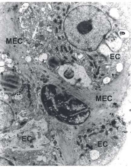
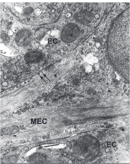
MEC exhibit expression of proteins typical for the contractile apparatus of smooth muscles cells, a-actin of smooth muscles (SMA), myosin heavy chains, a -ac-tinin, vinculin, and calponin. MEC cytoplasm is filled with bundles of actin microfilaments and myosin fila-ments responsible for cell contraction. Sub-membrane dense plaques and cytoplasmic dense bodies organize the spatial arrangement of the contractile apparatus. In MEC processes, bundles of actin microfilaments are concentrated on one surface of the cell bordering with basement lamina (Figs. 2, 3).
Unlike in smooth muscle cells, intermediate fila-ments consist mostly of cytokeratins (CK5, CK14, and CK17) which create a net around the nucleus, ex-tend towards the cell surface, and finally, as parallel bundles enter the processes, reinforcing their struc-ture. In particular, cytokeratins 5 and 14 are involved in the maintenance of MEC cytoarchitecture, as well as the formation of desmosomes and hemidesmo-somes [36].
Plasma membrane forms numerous invagina-tions (caveolae) and subsurface vesicles which are characteristic for both myoepithelial and smooth muscle cells (Fig. 3). The function of caveolae is debatable; they are supposed to act as a kind of storage for extracellular calcium ions, participate in the process of endocytosis, or mediate the transport of molecules between luminal cells and the extracellular matrix. Numerous caveolae ar-ranged in parallel rows and located between bun-dles of microfilaments at the basal cell surface bordering with basement lamina suggest their
Figure 1. Three-dimensional arrangement of myoepithelial cell morphology in whole mammary gland tissue from a lactating mouse, visualized by the fluorescent stain NBD-phallicidin, which binds specifically to actin filaments. The cells display long, thin processes that radiate from the cell body (¥600).
possible participation in MEC cytoskeleton arran-gement [45].
MEC oval nuclei are filled with dispersed chro-matin and clearly separated nucleoli. In perinuclear cytoplasm there are clusters of cellular organelles, cisterns of endoplasmic reticulum, numerous ribo-somes, well-developed Golgi apparatus, multiple vacuoles, and vesicles. Mitochondria are present in both the perinuclear part of the cell and in process-es (Fig. 3). Thprocess-ese MEC featurprocess-es point toward their activity in the synthesis of structural proteins of con-tractile apparatus, components of basement lami-na, and numerous regulatory and suppressor pro-teins (Table 1).
MYOEPITHELIAL CELLS
DURING LACTATION
The basic physiological function of MECs during lactation is milk ejection. Oxytocin triggers the con-tractions of MECs located around the milk-storing alveoli and excretory ducts, which leads to self-in-duced milk expulsion.
Although the mechanisms of MECs and smooth muscle cell contraction are very similar, their induc-Figure 3. A higher magnification of mammary epithelium. Myo-epithelial cell (MEC) is shown to be packed with microfilaments. In perinuclear cytoplasm there are clusters of cellular organelles, many ribosomes and multiple vacuoles. Plasma membrane forms numerous invaginations — caveolae and subsurface vesicles (arrows); EC — epithelial cells (¥14000).
tion is different. MEC contraction is stimulated by oxytocin binding and activation of signalling path-way mediated by Gaq-11 protein and phospholipase C. The breakdown of phosphatidylinositol bisphos-phate results in calcium concentration increase, phosphorylation of myosin, and finally the contrac-tion of the cell [50, 52]. Unlike in myocytes, the main source of calcium ions in MECs is the extracellular influx. In addition, intracellular compartments may be engaged in the maintenance of the constant cal-cium level during the contraction. In contrast to uter-ine smooth muscles, oxytocin binding by MECs is not accompanied by mitogen-activated protein kinase (MAPK) activation nor by prostaglandin release [55].
As demonstrated in some experiments, oxytocin availability is not the only prerequisite for MEC con-traction, and the induction itself requires other, as yet undefined factors, which might work in an auto/ /paracrine way. Oxytocin binding by MECs is accom-panied by an increase in cytoplasmic cAMP concen-tration; however, there is no evidence suggesting the direct dependence between elevated cAMP and myosin phosphorylation levels. These effects seem to be mediated by different signalling pathways in-cluding calcium increase, cAMP formation, and MAPK activation [55].
Morphological observations of MEC contrac-tion suggest that physiological concentracontrac-tions of oxytocin do not stimulate the contraction of all MECs in lactating mammary glands but only the cells surrounding the alveoli filled with milk. Milk in particular secretory alveoli is produced in an asynchronous way. If availability of oxytocin was the only contraction inducer of MECs, then all of them should contract independently of milk level, which seems to be irrational from a physiological point of view [43].
It is assumed that the modulator of MEC con-traction, working in an auto/paracrine way, is par-athormone related peptide (PTHrP), like in vascular smooth muscle cells. PTHrP is synthesized in lumi-nal cells and MECs of the lactating mammary gland, and its receptors are present only in MECs. In vascu-lar smooth muscle cells, the PTH/PTHrP 1 receptor, like other vasodilators, activates the phosphatidyli-nositol-Ca2+
Table 1. Proteins and biomolecules specific for myoepithelial cells (MEC)
Molecule Function Diagnostic or clinical significance References
Structural proteins
Smooth muscle actin (SMA) Involved in MEC contraction. SMA expression is observed in 95% [10] Identical as in smooth muscle of MECs of the normal mammary gland,
cells, myofibroblasts and and non-invasive cancers. No expression pericytes is detected in invasive cancers.
Diagnosis of invasiveness should be confirmed with a simultaneous expression of SMA and collagen IV
Smooth muscle myosin The main component of MEC Similar to SMA, a useful marker in [14] (heavy chains) — SMMHC contractile apparatus differentiation between invasive and
non-invasive breast cancers
Calponin Binds tropomyosin and F-actin. A useful marker differentiating MECs [14] Participates in MEC contraction from spindle cells of the stroma, and
invasive from non-invasive cancers
H caldesmon (HCD) Cytoskeletal protein binding to actin Strong expression is specific for [42] myoepithelial and smooth muscle
cells of small blood vessels
P-cadherin Is a Ca2+-dependent cell adhesive Expressed in MEC of the normal mammary [24]
molecule playing a key role in the gland, and occasionally in hyperplastic maintenance of mammary gland tissues, and some non-invasive cancers epithelium structure
Cytokeratins 5, 7, 14, 17 Specific components of MEC Enable identification of MECs in the [41, 78] intermediate filaments normal mammary gland and breast
cancers, and differentiation of MEC precursors during mammary gland morphogenesis.
Desmoglein (Dsg3) and Dsg3 and Dsc3 desmosomal Responsible for the positioning of [57] desmocollin (Dsc3) cadherins are specific for MEC MECs and maintenance of bilayer
desmosomes and hemidesmosomes structure of the mammary gland epithelium
Non-structural molecules
Maspin Mammary gland specific serpin Tumour suppressor protein, the [54] (serine proteases inhibitor) present expression of which decreases
exclusively in normal mammary gland with the level of malignancy. MEC, and breast cancer epithelial cells Maspin inhibits tumour growth
and invasiveness inducing apoptosis and inhibiting cell mobility and angiogenesis
p63, p73 Nuclear proteins showing close Proteins responsible for the maintenance [5, 82] homology to p53, p63, and p73 of progenitor cell populations in
expression in mammary gland mammary gland epithelium. Participate is comprised to MEC nuclei in mammary gland morphogenesis
and the maintenance of the normal structure of the gland
WT-1 (Wilms tumour 1) A transcription factor involved in WT-1 expression is constantly observed [19, 59] gene expression, similarly as p53 in MECs, whereas in breast cancers it is
negatively correlated with tumour progression
S 100 Protein belonging to the big family S 100 protein is constitutively expressed [18, 27] of proteins containing at least one in MECs and is often present in mammary
Ca2+ binding motif gland luminal cells. In breast cancers,
expression of S 100 protein is often elevated and associated with tumour progression and poor prognosis
Table 1 (continued). Proteins and biomolecules specific for myoepithelial cells
Molecule Function Diagnostic or clinical significance References
CD10 (CALLA — common Metalloendopeptidase present on The enzyme exhibits stable expression [44] acute lymphoblastic the surface of the cells responsible in normal mammary gland MECs. During
leukaemia antigen) for the inactivation of many biologically breast cancer growth and progression the active peptides. Present on the lateral number of CD10 positive MECs undergoes surface of MECs reduction, and the intensity of
immuno-cytochemical reaction decreases
CD44 The molecule secreted by MECs, Marker applied in breast cancer prognosis [2, 37] inhibits cancer cells adhesion and
migration
CD 109 Participates in TGF-b signalling Present in mammary gland MECs, but not [26] pathway inhibition expressed in secretory and ductal epithelial
cells. MEC marker applied in invasive cancer breast diagnosis
14-3-3 sigma The product of cancer suppressor gene Expressed mainly in MECs of benign and [63] transactivated by p53 in response to pre-invasive breast cancers. Applied in
DNA damage breast cancer prognosis
NRP-1 (neuropilin) A specific receptor for vascular MECs in hyperplastic and neoplastic [69] endothelial growth factor in MECs tissues of the mammary gland exhibit higher
NRP-1 expression than normal tissue
PTHrP/PTHrPR (parathyroid PTHrP is produced both by myoepithelial PTHrP inhibits the growth and branching of [16, 81] hormone-related protein/ and luminal cells, but in mammary excretory ducts during development. Exhibits
/parathyroid hormone-related glands only MECs exhibit its receptor proapoptotic and antiproliferative properties protein receptor) in hyperplastic tissues of the mammary gland
PTHrP on MEC cells leads to their relaxation by stop-ping the oxytocin triggered influx of calcium ions.
Another molecule that may be engaged in the regulation of MEC contraction is nitric oxide (NO). In mammary gland MEC, both in the resting and lactation periods, the activity of nitric oxide synthase (NOS-1), as well as the presence of NO receptor-soluble guanylyl cyclase (sGC), were observed. sGC, in an auto/paracrine way, catalyzes the conversion of GTP to cGMP [79]. In smooth muscle cells, the increase in cGMP concentration inhibits the influx of calcium ions and thereby the activity of Ca2+
--dependent myosin light chain kinase, which leads to the relaxation of cells [46]. Because an identical kinase is present in MEC, a similar mechanism of auto/paracrine regulation of contraction may oper-ate here.
An additional mechanism of the regulation of MEC contraction is variable and diversified localiza-tion of oxytocin receptors (OTR). The availability of OTR may be regulated via their localization in spe-cialized membrane microdomains such as lipid rafts and caveolae, which may be connected with the activation of different transduction pathways lead-ing to a diversified response of MECs to oxytocin signal [56].
THE ORIGIN AND DIFFERENTIATION
OF MYOEPITHELIAL CELLS
The mammary gland differentiates from ectoder-mal epithelium, which, during the embryonic period, forms the milk line. The epithelium of milk line invag-inates into mesenchyma forming primary and later secondary buds. These buds elongate and grow later-al branches forming a complex system of ducts end-ing with expanded terminal end buds (TEBs). The TEBs undergo intensive growth and differentiation. A cap cell layer surrounds the body cells constituting the cel-lular material from which basal MEC and luminal cells differentiate and are therefore thought to be multi-potent stem cells (Fig. 4). Some of them remain undif-ferentiated stem cells which settle in particular niches of excretory ducts and secretory units [80].
Intensive development of the mammary gland takes place during puberty, pregnancy (when the process is stimulated by systemic hormones, oestro-gens, progesterone, placental lactooestro-gens, and pro-lactin), and after childbirth, when the combination of systemic hormones, local growth factors, and milk ejection evokes further development of the gland structure [47].
Figure 4. The structure of terminal end buds (TEB) (A) and ductal and alveolar cells during pregnancy (B). A cap cell layer surrounds the body cells. The cap cells can take on either a myoepithelial lineage or a luminal epithelial lineage and therefore are thought to be multipotent stem cells. Differentiated myoepithelial and luminal epithelial cells line the neck of the TEB and the subtending duct. During midpregnancy the ducts are sur-rounded by a basal layer of overlapping myoepithelial cells, whereas the alveoli cells are sursur-rounded by a basket-like layer of myoepithelial cells.
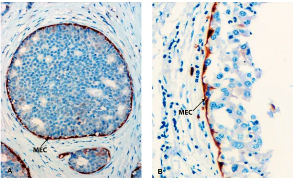
Figure 5. Myoepithelial cells (MEC) surrounding breast excretory ducts highlighted by maspin immunoreactivity. MECs are arranged in a continuous layer with their long axis being laid parallel to the ducts. Cross section (A), longitudinal section (B) (A and B ¥ 600).
every stage of development. The analysis of mam-mary gland ultrastructure demonstrated the pres-ence of three types of epithelial cells: luminal cells, myoepithelial cells, and basal pale cells, which, as the authors suggest, may constitute the population of stem cells. In human mammary glands, a gradual transition of pale cells into fully differentiated MECs was demonstrated, but there are no observations of the transition of these cells into differentiated luminal cells [64, 75].
Isolated suprabasal cells in three-dimensional (3D) cultures in laminin-rich media form structures similar to functional units of the mammary gland (commonly referred to as terminal duct lobular units [TDLUs]) with an internal layer of CK19 positive lu-minal cells and an external layer of CK14 and a-SMA positive cells similar to MECs. There is strong evi-dence that luminal epithelial and myoepithelial cells are derived from a suprabasal cell type [22].
The presence of cells with simultaneous CK19 and CK14 expression was observed in mammary gland epithelium. These cells located within duct bifurcations are relatively weakly differentiated and dye-resistant. Planted into pure fat tissue they form follicular and tubular structures, which proves that they have full potential of differentiation [3]. The analysis of human mammary gland specimens ex-hibited a population of bipotential CK5 positive cells differentiating either into luminal or myoepi-thelial cells [9].
Recently, several populations of stem cells differ-ing in determination level have been identified in human mammary gland. A helpful combination of markers, applied for the sorting of mammary gland stem cells, is made up of EpCam (epithelial adhesion molecules, also known as epithelial specific antigen — ESA), CD49f, and MUC1 (luminal cells-specific gly-coprotein). EpCam displays a strong expression in luminal cells and poor expression in basal cells, and the expression pattern for CD49f is reversed. Bipo-tential basal stem cells, capable of differentiation into luminal or myoepithelial cells, exhibit poor expres-sion of EpCam, strong expresexpres-sion of CD49f, and no expression of MUC1, whereas luminal progenitor cells display strong expression of EpCam and the presence of CD49f-
and MUC1+
[17, 72].
In a suspension of cells freshly isolated from mam-mary gland, 1% display the ability to proliferate and form three types of colonies in the medium. Most of them (70%) create tight clusters of cells displaying ex-pression of CK18, CK19, and MUC1 antigens specific for luminal cells, but no expression of CK14 typical for
basal cells. A further analysis showed the cells to present luminal cell phenotype, high expression of EpCam, and the presence of CD49f and MUC1 antigens. The second population, in terms of quantity (25%), is formed by bipotential progenitor cells with poor expression of EpCam, strong expression of CD49f, and no expression of MUC. These cells created colonies characterized by the presence of cells with CK14–, K18+, K19+, and MUC1 in their central area, and CK14+ basal cells in the peripheral area. Both cell types, as demonstrated using clonal analysis, descended from common progen-itor cells. The third type of cells included stem cells of MEC forming colonies consisting only of basal cells of CK14+, CK18–, CK19–, and MUC1-phenotypes. Precur-sors of MEC, as further analysis showed, originate di-rectly from bipotential stem cells [71].
MYOEPITHELIAL CELLS IN
MORPHOGENESIS AND
ORGANIZATION OF MAMMARY
GLAND STRUCTURE
The location of MEC between the luminal cells and the extracellular matrix suggests their active role in the exchange of information between the extra-cellular matrix and the luminal epithelium of the gland, and therefore their participation in the regu-lation of growth, morphogenesis, and the mainte-nance of the proper two-layered structure of the mammary gland (Fig. 5).
MECs show a strong expression of receptors of integrins and growth factors such as EGF and FGF-2, which may suggest their regulatory function. Activin, belonging to the TGF-b superfamily, is expressed only in MECs of mammary gland and plays a role in the regulation of ducts growth [38]. Maspin, being ex-clusively expressed in MECs, has been shown to play a significant role in the morphogenesis, development, and functioning of mammary gland. Maspin gene overexpression coupled with WAP promoter (respon-sible for the regulation of milk protein expression, and active since half way through gestation until the end of lactation) led to the inhibition of gland growth and disturbances in the formation of mature gland structures. Transgenic mice with maspin overexpres-sion had a decreased number of alveolar structures unable to synthesize milk components [83].
unable to synthesize oxytocin, demonstrated their inability to form the proper lobulo-alveolar structure of mammary gland in the period of puberty, and the impairment of the further development of the gland during the postpartum period [77]. Oxytocin injec-tion into non-lactating mice induces proliferainjec-tion and differentiation of MECs. This phenomenon was ob-served only in the mice treated previously with proges-terone and oestrogens. These findings suggest that the proliferational effect of oxytocin depends on the level of mammary gland development and works at the hormonal stage corresponding to the pregnancy period [58].
The expression of both oxytocin mRNA and pep-tide observed in MEC primary cultures [12] suggests that these cells may act as a local source of oxytocin synthesis, and therefore may be involved in auto/ /paracrine regulation of proliferation and differenti-ation of mammary gland cells at all stages of its development.
The integrity of mammary gland epithelium is maintained by a complex system of cell/cell and cell/ /extracellular matrix interactions mediated by cad-herins and integrins.
In mammary gland P-cadherins are located only in MECs and the cells of terminal end buds, while E-cadherins are exclusively in luminal cells [15]. Virgin mice with P-cadherin deficiency display accelerated mammary gland growth, and luminal cells initiate the synthesis of casein, likewise during the period of early gestation. It finally leads to hyperplasia and dysplasia of the gland epithelium. These observa-tions indicate that P-cadherins mediate interacobserva-tions between myoepithelial and luminal cells and partici-pate in the control over growth and differentiation of mammary gland epithelium [53].
In epithelial cells, b-catenin is a component of cadhercontaining intercellular junctions. The in-teractions with MECs trigger the Wnt/b-catenin sig-nalling pathway in luminal epithelium, the main mediators of which are Tcf transcription factors. The presence of both Tcf4 and Tcf1 was observed in mammary gland epithelium; however, only in MECs was the nuclear Tcf1 localization demonstrated [57]. A significant role in the formation of normal mammary gland structure is played by desmosomal cadherins: desmoglein (Dsg) and desmocollin (Dsc). They display different expression in luminal and myoepithelial cells. Dsg2 and Dsc2 are present in both layers of the cells, while Dsg3 and Dsc3 occur only in MECs. Co-culture of myoepithelial and lumi-nal cells form bilayered structures resembling their
in vivo arrangement. The introduction of specific
peptides inhibiting myoepithelial Dsc3 and Dsg3 to the co-culture causes distortions in the formation of the proper bilayer epithelium structure and dis-turbs the polarization of luminal cells [21]. These observations prove that MECs play a significant role in the arrangement of mammary gland structure and polarization of luminal cells via direct interactions between cells mediated by desmosomes [25].
Isolated mammary luminal cells in 3D cultures in collagen I gel form lumenless alveolar structures. Double labelling for MUC1/ESA and then for MUC1/ /occludin demonstrated that cells form clusters with reversed polarity. Introducing MECs to the culture caused the correct polarization of epithelial cells and the forming alveolar structures to have clearly dis-tinguished lumen.
In addition, the presence of laminin-1 (but not other laminin isoforms; 5, 10, or 11) in the culture gel al-lowed the formation of correct epithelium polariza-tion. MECs are the only cells synthesizing laminin-1 (among other main components of the basement lam-ina), and their introduction to the culture makes up for laminin-1 deficiency, which leads to the appropria-te formation of alveolar epithelial structures [22].
Although laminin-1 is the key regulator of cor-rect polarization of epithelium and the morphogen-esis of mammary gland, other molecules produced by MECs may also take part in this process.
Except for the occurrence of all integrins present in luminal cells, mammary gland MECs show an ex-clusive strong expression of a1b1 and a5b5 integrins. Inactivation of b1 integrin in virgin mice leads to disturbances in the formation of ducts and their branching, resulting in an improper arrangement of the lobulo-alveolar structure of the mammary gland during the gestation [20].
MYOEPITHELIAL CELLS AS
SUPPRESSORS OF BREAST CANCER
MECs present in normal mammary glands and benign forms of tumours are considered natural suppressors of breast cancer. They inhibit the growth of tumours and neoangiogenesis, induce apopto-sis, and limit the mobility of cancer cells. MEC locali-zation between the basement lamina and the layer of luminal cells suggests their paracrine action on adjacent luminal cells as well as on the connective tissue and endothelial cells [48].
A commonly accepted hypothesis states that with the increase in cancer malignancy the ratio of lumi-nal to myoepithelial cells rises, while the invasive cers are almost devoid of the latter. In invasive can-cers, MEC markers — CK14, CK17, and vimentin — are present in less than 20% of all tumours [76].
Although the presence of MECs is associated with the maintenance of ductal cancer in situ (DCIS) in the benign form even for a long period, some of these cancers undergo malignant transformation. Compar-ative analysis of gene expression has shown signifi-cant differences between MECs accompanying DCIS and MECs of normal mammary gland ducts. DCIS-as-sociated MECs exhibit lowered expression of oxytocin receptors, laminin-1, and thrombospondin, but high-er expression of chemokines responsible for cellular proliferation, migration, and invasiveness of cells such as SDF1/CXCL12 and CXCL14. Moreover, an increase in enzymes engaged in extracellular matrix degrada-tion, such as numerous metalloproteinases, as well as various epigenetic changes connected with higher levels of DNA methylation, were observed in these cells [1]. These properties of DCIS-associated MECs may sug-gest their paracrine effects on normal glandular epi-thelium resulting in malignant transformation.
Myoepithelial cells associated with DCIS lose con-tact with luminal cells, which leads to disturbances in luminal cell polarization and inhibition of the base-ment lamina component synthesis by MECs. In breast cancers with laminin-1 synthesis deficiency in MECs, even the presence of these cells was not correlated with better prognosis [39]. On the other hand, breast tumours with functioning MECs that synthesize base-ment lamina components do not give distant me-tastases and are correlated with better prognosis [32]. Therefore, it can be stated that the presence of MECs in breast tumours inhibits the alterations associated with malignant transformation [7].
A break of integrity of the MEC layer affects the genetic and functional changes in the luminal cells located above it. A decrease in expression of
oestro-genic receptors, higher frequency of heterozygosity occurrence, higher cellular proliferation index, and increase in expression of genes related to the active mobility of cells and angiogenesis have been ob-served in these cells.
Normal MEC and myoepithelial cell lines derived from benign breast tumours produce relatively high numbers of protease inhibitors and anti-angiogenic factors [48]. HMS1 mammary myoepithelial cells (the cell line derived from benign tumours) are charac-terized by a high ratio of proteinase inhibitors to proteinases, unlike mammary cancer cell lines where the number of proteinases significantly prevail their inhibitors. The analysis of the profile of inhibitors secreted by MECs revealed the presence of tissue proteases inhibitor (TIMP-1), plasminogen activator inhibitor, and trypsin inhibitors such as a 1-anti-trypsin [70].
Numerous studies point towards a significant role of fibroblasts in the progression and invasiveness of cancers. The majority of metalloproteinases (MMP) participating in cancer progression are fibroblasts produced in response to signals coming from can-cerous cells. MECs suppress the pro-invasive dialogue between cancerous and fibroblast cells by the inhibi-tion of MMP expression, the mechanism of which has not been elucidated. There are some suggestions pointing towards growth factors produced by MECs (TGF-b, FGF-2, activin, or components of Wnt signal-ling pathway) as the mediators reducing the expres-sion of pro-invasive MMP in fibroblasts [30].
A break of integrity of the MEC layer and its at-rophy in cancer tissue may be caused by an autoim-munological reaction and the impact of leukocytes and macrophages on the basement lamina, as well as their direct action on MECs. An increase in leuko-cytes and macrophage numbers is an evident fea-ture of in-situ cancer transition into infiltrated inva-sive forms, and it coincides with bad prognosis and higher mortality rates. Leukocytes are able to force the barrier of the basement lamina and the MEC layer thanks to the secretion of numerous proteas-es, effectively degrading their components [40].
capabili-ty of cancer cell migration. Immunoprecipitation of maspin in culture medium with specific anti-maspin antibodies abolishes the suppression effect of the medium on the invasiveness of cancerous cells [6].
In maspin-treated breast cancer cells, the profile of integrin expression on the surface changes in direc-tion promoting binding with collagen and fibronectin (increased expression of a3 and a5 integrins accom-panied by the decrease of others) [4]. Alterations caused by maspin also include proteins which are en-gaged in signalling pathways related to cell mobility (reduced activity of Rac1 kinase and increased activity of ERK1/2 and PI3K kinases) [49]. Another form of maspin impact on cancer cell decreased mobility is the inhibition of proteases engaged in plasminogen acti-vation (responsible for the digestion of the extracellu-lar matrix, which promotes the migration). The mecha-nism of maspin action consists of binding urokinase plasminogen activator (uPA), its precursor (pro-uPA), and its receptor (uPAR) [62].
Moreover, MECs are able to cleave the surface form of the CD44 molecule generating its soluble form, which inhibits the migration ability of adja-cent cancer cells [2].
MECs in normal mammary glands and in DCIS tumours separate the luminal epithelium from the blood vessels of the gland stroma, forming a barrier that is impenetrable to the vessels (Fig. 6). Myoepi-thelial cells (HMS) derived from benign mammary gland tumours (myoepithelioma) are characterised by high expression of active angiogenesis inhibitors such as TIMP-1, thrombospondin, or maspin, and low level of angiogenic factors. The comparison of xe-nografts of breast cancer tumours derived from myo-epithelial or luminal cells has shown an intensive synthesis of extracellular matrix components and very little neoangiogenesis in the first case, but almost ten times higher neoangiogenesis in the second case. Both HMS-1 myoepithelial cells and HMS-1 concen-trated culture medium significantly inhibit migration and proliferation of endothelial cells. This effect is enhanced by phorbol esters but stopped by cyclo-heximide and dexamethasone [48]. Immunoprecipi-tative analysis of the culture medium points to maspin and thrombospondin as the key molecules responsi-ble for angiogenesis inhibition by MECs [84].
Myoepithelial neoplasm-derived cells produced in culture significantly lower quantities of vascular endothelial growth factor and nitric oxide synthase activity in response to hypoxia, when compared with the cell line derived from luminal cancer [48]. This observation supports the hypothesis assuming that
MECs, even those subjected to malignant transfor-mation, exhibit natural anti-angiogenic capabilities. An important function of MECs in cancer suppres-sion is their involvement in steroid hormone metab-olism. Comparative studies on the influence of 17-b
estradiol on steroid sulphatase (STS) in normal hu-man MECs and breast cancer cells (MCF-7) have shown STS activity to be more than a hundred times higher in MECs [73]. Exposure to 17-b estradiol led to a 70% reduction in STS activity in MCF-7 cells, and a 9% activity increase in MECs, which suggests that MECs may participate in the conversion of precur-sors into active steroid hormones. The exposition of MECs to tamoxifen enhances the synthesis of maspin and inhibits maspin-dependent invasiveness of can-cer cells. The introduction of 17-b estradiol inhibits the effect of tamoxifen on MECs, which suggests that the mechanism of tamoxifen action is dependent on oestrogen receptors. Since MECs have ERb receptors (but not ERa), it might be concluded that tamoxifen-induced maspin secretion results from the triggering of the signalling pathway initiated by ERb receptors, and activation of transcription factors AP1 [61].
The presented data allow the notion suggesting that MECs have a genetic program preventing not only their own malignant transformation but also the transition of noninvasive tumours derived from luminal cells into malignant forms of breast cancer in an autocrine or paracrine way.
However, the changes of the genetic expression profile of MECs, which co-occur with the transfor-mation of benign cancers into their malignant forms, may modify MECs in such a way that allows them to enforce proliferation, migration, and invasiveness of cancerous cells [1].
BREAST CANCERS DERIVED FROM
MYOEPITHELIAL CELLS
The analysis of the genetic profile of breast can-cers allows their subdivision into four basic histoge-netic types: one normal breast tissue-like type, two luminal-like types, one ERBB2-overexpressing type, and one basal-like type. Each of the types is char-acterized by a diverse clinical response [67]. The basal--like type of breast cancer derived from MECs com-prises 2–18% of all invasive ductal breast cancers and can be identified with the use of markers spe-cific for MECs, such as cytokeratins (CK5, CK14, CK17), smooth muscle cells actin and myosin, and others, such as p63 or s100 protein [31].
mor-phological features. They usually are high-grade (III) tumours and often contain in their central area a non-cellular substance consisting of necrotic cells debris, collagen, and hyaline substance [74]. Besides the ex-pression of MEC markers, these cancers are also char-acterized by the lack of expression of progesterone (PR), oestrogenes (ER), and HER-2 receptors [29]. The microarray analysis of the immunophenotype and genetic expression profile shows many similarities between sporadic basal cancers and inherited cers with BRCA1 mutation [35, 67]. Basal breast can-cers, unlike luminal cancan-cers, are characterized by a high expression of gene coding for a2 and g2 laminin chains and â4 integrin subunits [51].
Further analysis of mammary gland basal can-cers has shown 82% frequency of TP53 mutation, while in luminal-type cancers it was only 13%. TP53 mutations are associated with poor prognosis and poor response to therapy [66]. Poor clinical progno-sis is also related to the high cancer grade and the lack of expression of steroid hormone receptors. In rare cases, basal cancers are connected with in-creased risk of brain metastases and higher mortal-ity rates, independently of the lymph node status and tumour size. Numerous clinical trials show that breast cancers with 5 and 17 cytokeratins expres-sion (specific for basal cells) exhibit poor prognosis and shorter survival rates.
However, some authors argue that such an in-terpretation is too simple because these might not be pure myoepithelial cancers, and the presence of MEC markers within the tumour may be caused by luminal cells exhibiting high phenotypic plasticity, or by stem cells with broad expression of both myo-epithelial and luminal cell markers [13].
The basal type of breast cancer with poor clin-ical prognosis, negative for oestrogen receptors, has markers of both luminal and myoepithelial cells, and MECs are partially differentiated, unlike luminal cells which are highly unorganised. It should be mentioned that MECs are highly resis-tant to malignant transformation, and even if they undergo the transformation the cancers derived from MECs are benign, except for malignant myo-epithelioma which is the least frequent form of breast cancer [34].
SUMMARY AND CONCLUSIONS
Although myoepithelial cells of mammary gland comprise the second population in the gland (with respect to cell number), they have not been the sub-ject of many scientific studies until recently when theirimportant role in the regulation of proliferation, dif-ferentiation, and activity of luminal cells, and mor-phogenesis of mammary gland have been observed. The layer of MECs and basal lamina (most of the components of which are produced by these cells) form a selective barrier regulating bidirectional ex-change of information between mammary epitheli-um and stroma cells (fibroblasts, endothelial cells). The regulation of luminal cell function and mam-mary gland development may result from the direct effect of the cells via integrins and cadherins of cell--cell junctions but may also be caused by the para-crine influence on neighbouring cells through nu-merous regulatory proteins.
MECs of normal mammary glands or benign breast tumours are natural cancer suppressors, re-sponsible for the maintenance of the proper struc-ture of the gland.
In numerous studies, the inhibitory effect of MECs on cancer growth, invasiveness, and angiogenesis has been demonstrated. The elucidation of the mech-anisms of MEC differentiation and their involvement in mammary gland morphogenesis and malignant transformation would allow better insight into breast cancer biology resulting in the improvement in cancer diagnosis and therapy.
REFERENCES
1. Allinen M, Beroukhim R, Cai L, Brennan C, Lahti-Domenici J, Huang H, Porter D, Hu M, Chin L, Richardson A, Schnitt S, Sellers WR, Polyak K (2004) Molecular characterization of the tumor microenvironment in breast cancer. Can-cer Cell, 6: 17–32.
2. Alpaugh ML, Lee MC, Nguyen M, Deato M, Dishakjian L, Barsky SH (2000) Myoepithelial-specific CD44 shedding contributes to the anti-invasive and antiangiogenic phenotype of myoepithelial cells. Exp Cell Res, 261: 150–158.
3. Alvi AJ, Clayton H, Joshi C, Enver T, Ashworth A, Vivanco MM, Dale TC, Smalley MJ (2003) Functional and molecular characterisation of mammary side popu-lation cells. Breast Cancer Res, 5: R1–R8.
4. Bailey CM, Khalkhali-Ellis Z, Seftor EA, Hendrix MJ (2006) Biological functions of maspin. J Cell Physiol, 209: 617–624.
5. Barbareschi M, Pecciarini L, Cangi MG, Macri E, Rizzo A, Viale G, Doglioni C (2001) p63, a p53 homologue, is a selective nuclear marker of myoepithelial cells of the human breast. Am J Surg Pathol, 25: 1054–1060. 6. Barsky SH, Karlin NJ (2006) Mechanisms of disease:
breast tumor pathogenesis and the role of the myo-epithelial cell. Nat Clin Pract Oncol, 3: 138–151. 7. Barsky SH, Karlin NJ (2005) Myoepithelial cells: autocrine
8. Benson GK, Folley SJ (1957) The effect of oxytocin on mammary gland involution in the rat. J Endocrinol, 16: 189–201.
9. Bocker W, Burger H, Buchwalow IB, Decker T (2005) Ck5-positive cells are precursor cells of glandular and myoepithelial cell lineages in the human breast epi-thelium. A new cell concept as a basis for a better un-derstanding of proliferative breast disease? Verh Dtsch Ges Pathol, 89: 45–47.
10. Bose S, Derosa CM, Ozzello L (1999) Immunostaining of Type IV collagen and smooth muscle actin as an aid in the diagnosis of breast lesions. Breast J, 5: 194–201. 11. Breton C, Di Scala-Guenot D, Zingg HH (2001)
Oxyto-cin receptor gene expression in rat mammary gland: structural characterization and regulation. J Mol Endo-crinol, 27: 175–189.
12. Cassoni P, Sapino A, Marrocco T, Chini B, Bussolati G (2004) Oxytocin and oxytocin receptors in cancer cells and proliferation. J Neuroendocrinol, 16: 362–364. 13. Clarke RB (2006) Ovarian steroids and the human breast:
regulation of stem cells and cell proliferation. Maturi-tas, 54: 327–334.
14. Dabbs DJ, Gown AM (1999) Distribution of calponin and smooth muscle myosin heavy chain in fine-needle aspiration biopsies of the breast. Diagn Cytopathol, 20: 203–207.
15. Daniel CW, Strickland P, Friedmann Y (1995) Expres-sion and functional role of E-and P-cadherins in mouse mammary ductal morphogenesis and growth. Dev Biol, 169: 511–519.
16. Dunbar ME, Dann P, Brown CW, Van Houton J, Dreyer B, Philbrick WP, Wysolmerski JJ (2001) Temporally regu-lated overexpression of parathyroid hormone-reregu-lated protein in the mammary gland reveals distinct fetal and pubertal phenotypes. J Endocrinol, 171: 403–416. 17. Eirew P, Stingl J, Raouf A, Turashvili G, Aparicio S,
Emerman JT, Eaves CJ (2008) A method for quantifying normal human mammary epithelial stem cells with in vivo regenerative ability. Nat Med, 14: 1384–1389. 18. Emberley ED, Murphy LC, Watson PH (2004) S100A7
and the progression of breast cancer. Breast Cancer Res, 6: 153–159.
19. Fabre A, McCann AH, O’Shea D, Broderick D, Keating G, Tobin B, Gorey T, Dervan PA (1999) Loss of heterozygo-sity of the Wilms’ tumor suppressor gene (WT1) in in situ and invasive breast carcinoma. Hum Pathol, 30: 661–665. 20. Faraldo MM, Taddei-De La Hosseraye I, Teuliere J, Deugnier MA, Moumen M, Thiery JP, Glukhova MA (2006) Mammary gland development: role of basal myoepithelial cells. J Soc Biol, 200: 193–198. 21. Garrod DR, Merritt AJ, Nie Z (2002) Desmosomal
cad-herins. Curr Opin Cell Biol, 14: 537–545
22. Gudjonsson T, Adriance MC, Sternlicht MD, Petersen OW, Bissell MJ (2005) Myoepithelial cells: their origin and function in breast morphogenesis and neoplasia. J Mammary Gland Biol Neoplasia, 10: 261–272. 23. Gudjonsson T, Ronnov-Jessen L, Villadsen R, Rank F,
Bissell MJ, Petersen OW (2002) Normal and tumor-de-rived myoepithelial cells differ in their ability to interact with luminal breast epithelial cells for polarity and base-ment membrane deposition. J Cell Sci, 115: 39–50.
24. Han AC, Soler AP, Knudsen KA, Salazar H (1999) Dis-tinct cadherin profiles in special variant carcinomas and other tumors of the breast. Hum Pathol, 30: 1035–1039.
25. Hardman MJ, Liu K, Avilion AA, Merritt A, Brennan K, Garrod DR, Byrne C (2005) Desmosomal cadherin mis-expression alters beta-catenin stability and epidermal differentiation. Mol Cell Biol, 25: 969–978.
26. Hasegawa M, Hagiwara S, Sato T, Jijiwa M, Murakumo Y, Maeda M, Moritani S, Ichihara S, Takahashi M (2007) CD109, a new marker for myoepithelial cells of mam-mary, salivary, and lacrimal glands and prostate basal cells. Pathol Int, 57: 245–250.
27. Jenkinson SR, Barraclough R, West CR, Rudland PS (2004) S100A4 regulates cell motility and invasion in an in vitro model for breast cancer metastasis. Br J Can-cer, 90: 253–262.
28. Jolicoeur F, Seemayer TA, Gabbiani G, Robidoux A, Gaboury L, Oligny LL, Schurch W (2002) Multifocal, nascent, and invasive myoepithelial carcinoma (malig-nant myoepithelioma) of the breast: an immunohis-tochemical and ultrastructural study. Int J Surg Pathol, 10: 281–291.
29. Jones C, Nonni AV, Fulford L, Merrett S, Chaggar R, Eusebi V, Lakhani SR (2001) CGH analysis of ductal car-cinoma of the breast with basaloid/myoepithelial cell differentiation. Br J Cancer, 85: 422–427.
30. Jones JL, Shaw JA, Pringle JH, Walker RA (2003) Prima-ry breast myoepithelial cells exert an invasion-suppres-sor effect on breast cancer cells via paracrine down-regulation of MMP expression in fibroblasts and tu-mour cells. J Pathol, 201: 562–572.
31. Jones S, Clark G, Koleszar S, Ethington G, Mennel R, Paulson S, Brooks B, Kerr R, Denham C, Savin M, White C, Blum J, Kirby R, Stone M, Pippen J, Kitchens L, George T, Cooper B, Peters G, Knox S, Grant M, Cheek H, Jones R, Kuhn J, Lieberman Z, Savino D, Rietz C (2001) Low pro-liferative rate of invasive node-negative breast cancer predicts for a favorable outcome: a prospective evalu-ation of 669 patients. Clin Breast Cancer, 1: 310–314 (discussion 315–317).
32. Kasami M, Olson SJ, Simpson JF, Page DL (1998) Main-tenance of polarity and a dual cell population in ade-noid cystic carcinoma of the breast: an immunohis-tochemical study. Histopathology, 32: 232–238. 33. Koukoulis GK, Howeedy AA, Korhonen M, Virtanen I,
Gould VE (1993) Distribution of tenascin, cellular fibronec-tins and integrins in the normal, hyperplastic and neo-plastic breast. J Submicrosc Cytol Pathol, 25: 285–295. 34. Lakhani SR, O’Hare MJ (2001) The mammary
myoepithe-lial cell: Cinderella or ugly sister? Breast Cancer Res, 3: 1–4. 35. Lakhani SR, Van De Vijver MJ, Jacquemier J, Anderson TJ, Osin PP, McGuffog L, Easton DF (2002) The pathology of familial breast cancer: predictive value of immunohis-tochemical markers estrogen receptor, progesterone re-ceptor, HER-2, and p53 in patients with mutations in BRCA1 and BRCA2. J Clin Oncol, 20: 2310–2318. 36. Lazard D, Sastre X, Frid MG, Glukhova MA, Thiery JP,
myofi-broblasts of normal and malignant human breast tis-sue. Proc Natl Acad Sci USA, 90: 999–1003.
37. Lee MC, Alpaugh ML, Nguyen M, Deato M, Dishakjian L, Barsky SH (2000) Myoepithelial-specific CD44 shedding is mediated by a putative chymotrypsin-like sheddase. Biochem Biophys Res Commun, 279: 116–123. 38. Liu QY, Niranjan B, Gomes P, Gomm JJ, Davies D,
Coombes RC, Buluwela L (1996) Inhibitory effects of activin on the growth and morpholgenesis of primary and transformed mammary epithelial cells. Cancer Res, 56: 1155–1163.
39. Malzahn K, Mitze M, Thoenes M, Moll R (1998) Biolo-gical and prognostic significance of stratified epithe-lial cytokeratins in infiltrating ductal breast carcinomas. Virchows Arch, 433: 119–129.
40. Man YG, Zhang Y, Shen T, Zeng X, Tauler J, Mulshine JL, Strauss BL (2005) cDNA expression profiling reveals elevated gene expression in cell clusters overlying fo-cally disrupted myoepithelial cell layers: implications for breast tumor invasion. Breast Cancer Res Treat, 89: 199–208.
41. McGowan KM, Coulombe PA (1998) Onset of keratin 17 expression coincides with the definition of major epi-thelial lineages during skin development. J Cell Biol, 143: 469–486.
42. Miettinen MM, Sarlomo-Rikala M, Kovatich AJ, Lasota J (1999) Calponin and h-caldesmon in soft tissue tumors: consistent h-caldesmon immunoreactivity in gas-trointestinal stromal tumors indicates traits of smooth muscle differentiation. Mod Pathol, 12: 756–762. 43. Moore DM, Vogl AW, Baimbridge K, Emerman JT (1987)
Effect of calcium on oxytocin-induced contraction of mammary gland myoepithelium as visualized by NBD--phallacidin. J Cell Sci, 88 (Part 5): 563–569.
44. Moritani S, Kushima R, Sugihara H, Bamba M, Kobayashi TK, Hattori T (2002) Availability of CD10 immunohistochemistry as a marker of breast myoepithe-lial cells on paraffin sections. Mod Pathol, 15: 397–405. 45. Nakano H, Furuya K, Furuya S, Yamagishi S (1997) In-volvement of P2-purinergic receptors in intracellular Ca2+ responses and the contraction of mammary myo-epithelial cells. Pflugers Arch, 435: 1–8.
46. Nakano H, Furuya K, Yamagishi S (2001) Synergistic effects of ATP on oxytocin-induced intracellular Ca2+ response in mouse mammary myoepithelial cells. Pflugers Arch, 442: 57–63.
47. Neville MC, McFadden TB, Forsyth I (2002) Hormonal regulation of mammary differentiation and milk secre-tion. J Mammary Gland Biol Neoplasia, 7: 49–66. 48. Nguyen M, Lee MC, Wang JL, Tomlinson JS, Shao ZM,
Alpaugh ML, Barsky SH (2000) The human myoepithe-lial cell displays a multifaceted anti-angiogenic pheno-type. Oncogene, 19: 3449–3459.
49. Odero-Marah VA, Khalkhali-Ellis Z, Chunthapong J, Amir S, Seftor RE, Seftor EA, Hendrix MJ (2003) Maspin re-gulates different signaling pathways for motility and adhesion in aggressive breast cancer cells. Cancer Biol Ther, 2: 398–403.
50. Olins GM, Bremel RD (1984) Oxytocin-stimulated myo-sin phosphorylation in mammary myoepithelial cells: roles of calcium ions and cyclic nucleotides. Endocri-nology, 114: 1617–1626.
51. Perou CM, Sorlie T, Eisen MB, van de Rijn M, Jeffrey SS, Rees CA, Pollack JR, Ross DT, Johnsen H, Akslen LA, Fluge O, Pergamenschikov A, Williams C, Zhu SX, Lon-ning PE, Borresen-Dale AL, Brown PO, Botstein D (2000) Molecular portraits of human breast tumours. Nature, 406: 747–752.
52. Pettibone DJ, Woyden CJ, Totaro JA (1990) Identification of functional oxytocin receptors in lactating rat mamma-ry gland in vitro. Eur J Pharmacol, 188: 235–241. 53. Radice GL, Ferreira-Cornwell MC, Robinson SD, Rayburn H,
Chodosh LA, Takeichi M, Hynes RO (1997) Precocious mammary gland development in P-cadherin-deficient mice. J Cell Biol, 139: 1025–1032.
54. Reis-Filho JS, Milanezi F, Silva P, Schmitt FC (2001) Maspin expression in myoepithelial tumors of the breast. Pathol Res Pract, 197: 817–821.
55. Reversi A, Cassoni P, Chini B (2005) Oxytocin receptor signaling in myoepithelial and cancer cells. J Mammary Gland Biol Neoplasia, 10: 221–229.
56. Reversi A, Rimoldi V, Brambillasca S, Chini B (2006) Effects of cholesterol manipulation on the signaling of the human oxytocin receptor. Am J Physiol Regul Integr Comp Physiol, 291: R861–R869.
57. Runswick SK, O’Hare MJ, Jones L, Streuli CH, Garrod DR (2001) Desmosomal adhesion regulates epithelial mor-phogenesis and cell positioning. Nat Cell Biol, 3: 823– –830.
58. Sapino A, Macri L, Tonda L, Bussolati G (1993) Oxyto-cin enhances myoepithelial cell differentiation and pro-liferation in the mouse mammary gland. Endocrinolo-gy, 133: 838–842.
59. Scharnhorst V, van der Eb AJ, Jochemsen AG (2001) WT1 proteins: functions in growth and differentiation. Gene, 273: 141–161.
60. Seitz PK, Cooper KM, Ives KL, Ishizuka J, Townsend CM, Jr., Rajaraman S, Cooper CW (1993) Parathyroid hormone-related peptide production and action in a myoepithe-lial cell line derived from normal human breast. Endo-crinology, 133: 1116–1124.
61. Shao ZM, Radziszewski WJ, Barsky SH (2000) Tamo-xifen enhances myoepithelial cell suppression of hu-man breast carcinoma progression in vitro by two dif-ferent effector mechanisms. Cancer Lett, 157: 133–144. 62. Sheng S (2006) A role of novel serpin maspin in tumor progression: the divergence revealed through efforts to converge. J Cell Physiol, 209: 631–635.
63. Simpson PT, Gale T, Reis-Filho JS, Jones C, Parry S, Steele D, Cossu A, Budroni M, Palmieri G, Lakhani SR (2004) Dis-tribution and significance of 14-3-3sigma, a novel myo-epithelial marker, in normal, benign, and malignant breast tissue. J Pathol, 202: 274–285.
64. Smith GH, Chepko G (2001) Mammary epithelial stem cells. Microsc Res Tech, 52: 190–203.
65. Sopel M, Lis A (2000) Coexpression of PTHrP and PTH/PTHrP receptor in a myoepithelial cell line derived from normal human breast. Folia Histochem Cytobiol, 38: 65–69. 66. Sorlie T, Perou CM, Tibshirani R, Aas T, Geisler S,
tumor subclasses with clinical implications. Proc Natl Acad Sci USA, 98: 10869–10874.
67. Sorlie T, Tibshirani R, Parker J, Hastie T, Marron JS, Nobel A, Deng S, Johnsen H, Pesich R, Geisler S, Demeter J, Perou CM, Lonning PE, Brown PO, Borresen--Dale AL, Botstein D (2003) Repeated observation of breast tumor subtypes in independent gene expression data sets. Proc Natl Acad Sci USA, 100: 8418–8423. 68. Srinivasan K, Strickland P, Valdes A, Shin GC, Hinck L
(2003) Netrin-1/neogenin interaction stabilizes multi-potent progenitor cap cells during mammary gland morphogenesis. Dev Cell, 4: 371–382.
69. Stephenson JM, Banerjee S, Saxena NK, Cherian R, Banerjee SK (2002) Neuropilin-1 is differentially ex-pressed in myoepithelial cells and vascular smooth muscle cells in preneoplastic and neoplastic human breast: a possible marker for the progression of breast cancer. Int J Cancer, 101: 409–414.
70. Sternlicht MD, Kedeshian P, Shao ZM, Safarians S, Barsky SH (1997) The human myoepithelial cell is a na-tural tumor suppressor. Clin Cancer Res, 3: 1949–1958. 71. Stingl J (2009) Detection and analysis of mammary
gland stem cells. J Pathol, 217: 229–241.
72. Stingl J, Eaves CJ, Zandieh I, Emerman JT (2001) Chara-cterization of bipotent mammary epithelial progenitor cells in normal adult human breast tissue. Breast Can-cer Res Treat, 67: 93–109.
73. Tobacman JK, Hinkhouse M, Khalkhali-Ellis Z (2002) Ste-roid sulfatase activity and expression in mammary myo-epithelial cells. J Steroid Biochem Mol Biol, 81: 65–68. 74. Tsuda H, Takarabe T, Hasegawa F, Fukutomi T,
Hirohashi S (2000) Large, central acellular zones indicating myoepithelial tumor differentiation in high-grade invasive ductal carcinomas as markers of predisposition to lung and brain metastases. Am J Surg Pathol, 24: 197–202. 75. Villadsen R, Fridriksdottir AJ, Ronnov-Jessen L,
Gudjonsson T, Rank F, LaBarge MA, Bissell MJ, Petersen OW
(2007) Evidence for a stem cell hierarchy in the adult human breast. J Cell Biol, 177: 87–101.
76. Wada T, Yasutomi M, Hashmura K, Kunikata M, Tanaka T, Mori M (1992) Vimentin expression in benign and malignant lesions in the human mammary gland. Anti-cancer Res, 12: 1973–1982.
77. Wagner KU, Young WS, 3rd, Liu X, Ginns EI, Li M, Furth PA, Hennighausen L (1997) Oxytocin and milk removal are required for post-partum mammary-gland develop-ment. Genes Funct, 1: 233–244.
78. Wetzels RH, Kuijpers HJ, Lane EB, Leigh IM, Troyanovsky SM, Holland R, van Haelst UJ, Ramaekers FC (1991) Basal cell-specific and hyperproliferation-re-lated keratins in human breast cancer. Am J Pathol, 138: 751–763.
79. Wockel A, Baum O, Planitzer G, Rothen-Rutishauser B, Gossrau R, Abou-Dakn M (2005) Constitutive coexpres-sion of nitric oxide synthase-1 and soluble guanylyl cy-clase in myoepithelial cells of mammary glands in mice. Cells Tissues Organs, 180: 178–184.
80. Woodward WA, Chen MS, Behbod F, Rosen JM (2005) On mammary stem cells. J Cell Sci, 118: 3585–3594. 81. Wysolmerski JJ, McCaughern-Carucci JF, Daifotis AG,
Broadus AE, Philbrick WM (1995) Overexpression of parathyroid hormone-related protein or parathyroid hormone in transgenic mice impairs branching mor-phogenesis during mammary gland development. De-velopment, 121: 3539–3547.
82. Yamamoto T, Oda K, Miyazaki K, Ichigotani Y, Takenouchi Y, Kamei T, Shirafuji N, Nimura Y, Hamaguchi M, Matsuda S (2001) p73 is highly expressed in myoepithelial cells and in carcinomas with metaplasia. Int J Oncol, 19: 271–276. 83. Zhang M, Magit D, Botteri F, Shi HY, He K, Li M, Furth P, Sager R (1999) Maspin plays an important role in mam-mary gland development. Dev Biol, 215: 278–287. 84. Zhang M, Volpert O, Shi YH, Bouck N (2000) Maspin is