O R I G I N A L A R T I C L E
Open Access
68
Ga-PSMA and
11
C-Choline comparison
using a tri-modality PET/CT-MRI (3.0 T)
system with a dedicated shuttle
Omar Alonso
1,2*†, Gerardo dos Santos
1,2†, Margarita García Fontes
1, Henia Balter
1and Henry Engler
1* Correspondence:alonso.om@ gmail.com
Omar Alonso and Gerardo dos Santos contributed equally to this work.
†Equal contributors
1Uruguayan Centre of Molecular
Imaging (CUDIM), Av. Ricaldoni 2010, 11600 Montevideo, Uruguay
2
Nuclear Medicine and Molecular Imaging Centre, Hospital de Clínicas, Universidad de la República, Av. Italia S/N, 11600 Montevideo, Uruguay
Abstract
Background:The aim of this study was to prospectively compare the detection rate of68Ga-PSMA versus11C-Choline in men with prostate cancer with biochemical recurrence and to demonstrate the added value of a tri-modality PET/CT-MRI system. Methods:We analysed 36 patients who underwent both11C-Choline PET/CT and 68
Ga-PSMA PET/CT scanning within a time window of 1-2 weeks. Additionally, for the 68
Ga-PSMA scan, we used a PET/CT-MRI (3.0 T) system with a dedicated shuttle, acquiring MRI images of the pelvis.
Results:Both scans were positive in 18 patients (50%) and negative in 8 patients (22%). Nine patients were positive with68Ga-PSMA alone (25%) and one with11C-Choline only (3%). The median detected lesion per patient was 2 for68Ga-PSMA (range 0-93) and 1 for11C-Choline (range 0-57). Tumour to background ratios in all concordant lesions (n= 96) were higher for68Ga-PSMA than for11C-Choline (110.3 ± 107.8 and 27.5 ± 17.1, mean ± S.D., for each tracer, respectivelyP= 0.0001). The number of detected lesions per patient was higher for11C-Choline in those with PSA≥3.3 ng/mL, while the number of detected lesions was independent of PSA levels for68Ga-PSMA using the same PSA cut-off value. Metastatic pelvic lesions were found in 25 patients (69%) with 68Ga-PSMA PET/CT, in 18 (50%) with 11C-Choline PET/CT and in 21 (58%) with MRI (3.0 T). MRI was very useful in detecting recurrence in cases classified as indeterminate by means of PET/CT alone at prostate bed.
Conclusions: In patients with prostate cancer with biochemical recurrence68Ga-PSMA detected more lesions per patient than11C-Choline, regardless of PSA levels. PET/CT-MRI (3.0 T) system is a feasible imaging modality that potentially adds useful relevant information with increased accuracy of diagnosis.
Keywords: Prostate cancer, Biochemical recurrence, 11C-Choline, 68Ga-PSMA, PET/ CT, PET/MRI
Background
Prostate cancer (PCa) is the second most common malignant tumour in man and one of the most common malignancies in the world (Jemal et al., 2011). Despite the good prognosis the mortality is relatively high especially in more ag-gressive tumours. Besides, in the period 1990-2010 there was an absolute in-crease of 100.000 deaths registered worldwide (Lozano et al., 2012).
© The Author(s). 2018Open AccessThis article is distributed under the terms of the Creative Commons Attribution 4.0 International
License (http://creativecommons.org/licenses/by/4.0/), which permits unrestricted use, distribution, and reproduction in any medium,
One of the key clinical issues of this disease is the detection of recurrent disease. To date this is a major challenge for all conventional imaging modalities (Kosuri et al., 2012). PET/CT with radioactively labelled Choline derivatives has found widespread use for the diagnosis of PCa recurrence in patients with biochemical failure.
However, numerous studies have been reported with a relatively low sensitivity and specificity, especially at low prostate specific antigen (PSA) levels (Schmid et al., 2005; Igerc et al., 2008; Kwee & DeGrado, 2008; Hacker et al., 2006; Husarik et al., 2008; Cimitan et al., 2006; Pelosi et al., 2008; Beauregard et al.,
2010; Heinisch et al., 2006; Steiner et al., 2009). New tracers with better diagnos-tic accuracy are needed.
The 68Ga-labelled HBED-CC conjugate of the prostate-specific membrane anti-gen (PSMA) specific pharmacophore based on urea-like structures Glu-NH-CO-NH-Lys (Ga-68 PSMA-HBED-CC) constitutes an attractive and alternative PET tracer for the evaluation of patients with PCa (Perera et al., 2016; von Eyben et al., 2016; Hillier et al., 2009; Eder et al., 2012; Schäfer et al., 2012; Bander,
2006; Liu et al., 1997; Sweat et al., 1998; Mannweiler et al., 2009; Eder et al.,
2013; Afshar-Oromieh et al., 2013). Some studies have demonstrated the diagnos-tic superiority of 68Ga-PSMA PET/CT in patients with PCa at biochemical relapse, making intra-individual comparisons with the previous gold standard: radiolabelled Choline (Afshar-Oromieh et al., 2012; Afshar-Oromieh et al., 2014; Schwenck et al., 2017). In this sense, a greater detection rate has been observed, especially evident in the group of patients with low levels of PSA (< 0.5 ng/mL) with the po-tential of changing clinical management (Schwenck et al., 2017).
Although there are studies that have compared these two radiotracers (with better results for PSMA), only one of them had a prospective design (Morigi et al., 2015).
In addition, the contribution of magnetic resonance imaging (MRI) in hybrid envi-ronments (PET/MRI and PET/CT-MRI) has the potential to increase the diagnostic ac-curacy of68Ga-PSMA scanning as reported by Afshar-Oriomieh et al. (Afshar-Oromieh et al.,2014) and by our group (Alonso et al.,2016) in a preliminary analysis performed in 24 patients that are also included in this study.
Thus, as improved molecular imaging of PCa is necessary, the aim of this study was to pro-spectively compare the detection rate of68Ga-PSMA versus11C-Choline in men with PCa with biochemical recurrence using a tri-modality PET/CT-MRI system.
Material and methods
Patient characteristics
Compliance with ethical standards
The study was approved by the Ethics Committee of the Uruguayan Centre of Molecu-lar Imaging (CUDIM) and conducted according to the Helsinki DecMolecu-laration and its subsequent amendments. Additionally, consent to publish and Informed Consents were obtained from all individual participants included in the study.
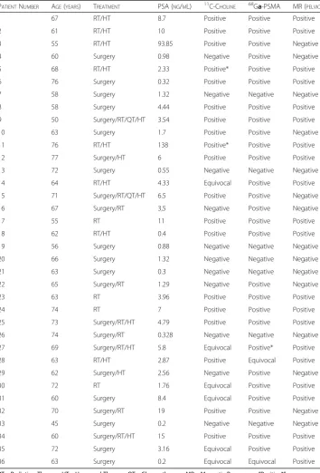
Table 1Patient characteristics and scan results
PATIENTNUMBER AGE(YEARS) TREATMENT PSA (NG/ML) 11C-CHOLINE 68Ga-PSMA MR (PELVIC)
1 67 RT/HT 8.7 Positive Positive Positive
2 61 RT/HT 10 Positive Positive Positive
3 55 RT/HT 93.85 Positive Positive Negative
4 60 Surgery 0.98 Negative Positive Negative
5 68 RT/HT 2.33 Positive* Positive Positive
6 76 Surgery 0.32 Positive Positive Positive
7 58 Surgery 1.32 Negative Negative Negative
8 58 Surgery 4.44 Positive Positive Positive
9 50 Surgery/RT/QT/HT 3.54 Positive Positive Positive
10 63 Surgery 1.7 Positive Positive Negative
11 76 RT/HT 138 Positive* Positive Positive
12 77 Surgery/HT 6 Positive Positive Positive
13 72 Surgery 0.55 Negative Negative Negative
14 64 RT/HT 4.33 Equivocal Positive Positive
15 71 Surgery/RT/QT/HT 6.5 Positive Positive Negative
16 67 Surgery/RT 3,5 Negative Positive Negative
17 55 RT 11 Positive Positive Positive
18 62 RT/HT 0.4 Positive Positive Positive
19 56 Surgery 0.88 Negative Negative Negative
20 66 Surgery 1.32 Negative Negative Negative
21 63 Surgery 0.3 Negative Negative Negative
22 65 Surgery/RT 1.29 Negative Positive Negative
23 63 RT 3.96 Positive Positive Positive
24 74 RT 7 Positive Positive Positive
25 73 Surgery/RT/HT 4.79 Positive Positive Positive
26 74 Surgery/RT 0.328 Negative Negative Negative
27 69 Surgery/RT/HT 5.8 Equivocal Positive* Positive
28 63 RT/HT 2.87 Positive Equivocal Positive
29 62 Surgery/HT 2.56 Negative Positive Negative
30 72 RT 1.76 Equivocal Positive Positive
31 60 Surgery 8.4 Equivocal Positive Positive
32 70 Surgery/RT 19 Positive Positive Negative
33 45 Surgery 0.2 Negative Negative Negative
34 60 Surgery/RT/HT 15 Positive Positive Positive
35 72 Surgery 3.16 Equivocal Positive Positive
36 63 Surgery 0.2 Equivocal Equivocal Positive
RT = Radiation Therapy; HT = Hormonal Therapy; QT = Chemo therapy; MR = Magnetic Resonance;“Positive*”= means
Radiopharmaceuticals
11C-Choline
Automatized synthesis of [11C] N-methyl Choline was performed from 11CO2 and
di-methyl-amino-ethanol (DMAE). Reaction and purification was done on a Sep-Pak Classic CM cartridge (Millipore). Radiochemical purity of11C-Choline was higher than 95%.
68Ga-PSMA
68
Ga-PSMA was produced using PSMA-11 (HBED-CC) from ABX as precursor and68Ga eluted from an68Ge/68Ga generator (ITG, Germany). The precursor (3,2 to 3,6 nmol) was dissolved in ultrapure water and mixed with 1,00 mL 0,25 M sodium acetate and 650 – 1450 MBq68GaCl3in 4 mL HCl 0.05 M. After 5 min of incubation at 100 °C,68Ga-PSMA
was purified by solid phase extraction (Sep-Pak C18 light), formulated with saline and sterilized by filtration. Radiochemical purity was 99.2 ± 1.7%,68Ge breakthrough was less than 2 × 10⁻⁴% and of specific activity was 170 ± 76 MBq/nmol.
Imaging
Patients did not need to fast and were allowed to take all their medications. Patient preparation included the placement of an i.v. line and hydration with at least 500 mL. Within 1-2 weeks all patients underwent a PET/CT scan with 11C-Choline and with
68
Ga-PSMA, randomly performed. PET/CT studies were performed with a 64-slice PET/CT (General Electric Discovery 690 VCT, 64 slices, Waukesha, WI, USA) immedi-ately after the injection of 6.0 MBq/kg 11C-Choline or 60 min after the administration of 2.0 MBq/kg 68Ga-PSMA. The images were acquired from skull to mid-thigh. The acquisition and processing parameters of PET images were the same for 11C-Choline and 68Ga-PSMA. CT parameters were as follows: tube voltage 120 kVp, autoMA 80-180 mA, index noise 30,“GE SmartMa dose modulation”, rotation time 0.8 s, rotation length - full helical thickness: 3.75 mm, Pitch 1.375:1 and speed 55 (mm/rot). PET data were acquired in 3D with time-of-flight correction with a scan duration of 3 or 4 min per bed position (11C-Choline and68Ga-PSMA, respectively) and with 11-slice overlap. Images were reconstructed using an ordered subset expectation maximization algo-rithm (OSEM) with time-of-flight correction (matrix size 128 × 128 pixels) with 2 itera-tions/24 subsets.
For the 68Ga-PSMA studies we used a dedicated shuttle for MRI pelvic co-registration: PET/CT-MRI (Discovery 750w 3.0 T, GE Healthcare, Waukesha, WI, USA). An MRI abbreviated protocol was planned with axial panoramic slices (T1 and T2 sequences) starting at the aortic bifurcation. High-resolution thin slices with T2 sequence of the prostate bed as well as focal and panoramic diffusion (DWI) and ADC maps were obtained. MRI images were performed during 30 min, starting 30 min after tracer injection. No contrast media was used for MR or CT scans. Only MRI images of the pelvis were acquired in this study. Whole body MRI was not performed.
Images were evaluated by two board-certified specialists in nuclear medicine and by two board-certified radiologists.
We measured the lesion to background ratio (lesion SUVmax/background SUVmax) in all coincident lesions to evaluate the impact on image interpretation. Gluteal muscula-ture was selected as background.
For PET scans, suspicious lesions were defined as any focal uptake, at one or more locations, higher than the nearby background or compared with normal tissue, exclud-ing joint processes and areas of physiological uptake. Lesions were defined as equivocal or indeterminate if the uptake was no typical for malignancy but nevertheless remains unclear or when the urinary physiological activity prevents a correct assessment.
Regarding MR evaluation local recurrence at the prostate gland was described in two clinical scenarios. In the case of patients treated with radiotherapy of the pros-tate gland, areas of restricted diffusion and low signal were considered as abnor-mal. In the case of those who underwent prostatectomy, a pathologic image with signal intensity similar to solid tissue and with restricted diffusion was interpreted as local recurrence. Bone metastases were described when there was a low intensity lesion in T1 and T2, or show restricted diffusion. Lymph nodes were considered positive if their size was > 12 mm.
This paper analyzes only detection rates of the radiopharmaceuticals. Validation of findings using histology and/or clinical follow-up is needed in order to calculate accur-acy diagnostic values.
Image analysis was performed using a General Electric AW 4.6 platform (HP Work-station and LINUX OS) and OsiriX v6.5.2 (OS X El Capitan).
Statistical analysis
Tumour to background ratio signal from the same lesions in both68Ga-PSMA and11 C-Choline studies were analysed using the Student paired t-test. Comparisons of the number of detected lesions per patient in each study, and with each radiotracer regarding PSA levels, were done with the two-sided Mann–Whitney test. Additionally, the number of pelvic lesions detected with Choline, PSMA and MRI were compared using the Kruskal-Wallis (ANOVA) and Fisher tests. APvalue < 0.05 was considered significant. For statis-tical analyses, we used GraphPad Prism version 7.00 for Mac OS X, La Jolla, CA, USA.
Results
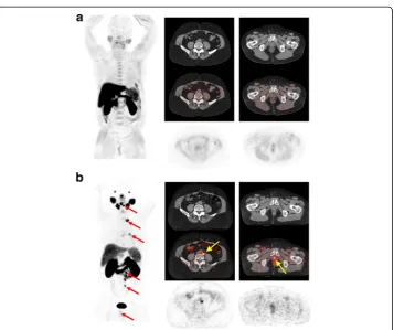
Overall detection rate was 75% (27/36) for 68Ga-PSMA and 53% (19/36) for11 C-Cho-line. Both scans were positive in 18 patients (50%) and negative in 8 patients (22%). Nine patients were positive with 68Ga-PSMA alone (25%) and one with 11C-Choline only (3%), (Figs. 1and2).
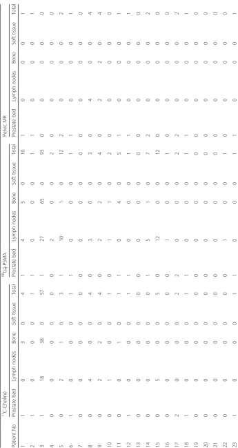
A total of 185 lesions were detected by at least one radiopharmaceutical: 183 for
68
Ga-PSMA and 98 for11C-Choline. Lesion localization for each tracer is described in Table2. The median detected lesion per patient was 2 for68Ga-PSMA (range 0-93) and 1 for11C-Choline (range 0-57). The difference was statistically significant (P= 0.023).
Tumour to background ratios in all concordant lesions (n= 96) were higher for68 Ga-PSMA than for11C-Choline (110.3 ± 107.8 and 27.5 ± 17.1, mean ± S.D., for each tracer, respectivelyP= 0.0001).
with PSA≥3.3 ng/mL(median value of our patient sample): 0.5 (0-3) versus 2 (0 -57), median (range), respectively, (P= 0.03). On the other hand, the number of detected lesions was independent of PSA levels for 68Ga-PSMA using the same PSA cut-off value: 1 lesion per patient if PSA≥3.3 (range 0–12) versus 3 lesion per patient if PSA < 3,3 (range 1-93),(P= 0.05).
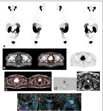
Concerning pelvic evaluation, metastatic lesions were found in 25 patients (69%) with68Ga-PSMA, 18 (50%) with 11C-Choline and 21 (58%) with MRI (3.0 T). Pelvic MRI detected 31 lesions: 20 at the prostate bed (local recurrence), 8 lymph nodes and 3 bone lesions, as shown in Table 2. Figures3and 4show68Ga-PSMA MIP and PET/ MRI images demonstrating coincident metastatic pelvic lymph nodes and a local re-lapse, respectively (11C-Choline images not shown). Besides, MRI was very useful in
Fig. 1Patient 1. A 67-year-old patient treated with radiotherapy and hormonotherapy with PSA relapse
detecting recurrence in cases classified as indeterminate by means of PET/CT alone at prostate bed as shown in Fig. 5. Nine patients in total were classified as indeterminate or equivocal (patients 5, 11, 14, 27, 28, 30, 31, 35 and 36), with at least one of the tracers: six patients with 11C-Choline (patients 5, 11, 14, 30, 31, and 35), one patient with 68Ga-PSMA (patient 28) and two patients with both tracers were indeterminate, (patients 27 and 36) as shown in Table 2. When PET/CT was unable to detect a local relapse due to physiological tracer activity in the bladder, the MRI component was clearly pathological.
Of these nine patients, four had bone or lymph node metastasis detected with at least one of the tracers as shown in Table 2 (patients 5, 11, 14 and 27), rendering the added value of MRI in those cases less important. It is well known that having a local recurrence in the prostatic bed is of minor importance if metastases are present at the same time.
No indeterminate or equivocal results were found at lymph nodes or bone lesions.
Discussion
After applying curative intent treatment for clinically localized PCa with surgery or radiotherapy, 15-30% of patients show biochemical progression (Cher et al., 1998), which precedes a clinically detectable recurrence at the pelvis or metastatic disease within a period of months or years (Moul et al., 2000; Roberts et al., 2001). Furthermore, the
Fig. 2Patient 5. A 68-year-old patient with PCa treated with radiotherapy and complete androgen blockade
detection of lesions associated with a recurrence of PCa in the context of a biochemical relapse constitutes a major challenge for all imaging modalities including Choline PET/CT (Schmid et al.,2005; Igerc et al.,2008; Kwee & DeGrado,2008; Hacker et al.,2006; Husarik et al.,2008; Cimitan et al.,2006; Pelosi et al.,2008; Beauregard et al.,2010; Heinisch et al.,
2006; Steiner et al.,2009).
The usefulness of PET/CT with radiolabelled Choline in the assessment of these patients has been amply demonstrated in several clinical studies (Perera et al., 2016; von Eyben et al., 2016; de Jong et al., 2003; Picchio et al., 2003; Rinnab et al., 2007), which have confirmed a preferential uptake by PCa, lymph nodes and their metastases (Hara et al., 1998; Kwee et al., 2005; Yamaguchi et al.,2005). However, Choline is not specific for prostate cancer either in intra prostatic or extra prostatic disease and a high affinity of the radiopharmaceutical has been evidenced by benign hyperplasia. As a con-sequence, discrimination between benign and malignant intraprostatic tissue is ham-pered by low specificity (Souvatzoglou et al., 2011; Farsad et al., 2005). Moreover, the sensitivity of Choline PET/CT in detecting locally recurrent PCa is low, notably in the case of low PSA values (Krause et al.,2008; Giovacchini et al., 2013; Castellucci et al.,
2009; Castellucci et al.,2011; Mamede et al.,2013).
PSMA is a transmembrane protein with a significantly increased expression in PCa cells (Silver et al.,1997), recently selected as a target for molecular imaging approaches (Mease et al., 2013). Several clinical studies showed additional advantages of 68 Ga-PSMA PET/CT in comparison with radioactively labelled analogues of Choline (Afshar-Oromieh et al.,2014; Eiber et al.,2015), a high image contrast due to low back-ground signal, sensitive detection of small lesions due to high radiotracer uptake, and improved detection rates of recurrent prostate cancer and metastases especially at low PSA levels. However, labelled PSMA is also not specific for prostate cancer and there is
Fig. 3Patient 8. A 58-year-old-patient with PSA relapse (PSA level 4.44 ng/ml) after radical prostatectomy.
increasing evidence suggesting that PSMA can be expressed in other solid tumours. This fact can limit the technique’s specificity (Silver et al.,1997).
We prospectively analysed 36 patients who underwent both11C-Choline PET/CT and
68
Ga-PSMA PET/CT analysis within a time window of 1-2 weeks. Additionally, for the
68
Ga-PSMA scan, we used a PET/CT-MRI (3.0 T) system with a dedicated shuttle, acquiring MRI images of the pelvis. In all cases, there was biochemical recurrence fol-lowing prior conventional treatment of PCa.
Although there is controversy about the influence of androgen deprivation therapy (ADT) in the detection rate of PET/CT with 68Ga-PSMA, it has been shown that the probability of a pathological 68Ga-PSMA-11 PET/CT scan is strongly associated with ongoing ADT (Afshar-Oromieh et al.,2017).
The use of ADT in our patient sample was not an exclusion criteria and its impact on the detection rate remains unknown.
In 75% of the patients at least one suspicious lesion for PCa was detected with
68
Ga-PSMA PET/CT whereas only 53% of the patients presented pathological find-ings with 11C-Choline PET/CT. Our results were like those reported in the litera-ture (Schmid et al., 2005; Igerc et al., 2008; Kwee & DeGrado, 2008; Hacker et al.,
2006; Husarik et al., 2008; Cimitan et al., 2006; Pelosi et al., 2008; Beauregard et al., 2010; Heinisch et al., 2006; Steiner et al., 2009; Afshar-Oromieh et al., 2013).
11
C-Choline PET/CT did not reveal any suspicious lesions in 17 patients, while
Fig. 4Patient 2. A 61-year-old-patient treated with radiotherapy and hormonotherapy with PSA relapse
only 9 patients presented without any pathological findings in 68Ga-PSMA PET/ CT.
68
Ga-PSMA PET/CT detected significantly more PCa lesions when compared to Choline. These data are also similar to our previous report concerning this PSMA lig-and (Afshar-Oromieh et al., 2013). In addition, the detection rates of Choline agree with the data reported in the literature (Schmid et al., 2005; Igerc et al., 2008; Kwee & DeGrado, 2008; Hacker et al., 2006; Husarik et al., 2008; Cimitan et al., 2006; Pelosi et al., 2008; Beauregard et al.,2010; Heinisch et al., 2006; Steiner et al., 2009). We also demonstrated a significantly higher tumour to background ratio with 68Ga-PSMA than with11C-Choline. Similar results have been reported in the literature (Afshar-Oromieh et al., 2014; Morigi et al., 2015).Therefore, 68Ga-PSMA PET/CT proved to be clearly superior in detecting PCa lesions compared to Choline-based PET/CT, especially at low PSA levels.
Fig. 5Patient 36. MIP68Ga-PSMA images (a) of a 68-year-old patient with PCa treated with surgery with current
Even though the literature sets the positivity of 68Ga-PSMA PET/CT dependent on the PSA-value (PSA < 0.5), in this study only 6 patients have PSA-values < 0.5 ng/mL, and 27 patients have PSA > 1 ng/mL (Table1).
As a result, the statistical cut-off point criteria was set according to the PSA values of the sample i.e. (median PSA value = 3.3 ng/mL). Unfortunately, our study did not in-clude enough patients with lower PSA values who can potentially benefit from salvage treatment.
Despite the small number of patient enrolled, this study reinforces the high detection rate for 68Ga-PSMA at low PSA levels, compared to11C-Choline in a prospective trial design.
In the last few years, the advent of new molecular imaging methods, such as MRI may provide clinicians with useful information that can have an impact on the management of PCa patients. Multiparametric MRI has shown high sensitivity and specificity for the detection of local and regional recurrence after treatment in PCa patients (de Rooij et al., 2014). PET/MRI with 68Ga-PSMA has recently been introduced with promising results. This combination allows excellent morphological detail, multi-parameter functional information and molecular information data. In this context, the tri-modality PET/CT–MRI system includes the transfer of the patient on a dedicated shuttle from one modality to another without changing pa-tient position (Veit-Haibach et al., 2013). This novel sequential imaging technique could lead to a significant improvement in the detection of PCa.
Afshar-Oromiehet al. (Afshar-Oromieh et al., 2014) and Eiber et al. (Eiber et al.,
2016) evaluated the feasibility of PET/MRI hybrid system with 68Ga-PSMA. In their studies, they demonstrated that prostate cancer was detected more easily and more accurately with the 68Ga-PSMA PET/MRI hybrid system, than with PET/CT, and with less radiation exposure. Consequently, this new technique could clarify inconclusive PET/CT results.
Regarding pelvic evaluation, metastatic lesions were found in 25 patients (69%) with 68Ga-PSMA, 18 (50%) with 11C-Choline and 21 (58%) with MRI (3.0 T). The detection rate of 68Ga-PSMA PET/CT was greater than MRI in the detection of pelvic lymph nodes as the literature has described (Gupta et al., 2017; Tulsyan et al., 2017). Advantages of the trimodality PET/CT-MRI system include a more accurate attenuation correction, reliable PET-quantification, superior soft tissue contrast and a higher imaging flexibility that improves diagnostic accuracy for PCa. (de Rooij et al., 2014; Veit-Haibach et al., 2013). The sequential acquisition of the techniques reduces the problems related to the lack of alignment and the change of position of the patient. An important finding in our study is that MRI was highly useful in detecting recurrence in cases classified as indeterminate at prostate bed by means of PET/CT alone, especially in patients with local recurrences detected near the bladder as described in the literature (Afshar-Oromieh et al.,
Conclusion
In patients with prostate cancer with biochemical recurrence 68Ga-PSMA detected more lesions than 11C-Choline regardless of PSA levels.PET/CT-MRI (3.0 T) system is a feasible imaging modality that potentially adds useful relevant information with increased accuracy of diagnosis.
Abbreviations
ADT:androgen deprivation therapy; DMAE: Di-methyl-amino-ethanol; MRI: Magnetic Resonance Imaging; PCa: Prostate Cancer; PSA: Prostate Specific Antigen; PSMA: Prostate-specific Membrane Antigen
Acknowledgements
The publication of this article was supported by funds of the European Association of Nuclear Medicine (EANM).
Funding
None.
Author’s contributions
Study conception and design: OA, GdS, MGF. Acquisition of data: OA, GdS, MGF, HB. Analysis and interpretation of data: OA, GdS, MGF, HB, HE. Drafting of manuscript: OA and GdS. Critical revision: OA, GdS, MGF, HB, HE. All authors read and approved the final manuscript.
Competing interests
We declare that all authors have no competing financial, professional or personal interests that might have influenced the performance of the work described in this manuscript.
Publisher’s Note
Springer Nature remains neutral with regard to jurisdictional claims in published maps and institutional affiliations.
Received: 10 October 2017 Accepted: 17 January 2018
References
Afshar-Oromieh A, Haberkorn U, Eder M, Eisenhut M, Zechmann C (2012) [68Ga]gallium-labelled PSMA ligand as superior PET tracer for the diagnosis of prostate cancer: comparison with 18F-FECH. Eur J Nucl Med Mol Imaging 39:1085–1086
Afshar-Oromieh A, Haberkorn U, Schlemmer HP et al (2014) Comparison of PET/CT and PET/MR hybrid systems using a 68Ga-labelled PSMA ligand for the diagnosis of recurrent prostate cancer: initial experience. Eur J Nucl Med Mol Imaging 41:887–897
Afshar-Oromieh A, Holland-Letz T, Giesel FL et al (2017) Diagnostic performance of 68Ga-PSMA-11 (HBED-CC) PET/CT in patients with recurrent prostate cancer: evaluation in 1007 patients. Eur J Nucl Med Mol Imaging 44(10):1781 Afshar-Oromieh A, Malcher A, Eder M, Eisenhut M, Linhart HG, Hadaschik BA et al (2013) PET imaging with a
[(68)Ga]gallium-labelled PSMA ligand for the diagnosis of prostate cancer: biodistribution in humans and first evaluation of tumour lesions. Eur J Nucl Med Mol Imaging 40:486–495
Afshar-Oromieh A, Zechmann CM, Malcher A et al (2014) Comparison of PET imaging with a (68)Ga-labelled PSMA ligand and (18)F-choline-based PET/CT for the diagnosis of recurrent prostate cancer. Eur J Nucl Med Mol Imaging 41:11–20
Alonso O, dos Santos G, Garcia-Fontes M, Engler H (2016) Prospective comparison of 11C-Choline versus 68Ga-PSMA using a tri-modality PET/CT-MR system for the diagnosis of prostate cancer patients with biochemical recurrence (abstract). J Nucl Med 57(Suppl 2):564
Bander NH (2006) Technology insight: monoclonal antibody imaging of prostate cancer. Nat Clin Pract Urol 3:216–225 Beauregard JM, Williams SG, Degrado TR, Roselt P, Hicks RJ (2010) Pilot comparison of fluorocholine and
F-fluorodeoxyglucose PET/CT with conventional imaging in prostate cancer. J Med Imaging Radiat Oncol 54:325–332 Castellucci P, Fuccio C, Nanni C, Santi I, Rizzello A, Lodi F et al (2009) Influence of trigger PSA and PSA kinetics on
11C-choline PET/CT detection rate in patients with biochemical relapse after radical prosta-tectomy. J Nucl Med 50: 1394–1400
Castellucci P, Fuccio C, Rubello D, Schiavina R, Santi I, Nanni C et al (2011) Is there a role for (11)C-choline PET/CT in the early detection of metastatic disease in surgically treated prostate cancer patients with a mild PSA increase < 1.5 ng/mL? Eur J Nucl Med Mol Imaging 38:55–63
Cher ML, Bianco FJ Jr, Lam JS, Davis LP, Grignon DJ, Sakr WA et al (1998) Limited role of radionuclide bone scintigraphy in patients with prostate specific antigen elevations after radical prostatectomy. J Urol 160:1387–1391 Cimitan M, Bortolus R, Morassut S, Canzonieri V, Garbeglio A, Baresic T et al (2006) 18F-fluorocholine PET/CT imaging
for the detection of recurrent prostate cancer at PSA relapse: experience in 100 consecutive patients. Eur J Nucl Med Mol Imaging 33:1387–1398
de Jong IJ, Pruim J, Elsinga PH, Vaalburg W, Mensink HJ (2003) Preoperative staging of pelvic lymph nodes in prostate cancer by 11C-choline PET. J Nucl Med 44(3):331–335
Eder M, Eisenhut M, Babich J, Haberkorn U (2013) PSMA as a target for radiolabelled small molecules. Eur J Nucl Med Mol Imaging 40(6):819–823
Eder M, Schäfer M, Bauder-Wüst U, Hull WE, Wängler C, Mier W et al (2012) (68) Ga-complex lipophilicity and the targeting property of a urea-based PSMA inhibitor for PET imaging. Bioconjug Chem 23:688–697
Eiber M, Maurer T, Souvatzoglou M et al (2015) Evaluation of hybrid 68Ga-PSMA ligand PET/CT in 248 patients with biochemical recurrence after radical prostatectomy. J Nucl Med 56:668–674
Eiber M, Weirich G, Holzapfel K, Souvatzoglou M, Haller B, Rauscher I et al (2016) Simultaneous 68Ga-PSMA HBED-CC PET/MR improves the localization of primary prostate cancer. Eur Assoc Urol 70(5):829–836 Farsad M, Schiavina R, Castellucci P et al (2005) Detection and localization of prostate cancer: correlation of
(11)C-choline PET/CT with histo-pathologic step-section analysis. J Nucl Med 46:1642–1649
Giovacchini G, Picchio M, Garcia-Parra R, Mapelli P, Briganti A, Montorsi F et al (2013) [11C]choline positron emission tomography/compute- rized tomography for early detection of prostate cancer recurrence in patients with low increasing prostate specific antigen. J Urol 189:105–110
Gupta M, Choudhury PS, Hazarika D et al (2017) A comparative study of 68Gallium-prostate specific membrane antigen positron emission tomography-computed tomography and magnetic resonance imaging for lymph node staging in high risk prostate cancer patients: an initial experience. W J Nucl Med 16(3):186–191
Hacker A, Jeschke S, Leeb K, Prammer K, Ziegerhofer J, Sega W et al (2006) Detection of pelvic lymph node metastases in patients with clinically localized prostate cancer: comparison of 18F-fluorocholine positron emission tomography-computerized tomography and laparoscopic radioisotope guided sentinel lymph node dissection. J Urol 176:2014–2018
Hara T, Kosaka N, Kishi H (1998) PET imaging of prostate cancer using carbon-11-choline. J Nucl Med 39:990–995 Heinisch M, Dirisamer A, Loidl W, Stoiber F, Gruy B, Haim S et al (2006) Positron emission tomography/
computed tomography with F-18- fluorocholine for restaging of prostate cancer patients: meaningful at PSA < 5 ng/ml? Mol Imaging Biol 8:43–48
Hillier SM, Maresca KP, Femia FJ, Marquis JC, Foss CA, Nguyen N et al (2009) Preclinical evaluation of novel glutamate-urea-lysine analogues that target prostate-specific membrane antigen as molecular imaging pharmaceuticals for prostate cancer. Cancer Res 69(17):6932–6940
Husarik DB, Miralbell R, Dubs M, John H, Giger OT, Gelet A et al (2008) Evaluation of [(18)F]-choline PET/CT for staging and restaging of prostate cancer. Eur J Nucl Med Mol Imaging 35:253–263
Igerc I, Kohlfürst S, Gallowitsch HJ, Matschnig S, Kresnik E, Gomez-Segovia I et al (2008) The value of 18F-choline PET/CT in patients with elevated PSA-level and negative prostate needle biopsy for localisation of prostate cancer. Eur J Nucl Med Mol Imaging 35:976–983
Jemal A, Bray F, Center MM, Ferlay J, Ward E, Forman D (2011) Global cancer statistics. CA Cancer J Clin 61:69–90 Kosuri S, Akhtar NH, Smith M, Osborne JR, Tagawa ST (2012) Review of salvage therapy for biochemically
recurrent prostate cancer: the role of imaging and rationale for systemic salvage targeted anti-prostate-specific membrane antigen radioimmunotherapy. Adv Urol 2012:921674
Krause BJ, Souvatzoglou M, Tuncel M, Herrmann K, Buck AK, Praus C et al (2008) The detection rate of [11C]choline-PET/CT depends on the serum PSA-value in patients with biochemical recurrence of prostate cancer. Eur J Nucl Med Mol Imaging 35:18–23
Kwee SA, Coel MN, Lim J, Ko JP (2005) Prostate cancer localization with 18fluorine fluorocholine positron emission tomography. J Urol 173(1):252–255
Kwee SA, DeGrado T (2008) Prostate biopsy guided by 18F-fluorocholine PET in men with persistently elevated PSA levels. Eur J Nucl Med Mol Imaging 35:1567–1569
Liu H, Moy P, Kim S, Xia Y, Rajasekaran A, Navarro V et al (1997) Monoclonal antibodies to the extracellular domain of prostatespecific membrane antigen also react with tumor vascular endothelium. Cancer Res 57:3629–3634
Lozano R, Naghavi M, Foreman K et al (2012) Global and regional mortality from 235 causes of death for 20 age groups in 1990 and 2010: a systematic analysis for the global burden of disease study 2010. Lancet 380:2095–2128 Mamede M, Ceci F, Castellucci P, Schiavina R, Fuccio C, Nanni C et al (2013) The role of 11C-choline PET imaging in the
early detection of recurrence in surgically treated prostate cancer patients with very low PSA level < 0.5 ng/mL. ClinNucl Med 38(9):e342–e345
Mannweiler S, Amersdorfer P, Trajanoski S, Terrett JA, King D, Mehes G (2009) Heterogeneity of prostate-specific membrane antigen (PSMA) expression in prostate carcinoma with distant metastasis. Pathol Oncol Res 15:167–172
Mease RC, Foss CA, Pomper MG (2013) PET imaging in prostate cancer: focus on prostate-specific membrane antigen. Curr Top Med Chem 13:951–962
Morigi JJ, Stricker PD, van Leeuwen PJ, Tang R, Ho B (2015) Prospective comparison of 18F-Fluoromethylcholine versus 68Ga-PSMA PET/CT in prostate cancer patients who have rising PSA after curative treatment and are being considered for targeted therapy. J Nucl Med 56(8):1185–1190
Moul JW et al (2000) Prostate specific antigen only progression of prostate cancer. J Urol 163:1632–1642
Pelosi E, Arena V, Skanjeti A, Pirro V, Douroukas A, Pupi A et al (2008) Role of whole-body 18F-choline PET/CT in disease detection in patients with biochemical relapse after radical treatment for prostate cancer. Radiol Med 113:895–904 Perera M, Papa N, Christidis D, Wetherell D et al (2016) Sensitivity, specificity, and predictors of positive
68Ga-prostate-specific membrane antigen positron emission tomography in advanced prostate cancer: a systematic review and meta-analysis. Eur Urol 70(6):926–937
Picchio M, Messa C, Landoni C, Gianolli L, Sironi S, Brioschi M et al (2003) Value of [11C]choline-positron emission tomography for re-staging prostate cancer: a comparison with [18F]fluorodeoxyglucose-positron emission tomography. J Urol 169(4):1337–1340
Roberts SG, Blute ML, Bergstralh EJ, Slezak JM, Zincke H (2001) PSA doubling time as a predictor of clinical progression after biochemical failure following radical prostatectomy for prostate cancer. Mayo Clin Proc 76:576–581 Schäfer M, Bauder-Wüst U, Leotta K, Zoller F, Mier W, Haberkorn U et al (2012) A dimerized urea-based inhibitor of the
prostate-specific membrane antigen for 68Ga-PET imaging of prostate cancer. EJNMMI Res 2:23
Schmid DT, John H, Zweifel R, Cservenyak T, Westera G, Goerres GW et al (2005) Fluorocholine PET/CT in patients with prostate cancer: initial experience. Radiology 235:623–628
Schwenck J, Rempp H, Reischl G et al (2017) Comparison of 68Ga-labelled PSMA-11 and 11C-choline in the detection of prostate cancer metastases by PET/CT. Eur J Nucl Med Mol Imaging 44(1):92–101
Silver DA, Pellicer I, Fair WR, Heston WD, Cordon-Cardo C (1997) Prostate-specific membrane antigen expression in normal and malignant human tissues. Clin Cancer Res 3:81–85
Souvatzoglou M, Weirich G, Schwarzenboeck S et al (2011) The sensitivity of [11C]choline PET/CT to localize prostate cancer depends on the tumor configuration. Clin Cancer Res Off J Am Assoc Cancer Res 17:3751–3759 Steiner C, Vees H, Zaidi H, Wissmeyer M, Berrebi O, Kossovsky MP et al (2009) Three-phase 18F-fluorocholine PET/CT in
the evaluation of prostate cancer recurrence. Nuklearmedizin 48:1–9
Sweat SD, Pacelli A, Murphy GP, Bostwick DG (1998) Prostate-specific membrane antigen expression is greatest in prostate adenocarcinoma and lymph node metastases. Urology 52:637–640
Tulsyan S, Das CJ, Tripathi M, Seth A, Kumar R, Bal C (2017) Comparison of 68Ga-PSMA PET/CT and multiparametric MRI for staging of high-risk prostate cancer68Ga-PSMA PET and MRI in prostate cancer. Nucl Med Commun 38(12): 1094–1102
Veit-Haibach P, Kuhn FP, Wiesinger F et al (2013) PET-MR imaging using a tri- modality PET/CT-MR system with a dedicated shuttle in clinical routine. MAGMA 26(1):25–35
von Eyben FE, Picchio M, von Eyben R, Rhee H, Bauman G et al (2016)
68Ga-LabeledProstate-specificMembraneAntigenLigandPositronEmissionTomography/ComputedTomographyforProstateCancer:a systematic review and meta-analysis. Eur Urol Focus http://dx.doi.org/10.1016/j.euf.2016.11.002