*Address for correspondence
E-mail: [email protected]
Eudragit
®Microparticles for the Release of Budesonide:
A Comparative Study
RITA CORTESI*, LAURA RAVANI, ENEA MENEGATTI, ELISABETTA ESPOSITO AND F. RONCONI1
Departments of Life Sciences and Biotechnology, and 1Physics, University of Ferrara, 44121-Ferrara, Italy
Cortesi, et al.: Eudragit® Microparticles for the Release of Budesonide
This study compares the behaviour of budesonide‑containing microparticles made of Eudragit®RS or Eudragit®RS/ Eudragit®RL 70:30 (w/w) prepared either by solvent evaporation or spray‑drying technique. The loading efficiency of budesonide within microparticles was about 72% for microparticles prepared by solvent evaporation and around 78% for spray‑dried microparticles. Thermal analyses were assessed to collect information about the structural stability of budesonide within the polymeric microspheres. The in vitro release was performed using simulating gastric (fasted state simulated gastric fluid) and intestinal (fasted state simulated intestinal fluid) fluids as the receiving solutions. After 3 h the drug release from Eudragit®RS/Eudragit®RL microparticles was about 6‑fold higher than that obtained in the case of monopolymer microparticles. Using fasted state simulated intestinal fluid the drug was released between 4 and 30% in both types of preparations. Eudragit®RS microparticles showed a better protection of the drug from gastric acidity than those of Eudragit®RS/Eudragit®RL allowing us to propose Eudragit®RS microparticles as a hypothetical system of colon specific controlled delivery.
Key words: Budesonide, controlled release, Crohn’s disease, microparticles
Research Paper
Inflammatory bowel diseases (IBD) are rare afflictions
of unknown aetiology affecting the intestines and characterized by a chronic course and recurrent inflammation. IBD consist of Crohn’s disease and ulcerative colitis, which have an onset of disease between 15 and 40 years. While ulcerative colitis
causes inflammation only in the colon (colitis) and/
or in the rectum (proctitis), Crohn’s disease may cause inflammation in the colon, rectum, small intestine (jejunum and ileum), and occasionally even in the stomach, mouth and oesophagus[1].
In the treatment of IBD, sustained release devices
like pellets, capsules or tablets have less efficiency
due to diarrhoea that enhances their elimination and reduces the time of drug release. Particularly, drug carrier systems with a size larger than 200 µm would be subjected to speedy bowel evacuation due to diarrhoea[2]. Therefore, a particulate system in the micron size range could be useful to design a suitable dosage form for IBD.
According to the different pathologic steps of Crohn’s disease[3], therapeutic studies have focused on different
classes of drug[4]. Corticosteroids typify the current first line treatment option in moderate‑severe Crohn’s
disease until resolution of symptoms[3‑7]. A comparison between budesonide (BD) standard formulation and a controlled release one demonstrated that the later, released a major fraction of active in the ileum and throughout the colon[8]. In particular, this drug has been widely used in polymeric microsystems such as poly(lactic‑co‑glycolic acid)‑microparticles (PLGA‑ microparticles)[9], Polylactic acid‑microparticles (PLA‑ microparticles)[10] and chitosan coated Ca‑alginate microparticles[11] to optimize the delivery to the inflamed colonic mucosa.
to the more readily permeable E‑RL (8.8‑12% ammonium groups). These copolymers are insoluble at physiological pH values, are able to swell
forming permeable films enabling sustained release
formulation manufacture[12]. Eudragit® polymers have been proposed in various studies for colon targeting[13] because of their low content in ammonium groups
allowing a low solubility in gastric fluids.
Summarizing, this study reports (a) the production by two different techniques of microparticles constituted of sole E‑RS or of a mixture of E‑RS and E‑RL[12,13], (b) the influence of preparation procedure on microparticle characteristics (i.e. morphology and encapsulation yield) and (c) the analysis the in vitro
drug release profiles.
MATERIALS AND METHODS
E‑RS and E‑RL were from Rohm GmbH (Darmstadt, Germany). BD and all other materials and solvents were from Sigma‑Aldrich, Germany.
Solvent evaporation method for microparticles: 500 mg of polymer (eventually added with 5 mg of BD) were dissolved in 5 ml of CH2Cl2. The mixture was emulsified with 100 ml of an aqueous phase containing 1% (w/v) of 88% hydrolyzed polyvinyl alcohol as dispersing agent. The emulsion was continuously magnetic stirred at 750 rpm for 3‑5 h.
Microparticles were isolated by filtration and left to
completely desiccate in Petri dishes[14]. Spray drying method for microparticles:
Microparticles were produced using a Mini Spray Dryer Model 190 (Büchi, Laboratoriums Technik AG, Germany). Briefly, 100 ml of E‑RS aqueous suspension (eventually containing 2 mg of BD) were withdrawn through a peristaltic pump at 10 ml/min
and sprayed with a 0.7 mm nozzle by mean of a flow
of compressed air (600l/h), in the drying chamber of the apparatus. The desiccating air was 130°[15].
Size and morphology analysis:
Size and size distribution of microspheres were determined using an average of 300‑400 measured microparticles, visualized by an optical microscope with a digital camera (Nikon Diaphot inverted microscope, Tokyo, Japan). The morphology of microspheres was evaluated by scanning electron microscopy observation using a SEM Zeiss EVO 40 (Carl Zeiss NTS GmbH, Oberkochen, Germany).
FTIR spectroscopy, thermal analysis and X‑ray diffraction:
IR spectra of pure drugs, polymers, microparticles and physical mixtures were obtained with a Perkin‑Elmer 1600 spectrophotometer (Monza, Italy) using potassium bromide disks containing about 10 mg of sample. The scanning range used was 4000‑500 cm–1 at a scan period of 1 min.
Temperature and enthalpy measurements of raw materials and samples were performed by means of a Netzsch model 409STA (Netzsch‑Gerätebau GmbH, Germany) equipped with a platinum furnace and platinum/rhodium sample carrier. All experiments were conducted at heating rates of 10°/min from 25 to 280°. Instrument calibration was performed with standard indium and zinc samples (purity>99.99%). Analyses were made in duplicate.
X‑ray diffractograms of BD, empty and drug loaded microparticles and physical mixtures between polymers and drug substance were recorded using Philips PW 1820, Netherlands. Samples were irradiated with monochromatized Cu‑Kα radiation and analysed between 2 and 60°. The range and the chart speed were 2×10⁴ cps and 10 mm/2θ, respectively. Drug content of microparticles:
About 5‑7 mg of microparticles were dissolved in 5 ml of mobile phase. Then, 20 µl of the solution were injected on a Platinum C18 A100 column (15×0.46 cm, 5 µm) in an HPLC
system (Jasco, Japan). The mobile phase used for the elution was acetonitrile/phosphate buffer (0.025M, pH 3.2) (60:40 v/v) at a flow rate of 0.8 ml/min. Under these conditions BD showed a limit of quantization of 10 ng/ml and a retention time of 4.2 min. The calibration curve was linear in the concentration range 10‑5000 ng/ml, r=0.9982.
In vitro release of BD:
Data analysis and statistics:
Statistical analysis was performed by the analysis of variance (ANOVA), followed by SPSS 11.5. The level
of significance was taken at P values<0.05.
RESULTS AND DISCUSSION
As previously reported, the present study describes the behaviour of two different poly(methacrylate) microparticle formulations designed for oral administration of BD obtained either by solvent evaporation (SE) and spray drying (SD). Particularly, BD‑containing microparticles were prepared using E‑RS or a mixture of E‑RS/E‑RL 70:30 (w/w).
In the first approach, microparticles were produced by
SE method[14]. As reported in Table 1, the percentage of recovery by weight of both types of BD‑containing microspheres was about 92%.
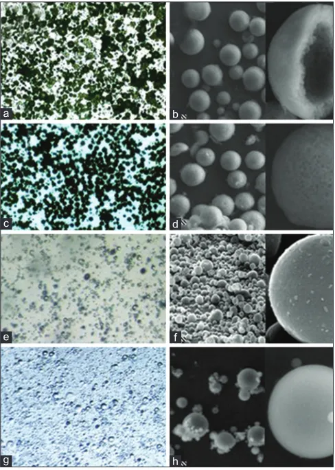
Dimensional analysis was performed by mean of both optical and scanning electron microscopy. This shows that BD‑containing microparticles have similar mean diameters and homogeneous size distribution, being 32.34±9.07 µm for E‑RS and 27.26±4.98 µm for E‑RS/E‑RL (Table 2). As shown in fig. 1, microparticles produced by SE are characterized by spherical shape and generally with porous surface. BD‑containing microparticles were also produced through SD technique[15]. Microparticles obtained by SD are usually organic solvent free with respect to other preparation methods often resulting in particles possibly contaminated by toxic organic solvents[16,17]. From data reported in fig. 1, Tables 1 and 2 it is evident that microparticles are spherically shaped with smooth surface and are characterized by smaller mean diameters with respect to microparticles produced by SE, being 4.60±2.65 µm for E‑RS and 3.75±2.12 µm for E‑RS/E‑RL. From SEM micrographs, it was observed that both processes yielded porous surface and spherical microspheres
with uniform particle size distribution. Microparticles obtained by SE showed a higher porous structure as compared to that of microparticles obtained by SD technique. The presence of a porous structure could be interesting for the release of the entrapped drug for the treatment of Crohn’s disease making this new formulation to be a good candidate for an application
in colon delivery. However, the recovery efficiency
of BD‑containing microspheres produced by SD was lower as compared to that of microparticle produced
Fig. 1: Optical and scanning electron micrographs.
Optical and scanning electron micrographs of budesonide‑containing microspheres obtained by solvent evaporation (a‑d) or spray‑drying (e‑h). Microparticles were composed of E‑RS (a, b, e, f) or E‑RS/E‑RL (c, d, g, h). Bar corresponds to 20 mm (panels b, d) or to 15 mm (panels f, h).
d
h c
g
b
f a
e
TABLE 1: CHARACTERISTICS OF BUDESONIDE‑CONTAINING EUDRAGIT® MICROPARTICLES PRODUCED BY
SOLVENT EVAPORATION (SE) OR SPRAY‑DRYING (SD) Polymer type (method of
production) composition (%)Polymer recovery*±SD (%)Microspheres loading (%w/w)±SDBudesonide Mean size±SD (µm)
E‑RS (solvent evaporation) 100 92.60±3.27 72.57±0.47 32.34±9.07
E‑RS (spray drying) 100 44.76±3.68 79.33±0.84 4.60±2.65
E‑RS/E‑RL (solvent evaporation) 70/30 92.18±2.57 71.72±0.78 27.26±4.98
E‑RS/E‑RL (spray drying) 70/30 26.65±6.84 78.31±1.22 3.75±2.12
E‑RS=Eudragit®RS, E‑RL=Eudragit®RL. Data are the mean of five independent experiments±SD. P<0.05,*Percentage of recovery with respect to the total amount
by SE being 44.76±3.68% for E‑RS and 26.65±6.84% for E‑RS/E‑RL (Table 1). This drawback can be possibly attributed to the polymer that tends to adhere to the desiccating chamber, minimizing the recovery of microspheres. In addition, while no differences in terms of morphology are evident between empty and drug‑loaded microparticles, the mean diameter of microspheres is affected by the presence of BD possibly due to an increase of the precursor emulsion droplet size during microparticle production[18,19].
Concerning the drug loading capacity it was found that in all cases BD encapsulation was around 72% for microparticles prepared by SE and around 80% for microparticles obtained by SD (Table 1)[20,21].
The physical state of BD within polymer matrices was studied by mean of FTIR and differential scanning calorimetry, IR spectrum of BD shows a C=C stretching band at 1666 cm‑1 and the O‑H stretching peak at 3490 cm‑1. These two peaks were still visible in the physical mixtures of BD with either E‑RS or E‑RS/E‑RL, whilst totally disappeared
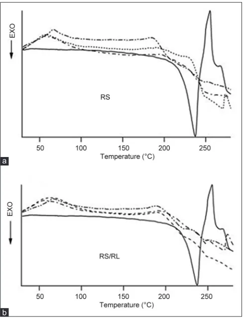
in the IR spectra of BD‑containing microspheres obtained by both preparation methods (data not shown). The thermal curves of BD and Eudragit® microparticles obtained by SE are shown in fig. 2,
the onset temperature of exothermic peaks is reported in Table 3. The sharp endothermic peak of pure drug was observed at 250° characteristic for the melting behaviour of BD. On the other hand, the thermograms of both types of BD‑containing microparticles resulted in a complete suppression of the endothermic peak of the drug, suggesting a homogeneous dissolution of the drug within the polymers. The same behaviour was obtained for physical mixture between BD and Eudragit® polymers. These results were further confirmed by X‑ray diffraction studies. The diffraction patterns of both types of BD loaded Eudragit microparticles did not contain any peaks associated with the crystalline molecule of the drug substance. In addition, no differences in terms of calorimetric peaks were appreciable for SD BD‑containing microparticles made of either E‑RS or E‑RS/E‑RL (data not shown). TABLE 2: SIZE DISTRIBUTION ANALYSIS OF
BUDESONIDE‑CONTAINING MICROPARTICLES PRODUCED BY SOLVENT EVAPORATION (SE) OR SPRAY‑DRYING (SD)
Parameter E‑RS
SE
E‑RS/E‑RL SE
E‑RS SD
E‑RS/E‑RL SD
Number of
microparticles 364 338 399 328
Size max. (µm) 81.0 40.7 16.19 10.52 Size min. (µm) 8.9 13.0 0.95 0.57
Mean 32.34 27.26 4.60 3.75
Median 30.5 27.2 3.81 3.58
Range 35.25 27.71 5.31 4.22
Standard deviation 9.07 4.98 2.65 2.12
Variance 198.03 24.81 7.01 4.44
Standard error 1.109 0.352 0.133 0.140
Skewness 0.475 −0.106 1.125 0.927
Kurtosis −0.235 0.264 0.965 0.566
E‑RS=Eudragit®RS, E‑RL=Eudragit®RL, SE=Solvent evaporation, SD=Spray‑drying.
Data are the mean of three independent analyses. P<0.05
TABLE 3: ONSET TEMPERATURE OF EXOTHERMIC PEAKS OF THERMOGRAMS REPORTED IN FIGURE 2*
Samples E‑RS(º) E‑RS/E‑RL 70:30(º)
BD 215 215
Empty polymeric microspheres 197 197 BD‑containing polymeric
microspheres
232 193
Polymer/BD physical mixture 188 192
BD=Budesonide, E‑RS=Eudragit®RS, E‑RL=Eudragit®RL.*Analyses were made
in duplicate
Fig. 2: DSC thermograms.
DSC thermograms of budesonide (BD) (——), empty polymeric microspheres (–––), BD‑containing microparticles (—–—) obtained by solvent evaporation and physical mixture of polymer plus BD (—–– —). Panel a: E‑RS. Panel b: E‑RS/RL.
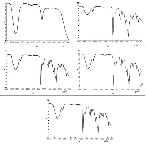
Thermal studies confirmed that there was no interaction between drug and polymer as endotherms of drug and polymers were separate in formulation prepared by SD or SE. However, from the analysis of onset temperature of exothermic peak (Table 3) it seems that E‑RS and BD are characterized by a higher intimate interaction as compared to that of E‑RS/E‑RL polymer composition and BD. IR spectra of BD and BD‑containing E‑RS or E‑RS/ E‑RL microparticles both in water and in phosphate
buffer (pH 3.2‑0.025 M). Particularly, the experiments were carried out both on microparticles obtained by
SE and by SD. As reported in the fig. 3, the presence of acidic conditions does not influence the signal of
the characteristic peak at 1635 nm of BD either alone or included in both type of SE microparticles. The same results were obtained for SD. Taken together these results it should be supposed that the slower release of BD in the acidic conditions could be
possibly ascribed to the inner morphology (fig. 1) and
(a)
(e)
(c) (d)
Fig. 3: IR specta of Budesonide and formulations.
IR spectra of pure budesonide (BD) (a) and BD‑containing microparticles of Eudragit®RS (b, c) or Eudragit® RS/RL (d, e) produced by solvent
evaporation. Spectra were performed in water (b, d) or in phosphate buffer (pH 3.2‑0.025M) (c, e).
to the mean size of microparticles that influences their surface specific area.
The complete release profile of BD from microspheres was determined by dialysis method. The profile and
kinetics of drug release are important to correlate the
in vitro and in vivo drug responses by comparing results of pharmacokinetics and dissolution profile patterns[22‑24]. Fig. 4 reports the release kinetics of BD from microparticles obtained either by SE or SD.
As clearly appreciable, both types of microparticles showed a controlled release as compared to the free
drug. In particular microparticles produced by SE (fig.
4a) showed within the 24 h for the mixture E‑RS/ E‑RL a release of BD reaching the 30% of total drug content higher with respect to E‑RS microparticles that showed a release of the 24% of the drug. In the case of microparticles produced by spray‑drying the release
of BD was surprisingly different (fig. 4b). Spray dried
E‑RS and E‑RS/E‑RL microparticles obtained showed BD release kinetics almost superimposable reaching a pseudo‑plateau after 24 h with a maximum of drug released of 25 and 32%, respectively. Concerning microparticles obtained by SD the release kinetics of BD from E‑RS shows after 24 h a release around 32‑35% without presenting a pseudo‑plateau. On the other hand, BD release from E‑RS/E‑RL reaches a pseudo‑plateau after 8 h showing a maximum of drug released of 15%. However, both types of preparation methods lead to microparticles characterized, within the first 2 h of release (when FASSGF was used), by a slower release of BD in the case of E‑RS as compared to that of E‑RS/E‑RL due to the different
polymer permeability in simulated gastric fluid. These
results could be explained considering the chemical structure of Eudragit®. E‑RL and E‑RS are synthetized from acrylic and methacrylic esters with high and low content of quaternary ammonium groups (8.8‑12 and 4.5‑6.8%, respectively) and results in different permeability behaviours. Due to the content of the quaternary ammonium groups, E‑RS is only slightly permeable; hence drug release is relatively retarded, whereas E‑RL is freely permeable, so that the release is less retarded. The combination of E‑RS and E‑RL increase the permeability of the obtained microparticles and determine a minor drug protection from the gastric ambient.
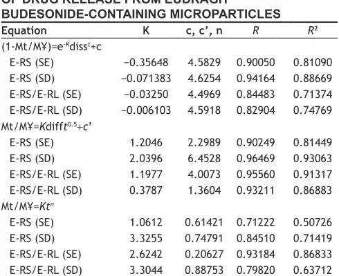
The release of a drug from microparticles can be described using some mathematical models
describing Fickian diffusive and dissolutive release mechanisms[22,25,26] and nonFickian release (Korsmeyer‑Peppas equation)[27,28]. The obtained data are reported in Table 4.
When the n value is >0.5, the release mechanism follows Fickian diffusion while for values comprised between 0.5 and 1 the release mechanism follows a nonFickian model. Correlation coefficients (R and R2) were used to evaluate the accuracy of the fit.
(a)
(b)
Fig. 4:In vitro release profiles of budesonide from microparticles.
On the basis of the value of R it appears that the release of BD from the prepared microspheres is more consistently diffusive rather than dissolutive[25,28]. These results are in agreement with the physicochemical characteristics of E‑RS and E‑RL.
Taking into consideration the above reported results the microparticles produced with E‑RS showed a better protection of the drug from gastric acidity than E‑RS/E‑RL microparticles and a hypothetical system
of colon specific controlled delivery.
REFERENCES
1. Podolsky DK. Inflammatory bowel disease. N Engl J Med 2002;347:417‑29.
2. Philip AK, Philip B. Colon targeted drug delivery systems: A review on primary and novel approaches. Oman Med J 2010;25:79‑87. 3. Hanauer SB, Sandborn W, Practice Parameters Committee of the
American College of Gastroenterology. Management of Crohn’s disease in adults. Am J Gastroenterol 2001;96:635‑43.
4. Crotty B, Hoang P, Dalton HR, Jewell DP. Salicylates used in
inflammatory bowel disease and colchicine impair interferon‑gamma
induced HLA‑DR expression. Gut 1992;33:59‑64.
5. Baumgart DC, Sandborn WJ. Inflammatory bowel disease: Clinical aspects and established and evolving therapies. Lancet 2007;369:1641‑57.
6. Rutgeerts P, Löfberg R, Malchow H, Lamers C, Olaison G, Jewell D,
et al. A comparison of budesonide with prednisolone for active Crohn’s
disease. N Engl J Med 1994;331:842‑5.
7. Campieri M, Ferguson A, Doe W, Persson T, Nilsson LG. Oral budesonide is as effective as oral prednisolone in active Crohn›s disease. The Global Budesonide Study Group. Gut 1997;41:209‑14. 8. Edsbäcker S, Larsson P, Wollmer P. Gut delivery of budesonide, a
locally active corticosteroid, from plain and controlled‑release capsules.
Eur J Gastroenterol Hepatol 2002;14:1357‑62.
9. Krishnamachari Y, Madan P, Lin S. Development of pH‑ and time‑dependent oral microparticles to optimize budesonide delivery to ileum and colon. Int J Pharm 2007;338:238‑47.
10. Martin TM, Bandi N, Shulz R, Roberts CB, Kompella UB. Preparation of budesonide and budesonide‑PLA microparticles using supercritical
fluid precipitation technology. AAPS Pharm Sci Tech 2002;3:E18.
11. Crcarevska MS, Dodov MG, Petrusevska G, Gjorgoski I, Goracinova K.
Bioefficacy of budesonide loaded crosslinked polyelectrolyte microparticles
in rat model of induced colitis. J Drug Target 2009;17:788‑802. 12. Kadian SS, Harikumar SL. Eudragit and its pharmaceutical
significance. PharmainfoNet, 2009. Available from: http:// w w w. p h a r m a i n f o . n e t / s a t i s h s i n g h k a d i a n / p u b l i c a t i o n s / eudragit‑and‑its‑pharmaceutical‑significance. [Last accessed 2 Sep 2012].
13. Paharia A, Yadav AK, Rai G, Jain SK, Pancholi SS, Agrawal GP. Eudragit‑coated pectin microspheres of 5‑fluorouracil for colon targeting. AAPS Pharm Sci Tech 2007;8:12.
14. Esposito E, Sebben S, Cortesi R, Menegatti E, Nastruzzi C. Preparation and characterization of cationic microspheres for gene delivery. Int J Pharm 1999;189:29‑41.
15. Esposito E, Roncarati R, Cortesi R, Cervellati F, Nastruzzi C. Production of Eudragit microparticles by spray‑drying technique:
Influence of experimental parameters on morphological and dimensional
characteristics. Pharm Dev Technol 2000;5:267‑78.
16. Jantratid E, Janssen N, Chokshi H, Tang K, Dressman JB. Designing biorelevant dissolution tests for lipid formulations: Case example–Lipid suspension of RZ‑50. Eur J Pharm Biopharm 2008;69:776‑85. 17. Palmieri GF, Bonacucina G, Di Martino P, Martelli S. Spray‑drying as
a method for microparticulate controlled release systems preparation: Advantages and limits. I. Water‑soluble drugs. Drug Dev Ind Pharm 2001;27:195‑204.
18. Cortesi R, Esposjto E, Luca G, Nastruzzi C. Production of lipospheres as carriers for bioactive compounds. Biomaterials 2002;23:2283‑94. 19. Esposito E, Cortesi R, Luca G, Nastruzzi C. Pectin‑based microspheres:
A preformulatory study. Ann N Y Acad Sci 2001;944:160‑79. 20. Jyothi NV, Prasanna PM, Sakarkar SN, Prabha KS, Ramaiah PS,
Srawan GY. Microencapsulation techniques, factors influencing
encapsulation efficiency. J Microencapsul 2010;27:187‑97.
21. Vehring R. Pharmaceutical particle engineering via spray drying. Pharm Res 2008;25:999‑1022.
22. Esposito E, Menegatti E, Cortesi R. Hyaluronan‑based microspheres as tools for drug delivery: A comparative study. Int J Pharm 2005;288:35‑49. 23. Zubiri D, Monge ME, Negri RM. New kinetic model of drug
release from swollen gels under non‑sink conditions. Colloids Surf A Physicochem Eng Asp 2006;273:165‑73.
24. Zahirul M, Khan I. Dissolution testing for sustained or controlled
release oral dosage forms and correlation with in vivo data: challenges
and opportunities. Int J Pharm 1996;140:131‑43.
25. Nastruzzi C, Esposito E, Cortesi R, Gambari R, Menegatti E. Kinetics of bromocriptine release from microspheres: Comparative analysis
between different in vitro models. J Microencapsul 1994;11:565‑74.
26. Peppas NA. Analysis of Fickian and non‑Fickian drug release from polymers. Pharm Acta Helv 1985;60:110‑1.
27. Korsmeyer RW, Gurny R, Doelker EM, Buri P, Peppas NA. Mechanism of solute release from porous hydrophilic polymers. Int J Pharm 1983;15:25‑35.
28. Siepmann J, Peppas NA. Modeling of drug release from delivery systems based on hydroxypropyl methylcellulose (HPMC). Adv Drug Deliv Rev 2001;48:139‑57.
TABLE 4: RELEASE KINETIC PARAMETERS
OF DRUG RELEASE FROM EUDRAGIT®
BUDESONIDE‑CONTAINING MICROPARTICLES
Equation K c, c’, n R R2
(1‑Mt/M¥)=e‑Kdisst+c
E‑RS (SE) −0.35648 4.5829 0.90050 0.81090 E‑RS (SD) −0.071383 4.6254 0.94164 0.88669 E‑RS/E‑RL (SE) −0.03250 4.4969 0.84483 0.71374 E‑RS/E‑RL (SD) −0.006103 4.5918 0.82904 0.74769 Mt/M¥=Kdifft0.5+c’
E‑RS (SE) 1.2046 2.2989 0.90249 0.81449 E‑RS (SD) 2.0396 6.4528 0.96469 0.93063 E‑RS/E‑RL (SE) 1.1977 4.0073 0.95560 0.91317 E‑RS/E‑RL (SD) 0.3787 1.3604 0.93211 0.86883 Mt/M¥=Ktn
E‑RS (SE) 1.0612 0.61421 0.71222 0.50726 E‑RS (SD) 3.3255 0.74791 0.84510 0.71419 E‑RS/E‑RL (SE) 2.6242 0.20627 0.93184 0.86833 E‑RS/E‑RL (SD) 3.3044 0.88753 0.79820 0.63712
E‑RS= Eudragit®RS, E‑RL=Eudragit®RL, K and c=Mathematical coefficients
obtained by plotting the linear forms of the indicated equations, R=Regression coefficient, R2=Squared regression coefficient, SE=Solvent evaporation,
SD=Spray‑drying