Comparison of in vitro static and dynamic
assays to evaluate the efficacy of an
antimicrobial drug combination against
Staphylococcus aureus
Diane C. BroussouID1,2, Pierre-Louis Toutain3, Fre´de´rique Woehrle´2, Farid El Garch2,
Alain Bousquet-Melou1, Aude A. Ferran
ID1*
1 UMR 1436 INTHERES, Universite´ de Toulouse, INRA, ENVT, Toulouse, France, 2 Vetoquinol SA, Lure, France, 3 Department of Veterinary Basics Sciences, Royal Veterinary College, London, United Kingdom
Abstract
An easily implementable strategy to reduce treatment failures in severe bacterial infections is to combine already available antibiotics. However, most in vitro combination assays are performed by exposing standard bacterial inocula to constant concentrations of antibiotics over less than 24h, which can be poorly representative of clinical situations. The aim of this study was to assess the ability of static and dynamic in vitro Time-Kill Studies (TKS) to iden-tify the potential benefits of an antibiotic combination (here, amikacin and vancomycin) on two different inoculum sizes of two S. aureus strains. In the static TKS (sTKS), performed by exposing both strains over 24h to constant antibiotic concentrations, the activity of the two drugs combined was not significantly different the better drug used alone. However, the dynamic TKS (dTKS) performed over 5 days by exposing one strain to fluctuating concen-trations representative of those observed in patients showed that, with the large inoculum, the activities of the drugs, used alone or in combination, significantly differed over time. Van-comycin did not kill bacteria, amikacin led to bacterial regrowth whereas the combination progressively decreased the bacterial load. Thus, dTKS revealed an enhanced effect of the combination on a large inoculum not observed in sTKS. The discrepancy between the sTKS and dTKS results highlights that the assessment of the efficacy of a combination for severe infections associated with a high bacterial load could be demanding. These situations proba-bly require the implementation of dynamic assays over the entire expected treatment dura-tion rather than the sole static assays performed with steady drug concentradura-tions over 24h.
Introduction
The recalcitrance of severe, chronic bacterial infections to antibiotic treatments, due to toler-ance or resisttoler-ance, is necessitating a search for new antimicrobial strategies. One therapeutic option to extend the spectrum, minimize the emergence of resistance or enhance the
a1111111111 a1111111111 a1111111111 a1111111111 a1111111111
OPEN ACCESS
Citation: Broussou DC, Toutain P-L, Woehrle´ F, El
Garch F, Bousquet-Melou A, Ferran AA (2019) Comparison of in vitro static and dynamic assays to evaluate the efficacy of an antimicrobial drug combination against Staphylococcus aureus. PLoS ONE 14(1): e0211214.https://doi.org/10.1371/ journal.pone.0211214
Editor: Karsten Becker, Universitatsklinikum
Munster, GERMANY
Received: June 5, 2018
Accepted: January 9, 2019
Published: January 23, 2019
Copyright:©2019 Broussou et al. This is an open access article distributed under the terms of the
Creative Commons Attribution License, which permits unrestricted use, distribution, and reproduction in any medium, provided the original author and source are credited.
Data Availability Statement: All relevant data are
within the paper and its Supporting Information files.
Funding: The authors received no specific funding
bactericidal activity, and which can be easily and rapidly implemented, is to use a combination of existing antimicrobials [1,2]. However, the potential advantage of the combination for man-aging an infection has to be assessedin vitrobefore its implementation in clinical practice. Currently, one of the most commonly usedin vitroassays to evaluate the efficacy of a combina-tion is the checkerboard assay (CA). This is based on a similar method to the one used for MIC determination and involves exposure of a standard inoculum of 5.7 log10CFU/mL [3] to
constant concentrations of one or two drugs followed by assessment of the turbidity after incu-bating the mixture for 16–20 hours. These CA results are used to classify the combination in one of three categories: synergy, indifference or antagonism [4]. Another more time-consum-ing method is to carry out static time-kill studies (sTKS) in which constant concentrations of drugs are used to monitor the kinetics of bacterial killing over time [5–8]. sTKS is a more quantitative approach than CA as the bacterial counts during exposure to the drugs alone or in combination are compared over 24 h. However, although both CA and sTKS assays are inex-pensive and quite easy to perform, their limitations include short duration of the assay and the exposure to constant concentrations of drugs. Indeed, due to the absence of broth renewal, both assays take less than 24 hours, which can prevent the detection of potential bacterial regrowth, predictive of a relapse in the patient [9], or of progressive activity leading to bacterial eradication after several days of treatment. Thus, an exposure to constant concentrations does not reflect the behaviour of drugs in patients since the phenomena of drug absorption, metab-olism and elimination lead to fluctuating concentrations over time and to a potential exposure of bacteria to sub-inhibitory concentrations which can promote the development of resistance [10]. A Hollow-Fiber (HF) infection model has been proposed to overcome these limitations [11]. This model is used in dynamic time-kill studies (dTKS) to follow the evolution of a bacte-rial population exposed to antibiotic concentrations that are similar to those observed in patients treated over several days.
Although many studies of antibiotic combinations have focused on standard inoculum sizes, infections that are refractory to treatments and require an improved antibiotic strategy are mainly associated with biofilms and a large bacterial load. Moreover, an inoculum effect that reduces antibacterial activity on large bacterial population has been described for many drugs including vancomycin against inoculum sizes ofStaphylococcus (S.) aureusstrains rang-ing from 8 to 9.5 log10CFU/mL [12,13]. We can therefore conclude thatin vitroassessments
of antibiotic combinations need to be performed in conditions that could more accurately pre-dict their efficacy against severe refractory infections associated with large inocula. Besides, the use of Mueller-Hinton Broth (MHB) was shown to be poorly predictive ofin vivooutcomes for some specific cases [14].
The aim of this study was not to promote the use of one specific combination but rather to detect discrepancies in the assessments of the efficacy of the same antibiotic combination by using different methods, culture media or inoculum sizes. Thus, we conducted the experi-ments with one selected combination to test our hypothesis which was the combination of vancomycin and an aminoglycoside recommended as the empirical treatment of some natural and prosthetic cardiac endocarditis [15]. The combination of vancomycin and amikacin was also previously investigated onS.aureusunder static conditions [16].
We compared the efficacy of this combination on standard (around 5 log10CFU/mL) and
large (around 9 log10CFU/mL) inocula of twoS.aureusstrains by performing CA, sTKS and
dTKS in different media. For sTKS, the inocula were exposed for 24 hours to the maximum free plasma concentrations obtained in patients at the current dosing regimen, i.e. after one adminis-tration of 15mg/kg amikacin [17] and/or 1 g of vancomycin by IV route [18]. For dTKS in a HF model, the inocula were exposed for 5 days to fluctuating concentrations, as could be observed in patients treated once a day with amikacin and/or every 12h with vancomycin [19,20].
specific roles of these authors are articulated in the ‘author contributions’ section.”
Competing interests: The authors have the
Material and methods
Bacterial strains
A Methicillin ResistantS.aureus(MRSA) ATCC 33591 and a Methicillin SusceptibleS.aureus
MSSA (MSSA) HG001, derived from NCTC 8325, were used for the CA and for sTKS with constant antibiotic concentrations. The MSSA strain HG001 was used for the dTKS with fluc-tuating antibiotic concentrations in the HF model. Stock cultures of the bacteria were pre-served at -80˚C in MHB (Sigma-Aldrich, Saint Quentin-Fallavier, France) supplemented with 15% glycerol. Before each assay, 10μL of the bacterial suspension was incubated overnight at 37˚C on a Mueller-Hinton agar plate.
Antimicrobial agents
Amikacin sulfate powder (Amikacine Mylan) and vancomycin chlorhydrate powder (Vanco-mycin Sandoz) for intravenous administration were used to prepare antibiotic stock solutions in water at desired concentrations and stored at -20˚C for less than 1 month.
Antibiotic solutions were thawed and diluted to desired concentrations just before use.
Minimal inhibitory concentration (MIC) determination
The MICs of vancomycin and amikacin for the MRSA and MSSA strains were performed in three independent experiments by broth microdilution with cation adjusted-MHB (Mueller-Hinton II, Sigma Aldrich, Saint-Quentin-Fallavier, France) according to the CLSI reference methods [3] and also in Roswell Park Medium Institute 1640 medium (RPMI) (Gibco, Ther-mofischer Scientific, MA, USA). Briefly, a bacterial suspension diluted in MHB or RPMI to give a final organism density of 5.7 log10CFU/mL was added to wells of a microtiter plate
con-taining serial 2-fold dilutions of vancomycin or amikacin. Bacterial growth was recorded after incubation for 18 hours at 35˚C.
Checkerboard assays (CA)
The combined effect of vancomycin and amikacin on the testedS.aureusstrains was first eval-uated by CA in MHB and RPMI [4]. Briefly, a bacterial suspension diluted in MHB or RPMI to give a final organism density of 5.7 log10CFU/mL was added to wells of a microtiter plate
containing serial 2-fold dilutions of amikacin along the ordinate and serial 2-fold dilutions of vancomycin along the abscissa. After incubation at 35˚C for 18 hours, the MIC of both drugs were determined in wells containing one drug and in wells containing the combination of both drugs. The Fractional Inhibitory Concentration (FIC) index was then calculated accord-ing toEq 1.
FIC index¼MIC of amikacin in combination
MIC of amikacin alone þ
MIC of vancomycin in combination
MIC of vancomycin alone ð1Þ
According to Odds [4], synergism is defined as an FIC index<0.5, the absence of interac-tion as an FIC index between 0.5 and 4 and antagonism as an FIC index>4. These assays were performed in triplicate.
sTKS (constant antibiotic concentrations)
were resuspended in fresh pre-warmed MHB or RPMI and diluted to obtain standard (5.0 log10CFU/mL) or large (9.0 log10CFU/mL) bacterial inocula in MHB or RPMI.
Each inoculum size was exposed to no drug (control), 18μg/mL of vancomycin, 70μg/mL of amikacin or both for 24 hours corresponding to the maximum free plasma concentrations of the antibiotics when administered at their current dosing regimen. The intravenous admin-istration of 1g of vancomycin is described to lead to a total peak concentration of 32μg/mL [21]. However, since vancomycin binding to plasma proteins is of 45% [22] and since bound vancomycin is not active on bacteria [23], the vancomycin tested concentration in thisin vitro
sTKS experiment was equal to the free maximal plasmatic concentration of 18μg/mL [18]. The administration of 15mg/kg of amikacin is described to achieve a free peak plasma concentra-tion between 60 and 80μg/mL with a negligible binding and we decided to reproduce a peak mean concentration of 70μg/mL [24]. So, in these sTKS experiments, that the highest free drug concentrations achievable in patients were maintained over 24 h in tubes whereas in patients, the concentrations would decrease due to elimination process. The activity of the antibiotics was considered as bactericidal when we observed a reduction of the bacterial burden of more than 3 log10CFU/mL. Samples were collected from each suspension at 0, 2, 4, 8 and 24h to
count the viable bacteria. All experiments were performed in duplicate.
dTKS (fluctuating antibiotic concentrations)
A Hollow-Fiber model was used to assess the antibacterial activity of the combination of ami-kacin and vancomycin onS.aureusduring exposure to fluctuating clinically-relevant antibi-otic concentrations. Basically, the HF model includes a cartridge with capillaries composed of a semipermeable polysulfone membrane. The pore size of the capillaries (42kDa) allows equili-bration of the concentrations of chemicals which circulate through the central and peripheral compartments by means of a peristaltic pump (Duet pump, FiberCell Systems, Inc., Frederick, MD, USA) while the bacteria stay confined to the extracapillary space in the peripheral compartment.
Twenty milliliters of a suspension containing 5.0 log10CFU/mL of MSSA were inoculated
into the extracapillary space of a hollow-fiber cartridge (C2011 polysulfone cartridge, FiberCell Systems, Inc., Frederick, MD, USA) and incubated at 37˚C. The antibiotics were added either 30 min later (standard inoculum assays) or 3 days later (large inoculum assays). For each experiment, the exposure to amikacin and/or vancomycin lasted 5 days to simulate the phar-macokinetic profiles of patients receiving the antibiotics at current dosing regimens i.e. 15mg/ kg amikacin once a day [24] and/or 1 g vancomycin twice a day [25] corresponding to maxi-mum free concentrations (Cmax) in the central compartment of 70μg/mL for amikacin and
18μg/mL for vancomycin by considering a plasma protein binding negligible for amikacin and equal to 45% for vancomycin. Previous studies showed that there were no issue with the drug binding to the polysulfone fibers [18,26,27]. The drugs were continuously diluted by means of a second peristaltic pump (MiniRythmic PN+, SMD, Fleury sur Orne, France) to simulate a mean elimination half-life of 4 hours, that could be observed for amikacin and vancomycin in patients [19,20].
The general design of the study is indicated inTable 1.
Bacterial counts in TKS
The sampled bacterial suspensions were centrifuged at 3000g for 5 min, the supernatant was discarded, and the pellet was resuspended in NaCl 0.9%. The suspension was then serially diluted and plated in triplicate on tryptic soy agar supplemented with magnesium sulfate and activated charcoal to prevent antibiotic carry-over effects. The colonies were counted after overnight incubation at 37˚C. The limit of detection (LOD) was 2.5 log10CFU/mL.
Statistical analysis
The bacterial counts obtained from 0 to 24 h (sTKS and in dTKS) and from 0 to 104h (dTKS) with the same strain, inoculum size and broth were analyzed with a two-way ANOVA to test the effect of the sampling time and of the treatment (amikacin and vancomycin alone or in combination) with repeated measures (Systat 12 software). When the two-way ANOVA gave a p-value<0.05 for interaction between the time and treatment factors or for the treatment fac-tor, a pairwise comparison of the different treatments was performed with a Tukey test with a correction for multiplicity.
Results
MIC
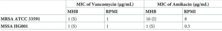
The MICs of vancomycin and amikacin for the MRSA and the MSSA strains in MHB and in RPMI are given inTable 2. The susceptible or intermediate status of both strains to both drugs, independently to the resistance to methicillin, was targeted to allow the detection of an advantage of the combination.
Based on the EUCAST breakpoints (standard determination with MHB) [28], both strains were considered as susceptible to vancomycin. The MSSA strain was considered as susceptible to amikacin and the MRSA strain showed an intermediate susceptibility to amikacin.
Checkerboard assays
To assess the bactericidal activity of a combination of amikacin and vancomycin onS.aureus, CA were performed in duplicate for the two strains in MHB and RPMI.
The FIC index of amikacin and vancomycin for each of the 2 tested strains, ranged between 1 and 1.5 in both MHB and RPMI, meaning that under the classical conditions of this CA, no interaction (no synergy or antagonism) was demonstrated between these two antibiotics [4].
sTKS
We then assessed the activity of constant concentrations of vancomycin at 18μg/mL and of amikacin at 70μg/mL, used alone or in combination, for 24hours in MHB and RPMI on a large initialS.aureusbacterial burden of 8.7±0.4 log10CFU/mL and on a standard inoculum
of 4.6±0.4 log10CFU/mL. The stability of both antibiotics in MHB and RPMI over 24h was
assessed and no significant degradation of the antibiotics was observed after 24h at 37˚C. The time-kill curves obtained by exposing the different inoculum sizes of the two strains to antibiotics are shown inFig 1.
In absence of antibiotic, the bacterial counts obtained after incubating the initial standard inocula for 24 hours, ranged between 7.8±0.1 log10CFU/mL and 8.3±0.1 log10CFU/mL for
Vancomycin alone was bactericidal on standard inocula of the two strains in MHB and in RPMI since it reduced the bacterial burden to below the LOD after 8 to 24h of antibiotic expo-sure. Exposure of a large inoculum to vancomycin for 24 h did not reduce the bacterial popula-tions except for the MRSA strain in RPMI where the bacterial population was reduced by 1.4 log10CFU/mL.
Amikacin alone was bactericidal on the standard inoculum of the MSSA strain and reduced the bacterial counts to below the LOD within less than 8h in both media. The reduction of the standard inoculum was smaller for the MRSA strain for which the bacterial counts were 4.8±0.1 and 5.4±0.5 log10CFU/mL in MHB and in RPMI respectively after 24h of exposure.
These counts were 3 log10CFU/mL less than the counts in control experiments but close to the
initial bacterial counts, thereby demonstrating a bacteriostatic effect. The bacterial populations in large inocula were not reduced by exposure for 24 h to amikacin alone except for the MSSA strain in RPMI where the population was reduced by 3.6 log10CFU/mL.
The combination of amikacin and vancomycin at constant concentrations over 24 hours never showed any significant difference from the better of the two drugs used alone. For the MSSA strain, the combination gave similar results to those obtained with amikacin alone. The bacterial loads after exposure of the standard inocula to the combination for 24 hours were below the LOD. For the large inocula, the bacterial counts were 8.0±0.4 log10CFU/mL and
5.1±0.6 log10CFU/mL after 24h in MHB and RPMI respectively. For the MRSA strain, the
combination gave similar results to vancomycin used alone. The bacterial counts after exposure of the standard inocula to the combination for 24 hours were 3.2±0.3 log10CFU/mL in MHB
and below the LOD in RPMI. For the large initial inocula, the bacterial counts were 8.8±0.5 log10CFU/mL and 7.1±0.1 log10CFU/mL after 24 hours in MHB and RPMI respectively.
dTKS
The HF model was applied to assess the efficacy of vancomycin and amikacin used alone or in combination under dynamic conditions over 5 days in RPMI on a standard inoculum of 5.4±0.3 log10CFU/mL and on a large inoculum of 9.4±0.3 log10CFU/mL.
Table 1. General design of the study with all the assays performed on each bacterial strain.
MSSA MRSA
MHB RPMI MHB RPMI
MIC SI SI SI SI
Checkerboard assay SI SI SI SI
sTKS SI and HI SI and HI SI and HI SI and HI
dTKS NA SI and HI NA NA
SI: Standard Inoculum, HI: High (Large) Inoculum, NA: Not Assessed
https://doi.org/10.1371/journal.pone.0211214.t001
Table 2. MICs (μg/mL) of vancomycin and amikacin for the MRSA and the MSSA strains in MHB and in RPMI.
MIC of Vancomycin (μg/mL) MIC of Amikacin (μg/mL)
MHB RPMI MHB RPMI
MRSA ATCC 33591 1 (S) 1 16 (I) 8
MSSA HG001 1 (S) 1 1 (S) 0.5
(S) classified as susceptible according to EUCAST breakpoints (I) classified as intermediate according to EUCAST breakpoints
In dTKS, the antibiotic concentrations fluctuated over time so as to resemble the concentra-tion profiles observed in patients receiving repeated recommended doses of amikacin and van-comycin over 5 days. Vanvan-comycin was administered twice a day at a Cmaxof 18μg/mL and
amikacin was administered once a day at a Cmaxof 70μg/mL. Both antibiotics were
continu-ously diluted to simulate an elimination half-life of 4 hours.
In the absence of antibiotic (control experiments), the standard inocula increased to 9.5±0.4 log10CFU/mL in 24h. Thereafter, all the inocula (standard and large) increased
slightly over the next 4 days to reach a final bacterial count of 10.8±0.2 log10CFU/mL at the
end of 5 days.
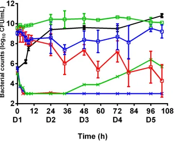
The time-kill curves for bacteria exposed in the HF model to amikacin and vancomycin alone or in combination are shown inFig 2.
On standard inocula, both antibiotics, alone or in combination, decreased the bacterial bur-den to below the LOD within less than 8h, as had been obtained with a constant antibiotic con-centration in sTKS. When exposed to amikacin alone or in combination, the bacterial
Fig 1. Time-kill curves of the 2 strains subjected to constant concentrations of antibiotic. Evolution of the bacterial
population (log10CFU/mL) for the MSSA (A and B) and MRSA (C and D) strains after exposure to 70μg/mL amikacin (blue) or 18μg/mL vancomycin (green) or the combination of both (red) over 24hours in MHB (A and C) and in RPMI (B and D). The marks represent the mean±SD of the bacterial counts for the different tested treatments (n = 2 for each treatment). Curves with open squares represent the bacterial counts on a large initial inoculum and curves with crosses represent the bacterial counts on a standard initial inoculum. (LOD = 2.5 log10CFU/mL). For the low inoculum of MSSA in MHB and RPMI (A and B), there was no difference between drugs alone or in combination. For the high inoculum of MSSA, there was no difference between drugs in MHB (A) but vancomycin led to significantly higher bacterial counts than amikacin and the combination in RPMI (B). For the low inoculum of MRSA, there was no difference between drugs alone or in combination in MHB (C) but amikacin led to significantly higher bacterial counts than vancomycin and combination in RPMI (D). For the high inoculum of MRSA, there was no difference between drugs in MHB (C) but amikacin led to significantly higher bacterial counts than the combination in RPMI (D).
population remained below the LOD until the end of the experiment whereas the bacterial population increased from 34h onwards until it was 6.4 log10CFU/mL at the end of the
experi-ment with vancomycin alone.
Large inocula of bacteria exposed to fluctuating concentrations of vancomycin alone were never reduced, as in sTKS. After exposure for 5 days, the population size in the HF model was comparable to that of control experiments, the final bacterial count being 10.0±0.1 log10
CFU/mL. Exposure to fluctuating concentrations of amikacin alone led to a slight reduction of the bacterial population over the first 24 hours, as had been observed with constant amikacin concentrations in sTKS. However, the reduction of 0.9 log10CFU/mL observed on the first
day in the HF model was quite slight and, after 5 days of treatment, the final bacterial count of 9.2±0.7 log10CFU/mL was very similar to that of the initial population and not much lower
than in control experiments.
When amikacin and vancomycin were used in combination in the HF model, a decrease of the bacterial population was observed over the first 24 hours, similar to the treatment with amikacin alone, with bacterial counts of around 8 log10CFU/mL. However, significant
differ-ent results (from amikacin alone) were obtained after exposure to the combination for 5 days,
Fig 2. Time-kill curves of the MSSA strain subjected to dynamic concentrations of antibiotic. Evolution of the
bacterial population (log10CFU/mL) after exposure to amikacin or vancomycin alone or the combination of both over 5 days in RPMI. The marks represent the mean±SD of the bacterial counts for the different tested treatments (black: control without antibiotic, green: vancomycin twice a day, blue: amikacin once a day, red: amikacin once a day and vancomycin twice a day. Curves with open squares represent the bacterial counts on a large initial inoculum and curves with crosses represent the bacterial counts on a standard initial inoculum. (n = 2 for each treatment on the large inocula and n = 1 for each treatment on the standard inocula) and LOD = 2.5 log10CFU/mL). The bacterial counts from 0 to 24 h after exposure to vancomycin were significantly higher (p<0.01) than those obtained with amikacin or the combination. The bacterial counts from 0 to 104 h were significantly different for each treatment (p<0.01).
the mean final bacterial count of 4.3±1.6 log10CFU/mL corresponding to an overall reduction
of more than 5 log10CFU/mL.
This observation of bactericidal activity with the combination after 5 days and no or only slight activity with both drugs used alone demonstrated a synergy between amikacin and van-comycin forS.aureusin the HF model.
Discussion
In this study, our objective was to compare the results of antibacterial efficacy of drug combi-nation obtained by performing checkerboard assays (CA), static Time-Kill studies (sTKS) or dynamic Time-Kill Studies (dTKS). For this comparison, we selected two antibiotics used for the management ofS.aureusinfection, namely amikacin and vancomycin. The activities of the two drugs, alone or in combination, were investigated on two different inoculum sizes of two
S.aureusstrains susceptible to vancomycin and susceptible or intermediate to amikacin. The efficacy of the antibiotic combination on the testedS.aureusstrains was found to differ depending on the inoculum size and the methods used.
The susceptibility of both strains to vancomycin was confirmed in sTKS by the systematic eradication of a standard inocula exposed to the concentrations of vancomycin achievable in patients, and in dTKS by a decrease of bacterial counts of the standard inocula below the LOD after 8 to 34 hours. In these dynamic experiments, slight bacterial regrowth was observed which attained 7 log10CFU/mL after 5 days, despite repeated administrations of vancomycin
twice a day. However, because no bacteria could be detected during 24 hours, we hypothesized that,in vivo, the immune system in immunocompetent patients could take over from antibi-otic action to eradicate the bacteria, prevent regrowth and lead to a bacterial cure. Testing two inoculum sizes revealed an important effect of inoculum size for vancomycin, since no bacteri-cidal activity (i.e. a reduction of more than 3 log10)was demonstrated on large bacterial inocula
of the two susceptible strains in either culture medium. One hypothesis for this effect is that bacteria are able to trap and rapidly decrease free vancomycin concentrations in the medium. Cuiet al. [29] described this phenomenon on MRSA and suggested that the thickened peptido-glycan layers of MRSA could trap free vancomycin. Yanagisawaet al. [30] also demonstrated that when 4μg/mL of vancomycin was added to a 7 log10CFU/mL culture of a MRSA strain,
the concentration of free vancomycin immediately fell from 4 to 3.5μg/mL suggesting that the initial trapping by bacteria occurred very rapidly. Otherin vitrostudies also confirmed this inoculum effect on vancomycin susceptible MSSA [31] and on vancomycin highly resistant MRSA strains [32].
For amikacin, the MSSA strain was classified as susceptible whereas the MRSA strain showed an intermediate susceptibility according to the EUCAST MIC breakpoint [28]. For aminoglycosides, thefCmax/MIC ratio (peak free plasma concentration divided by the MIC) is
considered as the best index to predict the efficacy of a treatment [33] and targeting a break-point value of more than 8 to 10 for this ratio is usually recommended to ensure clinical effi-cacy against the pathogen [34]. When the amikacin concentration tested in sTKS was 70μg/ mL, the ratios of the tested concentration over the MIC were 70 and 140 in MHB and RPMI respectively for the MSSA strain, i.e. far higher than the breakpoint value for clinical efficacy. Indeed, the standard bacterial inoculum was rapidly eradicated after exposure to amikacin in both sTKS and dTKS. For the MRSA strain, the fCmax/MIC ratios were 4.4 and 8.8 over the
Large inocula of MSSA and MRSA bacteria were not eradicated by amikacin in the sTKS. Thus, the classification of these bacteria as susceptible or intermediate to amikacin, based on the sole MIC determination, could not be predictive of an antibacterial effect on a large inocu-lum in sTKS. Similar results had been obtained in previous studies when a large inocuinocu-lum of susceptibleS.aureuswas challenged by another aminoglycoside [35,36].
Our results confirmed that no eradication of the bacterial burden was attained with mono-therapy in either medium when the inoculum was large.
To assess the potential interest of combining drugs, we first performed a CA which is a derivative of the MIC determination for drug combinations. This assay, classically carried out with a standard inoculum, indicated here indifference between amikacin and vancomycin. This corroborated the sTKS and dTKS results, indicating that the combination did not exhibit better antibacterial activity against standard inocula than the better drug used alone. For large inocula, the assessment of combination efficacy cannot be performed by a standardized CA since this method, like the MIC determination, relies on an assessment of turbidity after incu-bation for 16–20 h and the initial inoculum therefore needs to be clear and generally below 7 log10CFU/mL. This means that the efficacy of a combination or of one drug alone can only be
determined by TKS when the inoculum is large.
With standard inocula, amikacin and vancomycin alone or in combination decreased the bacterial burden to below the limit of detection in less than 24h in sTKS, except for amikacin on the MRSA strain. Reductions of large bacterial inocula were only observed after exposure of the MRSA strain to vancomycin alone or in combination and after exposure of the MSSA strain to amikacin alone or in combination. Thus, in sTKS, the combination never showed a significant different activity from the better drug used alone and never improved the efficacy or rate of bacterial killing over monotherapy, as already shown by the CA results.
concentrations over 5 days [26] but we did not explore further these hypotheses and were unable to discriminate between them.
In contrast, from the second to the fifth day, the vancomycin and amikacin combination produced a progressive decline of the bacterial population and a final reduction of 5.1 log10
CFU/mL, compared to the initial bacterial load. This late enhancement of the bactericidal activity might result from a sequential activity of amikacin and vancomycin, the activity of amikacin on the first day reducing the bacterial population and thereby improving the subse-quent efficacy of vancomycin. So, performing dTKS with fluctuations of antibiotic concentra-tions over several days might be helpful to understand therapeutic failures even though a limitation of dynamicin vitromodels is the absence of consideration of the immune system. However, by using a eukaryotic cell culture medium in sTKS experiments it should be possible to add components of the immune system in further studies.
Conclusion
The use of HFIM to assess the efficacy of antibiotic combinations is quite poorly represented in the literature and to our knowledge, this study is one the first comparing sTKS and dTKS with two inoculum sizes in different culture media. By exploring the combination of amikacin and vancomycin on twoS.aureusstrains, we showed that for low bacterial loads, all the tested methods including rapid methods such as MIC determination and CA led to similar results. However, for high bacterial loads, longer experiments with fluctuating concentrations of drugs revealed antibacterial effects varying over days which were not observed with shorter experi-ments. These results suggested that the efficacy of drug combination on infections associated with a high bacterial load could be difficult to assess and that the relevance of dynamic models to predictin vivoefficacy should be investigated to further promote the test of combinations in such models.
Author Contributions
Conceptualization: Diane C. Broussou, Alain Bousquet-Melou, Aude A. Ferran.
Formal analysis: Diane C. Broussou.
Investigation: Diane C. Broussou.
Methodology: Diane C. Broussou, Pierre-Louis Toutain, Alain Bousquet-Melou, Aude A.
Ferran.
Resources: Fre´de´rique Woehrle´, Farid El Garch.
Supervision: Pierre-Louis Toutain, Fre´de´rique Woehrle´, Farid El Garch, Alain
Bousquet-Melou, Aude A. Ferran.
Validation: Pierre-Louis Toutain, Aude A. Ferran.
Writing – original draft: Diane C. Broussou.
Writing – review & editing: Diane C. Broussou, Pierre-Louis Toutain, Fre´de´rique Woehrle´,
Farid El Garch, Alain Bousquet-Melou, Aude A. Ferran.
References
2. Deresinski S. Vancomycin in combination with other antibiotics for the treatment of serious methicillin-resistant Staphylococcus aureus infections. Clin Infect Dis Off Publ Infect Dis Soc Am. 2009; 49: 1072– 1079.https://doi.org/10.1086/605572PMID:19725789
3. Clinical and Laboratory Standards Institute. CLSI. Methods for Dilution Antimicrobial Susceptibility Tests f or Bacteria That Grow Aerobically; Approved Standard—Ninth Edition. CLSI document M07-A9. Clinical and Laboratory Standards Institute; 2012.
4. Odds FC. Synergy, antagonism, and what the chequerboard puts between them. J Antimicrob Che-mother. 2003; 52: 1.https://doi.org/10.1093/jac/dkg301PMID:12805255
5. Abreu AC, Serra SC, Borges A, Saavedra MJ, Salgado AJ, Simões M. Evaluation of the best method to assess antibiotic potentiation by phytochemicals against Staphylococcus aureus. Diagn Microbiol Infect Dis. 2014; 79: 125–134.https://doi.org/10.1016/j.diagmicrobio.2014.03.002PMID:24717959
6. Dong C-L, Li L-X, Cui Z-H, Chen S-W, Xiong YQ, Lu J-Q, et al. Synergistic Effect of Pleuromutilins with Other Antimicrobial Agents againstStaphylococcus aureus In Vitro and in an Experimental Galleria mel-lonella Model. Front Pharmacol. 2017; 8: 553.https://doi.org/10.3389/fphar.2017.00553PMID:
28874907
7. White RL, Burgess DS, Manduru M, Bosso JA. Comparison of three different in vitro methods of detect-ing synergy: time-kill, checkerboard, and E test. Antimicrob Agents Chemother. 1996; 40: 1914–1918. PMID:8843303
8. Laishram S, Pragasam A, Bakthavatchalam Y, Veeraraghavan B. An update on technical, interpretative and clinical relevance of antimicrobial synergy testing methodologies. Indian J Med Microbiol. 2017; 35: 445–468.https://doi.org/10.4103/ijmm.IJMM_17_189PMID:29405135
9. Canto´n R, Morosini M-I. Emergence and spread of antibiotic resistance following exposure to antibiot-ics. FEMS Microbiol Rev. 2011; 35: 977–991.https://doi.org/10.1111/j.1574-6976.2011.00295.xPMID:
21722146
10. Firsov AA, Smirnova MV, Lubenko IY, Vostrov SN, Portnoy YA, Zinner SH. Testing the mutant selection window hypothesis with Staphylococcus aureus exposed to daptomycin and vancomycin in an in vitro dynamic model. J Antimicrob Chemother. 2006; 58: 1185–1192.https://doi.org/10.1093/jac/dkl387
PMID:17028094
11. Cadwell JS. The Hollow Fiber Infection Model: Principles and Practice. Adv Antibiotics Antibodies. 2015; 1.
12. Rose WE, Leonard SN, Rossi KL, Kaatz GW, Rybak MJ. Impact of inoculum size and heterogeneous vancomycin-intermediate Staphylococcus aureus (hVISA) on vancomycin activity and emergence of VISA in an in vitro pharmacodynamic model. Antimicrob Agents Chemother. 2009; 53: 805–807.https:// doi.org/10.1128/AAC.01009-08PMID:19015334
13. LaPlante KL, Rybak MJ. Impact of high-inoculum Staphylococcus aureus on the activities of nafcillin, vancomycin, linezolid, and daptomycin, alone and in combination with gentamicin, in an in vitro pharma-codynamic model. Antimicrob Agents Chemother. 2004; 48: 4665–4672.https://doi.org/10.1128/AAC. 48.12.4665-4672.2004PMID:15561842
14. Buyck JM, Ple´siat P, Traore H, Vanderbist F, Tulkens PM, Van Bambeke F. Increased Susceptibility of Pseudomonas aeruginosa to Macrolides and Ketolides in Eukaryotic Cell Culture Media and Biological Fluids Due to Decreased Expression of oprM and Increased Outer-Membrane Permeability. Clin Infect Dis. 2012; 55: 534–542.https://doi.org/10.1093/cid/cis473PMID:22573850
15. Gould FK, Denning DW, Elliott TSJ, Foweraker J, Perry JD, Prendergast BD, et al. Guidelines for the diagnosis and antibiotic treatment of endocarditis in adults: a report of the Working Party of the British Society for Antimicrobial Chemotherapy. J Antimicrob Chemother. 2012; 67: 269–289.https://doi.org/ 10.1093/jac/dkr450PMID:22086858
16. Cokc¸a F, Arman D, Altay G. In vitro activity of vancomycin combined with rifampin, amikacin, ciprofloxa-cin or imipenem against methicillin-resistant and methicillin-susceptible Staphylococcus aureus. Clinical Microbiology and Infection. 1998: 657–659. PMID:11864265
17. Kato H, Hagihara M, Hirai J, Sakanashi D, Suematsu H, Nishiyama N, et al. Evaluation of Amikacin Pharmacokinetics and Pharmacodynamics for Optimal Initial Dosing Regimen. Drugs RD. 2017; 17: 177–187.https://doi.org/10.1007/s40268-016-0165-5PMID:28063020
18. Nicasio AM, Bulitta JB, Lodise TP, D’Hondt RE, Kulawy R, Louie A, et al. Evaluation of once-daily van-comycin against methicillin-resistant Staphylococcus aureus in a hollow-fiber infection model. Antimi-crob Agents Chemother. 2012; 56: 682–686.https://doi.org/10.1128/AAC.05664-11PMID:22083484
19. Adamis G, Papaioannou MG, Giamarellos-Bourboulis EJ, Gargalianos P, Kosmidis J, Giamarellou H. Pharmacokinetic interactions of ceftazidime, imipenem and aztreonam with amikacin in healthy volun-teers. Int J Antimicrob Agents. 2004; 23: 144–149.https://doi.org/10.1016/j.ijantimicag.2003.07.001
20. Matzke GR, Zhanel GG, Guay DRP. Clinical Pharmacokinetics of Vancomycin. Clin Pharmacokinet. 1986; 11: 257–282.https://doi.org/10.2165/00003088-198611040-00001PMID:3530582
21. Patel N, Pai MP, Rodvold KA, Lomaestro B, Drusano GL, Lodise TP. Vancomycin: We Can’t Get There From Here. Clin Infect Dis. 2011; 52: 969–974.https://doi.org/10.1093/cid/cir078PMID:21460308
22. Butterfield JM, Patel N, Pai MP, Rosano TG, Drusano GL, Lodise TP. Refining Vancomycin Protein Binding Estimates: Identification of Clinical Factors That Influence Protein Binding. Antimicrob Agents Chemother. 2011; 55: 4277–4282.https://doi.org/10.1128/AAC.01674-10PMID:21670191
23. Liu P, Mu¨ller M, Derendorf H. Rational dosing of antibiotics: the use of plasma concentrations versus tis-sue concentrations. Int J Antimicrob Agents. 2002; 19: 285–290.https://doi.org/10.1016/S0924-8579 (02)00024-9PMID:11978499
24. Ga´lvez R, Luengo C, Cornejo R, Kosche J, Romero C, Tobar E, et al. Higher than recommended amika-cin loading doses achieve pharmacokinetic targets without associated toxicity. Int J Antimicrob Agents. 2011; 38: 146–151.https://doi.org/10.1016/j.ijantimicag.2011.03.022PMID:21612894
25. Mandell LA, Wunderink RG, Anzueto A, Bartlett JG, Campbell GD, Dean NC, et al. Infectious Diseases Society of America/American Thoracic Society consensus guidelines on the management of commu-nity-acquired pneumonia in adults. Clin Infect Dis Off Publ Infect Dis Soc Am. 2007; 44 Suppl 2: S27– 72.https://doi.org/10.1086/511159PMID:17278083
26. Broussou DC, Lacroix MZ, Toutain P-L, Woehrle´ F, El Garch F, Bousquet-Melou A, et al. Differential Activity of the Combination of Vancomycin and Amikacin on Planktonic vs. Biofilm-Growing Staphylo-coccus aureus Bacteria in a Hollow Fiber Infection Model. Front Microbiol. 2018; 9: 572.https://doi.org/ 10.3389/fmicb.2018.00572PMID:29636741
27. Ferro BE, Srivastava S, Deshpande D, Sherman CM, Pasipanodya JG, van Soolingen D, et al. Amika-cin Pharmacokinetics/Pharmacodynamics in a Novel Hollow-Fiber Mycobacterium abscessus Disease Model. Antimicrob Agents Chemother. 2015; 60: 1242–1248.https://doi.org/10.1128/AAC.02282-15
PMID:26643339
28. The European Comittee on Antimicrobial Susceptibility Testing. Breakpoint tables for interpretation of MICs and zone diameters, version 8.0 [Internet]. 2018.http://www.eucast.org/clinical_breakpoints/. 29. Cui L, Murakami H, Kuwahara-Arai K, Hanaki H, Hiramatsu K. Contribution of a Thickened Cell Wall
and Its Glutamine Nonamidated Component to the Vancomycin Resistance Expressed by Staphylococ-cus aureus Mu50. Antimicrob Agents Chemother. 2000; 44: 2276–2285.https://doi.org/10.1128/AAC. 44.9.2276-2285.2000PMID:10952568
30. Yanagisawa C, Hanaki H, Matsui H, Ikeda S, Nakae T, Sunakawa K. Rapid Depletion of Free Vancomy-cin in Medium in the Presence ofβ-Lactam Antibiotics and Growth Restoration in Staphylococcus aureus Strains withβ-Lactam-Induced Vancomycin Resistance. Antimicrob Agents Chemother. 2009; 53: 63–68.https://doi.org/10.1128/AAC.00762-08PMID:18936187
31. Ekdahl C, Hanberger H, Ha¨llgren A, Nilsson M, Svensson E, Nilsson LE. Rapid decrease of free vanco-mycin in dense staphylococcal cultures. Eur J Clin Microbiol Infect Dis Off Publ Eur Soc Clin Microbiol. 2005; 24: 596–602.https://doi.org/10.1007/s10096-005-0011-0PMID:16187057
32. Sieradzki K, Tomasz A. Inhibition of cell wall turnover and autolysis by vancomycin in a highly vancomy-cin-resistant mutant of Staphylococcus aureus. J Bacteriol. 1997; 179: 2557–2566. PMID:9098053
33. Moore RD, Lietman PS, Smith CR. Clinical response to aminoglycoside therapy: importance of the ratio of peak concentration to minimal inhibitory concentration. J Infect Dis. 1987; 155: 93–99. PMID:3540140
34. Toutain PL, del Castillo JRE, Bousquet-Me´ lou A. The pharmacokinetic-pharmacodynamic approach to a rational dosage regimen for antibiotics. Res Vet Sci. 2002; 73: 105–114. PMID:12204627
35. McGrath BJ, Kang SL, Kaatz GW, Rybak MJ. Bactericidal activities of teicoplanin, vancomycin, and gentamicin alone and in combination against Staphylococcus aureus in an in vitro pharmacodynamic model of endocarditis. Antimicrob Agents Chemother. 1994; 38: 2034–2040. PMID:7811015
36. LaPlante KL, Rybak MJ. Impact of high-inoculum Staphylococcus aureus on the activities of nafcillin, vancomycin, linezolid, and daptomycin, alone and in combination with gentamicin, in an in vitro pharma-codynamic model. Antimicrob Agents Chemother. 2004; 48: 4665–4672.https://doi.org/10.1128/AAC. 48.12.4665-4672.2004PMID:15561842
37. Gumbo T, Pasipanodya JG, Nuermberger E, Romero K, Hanna D. Correlations Between the Hollow Fiber Model of Tuberculosis and Therapeutic Events in Tuberculosis Patients: Learn and Confirm. Clin Infect Dis. 2015; 61: S18–S24.https://doi.org/10.1093/cid/civ426PMID:26224768
38. Karlowsky JA, Zelenitsky SA, Zhanel GG. Aminoglycoside Adaptive Resistance. Pharmacother J Hum Pharmacol Drug Ther. 1997; 17: 549–555.