1
Time- and Dose-Dependent Effects of Ionizing Irradiation on the
Membrane Expression of Hsp70 on Glioma Cells
Helena Fellinger1, Stefan Stangl1, Alicia Hernandez1, Marija Pieper1, Maxim
Shevtsov1, Gabriele Multhoff11,2*
1Center for Translational Cancer Research (TranslaTUM), Radiation
Immuno-Oncology Group, Technical University of Munich (TUM), School of Medicine, Klinikum
rechts der Isar, Munich, Germany
2Department of Radiation Oncology, Technical University of Munich (TUM), School of
Medicine, Klinikum rechts der Isar, Munich, Germany
Corresponding author:
Prof. Dr. Gabriele Multhoff
Center for Translational Cancer Research (TranslaTUM)
Radiation Immuno-Oncology Group, Klinikum rechts der Isar
Einsteinstr. 25, 81675 Munich, Germany
Tel: +49 89 4140 4514
Fax: +49 89 4140 4299
Email: [email protected]
Key words: Radiotherapy, glioblastoma, membrane Hsp70 expression, dose and time kinetics, NK cell activity
2
Abstract
The major stress-inducible Hsp70 (HSPA1A) is overexpressed in highly aggressive
tumor cells including glioblastoma multiforme and presented on their plasma
membrane. Ionizing irradiation increases cytosolic and membrane-bound Hsp70
(mHsp70) on tumor cells. Depending on its intracellular or membrane localization,
Hsp70 either promotes tumor growth or serves as a target for the innate immune
system. To define the optimal window for a therapy with Hsp70-tageting NK cells the
kinetics of the mHsp70 density on the plasma membrane of human glioma cells (U87)
has been studied after radiotherapy by multiparameter flow cytometry. Cell cycle
dependent alterations in the mHsp70 expression were excluded by seeding different
cell numbers on day 0. Low dose irradiation (2 Gy) results in a slow upregulation of the
mHsp70 density in U87 cells which peaks on day 4 and starts to decline from day 7
onwards. In contrast, higher radiation doses (4 Gy, 6 Gy) resulted in a faster
upregulation of mHsp70 expression on days 2 and 1, respectively, followed by a
decline from day 5 onwards. Functionally, elevated mHsp70 densities correlate with
an improved lysis by Hsp70-targeting NK cells. In summary, the kinetics of changes in
the mHsp70 density upon irradiation on tumor cells is time and dose-dependent.
1. Introduction
The therapeutic application of ionizing radiation plays a pivotal role in the treatment of
solid tumors either alone or in combination with surgery and chemotherapy. By causing
DNA single- and double-strand breaks, predominantly mediated by reactive oxygen
species (ROS), ionizing radiation unfolds its cytotoxic activity against tumor cells [1].
Although the major goal of irradiation is the reduction in viable tumor mass to achieve
local tumor control and a decreased dissemination, also immunostimulatory effects
3
which dose and radiation regimen is optimal to induce abscopal anti-tumor immune
effects. Herein, the role of the major stress-inducible heat shock protein 70 (Hsp70) on
the membrane of highly aggressive tumor cells was analyzed as a radiation-inducible
target which can be recognized by Hsp70-peptide activated NK cells [3].
Beside its intracellular chaperoning functions including assisting proper protein folding,
assembly of nascent polypeptides, preventing protein aggregation and thereby
controlling the cellular protein homeostasis, members of the HSP70 family fulfil various
cytoprotective tasks upon environmental stress. An up-regulated Hsp70 expression
after radiation therapy enhances viability of tumor cells by fostering protein damage
repair and impairing apoptotic pathways [4]. By stimulating the overexpression of
anti-apoptotic proteins (Bcl-2/Bcl-xL), downregulating pro-anti-apoptotic Bax, Bcl-Xs and Bak [5]
or blocking TRAIL-induced apoptosis and formation of the death-inducing signaling
complex with death receptors DR4 and DR5, cytosolic Hsp70 promotes tumor cell
survival, protects against apoptosis and promotes tumor progression [6].
Apart from ionizing radiation also hyperthermia, hypoxia, heavy metals, glucose
deprivation and cytotoxic drugs can induce the Hsp70 expression and stimulate
antigen release and danger signal expression [7].
In contrast to intracellular Hsp70, extracellular and membrane-bound Hsp70 plays a
key role in stimulating both, adaptive and innate immune responses and thereby
initiating protective anti-tumor immunity [8]. By its exclusive expression on the surface
of tumor, but not normal cells [9], the membrane-bound form of Hsp70 (mHsp70)
provides a tumor-specific target for Hsp70-activated NK cells [10]. Therefore,
increasing the membrane density of Hsp70 on tumor cells by irradiation combined with
an NK cell-based immunotherapy targeting mHsp70 provides a novel strategy to
4
[11]. Herein, we were interested to identify the optimal radiation dose for up-regulating
mHsp70 as a target for NK cells.
2. Results
Membrane Hsp70 expression remains stable during a culture period of 7 days
The mHsp70 expression on tumor cells can be affected by the cell cycle. To maintain
cells in the exponential growth phase during a culture period of 7 days different cell
counts were seeded on day 0: 0.25 x 10⁶ cells were seeded for a culture period of 1-4
days, 0.125 x 10⁶ cells for 5 days, 0.06 x 10⁶ cells for 6 days and 0.01 x 10⁶ cells for 7
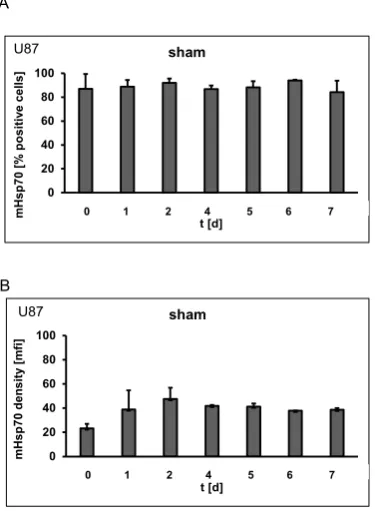
days. Representative data of the mHsp70 expression on U87 cells during a period of
7 days after sham irradiation (0 Gy) are summarized in Figure 1. No significant
differences in the percentage of positively stained cells (Fig. 1A) and in the mean
fluorescence intensity (mfi) (Fig. 1B) were detected over the whole period of time.
Similar results were obtained for untreated control cells (data not shown). The slightly
lower mHsp70 expression density on day 0 can be attributed to the short incubation
period of 5h after trypsin treatment.
Irradiation induces an increase in the mHsp70 density on different tumor cell lines
Although the percentage of mHsp70 positivity remains unaltered on U87 (Fig. 2A),
HeLa (Fig. 2B) and HepG2 (Fig. 2C) cells on day 1 after sham (0Gy), 4 Gy and 6 Gy
irradiation, the expression density of Hsp70 differed significantly upon radiation. A
radiation dose of 6 Gy nearly doubled the mean fluorescence intensity (mfi) values in
all three tumor cell lines. In U87 cells the mfi raised from 38.9±15.8 to 47.1±16.0 and
83.5±14.2 after irradiation with 4 Gy and 6 Gy, respectively (Fig. 2D, p=0.01). In HeLa
cells the mfi increased from 9.2±1.9 to 20.6±6.8 (Fig. 2E, p=0.03) on 1 day after a
5
22.0±2.6 (p=0.04) after a radiation dose of 4 Gy and to 22.3±4.6 (Fig. 2F, p=0.06) after
6 Gy. Similar results were obtained when untreated control cells were used instead of
sham treated cells (data not shown).
Irradiation-induced upregulation of mHsp70 is transient
Cell viability of U87 cells was not significantly affected by a radiation with 4 Gy and 6
Gy between days 1 to 7. A comparison of the mHsp70 densities on days 1, 4 and 7
after irradiation with 4 Gy and 6 Gy revealed significantly upregulated mfi values on
days 1 and 4 and a drop to initial levels on day 7 after irradiation. On day 1, significant
differences were detected in sham (0 Gy) versus 6 Gy irradiated cells and in 4 Gy
versus 6 Gy irradiated cells (p≤0.01), whilst no statistically significance was observed
in sham versus 4 Gy irradiated cells (Fig. 3A). On day 4 after irradiation, the mfi of
sham (0 Gy) treated cells was 41.9±0.8 compared to 79.3±12.8 and 75.1±7.7 after
irradiation with 4 Gy and 6 Gy. Significant differences were observed in sham versus
4 Gy and sham versus 6 Gy irradiated cells (p≤0.05), whilst no statstically significance
was observed in 4 Gy versus 6 Gy irradiated tumor cells (Fig. 3B). On day 7 after
irradiation, the mfi values of sham treated cells remained at 38.73±1.41 and the mfi of
4 Gy and 6 Gy irradiated cells dropped to initial levels (37.60±7.24 and 40.02±5.71,
respectively). No significant differences in the mfi values were observed in the
differently irradiated cells on day 7 (Fig. 3C).
Irradiation-induced effects on the mHsp70 density are dose- and time-dependent
The previous results indicated clear differences in the mHsp70 expression density on
U87 cells that depend on the recovery time after irradiation. Figure 4 summarizes the
dose- and time-dependent effects on the mHsp70 density. While mfi values of sham
(0 Gy) treated cells did not differ significantly over a period of 7 days when different
cell counts were seeded (Fig. 1B), an exposure to an irradiation dose of 2 Gy showed
6
5 that gradually decrease to 39.3±4.3 on day 7 (Fig. 4A). Cells that have been
irradiated with a higher dose of 4 Gy showed a similar kinetic to that after a 2 Gy
irradiation but the mHsp70 density reached a maximum in the mfi of 79.3±12.8 already
on day 4 instead of day 5. The drop in the mfi values thereafter occurred more rapid
than after an irradiation with 2 Gy (Fig. 4B). Following an irradiation with 6 Gy, mHsp70
expression density was significantly increased already on d1 compared to the sham (0
Gy) irradiated cells (23.3±3.7 versus 83.6±14.2; p≤0.05). After an irradiation with 6 Gy
the significantly elevated mHsp70 levels persisted over 3 days and dropped
significantly on day 5 (Fig. 4C). A comparison of all time points and all radiation doses
shown in Figure 4D reflects the different kinetics of the mHsp70 densities in U87 cells.
Increased mHsp70 expression densities are associated with an increased cytolytic activity mediated by TKD/IL-2 activated NK cells
The density of the mHsp70 expression plays a crucial role for the recognition by
TKD/IL-2 activated NK cells. Therefore, the cytolytic activity of Hsp70 peptide activated
NK cells was tested against sham (0 Gy) and irradiated (4 Gy) tumor cells. The
increase in the mHsp70 density on day 4 after irradiation is shown in Figure 5A. The
lysis of irradiated (4 Gy) tumor cells by TKD/IL-2 activated NK cells is significantly
higher than that of sham (0 Gy) irradiated cells at E:T ratios ranging between 50:1 and
3:1 (Fig. 5B; p≤0.05).
3. Discussion
Compared to normal cells, highly aggressive tumor cells frequently exhibit higher
cytosolic Hsp70 levels which are further upregulated upon environmental stress [12].
Intracellularly, Hsp70 ensures proper protein folding and transport [10] and interferes
with both, intrinsic and extrinsic apoptosis pathway to avoid cell death and thereby
7
released in lipid microvesicles such as exosomes [14-15] and is expressed on the
plasma membrane of tumor but not normal cells [16]. While cytosolic Hsp70 is mainly
responsible for maintaining protein homeostasis [4], mHsp70 mediates dual functions,
on the one hand it stabilizes lysosomal and plasma membranes and thereby protects
tumor cells from apoptosis [17], on the other hand it provides a tumor-specific target
for NK cells that have been pre-activated with an immunogenic Hsp70 peptide TKD
plus low dose IL-2 (TKD/IL-2) [8,18]. The specific membrane localization of Hsp70 on
tumor but not normal cells can be explained by an interaction of Hsp70 with
tumor-specific lipid compounds such as globoyltriaosylceramide Gb3 [16,19-20]. In vitro lipid
copellation assays revealed that recombinant Hsp70 interacts with the tumor-specific
lipid raft component globoyltriaosylceramid Gb3 or with the non-raft lipid compound
phosphatidylserine (PS) which translocates to the outer plasma membrane leaflet upon
stress [21]. Previously, we could demonstrate that environmental stress not only
enhances the synthesis of Hsp70 in the cytosol, but also increases the expression
density of mHsp70 on tumor cells [3,20,22]. Since mHsp70 serves as a target for
TKD/IL-2 stimulated NK cells [10] in vitro [23] and in vivo [24], time- and
dose-dependent effects of irradiation were studied with respect to the mHsp70 density on
human tumor cells. A better understanding of the mHsp70 expression kinetics following
irradiation might help to identify an optimal therapeutic window for combination
therapies consisting of ionizing irradiation and Hsp70 targeting NK cell-based immune
therapies. Herein, we could demonstrate that the mHsp70 density correlates the
cytolytic activity of Hsp70-targeting NK cells.
Glioblastoma multiforme (GBM) the most common malignant neoplasia of the brain in
adults is associated with high mortality rates. Despite multimodal treatment strategies
consisting of surgery, radiotherapy and systemic chemotherapy using Temozolomide
8
Its intratumoral heterogeneity, concerning genetic alterations and morphology as well
as the infiltrative growth and diffuse dissemination into the brain parenchyma often
hinder a successful treatment of GBM. Therefore, there is a high medical need for
innovative treatment strategies for glioblastoma. The mHsp70 density as a potential
NK cell target was measured in the human glioblastoma cell line U87 at different time
points after irradiation with doses in the range of 2 Gy to 6 Gy. To rule out cell density
dependent effects on the mHsp70 expression and to maintain cells in the exponential
growth phase, tumor cells were seeded at different cell densities which were adapted
to the different culture periods. In sham as well as non-treated tumor cells the
proportion of mHsp70 positive cells remained stable at nearly 100% with a relatively
low mfi value over the whole culture period of 7 days. These findings are in line with
previous results obtained with other tumor types such as epithelial tumor cells of the
head and neck area [26].
Environmental stress such as ionizing irradiation at sublethal doses initiates the
synthesis of Hsp70 in tumor and normal cells [10], but only tumor cells show an
up-regulated mHsp70 expression [17]. Herein, we have shown for the first time that the
kinetics of the mHsp70 expression is dependent on the irradiation dose. While the
maximum mHsp70 expression is reached on day 5 after an irradiation with 2 Gy, the
peak mHsp70 expression is detected already on day 1 after an irradiation with 6 Gy.
Furthermore, after low irradiation doses the decrease to initial mHsp70 levels occurs
earlier than after a higher irradiation dose. This means that a low dose irradiation might
be associated with a later onset and a shorter peak of the mHsp70 expression
compared to a high dose irradiation. These findings are in accordance to results of
Diller et al. who showed that the kinetics of the heat shock protein synthesis is transient
9
The intracellular Hsp70 levels remain unaltered in sham treated tumor cells, whilst
during irradiation with a sublethal dose of 2 Gy and directly after irradiation with lethal
doses [28] results in a considerable increase in the Hsp70 synthesis [2,10,16,29]. In
line with our assumption that synthesis and expression of Hsp70 is dependent on dose-
and time-dependent factors it can be assumed that cytoprotective repair mechanisms
that depend on the anti-apoptotic molecular chaperone Hsp70 are also related to the
applied stress. Our findings indicate that a high, but yet non-lethal irradiation dose of 6
Gy induces an early onset of the mHsp70 expression after 1 day with a sustained
overexpression that persists for 3 days, whereas lower irradiation doses (below 4 Gy)
induce a slower up-regulation kinetics with an earlier drop. In an effort to define the
optimal timing for the application of ex vivo stimulated mHsp70 targeting NK cells after
radiotherapy, it is important to determine the maximum density of mHsp70 on tumor
cells. In the case of U87 glioblastoma cells an irradiation dose between 4 Gy and 6 Gy
might be optimal to achieve a relative long-lasting upregulation of the mHsp70
expression, in vitro. However, future preclinical studies using fractionated irradiation
protocols are necessary to define the optimal irradiation dose which is suitable for an
in vivo application of mHsp70-targeting immune cell-based therapies.
There is strong evidence that a lethal dose of 10 Gy which drastically harms tumor cell
survival cannot be compensated by cytoprotective repair mechanisms that include the
cytosolic overexpression of Hsp70 [30]. After lethal irradiation Hsp70 will be
externalized by dying tumor cells as a danger associated molecular pattern (DAMP)
with the capacity to initiate anti-tumor immune responses [1,5,31] Hsp70-chaperoned
tumor peptides can induce CD8+ T cell mediated immune responses after cross
presentation of immunogenic tumor antigens on MHC class I molecules [32], whereas
peptide-free Hsp70 in the context of pro-inflammatory cytokines such as IL-2 can
10
remaining therapy-resistant mHsp70 positive tumor cells [14,23]. In summary, the
applied radiation dose and time kinetics play a critical role in optimizing
radiation-induced effects by increasing the density of mHsp70 expression on the surface of
surviving residual tumor cells as a target for immune cells. Membrane-bound Hsp70
plays a crucial role in stimulating the innate and adaptive immune system as it is
selectively expressed on the cell surface of tumor, but not normal cells [33] and
therefore serves as a tumor-specific recognition structure [19]. We could show that
Hsp70-targeting NK cells are capable to specifically recognize and kill tumor cells
presenting Hsp70 on their plasma membrane via granzyme B mediated apoptosis [9].
4. Material and Methods Cell line and culture conditions
The epithelial human primary glioblastoma cell line U87 MG (ATCC HTB-14) and the
human hepatocellular carcinoma cell line HepG2 (ATCC HB-8065) were grown in
complete growth medium, consisting of Dulbecco`s Eagle`s Minimum Essential
Medium (DMEM) (Sigma-Aldrich, Steinheim Germany) supplemented with 10% heat
inactivated fetal calf serum (FCS) (Sigma-Aldrich), 1% antibiotics (10,000 IU/ml
penicillin, 10 mg/ml streptomycin, Sigma-Aldrich), L-glutamine (Sigma-Aldrich), MEM
non-essential amino acid solution 100x Aldrich) and sodium pyruvate
(Sigma-Aldrich).The epithelial human cervix carcinoma cell line HeLa (ATCC CCL-2) was
grown in complete growth medium, consisting of RPMI-1640 (Sigma-Aldrich) Germany)
supplemented with 10% heat inactivated FCS (Sigma-Aldrich), 1% antibiotics (10,000
IU/ml penicillin, 10 mg/ml streptomycin, Sigma-Aldrich), L-glutamine (Sigma-Aldrich)
and sodium pyruvate (Sigma-Aldrich). After reaching confluency, adherent growing
tumor cells were trypsinized for 2 minutes at 37°C in trypsin ethylene
11
were seeded in 15 ml supplemented medium in T-75 ventilated culture flasks. Tumor
cells were routinely checked for mycoplasma contamination.
Irradiation
Tumor cells were irradiated with a single dose of 0 Gy (sham), 2 Gy, 4 Gy and 6 Gy
using the Gulmay RS225A irradiation machine (Gulmay Medical Ltd., Camberley, UK)
at a dose rate of 0.90 Gy/min (15 mA, 200 kEV) or were kept untreated.
Flow cytometry of mHsp70
The mHsp70 expression was determined by flow cytometry on a FACSCalibur flow
cytometer (BD Biosciences). Single cell suspensions of sham irradiated and irradiated
U87 cells (0.4 x 10⁶ cells per vial) were collected at different time-points after radiation.
After a washing step in phosphate-buffered saline (PBS)/10% fetal calf serum (FCS)
cells were incubated either with fluorescein-isothiocyanate (FITC)-conjugated mouse
monoclonal antibody specific for mHsp70 (cmHsp70.1, IgG1, multimmune GmbH,
Munich, Germany) or with an isotype matched FITC-labeled control antibody on ice in
the dark for 30 min. Only viable cells (propidium iodide negative cells) were gated and
the proportion of positively stained cells and mean fluorescence intensity (mfi) values
were analyzed on a FACSCalibur flow cytometer (Becton Dikinson, Heidelberg,
Germany.) The mfi is a relative value of the total fluorescence intensity of
cmHsp70.1-FITC antibody stained, viable cells subtracted by the intensity of the signal intensity
obtained after staining of the cells with an isotype-matched IgG1-FITC control antibody.
Fluorescence data were analyzed and plotted by using CellQuest software (Becton
Dickinson, Heidelberg, Germany).
Europium assay
The cytolytic activity of human NK cells cultured in RPMI-1640 medium supplemented
12
10 mg/ml streptomycin), L-glutamine, sodium pyruvate and Hsp70 peptide TKD (2
µg/ml) and IL-2 (100 IU/ml) at a cell density of 5x10⁶ peripheral blood lymphocytes
(PBL) for 4 days was determined against sham (0 Gy) and 4 Gy irradiated tumor cells
at different effector to target (E:T) ratios ranging from 50:1 to 3:1 by using a standard
3.5-hour Europium assay. Mean values of an experiment in triplicates are shown. The
specific lysis was calculated according to the equation: (experimental release –
spontaneous release) / (maximum release – spontaneous release).
Statistical analysis
Each sample was measured in at least 3 independent experiments. The Gaussian
distribution of the data was tested by using the Normality test. Comparative analysis
was performed by using the Student t-test, evaluating the relevance between variables.
Data were presented as mean values with standard deviation. Differences in p values
≤0.05 were considered as statistically significant.
5. Conclusions
In contrast to intracellular Hsp70, extracellular and mHsp70 play key roles in
stimulating both, adaptive and innate immune responses and thereby might provide
protective anti-tumor immunity [8]. By its exclusive expression on the surface of tumor
cells, but not normal cells [9] mHsp70 serves as a tumor-specific target for activated
NK cells [10]. In this study we present strong evidence that the radiation dose plays a
pivotal role on the kinetics of the mHsp70 density of human glioblastoma cells. A low
dose irradiation is associated with a later onset and a shorter peak of the mHsp70
expression compared to a higher irradiation dose. Therefore, hypofractionated
irradiation schemes with higher doses might be beneficial to generate an extended
13 Acknowledgements
We thank Anett Lange for editorial assistance. Work was funded by the Deutsche
Forschungsgemeinschaft DFG (SFB824/3, INST 411/36FUGG, INST 95/980-1FUGG,
STA1520/1-1), BMBF (01GU823, 02NUK038A), BMWi (ZF4320102CS7,
14 Figure Legends
Figure 1. Percentage (A) and mean fluorescence intensity (mfi) (B) of mHsp70 positive U87 glioblastoma cells after sham irradiation (0 Gy) on days 0, 1, 2, 4, 5, 6 and 7. Bars
represent the mean value and the corresponding standard error of the mean (SEM) of
n=3 independent experiments.
Figure 2. Percentage of mHsp70 positive tumor cells U87 (A), HeLa (B) and HepG2
(C) cells on day 1 after irradiation with 0 Gy (sham), 4 Gy and 6 Gy. Mean fluorescence intensity (mfi) of mHsp70 on U87 (D), HeLa (E) and HepG2 (F) cells on day 1 after irradiation with 0 Gy (sham), 4 Gy and 6 Gy. Bars represent the mean value and the
corresponding standard error of the mean (SEM) of n=3 (U87), n=4 (HeLa), and n=3
(HepG2) independent experiments. Significance: *p≤0.05, **p≤0.01.
Figure 3. Mean fluorescence intensity (mfi) ofmHsp70 in U87 cells after sham (0 Gy), 4 Gy and 6 Gy irradiation on days 1 (A), 4 (B) and 7 (C). Bars represent the mean values and the corresponding standard error of the mean (SEM) of n=3 independent
experiments. Significance: *p≤0.05, **p≤0.01.
Figure 4. Dose- and time-dependent radiation effects on the mHsp70 mean fluorescence intensity (mfi) on human glioblastoma cells U87 over 7 days (d0-d7) with
different radiation doses 2 Gy (A), 4 Gy (B), 6 Gy (C). Bars represent the mean values and the corresponding standard error of the mean (SEM) of n=3 independent
experiments. Significance: *p≤0.05. (D) Summary of the kinetics of the mHsp70 density
after sham (0 Gy), 2 Gy, 4 Gy, 6 Gy) over a period of 7 days (d0-d7). For clarity
statistical significance was only shown in Figures 4 A, B, C but not in Figure 4D.
15
day 4 were used as target cells. (A) mHsp70 mean fluorescence intensity (mfi) on sham (0 Gy) and 4 Gy irradiated tumor target cells. (B) Lytic activity of NK cells against sham (0 Gy) and 4 Gy irradiated tumor target cells. The effector to target (E:T) ratios
are ranging from 50:1 to 3:1. The results represent mean values and the corresponding
standard error of the mean (SEM) of n=2 independent experiments in triplicates.
16 References
1. Formenti, S.C.; Demaria, S. Combining radiotherapy and cancer immunotherapy: A paradigm shift. J Natl Cancer Inst 2013,105, 256-265.
2. Pasi, F.; Paolini, A.; Nano, R.; Di Liberto, R.; Capelli, E. Effects of single or combined treatments with radiation and chemotherapy on survival and danger signals expression in glioblastoma cell lines. Biomed Res Int 2014,2014, 453497.
3. Multhoff, G.; Hightower, L.E. Distinguishing integral and receptor-bound heat shock protein 70 (hsp70) on the cell surface by hsp70-specific antibodies. Cell Stress.Chaperones. 2011,16, 251-255.
4. Radons, J. The human hsp70 family of chaperones: Where do we stand? Cell Stress Chaperones 2016,21, 379-404.
5. Beere, H.M.; Wolf, B.B.; Cain, K.; Mosser, D.D.; Mahboubi, A.; Kuwana, T.; Tailor, P.; Morimoto, R.I.; Cohen, G.M.; Green, D.R. Heat-shock protein 70 inhibits apoptosis by preventing recruitment of procaspase-9 to the apaf-1 apoptosome. Nat.Cell Biol. 2000,2, 469-475.
6. Kumar, S.; Stokes, J., 3rd; Singh, U.P.; Scissum Gunn, K.; Acharya, A.; Manne, U.; Mishra, M. Targeting hsp70: A possible therapy for cancer. Cancer Lett 2016,374, 156-166.
7. Craig, E.A. The stress response: Changes in eukaryotic gene expression in response to environmental stress. Science 1985,230, 800-801.
8. Shevtsov, M.; Multhoff, G. Heat shock protein-peptide and hsp-based immunotherapies for the treatment of cancer. Front Immunol 2016,7, 171.
9. Radons, J.; Multhoff, G. Immunostimulatory functions of membrane-bound and exported heat shock protein 70. Exerc.Immunol.Rev. 2005,11, 17-33.
10. Multhoff, G.; Pockley, A.G.; Schmid, T.E.; Schilling, D. The role of heat shock protein 70 (hsp70) in radiation-induced immunomodulation. Cancer Letters 2015,356, 179-184. 11. Kokowski, K.; Stangl, S.; Seier, S.; Hildebrandt, M.; Vaupel, P.; Multhoff, G.
Radiochemotherapy combined with nk cell transfer followed by second-line pd-1 inhibition in a patient with nsclc stage iiib inducing long-term tumor control: A case study. Strahlenther Onkol 2019,195, 352-361.
12. Schmid, T.E.; Multhoff, G. Radiation-induced stress proteins - the role of heat shock proteins (hsp) in anti- tumor responses. Curr.Med.Chem. 2012,19, 1765-1770.
13. Multhoff, G. Heat shock protein 70 (hsp70): Membrane location, export and immunological relevance. Methods 2007,43, 229-237.
14. Gastpar, R.; Gehrmann, M.; Bausero, M.A.; Asea, A.; Gross, C.; Schroeder, J.A.; Multhoff, G. Heat shock protein 70 surface-positive tumor exosomes stimulate migratory and cytolytic activity of natural killer cells. Cancer Res. 2005,65, 5238-5247.
15. Ireland, H.E.; Leoni, F.; Altaie, O.; Birch, C.S.; Coleman, R.C.; Hunter-Lavin, C.; Williams, J.H. Measuring the secretion of heat shock proteins from cells. Methods 2007,43, 176-183. 16. Vega, V.L.; Rodriguez-Silva, M.; Frey, T.; Gehrmann, M.; Diaz, J.C.; Steinem, C.; Multhoff, G.;
Arispe, N.; De Maio, A. Hsp70 translocates into the plasma membrane after stress and is released into the extracellular environment in a membrane-associated form that activates macrophages. J.Immunol. 2008,180, 4299-4307.
17. Murakami, N.; Kuhnel, A.; Schmid, T.E.; Ilicic, K.; Stangl, S.; Braun, I.S.; Gehrmann, M.; Molls, M.; Itami, J.; Multhoff, G. Role of membrane hsp70 in radiation sensitivity of tumor cells. Radiat Oncol 2015,10, 149.
18. Hantschel, M.; Pfister, K.; Jordan, A.; Scholz, R.; Andreesen, R.; Schmitz, G.; Schmetzer, H.; Hiddemann, W.; Multhoff, G. Hsp70 plasma membrane expression on primary tumor biopsy material and bone marrow of leukemic patients. Cell Stress.Chaperones. 2000,5, 438-442. 19. Multhoff, G.; Mizzen, L.; Winchester, C.C.; Milner, C.M.; Wenk, S.; Eissner, G.; Kampinga,
17
20. Gehrmann, M.; Marienhagen, J.; Eichholtz-Wirth, H.; Fritz, E.; Ellwart, J.; Jaattela, M.; Zilch, T.; Multhoff, G. Dual function of membrane-bound heat shock protein 70 (hsp70), bag-4, and hsp40: Protection against radiation-induced effects and target structure for natural killer cells. Cell Death.Differ. 2005,12, 38-51.
21. Schilling, D.; Gehrmann, M.; Steinem, C.; De, M.A.; Pockley, A.G.; Abend, M.; Molls, M.; Multhoff, G. Binding of heat shock protein 70 to extracellular phosphatidylserine promotes killing of normoxic and hypoxic tumor cells. FASEB J. 2009,23, 2467-2477.
22. Ferrarini, M.; Heltai, S.; Zocchi, M.R.; Rugarli, C. Unusual expression and localization of heat-shock proteins in human tumor cells. Int.J.Cancer 1992,51, 613-619.
23. Multhoff, G.; Pfister, K.; Gehrmann, M.; Hantschel, M.; Gross, C.; Hafner, M.; Hiddemann, W. A 14-mer hsp70 peptide stimulates natural killer (nk) cell activity. Cell Stress.Chaperones.
2001,6, 337-344.
24. Stangl, S.; Wortmann, A.; Guertler, U.; Multhoff, G. Control of metastasized pancreatic carcinomas in scid/beige mice with human il-2/tkd-activated nk cells. J.Immunol. 2006,176, 6270-6276.
25. Stupp, R.;Hegi, M.E.;Mason, W.P.;Van Den Bent, M.J.;Taphoorn, M.J.;Janzer, R.C.;Ludwin, S.K.;Allgeier, A.;Fisher, B.;Belanger, K.; et al. Effects of radiotherapy with concomitant and adjuvant temozolomide versus radiotherapy alone on survival in glioblastoma in a
randomised phase iii study: 5-year analysis of the eortc-ncic trial. Lancet Oncol 2009,10, 459-466.
26. Kleinjung, T.;Arndt, O.;Feldmann, H.J.;Bockmuhl, U.;Gehrmann, M.;Zilch, T.;Pfister,
K.;Schonberger, J.;Marienhagen, J.;Eilles, C.; et al. Heat shock protein 70 (hsp70) membrane expression on head-and-neck cancer biopsy-a target for natural killer (nk) cells.
Int.J.Radiat.Oncol.Biol.Phys. 2003,57, 820-826.
27. Diller, K.R. Stress protein expression kinetics. Annu.Rev.Biomed.Eng 2006,8, 403-424. 28. Issels, R.D.; Meier, T.H.; Muller, E.; Multhoff, G.; Wilmanns, W. Ifosfamide induced stress
response in human lymphocytes. Mol.Aspects Med. 1993,14, 281-286.
29. Paolini, A.; Pasi, F.; Facoetti, A.; Mazzini, G.; Corbella, F.; Di Liberto, R.; Nano, R. Cell death forms and hsp70 expression in u87 cells after ionizing radiation and/or chemotherapy. Anticancer Res 2011,31, 3727-3731.
30. Rylander, M.N.; Feng, Y.; Bass, J.; Diller, K.R. Thermally induced injury and heat-shock protein expression in cells and tissues. Ann N Y Acad Sci 2005,1066, 222-242.
31. Pockley, A.G.; Henderson, B. Extracellular cell stress (heat shock) proteins-immune responses and disease: An overview. Philos Trans R Soc Lond B Biol Sci 2018,373,
32. Srivastava, P. Interaction of heat shock proteins with peptides and antigen presenting cells: Chaperoning of the innate and adaptive immune responses. Annu Rev Immunol 2002,20, 395-425.
18 0 20 40 60 80 100
0 1 2 4 5 6 7
m H sp 70 [ % p osi tiv e cel ls] t [d] sham
0 1 2 4 5 6 7
0 20 40 60 80 100
1 2 3 4 5 6 7
m H sp 70 d en si ty [m fi] t [d] sham
0 1 2 4 5 6 7 U87
A
B
Figure 1