R E S E A R C H
Open Access
Prenatal muscle development in a
mouse model for the secondary
dystroglycanopathies
Jihee Kim, Mark Hopkinson, Manoli Kavishwar, Marta Fernandez-Fuente and Susan Carol Brown
*Abstract
Background:The defective glycosylation ofα-dystroglycan is associated with a group of muscular dystrophies that are collectively referred to as the secondary dystroglycanopathies. Mutations in the gene encoding fukutin-related protein (FKRP) are one of the most common causes of secondary dystroglycanopathy in the UK and are associated with a wide spectrum of disease. Whilst central nervous system involvement has a prenatal onset, no studies have addressed prenatal muscle development in any of the mouse models for this group of diseases. In view of the pivotal role ofα-dystroglycan in early basement membrane formation, we sought to determine if the muscle formation was altered in a mouse model of FKRP-related dystrophy.
Results:Mice with a knock-down in FKRP (FKRPKD) showed a marked reduction inα-dystroglycan glycosylation and reduction in laminin binding by embryonic day 15.5 (E15.5), relative to wild type controls. In addition, the total number of Pax7+progenitor cells in the FKRPKDtibialis anterior at E15.5 was significantly reduced, and myotube cluster/myofibre size showed a significant reduction in size. Moreover, myoblasts isolated from the limb muscle of these mice at E15.5 showed a marked reduction in their ability to form myotubes in vitro.
Conclusions:These data identify an early reduction of lamininα2, reduction of myogenicity and depletion of Pax7+progenitor cells which would be expected to compromise subsequent postnatal muscle growth and its ability to regenerate postnatally. These findings are of significance to the development of future therapies in this group of devastating conditions.
Keywords:Dystroglycanopathy, Pax7, Congenital muscular dystrophy, Fukutin-related protein, Laminin, Dystroglycan
Background
Dystroglycan is a key component of the dystrophin-glycoprotein complex (DGC) which connects the extracel-lular matrix to the muscle fibre cytoskeleton [1]. It is com-posed of two non-covalently associated sub-units,αandβ. α-dystroglycan is subject to extensive O-mannosylation which is responsible for binding to extracellular matrix proteins such as laminin, perlecan and agrin [2], in addition to neurexin in the brain [3], pikachurin in the eye [4] and most recently, slit [5] in the nervous system. β -dystroglycan is a transmembrane protein which links
α-dystroglycan to dystrophin and utrophin on the
cytoplasmic side of the sarcolemma [6]. The abnormal glycosylation of this protein is the underlying cause for a subset of inherited autosomal recessive muscular dystrophies, collectively known as the secondary dys-troglycanopathies [7, 8].
To date, up to 18 genes have been implicated in the secondary dystroglycanopathies. Mutations in the fukutin-related protein (FKRP) gene are a common cause of secondary dystroglycanopathy and are associated with a wide range of clinical features, including severe structural brain involvement resembling Walker–Warburg syn-drome (WWS) and muscle-eye-brain disease (MEB), to congenital muscular dystrophy without brain involvement (MDC1C) and adult-onset limb-girdle muscular dystrophy (LGMD2I) [9–23]. LGMD2I represents the most common form of LGMD in the UK [8]. Phenotypic variability is not
* Correspondence:[email protected]
Department of Comparative Biomedical Sciences, Royal Veterinary College, University of London, London, UK
unique for mutations in the FKRP gene, and a similar spectrum of clinical involvement has been observed for most of the other dystroglycanopathy genes [24].
Muscle differentiation is accompanied by marked changes in the glycosylation of α-dystroglycan [25, 26] mediated at least in part by the coordinated upregula-tion of like-acetylglucosaminyltransferase (LARGE), a bifunctional glycosyltransferase which is known to be responsible for the post-translational addition of the poly-saccharide repeating unit [-3-xylose-α1,3-glucuronic acid-β1-] onα-dystroglycan [27]. Blocking this process leads to a less compact basement membrane, immature neuro-muscular junctions and abnormal muscle predisposed to dystrophy [27]. However, the impact of altered/reduced glycosylation of α-dystroglycan on prenatal muscle de-velopment has not yet been evaluated despite the im-plications that this might have for muscle mass in dystroglycanopathy patients [28].
Myogenesis in the mouse may be divided into two main stages; the first is referred to as primary (embry-onic) myogenesis. During this phase, primary myotubes are generated through myoblast proliferation and fusion. The primary myotubes make an appearance between E11 and E14 in mice, extending from tendon to tendon, and the majority initially expresses the slow myosin heavy chain (MHC) although expression may switch later depending on the individual muscle [29]. Groups of primary myotubes are initially surrounded by a single basement membrane but subsequently separate and be-come surrounded by their own basement membrane [30]. The primaries act as a scaffold for subsequent gen-erations of myoblasts that fuse to form secondary, and in the larger muscles, tertiary myotubes [30]. The sec-ondary myogenesis in the mouse takes place between E14.5 and E17.5. The early secondary myotubes initially form close to the neuromuscular junctions of the primary [31]. The majority of fibres in the adult muscle originate from the secondary and tertiary myotubes, and final fibre number is determined around the time of birth [30].
We previously generated a FKRP-deficient mouse
which has a knock-down in the expression of theFKRP gene (FKRPKD). These mice die around the time of birth or within the first 24 h due to central nervous system in-volvement [32, 33] since restoration of FKRP in all neural tube-derived cells prevents perinatal lethality [34]. In the present work, we used the FKRPKDmouse to examine the role of α-dystroglycan glycosylation on primary and secondary myogenesis. On the basis of the previous work which suggests that a deficiency of fuku-tin is associated with an impairment of myoblast
precur-sor cell proliferation, differentiation and muscle
regeneration [35], our hypothesis was that a reduction in α-dystroglycan glycosylation would be associated with a reduction in Pax7+ progenitor cells. Since FKRPKDmice
are not associated with an overt muscle pathology at birth [32], we further hypothesized that the initial diver-gence between FKRPKD and wild type would take place during the later stages of muscle development. An evalu-ation of the tibialis anterior (TA) and extensor digitorum longus (EDL) at E15.5 and P0 (at birth) indicated that
both these muscles showed a reduction in α
-dystroglycan glycosylation and laminin binding in the FKRPKD compared to wild types. Moreover, there was a significant reduction in myotube cluster size at E15.5 in the TA, implying that the secondary myogenesis was al-tered. This was further supported by the finding that there was a reduction in the myogenicity of cultures de-rived from the mice at this age. Associated with this was a significant reduction in the number of Pax7+ progeni-tor cells at E15.5 and P0 in the TA. Whilst differences were also noted in the EDL, the data did not achieve statistical significance. Indeed, this might be expected given that individual muscles often differ with respect to their stage of development during embryogenesis. Over-all, these findings suggest that defects inα-dystroglycan glycosylation are associated with an early reduction in the number of Pax7+ progenitor cells which would be expected to compromise subsequent postnatal muscle growth and regeneration.
Methods Mice
taken from the entire lower hindlimb and sections of the midbelly region of the EDL and TA selected for immunolabelling.
Genotyping
Genotyping was carried out by PCR using three primers. Primers: FKRP forward (GTTGTGCTTAAACCACCTTC) and FKRP reverse (CTAGGAGGTTGAGGATGATGG) were used for detecting the wild type allele, and FKRP-Neo forward (GGTGGGATTAGATAAATGCC) and FKRP re-verse were used for detecting the mutant allele.
Alcian blue/alizarin red staining
Alcian blue stains the cartilage (blue) whilst alizarin red stains the ossified bone (red). Newborn (P0) pups were culled by cervical dislocation, and the skin, viscera and adipose tissue were removed. Bodies were placed in acet-one for 2 days and then stained with 0.3 % (w/v) alcian blue 8GS in 70 % ethanol (1 volume), 0.1 % (w/v) alizarin red S in 95 % ethanol (1 volume), acetic acid (1 volume) and 70 % ethanol (17 volumes). Filtered stock solutions of alizarin red and alcian blue were made in ad-vance. The following staining samples were washed in dis-tilled water and then cleared in 1 % (w/v) solution of potassium hydroxide for 12–48 h. Once the skeleton was clearly visible, samples were cleared with 20, 50 and 80 % glycerol in 1 % potassium hydroxide. Once cleared, all samples were stored in 100 % glycerol.
Measurements of tibia and femur length by CT scan Tibia and femur length was measured using micro-computed tomography data of scanned newborns. Newborn (P0) pups underwent 180° scans in a Skyscan 1172F scanner with a 10 MP X-ray detector (Bruker, Kontich, Belgium). The X-ray source settings were at 40 kV and 200 mA, a 0.5-mm aluminium filter was used with a 6-μm pixel size and an exposure time of 2300 ms. Projection images were reconstructed into tomograms using NRecon (Skyscan) and then volume rendered using CTVox (Skyscan) to give 3D models which were measured using ImageJ [37].
Immunohistochemistry
Prior to immunolabelling, all slides were allowed to air-dry. Unfixed sections were used throughout this study with the exception of the Ki67 (DAKO), Pax7 (DSHB) and IIH6 (Millipore) where sections were fixed in 4 % paraformaldehyde (PFA) for 10 min, washed and per-meabilised in 0.1~0.5 % triton in phosphate-buffered so-lution (PBS) for 10–15 min. After washing, sections were blocked in 10 % horse serum (PAA) in PBS (10 % horse serum in PBS + 5 %BSA for IIH6) for 1 h prior to incubation with adequate primary antibodies at room temperature for 1 h (with the exception of the IIH6
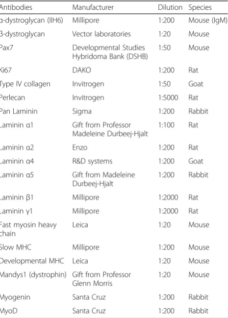
antibody which was incubated overnight at 4 °C). To minimise background staining when labelling for Pax7 and Ki67, the Mouse On Mouse immunodetection kit was used (Vector Laboratories). All the other primary antibodies (listed in Table 1) were applied to the sec-tions after blocking in 10 % horse serum in PBS for 1 h at room temperature. All dilutions were made up with PBS. The primary antibodies were incubated for 1 h at room temperature and the secondary antibodies for 30 min at room temperature followed by another 15 min incubation with streptavidin 488/594 (room temperature, Invitrogen) if a biotinylated secondary antibody was used. Nuclei were labelled with Hoechst 33342 trichloride (DAPI) (Sigma-Aldrich), which was diluted at 1:2000 with PBS. Controls for background included an incubation with the secondary antibody. In between all incubations, slides were washed in PBS three times for 5 min each time. The slides were then mounted with cover slides using Hydromount (Diag-nostic laboratories, UK). Immunolabelled slides were
viewed under epifluorescent microscope (Leica
DM4000B interfaced with a Zeiss AxioCam MRm monochrome camera.
Table 1Antibodies used in immunohistochemistry
Antibodies Manufacturer Dilution Species
α-dystroglycan (IIH6) Millipore 1:200 Mouse (IgM)
β-dystroglycan Vector laboratories 1:20 Mouse Pax7 Developmental Studies
Hybridoma Bank (DSHB)
1:50 Mouse
Ki67 DAKO 1:200 Rat
Type IV collagen Invitrogen 1:50 Goat
Perlecan Invitrogen 1:5000 Rat
Pan Laminin Sigma 1:200 Rabbit
Lamininα1 Gift from Professor Madeleine Durbeej-Hjalt
1:100 Rat
Lamininα2 Enzo 1:200 Rat
Lamininα4 R&D systems 1:200 Goat Lamininα5 Gift from Madeleine
Durbeej-Hjalt
1:200 Rabbit
Lamininβ1 Millipore 1:2000 Rat Lamininγ1 Millipore 1:2000 Rat Fast myosin heavy
chain
Leica 1:20 Mouse
Slow MHC Millipore 1:200 Mouse
Developmental MHC Leica 1:20 Mouse
Mandys1 (dystrophin) Gift from Professor Glenn Morris
1:20 Mouse
Myogenin Santa Cruz 1:200 Rabbit
Western blotting and laminin overlay assay
The muscle was dissected from the entire limb and cell proteins extracted in lysis buffer, radio immunoprecipita-tion assay (RIPA) buffer consisting of 50 mM Tris-HCl pH 7.5, 150 mM NaCl, 1 mM EDTA, 1 % Triton X-100, 0.1 % SDS and 1 mM azide, plus a cocktail of protease inhibitors (Roche). Protein concentration was measured using the BCA Protein Assay Kit (Thermo Scientific Pierce). A 30μg of soluble proteins was resolved using a NuPAGE Pre-cast gel (3–8 % Tris-acetate; Invitrogen) and then transferred electrophoretically to Hybond-PVDF membrane (GE Healthcare, UK). PVDF membrane were blocked in 5 % dried non-fat milk in phosphate-buffered saline buffer, and then probed with the primary antibodies: anti-mouseα-DG IIH6 (Millipore, UK, cat, 05-593) anti-mouseβ-DG (Vector Labs, UK), at room temperature for 1 h. After washing, they were incubated with the appropri-ate horseradish peroxidase (HRP)-conjugappropri-ated secondary antibody for 1 h: anti-mouse-IgM or anti-mouse-IgG (both from Jackson ImmunoResearch, USA). After wash-ing, the membranes were visualized using enhanced chemiluminescence (ECL) substrate (Thermo scientific Pierce) and imaged using a Bio-Rad ChemiDoc MP imaging system.
For the laminin overlay assay, PVDF membranes were blocked for 1 h in laminin-binding buffer (LBB; 10 mM triethanolamine, 140 mM NaCl, 1 mM MgCl2, 1 mM CaCl2, pH 7.6) containing 5 % non-fat dry milk followed by
incubation of 1 μg/ml mouse Engelbreth–Holm–Swarm
(EHS) laminin protein (Invitrogen) in LBB overnight on a roller at 4 °C. The membranes were washed and incubated with rabbit anti-laminin (Sigma) followed by HRP-anti-rabbit IgG (Jackson ImmunoResearch). Blots were visual-ized using enhanced chemiluminescence (ECL) substrate (Thermo scientific Pierce) and imaged using a Bio-Rad ChemiDoc MP imaging system.
Tissue culture
All muscles were removed from the limbs of E15.5 em-bryos and digested with freshly made enzyme solution. The enzyme solution consisted of phosphate-buffered saline (PBS; Gibco) mixed with 0.5 mg/ml w/v collage-nase (Sigma–Aldrich), 1 mg/ml w/vBovine serum albu-min (BSA; Sigma–Aldrich) and 6 %v/vTrypsin-ethylene diamine tetra acetic acid (EDTA; Invitrogen). This was incubated with the muscle following trituration for 30 min at 37 °C. After 30 min, the enzyme solution was removed and discarded in order to dispose of fibroblasts. The second and third digestions were then undertaken, and the cells from each were pooled and placed in growth medium (GlutaMAX DMEM (Gibco)) contain-ing 1 % penicillin–streptomycin–neomycin (PSN; Gibco) and 20 % fetal bovine serum (FBS). This cell solution was then plated onto Matrigel (1 mg/ml) coated plate or
chamber slides directly to facilitate proliferation. Differ-entiation was initiated by switching to 10 % FBS after 48 h (Gibco).
Quantification and statistical analysis
ImageJ was used to measure myotube clusters/fibre size [37]. This was done manually using a light pen on im-ages (resolution 150 pixels/in.) at either ×10 or ×20 magnification.
The expression of the transcription factor Pax7 was assessed across the entire cross-sectional area of the muscle. The quantification of the number of Pax7+ nu-clei was expressed as the number per fibre in the TA and EDL at E15.5 and P0.
Data were analysed using GraphPad Prism 5 software. Confirmation of normality was undertaken using Kol-mogorov–Smirnov test; unpairedttests were performed to compare wild type and mutant animals and ANOVA for comparisons between wild type, heterozygotes and mutants.
Results
Litter size and longitudinal growth were not altered in the newborn FKRPKDmice
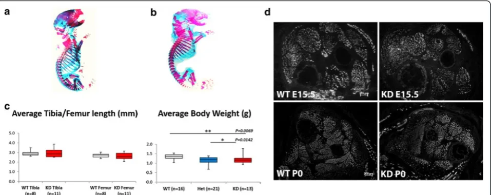
A comparison of alcian blue/alizarin red-stained wild type and FKRPKD pups at P0 which showed the extent of the ossified bone (red) and cartilage (blue) indicated no significant differences with respect to skeletal devel-opment between the two genotypes (Fig. 1a, wild type and 1B FKRPKD). Measurements of the tibia and femur using computed tomography as an indirect measure-ment of muscle length showed no significant differences between WT and FKRPKD(Fig. 1c). Body weight was sig-nificantly lower in the FKRPKDat P0 relative to wild type as previously reported, whilst litter size influenced this parameter, overall litter size was not significantly differ-ent between WT and FKRPKD.
A reduction inα-dystroglycan glycosylation does not alter muscle patterning but is associated with an alteration in laminin immunobinding by E15.5 and P0
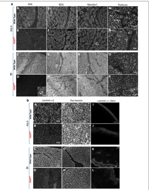
Transverse sections through the entire lower hindlimb at E15.5 and P0 of both FKRPKDand wild type immuno-labelled for slow myosin heavy chain showed no alter-ation in muscle patterning (Fig. 1d). In the tibialis anterior (TA) and extensor digitorum longus (EDL), a population of the larger primary myotubes could be clearly identified. The majority of these primaries at E15.5 were surrounded by smaller secondaries forming myotube clusters which were enclosed within a single basement membrane delineated by immunolabelling for laminin, perlecan or collagen type IV. In wild type mice immunolabelling for IIH6 (identifies the laminin binding
basement membrane markers (Fig. 2a). However, in the FKRPKDat E15.5 and P0, although there was some vari-ation between pups most likely due to the model being a hypomorph, overall, there was a marked reduction in IIH6 immunolabelling. These observations therefore in-dicate that glycosylated α-dystroglycan is present from the earliest stages of myotube formation in wild type mice and is markedly reduced in the FKRPKD. However, β-dystroglycan was unchanged between wild type and FKRPKDas was dystrophin which marks the sarcolemma of both primary and the early secondaries (Fig. 2a). Per-lecan, an additional ligand of α-dystroglycan also dependent on its proper glycosylation showed no differ-ence between mutant and wild type (Fig. 2a).
In order to make an assessment of the relative molecu-lar mass and laminin binding properties during muscle develoImmunolabelling using an antibodypment, we next compared α-dystroglycan at E15.5 and P0 in wild
type and FKRPKD mice using Western blotting. This
showed that, as has been previously observed during de-velopment of the chick and human muscle, the relative molecular mass ofα-dystroglycan in wild type muscle is reduced in fetal relative to the adult mouse muscle (Fig. 3b) [25, 26]. Protein loading was assessed using Amido Black staining of the PVDF membrane (Fig. 3a). In agreement with the immunolabelling, only residual IIH6 labelling was observed following Western blot ana-lysis of FKRPKD at either E15.5 or P0. A laminin overlay assay further showed that whilstα-dystroglycan was able
to bind laminin at both E15.5 or P0 in the wild type, no binding was observed in the FKRPKD (Fig. 3c). Taken overall, these observations imply thatα-dystroglycan gly-cosylation plays a role in basement membrane assembly during the early stages of fibre formation.
Expression of lamininα2 is reduced early during secondary myogenesis
(See figure on previous page.)
Fig. 2Immunolabelling ofα- andβ-dystroglycan, dystrophin and perlecan.aTransverse sections through the TA and EDL at E15.5 and P0 immunolabelled forα- (A-D) andβ-dystroglycan (E-H), dystrophin (Mandys1, I-L) and perlecan (M-P).α-dystroglycan (IIH6) showed a clear delineation of the basement membrane around clusters of primary and secondary myotubes at E15.5 (A) and individual muscle fibres at P0 (C), although some clusters are still evident. There was an almost total absence of IIH6 in the FKRPKD at both time points (B,D). Residual staining of the neuromuscular junction in the FKRPKD at P0 is shown in D as an inserted image.β-dystroglycan, dystrophin and perlecan immunolabelling were not markedly different between FKRPKDand wild type at either time point (E-P)
Fig. 3IIH6 Western blot and laminin overlay at E15.5 and P0.aShows an amido black stain of the transfer membrane indicating equivalent loading in all lanes.bWestern blot for IIH6 in the hindlimb muscle at E15.5 and P0, the relative molecular mass ofα-dystroglycan in the wild type is reduced compared to the adult mouse muscle and only residual IIH6 labelling was observed in FKRPKD.cAt both E15.5 and P0,α-dystroglycan
development there is no compensatory increase in either laminin α4 or α5 chains which is sometimes evident in adult muscle. However, given that γ1 levels were similar in FKRPKD and wild type, it remains a possibility that anotherα-chain at least partially compensates for the re-duction in lamininα2.
Primary myotube number and maturation is not altered in FKRPKDmuscle
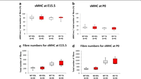
Primary myotubes differ from secondary myotubes in their size, number, development and expression of myosin chain isoforms at E15.5 in the mouse [38, 39]. The major-ity of slow myotubes initially express slow myosin [40] consequently we initially evaluated the number of slow myosin expressing myotubes in both the EDL and TA. Using this criteria there were no significant differences be-tween FKRPKD and wild type with respect to primary myotube number (Fig. 5) indicating that reduced α -dystroglycan glycosylation did not influence primary myo-tube numbers. However, it was formally possible that an
alteration in the proportion of primary myotubes express-ing slow myosin could have influenced this result, conse-quently we next checked this parameter. As can be seen from Fig. 5 this proportion decreased between E15.5 and P0 in both the FKRPKDand wildtype as myosin compos-ition changes with maturity depending on the muscle. Nonetheless, the proportion of slow myosin expressing myotubes was not significantly different between the FKRPKD and wild type at either age. This indicates that myotube maturation as determined by slow myosin heavy chain expression was not altered by a reduction in α -dystroglycan glycosylation and reduction in laminin α2. Interestingly total fibre number in the TA was not signifi-cantly different which contrasts with measurements taken from midpoint along the limb at P0 shown in Fig. 6 where there was a significant increase in the number of smaller secondary myotubes in the FKRPKDrelative to wild type. Secondary myotubes initially form close to early neuro-muscular junctions, this result may therefore reflect the distance these sections were from the innervation zone. Fig. 4Immunolabelling of lamininsα4,α5γ1 andβ1 at E15.5 and P0. Immunolabelling with chain-specific antibodies raised against lamininα4 (A,B,C,D),α5 (E,F,G,H) andγ1 (I,J,K,L), also indicated no clear reduction in FKRPKDrelative to wild type at either E15.5 or P0. However, laminin
Myotube cluster size at E15.5 is reduced but total fibre number at P0 is increased across the mid belly region of the muscle of FKRPKDrelative to wild type
Substantial myoblast proliferation fuels the formation of secondary myotubes. In view of the well recognized mitogenic effects of laminin we next evaluated myotube cluster size (one primary myotube and between one and two secondary myotubes enclosed within a single base-ment membrane) (Fig. 6a). Secondary myotubes initially form in the midbelly region of the muscle and with mat-uration extend the length of the muscle. In order to make a precise comparison between wild type and FKRPKD mice, our measurements were therefore under-taken across the midbelly region of each muscle. At E15.5 average myotube cluster size was significantly re-duced in the TA but not the EDL of the FKRPKDrelative to wild type (Fig. 6b). Further analysis of the size distri-bution of these clusters showed that in both muscles there was a shift towards smaller clusters in the FKRPKD relative to wild type (Fig. 6e). Since primary myotube number was not significantly different these observations were indicative of an alteration in secondary myogenesis (Fig. 6c).
However, at P0, when many but not all secondary myotubes had attained their own basal lamina, total fibre
number was significantly increased in the TA but not
the EDL of the FKRPKD compared to wild type at P0
(Fig. 6d). Analyses of the fibre size distribution showed this to be due to a significantly higher number of smaller fibres in the FKRPKD relative to wild type in the TA. These counts were taken from sections calculated to come from a region midway between the patella and ankle joint of the hindlimb (Fig. 6f ).
FKRPKDmice show a reduced number of Pax7+progenitor cells in the TA muscle but not the EDL
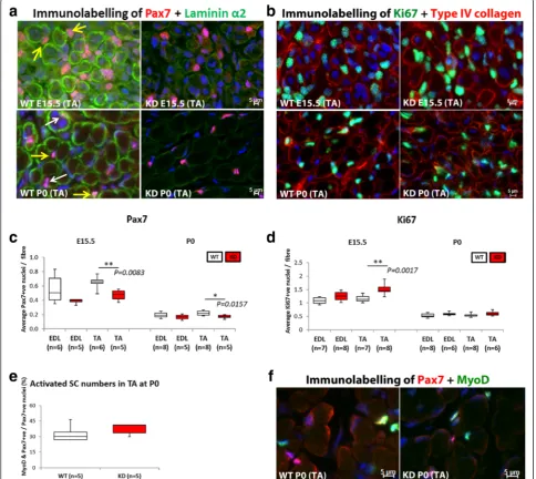
In view of the increase in myotube cluster size we next evaluated the expression of the transcription factor Pax7 which directs the differentiation of myogenic progenitor cells down the muscle lineage (Fig. 7a, b). Counts were made across the entire cross sectional area of the muscle. Quantification of the number of Pax7+ nuclei (expressed as the number per fibre) in the TA and EDL at E15.5 and P0 is shown in Fig. 7c where it can be seen that the number of Pax7+nuclei per fibre was significantly reduced in the FKRPKD TA at E15.5 and P0 (p= 0.0083 andp= 0.0157 respectively). Interestingly as with most of the other quantitative analyses there was a trend for the number of Pax7+ nuclei per fibre to be reduced in the EDL but this did not achieve statistical significance. Whilst Fig. 5Slow myosin heavy chain expression. The majority of primary myotubes initially express slow myosin. Double labelling for slow myosin and laminin/perlecan and counts of the entire muscle enabled a comparison of the percentage of primary myotubes that expressed slow myosin in the TA and EDL at E15.5 and P0 (a,b, respectively). These analyses showed no significant differences in the percentage of slow myosin positive fibres between wild type and FKRPKDindicating that primary myotube maturation was not altered by the reduction inα-dystroglycan glycosylation.
The total fibre number was also not significantly altered between wild type and FKRPKD(c,d). These counts were made on sections taken from the
Fig. 6Fibre size and fibre numbers at E15.5 and P0. Double immunolabelling of slow myosin with laminin (a). At E15.5, many myotube clusters are outlined by perlecan immunolabelling. These clusters consist of one primary and one or more smaller secondary myotubes (white arrow) together with a number of secondary myotubes/fibres that have attained their own basement membrane (yellow arrows). Fibre size (area) therefore refers to measurements of either myotube clusters or individual myotubes/fibres that have attained their own basement membrane. There are many single myotubes enclosed within their own basement membrane in the FKRPKDat E15.5 which are less prominent in the wild
type (yellow arrows). At P0, a population of smaller myotubes/fibres were still enclosed within the primary myotube basement membrane, this was more evident in the FKRPKDat P0 (
red arrows). This was reflected by a significant reduction in fibre size in the TA muscle in the FKRPKD
compared to wild type (b). Fibre number (includes both myotube clusters and myotubes/fibres with their own basement membrane) at E15.5 was not significantly different between FKRPKDand wild type as was indicated in Fig. 5 where different sections along the mid-third of the muscle
length were analysed (c). A comparison of the size distribution graph clearly shows a shift towards smaller clusters in FKRPKDrelative to wild type
at E15.5 attributed to a population of smaller secondaries attaining their own basement membrane (e). At P0, in sections taken at the midpoint region of the limb fibre counts showed a significant increase in fibre number in the TA but not in the EDL of the FKRPKDcompared to wild type
at P0 (d). The size distribution graph showed this to be due to an increase (p= 0.0547) in number of smaller fibres in the FKRPKDat P0 compared
the majority of Pax7+ nuclei appeared to be within the confines of the basal lamina at P0 - as delineated by lam-inin immunolabelling, there were also many whose loca-tion was ambiguous and so we could not accurately quantify whether satellite cell homing itself had been dis-turbed (Fig. 7a).
Interestingly the proportion of Pax7+/MyoD+ nuclei in the FKRPKD TA was similar to wild type at P0 indicating that myogenic differentiation was not delayed (Fig. 7e, f ).
However, the number of Pax7+ nuclei/fibre was signifi-cantly reduced in the FKRPKDTA compared to wild type.
The number of Ki67 positive nuclei in the FKRPKDwas increased relative to wild type in the TA but not the EDL at E15.5
In view of the reduction in Pax7 positive nuclei per fibre we next sought to determine if cellular proliferation was altered. Labelling for Ki67 (cell proliferation marker) Fig. 7Pax7 positive progenitor cell quantification.ashows double immunolabelling for Pax7 and lamininα2 in the TA, whilstbshows Ki67+ and type IV collagen in the TA.White arrowindicates Pax7+nuclei laying beneath the basal lamina whilst theyellow arrowshows Pax7+nuclei outside of the basal lamina (P0). However, many nuclei were in an ambiguous position and so only, the total number of Pax7+nuclei was counted. These
counts expressed as the number of Pax7+nuclei per fibre showed a significant reduction in the TA at E15.5 and P0 (
p= 0.0083 andp= 0.0157, respectively) in the FKRPKDrelative to wild type (c). There was however, a significant increase (p= 0.0017) in the number of Ki67 (cell proliferation marker) positive nuclei was observed in the TA of the FKRPKDat E15.5 but not at P0 (d). The proportion of Pax7 positive nuclei that were also
positive nuclei showed a significant increase (p= 0.0017) in the TA of the FKRPKD but not the EDL at E15.5. At birth these differences were no longer evident (Fig. 7d).
Myoblasts isolated from E15.5 embryos show reduced myogenicity relative to wild type
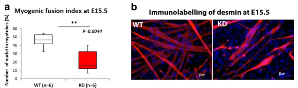
Since our data indicated an alteration in secondary myo-genesis reflected by the reduction in cluster size, we undertook primary cell culture by isolating myoblasts at E15.5, culturing these cells on a matrigel coated sub-strate and allowing them to fuse. In agreement with the reduction in myotube cluster size FKRPKD cells showed a significant reduction in the fusion index (Fig. 8a).
Discussion
The secondary dystroglycanopathies are associated with a wide clinical spectrum and a proportion of patients show defects in the central nervous system in addition to a muscular dystrophy. However, whilst neuronal mi-gration defects together with pial basement membrane defects occur prenatally, no previous reports have docu-mented a prenatal onset for the disease in muscle. In this study we hypothesized that, in view of the pivotal role played by α-dystroglycan in basement membrane organisation via its binding to ligands such as laminin and perlecan [2], muscle development would be altered such that fibre number, size and satellite cell number would be reduced. Since we and others have previously shown that the extent ofα-dystroglycan glycosylation in-creases with gestational age [25, 26] in both chick and human muscle we further reasoned that the alterations would be confined to the later stages of myogenesis.
Dystroglycan is essential for the initial binding of lam-inin on the cell surface, whereasβ1 integrins and perle-can are subsequently required for laminin matrix assembly [41]. In the present study we show that
organisation of the basement membrane as determined by IIH6 and laminin α2 immunolabelling is altered by E15.5 in the FKRPKDmice. This corresponds to a period when myoblasts align and fuse along the surface of the primary myotube and all are enclosed within a common basal lamina [30]. Since the majority of primary myo-tubes label for slow myosin heavy chain during the early stages [42] we used this as a marker for primary myo-tube formation. Primary myomyo-tube number was not sig-nificantly different in either the TA or EDL and muscle length as determined by the length of femur and tibia was also not significantly different between the FKRPKD or wild type littermates. Primary myotubes extend the full length of the muscle from their earliest stages of for-mation, and these observations therefore imply that early myotube formation and differentiation are not altered by defects in α-dystroglycan which are nonetheless present by this stage. Indeed the proportion of slow myosin heavy chain positive myotubes was not significantly dif-ferent between wild type and FKRPKD and the expres-sion patterns of fast MHC and developmental MHC was also the same between the two groups at both E15.5 and P0 which further suggested no obvious delay in matur-ation (data not shown).
Myotube clusters are a characteristic of muscle devel-opment mid gestation and they usually consist of a sin-gle primary surrounded by one or more (depending on the muscle) secondaries. Cluster size was found to be significantly reduced in the TA but not the EDL at E15.5. Since this parameter also showed a reduction in the EDL which was not statistically significant we believe that this time point represents the initial point of diver-gence in the hindlimb muscles. The isolation of myo-blasts from E15.5 hindlimbs showed a reduction in the fusion index which together with the reduction in clus-ter size suggests that a reduction in α-dystroglycan
Fig. 8Myoblast fusion at E15.5. Myoblasts were isolated from the limbs at E15.5 for the further evaluation of reduction in cluster size that we observed on transverse sections. The myogenic fusion index calculated by counting the proportion of nuclei contained within myotubes showed a significant reduction in FKRPKDrelative to wild type (a). Desmin immunolabelling further showed that the number of myotubes that formed
4–5 days after plating on Matrigel pre-coated coverslips was reduced in the FKRPKD(
glycosylation impacts upon secondary myotube forma-tion. Interestingly the number of Ki67 positive proliferat-ing cells in the TA was increased at E15.5 but not P0 suggesting a transitory increase in the division of cells not yet committed to the myogenic lineage. Given that fibre number was slightly increased in the FKRPKD at birth we assume that these cells do subsequently con-tribute to fibre formation.
α-dystroglycan binds to LG domain containing pro-teins of the extracellular matrix, including laminin and perlecan. At E15.5 and P0, immunolabelling of laminin α2 showed a reduction in FKRPKD mice relative to con-trols. However, there was no apparent alteration in per-lecan despite previous data showing the existence of a trimolecular arrangement between α-dystroglycan, perle-can and laminin, with perleperle-can binding toα-dystroglycan indirectly [43]. This suggests that the incorporation of laminin into the basement membrane during development is more dependent on α-dystroglycan glycosylation than perlecan, indeed the latter may have other binding part-ners that are sufficient to facilitate its incorporation. La-belling for type IV collagen (data not shown) displayed no clear difference in FKRPKDmice relative to controls at ei-ther E15.5 or birth showing that the oei-ther aspects of the basement membrane were unaltered by the changes inα -dystroglycan glycosylation. This is in contrast to the obser-vations made in dystroglycan-null embryoid bodies where the expression pattern of type IV collagen has been shown to be disrupted [44]. This may reflect tissue-specific
differ-ences or may reflect the absence of both α and β
-dystroglycan in these cells.
Fibre number at birth showed a significant increase in the FKRPKD TA but not EDL. This may be interpreted in several ways namely that there is a more rapid breakup of the clusters due to a weakened basement membrane or alternatively an increase in branching of the secondary myotubes. Branching is normally a re-sponse to repeated injury/overloading [45]; however, this has also been reported in dystrophin-deficient mdx and caveolin-3-deficient mouse muscle during development [46]. Indeed, it is possible that the lack ofα-dystroglycan glycosylation and the reduction in lamininα2 deposition provide inappropriate cues for the early secondary and tertiary myotubes and they branch.
Immunolabelling using an antibody to pan laminin showed no apparent difference between the two groups at both time points (Fig. 2b). Since pan laminin recog-nises several laminin chains including β1 and γ1, this observation strongly suggested that there might be a partial compensation by other laminin isoforms prior to birth for the loss of lamininα2. Lamininα5 is often up-regulated in neuromuscular conditions and thought to compensate for other α-chains. However, labelling with antibodies to laminin α4,α1,α5 and γ1 failed to detect
any significant differences between FKRPKD and con-trols. β1 on the other hand showed a subtle reduction suggesting that the expression of laminin β1 was rather influenced by the loss of lamininα2 and implies thatβ2 may partially compensate. However, real-time PCR indi-cated no significant differences between FKRPKD and wild type, although it was noted that there was a trend for all to be reduced in the FKRPKD at E15.5. Indeed, it could be that a reduction in expression levels of ligand proteins is only manifested later on during development when there has been a long-term reduction in the rele-vant receptor.
Satellite cells are first evident around E17 and typically identified using the paired box transcription factor, Pax7. We have therefore referred to those nuclei expressing Pax7 as progenitor cells rather than satellite cells. Whilst they are traditionally thought to be derived from the so-mites [47], there is now evidence to suggest that some adult satellite cells may have alternative origins other than the dermomyotome-derived Pax3+/Pax7+ progeni-tor cells [48]. The dynamic interaction between the extracellular matrix and the satellite cell plays a key role in determining activity [48, 49] and in agreement with this, we have previously shown the satellite cell niche to be disturbed in the dystroglycanopathies [50]. A series of transcription factors drive the progression from quies-cence to activation, proliferation, and differentiation/ self-renewal. Quiescent satellite cells are characterised by their expression of Pax7 and Myf5 but not MyoD or Myogenin. The initiation of differentiation and fusion is marked with the expression of Myogenin, which in con-cert with MyoD will activate muscle-specific genes [48]. In the present study, we observed that the proportion of activated progenitor cells (Pax7+/MyoD+) was not sig-nificantly different in the TA between FKRPKD and wild type at P0. This indicates that the differentiation itself has not been delayed as is supported by the pattern of myosin heavy chain expression. However, the absolute number of Pax7 positive cells was decreased in the FKRPKD TA relative to the wild type indicating that the number of committed progenitor/satellite cells has been reduced in the mutant which would be expected to com-promise not only postnatal growth but also the regen-erative capacity of the muscle.
muscular dystrophy whilst the second, deleted fukutin in muscle precursor cells using the Myf5 promoter. The earlier deletion resulted in a mouse with a more severe form of muscular dystrophy. In the present paper as-suming that fukutin and FKRP have a similar function, the reduction in the number of Pax7 progenitor cells identifies a possible mechanism behind previous obser-vations of a regeneration disorder [35, 51]. Another in-teresting aspect is the role of the CT carbohydrate antigen {GalNAcβ1,4[NeuAcα2,3]Galβ1(-3GalNAc or
-4GlcNAc)} on α-dystroglycan which when expressed
outside its usual synaptic location leads to an accumula-tion of satellite cells, reduced myofibre size and altered neuromuscular junctions [52]. This work would seem to indicate as did the work of Ross et al. [50] that alter-ations in the glycosylation pattern ofα-dystroglycan can directly impact upon the stem or satellite cell population with clear implications for postnatal muscle growth.
Conclusions
In summary, these data show for the first time a prenatal onset with regard to the disease process and identify an early reduction of laminin α2, reduction of myogenicity and depletion of Pax7 positive progenitor cells which would be expected to compromise subsequent postnatal muscle growth and its ability to regenerate postnatally. These findings are of significance to the development of future therapies in this group of devastating conditions.
Abbreviations
DAGC:dystrophin-glycoprotein complex; EDL: extensor digitorum longus; EDTA: ethylene diamine tetra acetic acid; FKRP: fukutin-related protein; FKRPKD: FKRP knock-down; LARGE: like-acetylglucosaminyltransferase;
LGMD2I: Limb-girdle muscular dystrophy type 21; MDC1C: congenital muscular dystrophy type 1C; MEB: muscle-eye-brain disease; TA: tibialis anterior; WWS: Walker–Warburg syndrome.
Competing interests
The authors declare that they have no competing interests.
Authors’contributions
JK contributed to the design of the study, acquisition of data, analysis and interpretation, has been involved in drafting the manuscript and has given final approval of the version to be published. MFF contributed to data acquisition and critically revising the manuscript. MK contributed to data acquisition and revising the manuscript. MH contributed to data acquisition and critically revising the manuscript. SCB contributed to the design of the study, drafting and revising the manuscript. All authors have given final approval of the version to be published and agree to be accountable for all aspects of the work in relation to accuracy.
Acknowledgements
We are grateful to the Muscular Dystrophy UK (JK) and the Muscular Dystrophy Association, USA (MF). We thank Professor Madeleine Durbeej-Hjalt (Lund University) for her generous donation of lamininα1 andα5 antibodies.
Received: 2 September 2015 Accepted: 5 January 2016
References
1. Campbell KP, Stull JT. Skeletal muscle basement membrane-sarcolemma-cytoskeleton interaction minireview series. J Biol Chem. 2003;278:12599–600. 2. Michele DE, Barresi R, Kanagawa M, Saito F, Cohn RD, Satz JS, et al.
Post-translational disruption of dystroglycan-ligand interactions in congenital muscular dystrophies. Nature. 2002;418:417–22.
3. Sugita S, Saito F, Tang J, Satz J, Campbell K, Sudhof TC. A stoichiometric complex of neurexins and dystroglycan in brain. J Cell Biol. 2001;154:435–45. 4. Sato S, Omori Y, Katoh K, Kondo M, Kanagawa M, Miyata K, et al. Pikachurin,
a dystroglycan ligand, is essential for photoreceptor ribbon synapse formation. Nat Neurosci. 2008;11:923–31.
5. Wright KM, Lyon KA, Leung H, Leahy DJ, Ma L, Ginty DD. Dystroglycan organizes axon guidance cue localization and axonal pathfinding. Neuron. 2012;76:931–44.
6. Blake DJ, Weir A, Newey SE, Davies KE. Function and genetics of dystrophin and dystrophin-related proteins in muscle. Physiol Rev. 2002;82:291–329. 7. Wells L. The o-mannosylation pathway: glycosyltransferases and proteins
implicated in congenital muscular dystrophy. J Biol Chem. 2013;288:6930–5. 8. Muntoni F, Torelli S, Wells DJ, Brown SC. Muscular dystrophies due to
glycosylation defects: diagnosis and therapeutic strategies. Curr Opin Neurol. 2011;24:437–42.
9. Stevens E, Carss KJ, Cirak S, Foley AR, Torelli S, Willer T, et al. Mutations in B3GALNT2 cause congenital muscular dystrophy and hypoglycosylation of alpha-dystroglycan. Am J Hum Genet. 2013;92:354–65.
10. Buysse K, Riemersma M, Powell G, van Reeuwijk J, Chitayat D, Roscioli T, et al. Missense mutations in beta-1,3-N-acetylglucosaminyltransferase 1 (B3GNT1) cause Walker-Warburg syndrome. Hum Mol Genet. 2013;22:1746–54. 11. Cirak S, Foley AR, Herrmann R, Willer T, Yau S, Stevens E, et al. ISPD gene
mutations are a common cause of congenital and limb-girdle muscular dystrophies. Brain. 2013;136:269–81.
12. Raphael AR, Couthouis J, Sakamuri S, Siskind C, Vogel H, Day JW, et al. Congenital muscular dystrophy and generalized epilepsy caused by GMPPB mutations. Brain Res. 2014;1575:66–71.
13. de BD B-V, Currier S, Steinbrecher A, Celli J, van Beusekom E, van der Zwaag B, et al. Mutations in the O-mannosyltransferase gene POMT1 give rise to the severe neuronal migration disorder Walker-Warburg syndrome. Am J Hum Genet. 2002;71:1033–43.
14. van Reeuwijk J, Janssen M, van den Elzen C, de BD B-V, Sabatelli P, Merlini L, et al. POMT2 mutations cause alpha-dystroglycan hypoglycosylation and Walker-Warburg syndrome. J Med Genet. 2005;42:907–12.
15. Brockington M, Blake DJ, Prandini P, Brown SC, Torelli S, Benson MA, et al. Mutations in the fukutin-related protein gene (FKRP) cause a form of congenital muscular dystrophy with secondary laminin alpha2 deficiency and abnormal glycosylation of alpha-dystroglycan. Am J Hum Genet. 2001;69:1198–209.
16. Yoshida A, Kobayashi K, Manya H, Taniguchi K, Kano H, Mizuno M, et al. Muscular dystrophy and neuronal migration disorder caused by mutations in a glycosyltransferase, POMGnT1. Dev Cell. 2001;1:717–24.
17. Kobayashi K, Nakahori Y, Miyake M, Matsumura K, Kondo-Iida E, Nomura Y, et al. An ancient retrotransposal insertion causes Fukuyama-type congenital muscular dystrophy. Nature. 1998;394:388–92.
18. Brockington M, Yuva Y, Prandini P, Brown SC, Torelli S, Benson MA, et al. Mutations in the fukutin-related protein gene (FKRP) identify limb girdle muscular dystrophy 2I as a milder allelic variant of congenital muscular dystrophy MDC1C. Hum Mol Genet. 2001;10:2851–9.
19. Longman C, Brockington M, Torelli S, Jimenez-Mallebrera C, Kennedy C, Khalil N, et al. Mutations in the human LARGE gene cause MDC1D, a novel form of congenital muscular dystrophy with severe mental retardation and abnormal glycosylation of alpha-dystroglycan. Hum Mol Genet. 2003;12:2853–61. 20. Lefeber DJ, Schonberger J, Morava E, Guillard M, Huyben KM, Verrijp K, et al.
Deficiency of Dol-P-Man synthase subunit DPM3 bridges the congenital disorders of glycosylation with the dystroglycanopathies. Am J Hum Genet. 2009;85:76–86.
21. Roscioli T, Kamsteeg EJ, Buysse K, Maystadt I, van Reeuwijk J, van den Elzen C, et al. Mutations in ISPD cause Walker-Warburg syndrome and defective glycosylation of alpha-dystroglycan. Nat Genet. 2012;44:581–5.
22. Willer T, Lee H, Lommel M, Yoshida-Moriguchi T, de Bernabe DB, Venzke D, et al. ISPD loss-of-function mutations disrupt dystroglycan O-mannosylation and cause Walker-Warburg syndrome. Nat Genet. 2012;44:575–80. 23. Yang AC, Ng BG, Moore SA, Rush J, Waechter CJ, Raymond KM, et al.
with dystroglycanopathy-type congenital muscular dystrophy. Mol Genet Metab. 2013.
24. Godfrey C, Clement E, Mein R, Brockington M, Smith J, Talim B, et al. Refining genotype phenotype correlations in muscular dystrophies with defective glycosylation of dystroglycan. Brain. 2007;130:2725–35. 25. Brown SC, Torelli S, Brockington M, Yuva Y, Jimenez C, Feng L, et al.
Abnormalities in alpha-dystroglycan expression in MDC1C and LGMD2I muscular dystrophies. Am J Pathol. 2004;164:727–37.
26. Leschziner A, Moukhles H, Lindenbaum M, Gee SH, Butterworth J, Campbell KP, et al. Neural regulation of alpha-dystroglycan biosynthesis and glycosylation in skeletal muscle. J Neurochem. 2000;74:70–80.
27. Goddeeris MM, Wu B, Venzke D, Yoshida-Moriguchi T, Saito F, Matsumura K, et al. LARGE glycans on dystroglycan function as a tunable matrix scaffold to prevent dystrophy. Nature. 2013;503:136–40.
28. Di Costanzo S, Balasubramanian A, Pond HL, Rozkalne A, Pantaleoni C, Saredi S, et al. POMK mutations disrupt muscle development leading to a spectrum of neuromuscular presentations. Hum Mol Genet. 2014;23:5781–92.
29. Matsakas A, Otto A, Elashry MI, Brown SC, Patel K. Altered primary and secondary myogenesis in the myostatin-null mouse. Rejuvenation Res. 2010;13:717–27.
30. Ontell M, Kozeka K. The organogenesis of murine striated muscle: a cytoarchitectural study. Am J Anat. 1984;171:133–48.
31. Duxson MJ, Usson Y, Harris AJ. The origin of secondary myotubes in mammalian skeletal muscles: ultrastructural studies. Development. 1989;107:743–50.
32. Ackroyd MR, Skordis L, Kaluarachchi M, Godwin J, Prior S, Fidanboylu M, et al. Reduced expression of fukutin related protein in mice results in a model for fukutin related protein associated muscular dystrophies. Brain. 2009;132:439–51.
33. Ackroyd MR, Whitmore C, Prior S, Kaluarachchi M, Nikolic M, Mayer U, et al. Fukutin-related protein alters the deposition of laminin in the eye and brain. J Neurosci. 2011;31:12927–35.
34. Whitmore C, Fernandez-Fuente M, Booler H, Parr C, Kavishwar M, Ashraf A, et al. The transgenic expression of LARGE exacerbates the muscle phenotype of dystroglycanopathy mice. Hum Mol Genet. 2013. 35. Kanagawa M, Yu CC, Ito C, Fukada SI, Hozoji-Inada M, Chiyo T, et al.
Impaired viability of muscle precursor cells in muscular dystrophy with glycosylation defects and amelioration of its severe phenotype by limited gene expression. Hum Mol Genet. 2013;22:3003–15.
36. Whitmore C, Fernandez-Fuente M, Booler H, Parr C, Kavishwar M, Ashraf A, et al. The transgenic expression of LARGE exacerbates the muscle phenotype of dystroglycanopathy mice. Hum Mol Genet. 2014;23:1842–55. 37. Schneider CA, Rasband WS, Eliceiri KW. NIH Image to ImageJ: 25 years of
image analysis. Nat Methods. 2012;9:671–5.
38. Hawke TJ, Garry DJ. Myogenic satellite cells: physiology to molecular biology. J Appl Physiol. 2001;91:534–51.
39. Miller JB, Schaefer L, Dominov JA. Seeking muscle stem cells. Curr Top Dev Biol. 1999;43:191–219.
40. Narusawa M, Fitzsimons RB, Izumo S, Nadal-Ginard B, Rubinstein NA, Kelly AM. Slow myosin in developing rat skeletal muscle. J Cell Biol. 1987;104:447–59. 41. Hohenester E, Yurchenco PD. Laminins in basement membrane assembly.
Cell Adhes Migr. 2013;7:56–63.
42. Schiaffino S, Reggiani C. Fiber types in mammalian skeletal muscles. Physiol Rev. 2011;91:1447–531.
43. Kanagawa M, Michele DE, Satz JS, Barresi R, Kusano H, Sasaki T, et al. Disruption of perlecan binding and matrix assembly by post-translational or genetic disruption of dystroglycan function. FEBS Lett. 2005;579:4792–6. 44. Henry MD, Campbell KP. A role for dystroglycan in basement membrane
assembly. Cell. 1998;95:859–70.
45. Chan S, Head SI. The role of branched fibres in the pathogenesis of Duchenne muscular dystrophy. Exp Physiol. 2011;96:564–71.
46. Merrick D, Stadler LK, Larner D, Smith J. Muscular dystrophy begins early in embryonic development deriving from stem cell loss and disrupted skeletal muscle formation. Dis Model Mech. 2009;2:374–88.
47. Armand O, Boutineau AM, Mauger A, Pautou MP, Kieny M. Origin of satellite cells in avian skeletal muscles. Arch Anat Microsc Morphol Exp. 1983;72:163–81. 48. Yin H, Price F, Rudnicki MA. Satellite cells and the muscle stem cell niche.
Physiol Rev. 2013;93:23–67.
49. Montarras D, L’Honore A, Buckingham M. Lying low but ready for action: the quiescent muscle satellite cell. Febs J. 2013;280:4036–50.
50. Ross J, Benn A, Jonuschies J, Boldrin L, Muntoni F, Hewitt JE, et al. Defects in glycosylation impair satellite stem cell function and niche composition in the muscles of the dystrophic Large(myd) mouse. Stem Cells. 2012;30:2330–41. 51. Beedle AM, Turner AJ, Saito Y, Lueck JD, Foltz SJ, Fortunato MJ, et al. Mouse
fukutin deletion impairs dystroglycan processing and recapitulates muscular dystrophy. J Clin Invest. 2012;122:3330–42.
52. Xia B, Hoyte K, Kammesheidt A, Deerinck T, Ellisman M, Martin PT. Overexpression of the CT GalNAc transferase in skeletal muscle alters myofiber growth, neuromuscular structure, and laminin expression. Dev Biol. 2002;242:58–73.
• We accept pre-submission inquiries
• Our selector tool helps you to find the most relevant journal • We provide round the clock customer support
• Convenient online submission • Thorough peer review
• Inclusion in PubMed and all major indexing services • Maximum visibility for your research
Submit your manuscript at www.biomedcentral.com/submit