Igdam Tobbia - igdam.tobbia@hse.ie; Fadel Benani - fadel.bennani@nuigalway.ie; Kevin M Barry - kevin.barry@hse.ie * Corresponding author
Abstract
Fibroadenomas are common benign breast tumours that display a characteristic pathological morphology, although several epithelial and stromal variations exist. A very rare histological finding is the presence of multinucleated giant cells throughout the stroma of a benign fibroadenoma. Cells of this type, which are more commonly found incidentally within the interlobular stroma of breast tissue, are benign and should not be mistaken for malignant cells on microscopic examination. Unfortunately a lack of awareness of this pathological entity can lead to diagnostic confusion amongst pathologists resulting in the multinucleate giant cells being mistaken for highly mitotic cells and consequently the fibroadenoma being mistaken for a malignant lesion. This may have serious implications for the subsequent management of the patient. The presence of this unusual cell type in the stroma does not alter the prognosis of otherwise benign lesion. We encountered two such cases at our institution in a six month period recently. We present their histories along with relevant radiological, microscopic and immunohistochemical features, followed by a discussion of this unusual pathological entity.
Case Presentation
A 42 year old female was referred to the Breast Clinic for assessment of a palpable right breast lump. She had detected the breast lump six weeks previously during rou-tine self examination and did not complain of any mast-algia, nipple discharge, skin changes or systemic symptoms. She had no personal or family history of breast cancer and had never used the oral contraceptive pill (OCP) or hormone replacement therapy. Clinical exami-nation revealed a non-tender, mobile 2 cm solid mass in the upper outer quadrant of the right breast. Mammogra-phy and UltrasonograMammogra-phy confirmed the presence of a 2
cm solid mass in the right upper quadrant (Figures 1a, 1b). Core biopsy demonstrated fibroadipose tissue with stromal calcification. Given the clinical and pathological findings the patient opted for surgical excision of the lesion. Gross examination of the specimen revealed a well circumscribed firm nodule measuring 2.5 × 2.0 cm. The cut surface was firm and tan-gray in colour, with a whorled appearance. Microscopically the tumour shows a benign epithelial component with elongated, branching ducts and cellular stroma. The stroma was composed of cells with giant nuclei some of which are multi-nucleated. Mitosis of these cells was not seen (Figures 2a, 2b, 2c). The Published: 1 August 2008
Diagnostic Pathology 2008, 3:33 doi:10.1186/1746-1596-3-33
Received: 25 June 2008 Accepted: 1 August 2008
This article is available from: http://www.diagnosticpathology.org/content/3/1/33
© 2008 Heneghan et al; licensee BioMed Central Ltd.
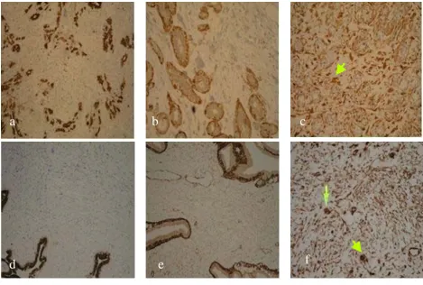
stromal cells stained negative for the Estrogen and Proges-terone receptors (ER, PR respectively) Pancytokeratin (AE1/3 & CAM 5.2), Muscle Specific Actin, S100 and desmin, and stained positive for Vimentin; a general mes-enchymal marker and suggestive of cells of myofibroblas-tic origin (Figures 3a, 3b, 3c). The conclusive diagnosis was that of a fully excised benign fibroadenoma, with multinucleated giant cells throughout its stroma. She made an uneventful postoperative recovery and follow-up has shown no recurrence of the lesion.
The second case is that of a 48 year old lady referred to the Breast Clinic with a two month history of a left breast lump and mastalgia. She denied nipple discharge, nipple inversion or skin changes. She had no relevant past medi-cal history, had never used the OCP and had no family history of breast cancer. On examination, a 1.5 cm tender solid mass was palpable in the upper inner quadrant of the left breast. Ultrasonography revealed the presence of a number of small benign cysts with a single solid lobulated mass lesion at 12 o'clock measuring 17 mm in diameter
Radiology images from 2 cases presenting with a breast mass
Figure 1
Radiology images from 2 cases presenting with a breast mass. a. Case 1. Mammogram of right breast. b. Case 1. Ultra-sound of right breast. c. Case 2. Mammogram of left breast. d. Case 2. UltraUltra-sound of left breast.
b
a
(Figure 1c). Mammography confirmed the presence of a smooth mass measuring 2 cm in diameter in the retro-are-olar region of left breast (Figure 1d).
Ultrasound guided tru-cut biopsy was performed. Histo-logical analysis demonstrated cores and fragments of fibroadenomatous breast tissue, with numerous uni-formly giant and multi-nucleated cells intermingled with fibroblasts throughout the stroma, (Figures 2d, 2e, 2f).
Immunohistochemistry staining of these giant cells was negative again for ER & PR status, pancytokeratin (AE1/3 & CAM 5.2) and muscle specific actin (Fig 3d, 3e) as well as for S100 protein and desmin. They showed only posi-tivity with vimentin (Figures 3f). No malignant changes were seen and a diagnosis of benign fibroadenoma of the left breast with a multi-nucleated giant cell stroma was made. The patient declined surgical excision of the lesion.
Discussion
In 1979 Rosen first described the presence of Multinucle-ated Stromal Giant Cells (MSGCs) in the breast, as an inci-dental finding in breast specimens from 14 patients with breast carcinoma [1]. The tissue foci containing these atypical MSGCs were located in otherwise normal areas of the mammary gland and were usually distinct from the carcinomata. Rosen concluded that these cells represented a non-neoplastic and possibly reparative process. Subse-quently MSGCs have been described in several other breast lesions raising an interesting differential diagnosis, mainly with benign disorders but also on occasion in association with malignant lesions [2-5]. MSGCs similar to those occurring in breast tissue have also been found to occur in the vagina, uterine cervix, nasal polyps, urinary bladder epithelium, anus, and in lesions of the oral cavity. Their presence in other benign tumours has also been described, including pleomorphic lipomas, leiomyomas
Microscopy of breast core biopsies
Figure 2
Microscopy of breast core biopsies. a. Case 1 (H&E stain, × 200). b. Case 1 (H&E stain, × 200). c. Case 1 (H&E stain, × 400). d. Case 2 (H&E stain, × 200). e. Case 2 (H&E stain, × 200). f. Case 2 (H&E stain, × 400).
b
c
a
and fibromas, schwannomas, and in variants of der-matofibromas with atypical cells. The pathogenesis of MSGCs in breast tissue and in the lower female genital tract is unclear. Rosen postulated that the MSGCs in mam-mary tissue may have been related to alterations in hor-mone levels during the perimenopausal period. Indeed all 14 patients in whom these cells were found in Rosen's case series, were aged between 40–50 years. It has also been noted that MSGCs in the female genital tract are associated with pregnancy and exogenous progestins.
Diagnostic pitfalls
Due to its rarity, few cases of MSGCs in benign breast tumours have been described cytologically. Hence recog-nition and correct interpretation of their presence is diffi-cult, yet crucial to forming an accurate diagnosis. Incorrect interpretation of these unusual cells as malignant cells can lead to misdiagnosis of more sinister conditions, such as malignant phyllodes tumor and metaplastic carcinoma.
Consequently treatment of a lesion bearing MSGCs could potentially be misguided. Another diagnostic pitfall to be aware of upon identifying multinucleated giant cells in breast lesions is their resemblance histologically to osteo-clast-like giant cells which are also infrequently reported to occur in a similar spectrum of both benign and malig-nant breast lesions [6-9]. A distinction must be made between these two cell types as it has diagnostic and prog-nostic implications. In fact osteoclast-like giant cells in association with malignant breast epithelial cells is indic-ative of a mammary carcinoma with postulated poor prognosis. Several reports have noted a less favourable outcome for patients with mammary carcinomas contain-ing osteoclast-like giant cells when compared with con-ventional ductal adenocarcinoma [9].
In a review article on this subject in 2004, Cai states that it is the combination of osteoclast-like giant cells and can-cer cells in association, which allows the diagnosis of this
Immunohistochemical stains on core breast biopsy tissue
Figure 3
Immunohistochemical stains on core breast biopsy tissue. a. Case 1: Pancytokeratin. b. Case 1: SMA. c. Case 1: Vimen-tin (arrow marks mulVimen-tinucleated giant cells in stroma). d. Case 2: PancytokeraVimen-tin. e. Case 2: SMA. f. Case 2: VimenVimen-tin (arrow marks multinucleated giant cells in stroma).
a b c
e
f
tion of any osteoclast-like giant cells in a breast biopsy, to avoid a false negative diagnosis [10]. Several cytological differences are noted between MSGCs and osteoclast-like giant cells to help in their differentiation. Multinucleated stromal cells have multiple overlapping nuclei and scant cytoplasm, and they are usually located within the stroma, with no association with the epithelial cells [11]. In con-trast, osteoclast-like giant cells have over 20 centrally located nuclei, and are found primarily in the advancing edges of the infiltrating tumour mass or in the lumen of the cribriform epithelial ducts, and in fine needle aspira-tion smears the osteoclast-like giant cells are intimately admixed with malignant epithelial cells [12]. In phyllodes tumors for example, which are uncommon fibroepithelial neoplasms with a prominent stromal component and where MSGCs may be seen on rare occasions, these partic-ular multinucleated stromal cells are different from osteo-clast-like giant cells, showing a linear nuclear arrangement or floret-like pattern [3,6,13,14].
The immunohistochemical staining pattern is also differ-ent for MSGCs and osteoclast-like giant cells. Tse described the immunohistochemical profile of the infre-quent MSGCs in phyllodes tumours, commenting that both the MSGCs and stromal cells expressed vimentin strongly but not desmin; and in some but not all fibroep-ithelial tumours, both MSGCs and stromal cells expressed actin weakly. These features suggest that MSGCs may be off myofibroblastic differentiation [3].
Indeed both of our cases stained strongly positive for vimentin though negative for actin, indicating likely fibroblastic origin. On the other hand, osteoclast-like giant cells have an immunohistochemistry profile typical of histiocytic differentiation, staining positive for the typ-ical osteoclast markers, CD68, CD1a, tartrate-resistant acid phosphatase (TRAP), and negative for cytokeratin, epithelial membrane antigen, lysozyme, and estrogen and progesterone receptors. Their staining pattern thus rejects any epithelial, endothelial or trophoblastic origin of these cells [15].
the histopathological and immunohistochemical features of breast spindle cell tumours, then suggests that the Vimentin/CD34 positive fibroblast of mammary stroma could be the result of differentiation from a pluripotential mesenchymal precursor cell with the potential to differen-tiate toward several mesenchymal lines [18]. MSGCs also show inconsistent focal positive staining with histiocytic markers such as alpha-1-antitrypsin, alpha-1-antichymot-rypsin, HAM-56, CD34, and CD68. These markers of his-tiocytic differentiation are more consistently found positive in osteoclast-like giant cells. As shown in our two cases MSGCs stain negatively with immunoperoxidase stains for oestrogen and progesterone receptors, cytokerat-ins (AE 1/3 and CAM 5.2), S100 protein, muscle specific actin and desmin, further supporting their fibroblastic ori-gin.
Differential Diagnosis
The differential diagnosis upon recognition of MSGCs in breast lesions also includes Breast spindle cell tumours (BSCTs), and other hamartomas of the breast. BSCTs are a heterogeneous group including benign and malignant lesions, with different therapeutic and prognostic implica-tions. Cytological examination followed by immunohis-tochemistry staining is critical to differentiate BSCTs from other breast lesions as well as to categorise the individual subtypes of spindle cell tumour. An accurate diagnosis is essential in order to appropriately manage the lesion and predict the prognosis for the patient [18]. For example with regard to myoepithelial tumours which are a subtype of BSCT composed of a dominant to pure population of myoepithelial cells, immunohistochemistry stains typi-cally show positivity for actin, S-100, cytokeratin, p63 and CD10, all of which are consistent with a myoepithelial origin. These may be differentiated from fibroepithelial tumours on the basis that the spindle cells stain negative for CD34. Differentiation of myoepitheliomas into benign and malignant lesions is based on further cyto-logic features. Those features observed only in malignant myoepithelial lesions include pleomorphism, coarse nuclear chromatin, prominent nucleoli, high mitotic activity and tumour necrosis [21].
Hamartomas are breast lesions with varying amounts of benign epithelial elements, fibrous tissue, and fat and in rare cases the occurrence of giant cells has been noted. They lack a distinctive pathological appearance however most authors agree on a general characteristic pattern of interlobular fibrosis, which is defined as the presence of lobules within a fibrotic stroma, which surrounds and extends to between individual lobules and obliterates the usual interlobular specialised loose stroma [22]. Unfortu-nately however this pattern is not unique to hamartomas. Other commonly described yet nonetheless inconsistent features of hamartomas include the presence of varying proportions of pseudo-angiomatous stroma, and adipose tissue within the stroma [23]. Epithelial changes such as hyperplasia without atypia, cystic change, apocrine meta-plasia, and adenosis have been described in a smaller pro-portion of hamartomas [23,24], as have other rare features including microcalcification, myoid differentia-tion, stromal oedema, and stromal giant cells. Further-more, there are occasional case reports of coincidental in-situ, or invasive ductal or lobular carcinoma occurring in hamartomas [25]. Hence the correct identification of hamartomas is important because there are the problems of recurrence and coincidental epithelial malignancy. However diagnosis is difficult, and not reliant solely on any one histopathological technique or immunohisto-chemistry stain, due to the presence of the various ele-ments. A complete triple assessment of these lesions is warranted to formulate the correct diagnosis, including correlation of the clinical impression with the
radiologi-cally distinct imaging findings and the above pathological features.
Conclusion
The presence of these pleomorphic, multinucleated large cells in small specimens such as those obtained at fine needle aspiration or core biopsy, may be misinterpreted as a malignant process. The typical benign cytoarchitecture of a fibroadenomas, in a background of naked multiple nuclei indicates the benign nature of the lesion and has no known malignant potential. Correct recognition and the ability to differentiate these cells from malignant cells are dependent on a combination of conventional diagnostic pathological techniques: H&E and IHC staining using a small panel of antibodies. Awareness of this phenomenon is critical in facilitating accurate diagnosis and appropriate management of the patient.
Competing interests
The authors declare that they have no competing interests.
Authors' contributions
HH is primarily responsible for drafting, literature search, submission and revising the manuscript. SM was involved in drafting and revising the manuscript, FB and IT evalu-ated the immunohistochemical stainings and confirmed the diagnoses, FB also was involved in revising the manu-script, MC performed and interpreted the radiological investigations for the patients. KB performed the surger-ies, supplied relevant clinical information about the patients and directed management of the patients clini-cally. All authors read and approved the final manuscript.
Authors’ consent to Publication (copyright)
I Helen Heneghan, the Corresponding Author have the right to grant on behalf of all authors of this manuscript, and do grant on behalf of all authors, an exclusive licence (or non-exclusive for government employees) on a world-wide basis to the BMJ Publishing Group Ltd and its Licen-sees to permit this article (if accepted) to be published in JCP and any other BMJPGL products to exploit all subsid-iary rights, as set out in your licence.
References
1. Rosen P: Multinucleated mammary stromal giant cells – A benign lesion that simulates invasive carcinoma. Cancer 1979,
44(4):1305-1308.
2. Berean K, Tron VA, Churg A, Clement PB: Mammary fibroade-noma with multinucleated stromal giant cells. American Journal of Surgical Pathology 1986, 10(11):823-827.
3. Tse G, Law BK, Chan KF, Mas TK: Multinucleated stromal giant cells in mammary phyllodes tumours. Pathology 2001,
33(2):153-156.
Publish with BioMed Central and every scientist can read your work free of charge
"BioMed Central will be the most significant development for disseminating the results of biomedical researc h in our lifetime."
Sir Paul Nurse, Cancer Research UK
Your research papers will be:
available free of charge to the entire biomedical community
peer reviewed and published immediately upon acceptance
cited in PubMed and archived on PubMed Central
yours — you keep the copyright
Submit your manuscript here:
http://www.biomedcentral.com/info/publishing_adv.asp
BioMedcentral
with osteoclast-like giant cells diagnosed by fine-needle aspi-ration biopsy: review of the cytologic literature and distinc-tion from other mammary lesions containing giant cells. Diagnostic Cytopathology 2004, 30(6):396-400.
11. Rosen P: Rosen's Breast Pathology Philadelphia: Lippincott Williams & Wilkins; 2001.
12. Sugano I, Nagao K, Kondo Y, Nabeshima S, Murakami S: Cytologic and Ultrastructural studies of a rare breast carcinoma with osteoclast-like giant cells. Cancer 1983, 52(1):74-78.
13. Shabb N: Phyllodes tumor. Fine needle aspiration cytology of eight cases. Acta cytologica 1997, 41(2):321-326.
14. Tse G, Ma TK, Pang LM, Cheung H: Fine needle aspiration cyto-logic features of mammary phyllodes tumors. Acta cytologica
2002, 46(5):855-863.
15. Gjerdrum L, Lauridsen MC, Sørensen FB: Breast carcinoma with osteoclast-like giant cells: morphological and ultrastructural studies of a case with review of the literature. The Breast 2001,
10(3):231-236.
16. Fuchs E, Weber K: Intermediate filaments: structure, dynam-ics, function, and disease. Annual Review of Biochemistry 1994,
63:345-382.
17. Goldman R, Khuon S, Chou YH, Opal P, Steinert PM: The function of intermediate filaments in cell shape and cytoskeletal integrity. Journal of Cell Biology 1996, 134(4):971-983.
18. Abd el-All HS: Breast spindle cell tumours: about eight cases. Diagnostic Pathology 2006, 1(13):1-9.
19. Bondeson L: Aspiration cytology of breast carcinoma with multinucleated reactive stromal giant cells. Acta cytologica
1984, 28:313-316.
20. Gupta R: Aspiration cytodiagnosis of a rare carcinoma of breast with bizarre malignant giant cells. Diagnostic Cytopathol-ogy 1996, 15:66-69.
21. Lingamfelter D, Chen Y, Kure K, Lankachandra K: Infiltrating myoepithelial carcinoma of the breast, a case report and cytologic-histologic correlation. Diagnostic Pathology 2008,
3(7):1-5.
22. Tse GM, Law BK, Ma TK, Chan AB, Pang LM, Chu WC, Cheung HS:
Hamartoma of the breast: a clinicopathological review. Jour-nal of Clinical Pathology 2002, 55:951-954.
23. Fisher CJ, Hanby AM, Robinson L, Millis RR: Mammary Hamar-toma – review of 35 cases. Histopathology 1992, 20:99-106. 24. Daya D, Trus T, D'Souza TJ, Minuk T, Yemen B: Hamartoma of the
breast, an underrecognized breast lesion. American Journal of Clinical Pathology 1995, 103:685-689.