Open Access
Methodology
Systematic comparison of RNA extraction techniques from frozen
and fresh lung tissues: checkpoint towards gene expression studies
Jai Prakash Muyal
†1,5, Vandana Muyal
†1, Brajesh Pratap Kaistha
2,
Carola Seifart
3and Heinz Fehrenbach*
4Address: 1Clinical Research Group "Chronic Airway Diseases", Faculty of Medicine, Philipps-University Marburg, Marburg, Germany, 2Dept. of
Internal Medicine, Division of Nephrology, University Hospital Giessen & Marburg GmbH, Marburg, Germany, 3Dept. of Internal Medicine,
Division of Respiratory Medicine, University Hospital Giessen & Marburg GmbH, Marburg Hessen, Germany, 4Section of Experimental
Pneumology, Research Center Borstel, Leibniz Center for Medicine and Biosciences, Borstel, Germany and 5School of Biotechnology, Gautam
Buddha University, Greater Noida – 201308, India
Email: Jai Prakash Muyal - [email protected]; Vandana Muyal - [email protected];
Brajesh Pratap Kaistha - [email protected]; Carola Seifart - [email protected]; Heinz Fehrenbach* - [email protected]
* Corresponding author †Equal contributors
Abstract
Background: The reliability of gene expression profiling-based technologies to detect transcriptional
differences representative of the original samples is affected by the quality of the extracted RNA. It strictly depends upon the technique that has been employed. Hence, the present study aimed at systematically comparing silica-gel column (SGC) and guanidine isothiocyanate (GTC) techniques of RNA isolation to answer the question which technique is preferable when frozen, long-term stored or fresh lung tissues have to be evaluated for the downstream molecular analysis.
Methods: Frozen lungs (n = 3) were prepared by long-term storage (2.5 yrs) in -80°C while fresh lungs
(n = 3) were harvested and processed immediately. The purity and quantification of RNA was determined with a spectrophotometer whereas the total amounted copy numbers of target sequences were determined with iCycler detection system for assessment of RNA intactness (28S and 18S) and fragment sizes, i.e. short (GAPDH-3' UTR), medium (GAPDH), and long (PBGD) with 200 bp, 700 bp, and 1400 bp distance to the 3'ends of mRNA motif, respectively.
Results: Total yield of RNA was higher with GTC than SGC technique in frozen as well as fresh tissues
while the purity of RNA remained comparable. The quantitative reverse transcriptase-polymerase chain reaction data revealed that higher mean copy numbers of 28S and a longer fragment (1400 bp) were obtained from RNA isolated with SGC than GTC technique using fresh as well as frozen tissues. Additionally, a high mean copy number of 18S and medium fragment (700 bp) were obtained in RNA isolated with SGC technique from fresh tissues, only. For the shorter fragment, no significant differences between both techniques were noticed.
Conclusion: Our data demonstrated that although the GTC technique has yielded a higher amount of
RNA, the SGC technique was much more superior with respect to the reliable generation of an intact RNA and effectively amplified longer products in fresh as well as in frozen tissues.
Published: 24 March 2009
Diagnostic Pathology 2009, 4:9 doi:10.1186/1746-1596-4-9
Received: 12 March 2009 Accepted: 24 March 2009
This article is available from: http://www.diagnosticpathology.org/content/4/1/9
© 2009 Muyal et al; licensee BioMed Central Ltd.
Background
DNA-microarray and quantitative reverse transcriptase-polymerase chain reaction (qRT-PCR) are two powerful techniques widely used in functional genomics for the analysis of gene expression profiles. These techniques are highly dependent on the quality of total RNA [1-3] obtained from either fresh or frozen biological samples.
RNA is a particularly labile bio-molecule and is much more susceptible to degradation by endogenous- and exogenous-nucleases and to non-specific degradation by divalent cations, heat, elevations in pH, and storage of tis-sue or cells over extended periods prior to RNA extrac-tions, which result in falsely altered gene expression patterns [4]. Particularly, many efforts are currently being made to circumvent the problem associated with contin-ues fragmentation and degradation of total RNA over-time. Total RNA can be isolated from archival tissue sam-ples for example, by the formalin-fixed paraffin-embed-ded tissue-based technique [5]. However, this technique poses many problems due to the fact that formalin fixa-tion cross-links nucleic acids and proteins. Further, mono-methylol is added to the amino groups for all four RNA bases (N-CH2OH) and subsequently methylene bridges are formed between neighbouring bases that resulted to continue degradation over-time [6]. Alternatively, imme-diate freezing of fresh tissue samples in liquid nitrogen and subsequent storage at -80°C until analysis preserves RNA integrity. However, this procedure is not routinely performed.
Obtaining high quality RNA is mandatory and depends upon the technique that has been employed [7]. Gener-ally, the high quality of total RNA is assessed on the basis of RNA intactness and purity, respectively. The intactness of RNA is assessed by analyzing 28S and 18S subunits of
ribosomal RNA either on micro-capillary chip's electro-phoresis with fluorescent detection (Bioanalyzer, Agilent Technologies) or on ethidium stained agarose gel electro-phoresis [8-11] while the purity is determined by calculat-ing the ratio at absorption (A)260 nm/A280 nm by spectrophotometer [12]. Purity is considered to be ade-quate if the ratio is greater than 1.8 [13], and hence, rec-ommended for introduction into the downstream molecular analysis.
Today, several other new techniques and kits are being offered for RNA isolation [14-17], which have their own respective principle and methodology and are believed to perform equally well. The two most reliable and widely used techniques for high throughput RNA isolation are 1) guanidine isothiocyanate-phenol:chloroform (GTC)-based RNA isolation technology and 2) Silica-gel column (SGC)-based RNA isolation technology. The GTC tech-nique for isolation of RNA, which was developed by Chomczynski and Sacchi [18], is very popular because it requires much less time than other classical methods (e.g., CsCl2 ultracentrifugation). Moreover, GTC salt denatures the cellular proteins and inactivates RNases ensuring that isolated RNA is not degraded. Many commercial reagents (e.g. Trizol®, RNAzol™, RNAwiz™) are based on this
prin-ciple. In contrast, the principle of SGC technology (Qia-gen RNeasy Mini column) is a combination of the selective binding properties of a silica-based membrane with the speed of microspin technology, which allows saving time, money, and efficient use of small and pre-cious biological samples. The comparison between GTC and SGC techniques are summarized in Table 1. Thus, when preparing the frozen or fresh samples for investiga-tion of gene expression profiling, it is essential to briefly evaluate the technique of RNA isolation, especially in
clin-Table 1: Comparison of GTC-based versus SGC-based techniques
GTC SGC
works best either with high (300 mg) or very low (20 mg) amount of tissue, because of phenol-chloroform follows by ethanol precipitation steps and uses of denaturants and RNase inhibitors
works best with low (<100 mg) amount of tissue, because filters may clog resulting in low yield of total RNA
Several chemical treatment steps in protocol may affect RNA quality Limited steps in protocol may not have influences on RNA intactness
Complex protocol, takes 4 hrs Simple protocol, takes only 20 min
chemically based aqueous-phase procedure, increased risk of cross contamination
No phase separation step present, low risk of cross contamination
Use of hazardous chemicals like phenol and chloroform requires specific safety rules in the laboratory
Absolutely safe while handling.
ical applications with limited tissue material in terms of their reproducibility and reliability, respectively.
Methods
Experimental design
In the present study, we have tested a strategy to store lung tissues for the period of more than 2.5 yrs without any chemical treatment or processing, and investigated which RNA extraction technique (i.e. SGC and GTC) is prefera-ble in terms of RNA recovery, purity, intactness, and amplification of various fragment sizes. A similar investi-gation was performed with fresh lung tissues.
Lung tissues were harvested from 4 months old male C57BL mice. Lung samples were either immediately dipped into liquid nitrogen, cut down into small fine pieces on sterile plate on ice using sterile blade and frozen at -80°C for 2.5 years until processing and analysis (n = 3 mice). Fresh lung tissue was processed and analysed immediately (n = 3 mice).
The step by step experimental design for the systematic comparison of SGC and GTC techniques using frozen and fresh samples is given in Figure 1. Total RNA recovery was calculated from the RNA concentration measured by absorbance at 260 nm (A260) whereas purity was deter-mined as per discussed above. The importance of RNA
integrity and various fragment sizes were determined with qRT-PCR. For the assessment of RNA integrity, we used 28S and 18S subunits of ribosomal RNA, an internal con-trol for RNA intactness. On the other hand, for the assess-ment of various fragassess-ment sizes of RNA, an amplification factor of Glyceraldehydes 3-phosphate dehydrogenase-3' un-translated region (GAPDH-3' UTR; short fragment), Glyceraldehyde 3-phosphate dehydrogenase (GAPDH; medium fragment) and Porphobilinogen deaminase (PBGD; long fragment), the respective distance of which to the 3'ends of messenger RNA (mRNA) were 200 bp, 700 bp, and 1400 bp (Figure 2), was taken in this study.
Materials and methods
Frozen- & Fresh- Lung Tissue Homogenization
The chopped pieces of lung tissues from frozen and fresh was collected in a 2.0 ml eppendorf tube, 1.9 ml of lyses buffer {RLT or 4 M GTC (4 M 25 mM Na-3-citrate: 0.5% laurylsarcosin, 100 mM Tris-Hcl)} was added, and with the help of a disposable syringe and a 20 G sterilized nee-dle, tissue pieces were disrupted. The lysed-disrupted sam-ples were kept at room temperature (RT) for 10 min, centrifuged for 15 min at 12,000 rpm to get rid off cellular debris, and supernatant was collected. In order to investi-gate the reproducibility of each method, collected super-natant was then divided into three equal volumes, i.e. 600
μl.
Total cellular RNA Isolation, RNA Quantity, RNA purity, and RNA Quality Measurement
Collected supernatant was directly applied on the RNeasy column (SGC; Qiagen, Hilden, Germany) and total cellu-lar RNA was isolated as per the steps given for the Qiagen RNA Miniprep kit. In contrast, using GTC technique col-lected supernatant was mixed with 200 μl of chloroform (Sigma-Aldrich, Steinheim, Germany) and 80 μl of 2 M NaoAc. The samples were vortexed and centrifuged at 4°C for 15 min at 12,000 rpm. The upper phase was
trans-Schematic representation of experimental plan
Figure 1
Schematic representation of experimental plan. A
tis-sue pool has been created by combining lung 1, lung 2, and lung 3, respectively, which were obtained from either frozen or fresh samples. Pool of frozen or fresh tissue was divided in two equal parts, one for the SGC and another for the GTC technique. To test technique's reproducibility, each divided part was sub-divided into three identical parts (triplicate approach).
3X 0. 6 ml tissue‘s pool
1 2 3
Method 1: SGC 90 mg / 1. 8 ml lyses buffer
Lung 1 Lung 2 Lung 3
Method 2: GTC Fr ozen lungs (2.5 yr )
tissue‘s pool
Method 1: SGC
Lung 1 Lung 2 Lung 3
Method 2: GTC Fr esh lungs (0 yr )
A
B
90 mg / 1. 8 ml lyses buffer
90 mg / 1. 8 ml lyses buffer
90 mg / 1. 8 ml lyses buffer
3X 0. 6 ml
1 2 3
3X 0. 6 ml
1 2 3
3X 0. 6 ml
1 2 3
Schematic representation of assessment for RNA intactness
Figure 2
Schematic representation of assessment for RNA
intactness. The location of the target sequences (i.e.
GAPDH-3' UTR-200 bp, GAPDH-700 bp, and PBGD-1400 bp) on the transcripts.
1400 bp
700 bp
180 bp Eukaryotic mRNA
5‘ 3‘
UTR UTR
GAPDH-3‘UTR GAPDH
ferred carefully into fresh 1.5 ml eppendorf tubes. For RNA precipitation, 500 μl of cold isopropanol (Sigma-Aldrich, Steinheim, Germany) with 70 μl of 2 M NaoAc was added to the supernatants and incubated for 2 hrs at -20°C. Samples were centrifuged at 4°C for 15 min at 12,000 rpm. The obtained pellets were washed with 500
μl of 70% chilled ethanol and centrifuged again at 4°C for 15 min at 12,000 rpm. The washed pellets were then air dried and finally dissolved in 40 μl of RNase free water.
The total RNA quantification and purity was determined with an Ultraspec 2100-spectrophotometer (Amersham Pharmacia Biotech, Cambridge, UK).
For total RNA quality determination, 1.2% of agarose gel was prepared by dissolving 1.2 g of agarose powder (Roth, Karlsruhe, Germany) in 100 ml of 1× Tris-Borate-EDTA buffer (Sigma-Aldrich, Steinheim, Germany). The mix was cooked until get boiled, and then 5 μl of 1% ethidium bromide (Roth, Karlsruhe, Germany) was added, mixed, and poured in to gel electrophoresis unit. RNA samples (1.0 μg each) were mixed with 2 μl of gel loading solution (Sigma-Aldrich, Steinheim, Germany) and were directly applied on a gel's wells. The integrity of total RNA was assessed on the basis of visualisation of 28S and 18S ribosomal RNA subunits under gel documentation system 2000 (Bio-Rad, München, Germany) as a band at posi-tions 4.8 kb and 1.8 kb, respectively.
First Strand cDNA Synthesis, Real-time PCR, and Analysis
First-strand cDNA synthesis was performed using 0.5 μg RNA in a total reaction of 20 μl using 1 μl oligo (dT)12–18
primer (Invitrogen, Karlsruhe, Germany) and Super-script™ II Reverse Transcriptase kit (Invitrogen, Karlsruhe, Germany). The reaction was incubated at 65°C for 5 min, at 37°C for 60 min, and at 72°C for 15 min in RoboCy-cler® Gradient 40 (Startagene, Heidelberg, Germany). The
cDNA qualities for all the samples were evaluated by per-forming GAPDH qualitative PCR (data not shown). The qRT-PCR for determining the amplification factor of tar-get genes i.e. 28S, 18S, GAPDH-3' UTR, GAPDH, and PBGD were performed in a 96-well format iCycler Detec-tion System (Bio-Rad, Hercules, USA). The reacDetec-tions (20
μl) were set up with the SYBR™ Green PCR mix (ABgene®
Thermo Fischer Scientific, UK) according to the manufac-turer's protocol. The thermal cycle conditions used for all reactions were as follows: Step 1: 95°C 15 min; followed by 40 cycles of Step 2: 95°C 50 sec; sequence-specific oli-gonucleotide primer's annealing temperature (Table 2) 40 sec; 72°C 40 sec; Step 3: 72°C 5 min. Due to the non-selective double stranded DNA binding of the SYBR™ Green I dye, melting curve analysis were performed to confirm the exclusive amplification of the expected PCR product.
The total amount of target gene copy numbers were deter-mined by applying the formula 2 raised to the power mean of cycle threshold (2-mean CT). Unless stated
other-wise, mean values ± s.d. are given. Since the aim of this study was to determine the most appropriate technique to isolate RNA, out of two tested techniques (i.e. SGC and GTC) in frozen- and fresh- lung samples, respectively, t-test (and non-parametric t-tests) was performed to deter-mine the level of significance of differences between SGC
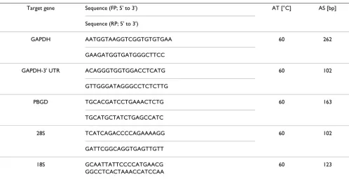
Table 2: Primer details: Sequence, annealing temperature (AT), and amplicon size (AS) of target sequence
Target gene Sequence (FP; 5' to 3') AT [°C] AS [bp]
Sequence (RP; 5' to 3')
GAPDH AATGGTAAGGTCGGTGTGTGAA 60 262
GAAGATGGTGATGGGCTTCC
GAPDH-3' UTR ACAGGGTGGTGGACCTCATG 60 102
GTTGGGATAGGGCCTCTCTTG
PBGD TGCACGATCCTGAAACTCTG 60 163
TGCATGCTATCTGAGCCATC
28S TCATCAGACCCCAGAAAAGG 60 102
GATTCGGCAGGTGAGTTGTT
18S GCAATTATTCCCCATGAACG 60 123
and GTC. If normality and equal variance were not given at p > 0.1, Mann-Whitney rank sum test was used using GraphPad Prism 4 software program (San Diego CA, USA). Values of p < 0.05 were considered to be significant.
Results
Comparison of Two Techniques for Total RNA Isolation from Frozen and Fresh Lung Tissues
Firstly, the GTC and SGC techniques for RNA extraction were tested to assess, which technique is the most efficient and reproducible in terms of total yield and purity. RNA isolated with GTC technique was characterized by a signif-icantly higher mean concentration {ng/total volume (
μl)} in comparison with RNA isolated by the SGC tech-nique, which was true for both in frozen (p < 0.05; 2.6×) and in fresh (p < 0.05; 1.9×) lung tissues (Figure 3A).
The purity of the extracted RNA was comparable through-out the samples and was close to a ratio (A260/A280) of 1.8 with both, the GTC and the SGC technique, in frozen and fresh lung tissues, respectively (Figure 3B). A ratio close to 1.8 indicates that there were only limited protein contam-inations.
Quantitative PCR: Amplification factor
Secondly, we evaluated the total RNA integrity on 1.2% agarose gel electrophoresis. All samples clearly demon-strated a visible intact band at two different positions, i.e. 4.8 kb and 1.8 kb, which represent 28S and 18S ribosomal RNA subunits, respectively (Figure 4).
RNA integrity was further evaluated by determining an amplification factor of RNA's internal quality controls (i.e. 28S and 18S). qRT-PCR revealed that the mean copy numbers of 28S sequence was significantly higher in RNA isolated with the SGC compared to the GTC technique (Figure 5A). This difference was more pronounced in fro-zen (p < 0.01; 123×) than in fresh (p < 0.03; 7.1×) lung
tis-sues. For the 18S sequence, RNA isolated from fresh lung tissues with SGC technique yielded higher mean copy numbers of 18S (p < 0.05; 1.6×) than RNA isolated from frozen lung tissues with GTC technique (Figure 5B).
Lastly, to assess, which RNA extraction technique would reliably conserve a longer fragment from poly-A tail of mRNA, qRT-PCR was performed to determine the short (GAPDH-3' UTR, 200 bp), medium (GAPDH, 700 bp), and long (PBGD, 1400 bp) fragment sizes of RNA, respec-tively. The qRT-PCR for the GAPDH-3'UTR revealed that both techniques have the potential to amplify fragments of a size <200 bp in comparable manner (Figure 6A). RNA isolated with the SGC technique from fresh lung tissues exhibited significantly more copy numbers of the medium product (GAPDH) than RNA isolated with the GTC based technique (Figure 6B; p < 0.02; 2.6×), whereas no difference was seen in RNA extracted from frozen lung. The RNA fragment of 1400 bp of size (PBGD; longer) showed a clear reduction in the copy numbers of PBGD in
Total yield and purity performance of RNA isolated from fro-zen and fresh lung tissues with SGC- and GTC-based tech-niques
Figure 3
Total yield and purity performance of RNA isolated from frozen and fresh lung tissues with SGC- and
GTC-based techniques. (A) Yield of total RNA in
nano-gram (ng), which was significantly higher in frozen and fresh lung tissue with GTC- than SGC-based isolation, respec-tively. (B) Total RNA purity assessment on the basis of A260/ A280.
Frozen Fresh 0 5000 10000 15000 20000 25000 30000 35000 40000 45000 SGC GTC T o ta l yie ld ( n g )
Frozen Fresh 0.0 0.3 0.6 0.9 1.2 1.5 1.8 SGC GTC A26 0n m / A280 n m
A * * B
Total RNA quality assessment on the basis of 18S and 28S rRNA
Figure 4
Total RNA quality assessment on the basis of 18S
and 28S rRNA. On left side, lane # 1 shows DNA ladder
(0.5 kb – 7.0 kb). The ethidium bromide-staining pattern of intact total RNA shows clearly defined 18S and 28S ribos-omal RNA bands [lane # 3–5 (GTC; fresh); lane # 6–8 (SGC; fresh sample); lane # 9–11 (GTC; frozen); and lane # 12–14 (SGC; frozen).
Assessment of total RNA quality
Figure 5
Assessment of total RNA quality. An amplification factor
of 28S and 18S subunits of rRNA was determined; a higher copy numbers of 28S (A) was obtained with SGC method in frozen and fresh lung tissue compared with the GTC method. No significant difference was observed for 18S (B) in RNA isolated from frozen lung, however, more copy num-bers of 18S was obtained from fresh lung tissues.
RNA preparations with GTC than SGC using frozen (p < 0.05; 0.09×) and fresh (p < 0.02; 0.6×) lung tissues (Figure 6C). This indicates that smaller products were conserved when RNA samples were isolated with the GTC technique from frozen as well as fresh lung tissues compared to SGC based technique. Further, a higher and equivalent copy numbers of PBGD were obtained with the SGC-based method in frozen and fresh lung tissues than GTC tech-nique.
Discussion
The demand for high quality of RNA has been dramati-cally increased with the extensive application of DNA-microarray technology and real-time PCR-based expres-sion profiling. A number of RNA extraction techniques are available to generate high quality RNA for use in gene expression profiling experiments [19]. Several attempts have been made to isolate RNA from formalin-fixed par-affin-embedded tissue using modifications of currently available techniques for RNA extraction [20]. However, the interpretation of results is often difficult and therefore, alternative methods for long-term storage of tissues prior to gene expression profiling are greatly required. The objective of the current study was to investigate which RNA extraction technique (i.e. GTC and SGC) is better and convenient if long-term stored frozen (2.5 yrs) and
stream applications. The comparison between the tech-niques was assessed in terms of RNA recovery, purity, intactness, and amplification of various fragment sizes of RNA (Figure 2).
The first step of evaluation after RNA extraction is the measurement of total recovery followed by the determina-tion of purity and intactness of an isolated RNA. Our results demonstrate that irrespective of using RNA from frozen or fresh samples, the total recovery of RNA was higher with GTC- than SGC-based techniques (Figure 3A). Similar findings were obtained by Xiang and co-workers, who compared three different methods {i.e. (1) GTC, (2) Trifast, and (3) SGC techniques} for RNA extraction using sputum samples [21]. Our data are in accordance with their findings of the mean RNA concentration (i.e. recov-ery) obtained with the GTC-based technique being signif-icantly higher than with the SGC-based technique with RNA purity being equivalent. We suggest that one reason for this difference in total yield may be the fact that the SGC technique uses a silica-gel membrane, which may get clogged with tissue lysate. Additionally, in the GTC-based technique phenol-chloroform and ethanol precipitation steps are implemented, which tend to concentrate RNA in a more robust manner than any other technique because a unique cocktail of denaturants and RNase inhibitors is used. This cocktail results into less degradation of RNA [22] and hence may contribute to increased total recovery compared to the SGC-based technique.
Next, the most important parameter in the evaluation procedure is to assess the intactness of RNA, which can be determined by analyzing the 18S and 28S subunits of ribosomal RNA on either the ethidium bromide-stained agarose gel [23,24] or micro-capillary electrophoresis traces [19,25]. Qualitative RNA analysis revealed clear bands of 28S and 18S in the agarose gel with no discerni-ble difference in intensity or overall electrophoretic pat-tern between the different samples (Figure 4). We further validated RNA intactness by using qRT-PCR, because of its high sensitivity, good reproducibility, and the wide quan-tification range for determination of an amplification fac-tor of 28S and 18S, respectively. Our qRT-PCR data revealed that SGC technique has a potential to generate higher copy numbers of 28S in RNA isolated from frozen and fresh lung tissues than GTC technique. On the other hand, a high copy number of 18S was observed in RNA isolated from fresh tissue with SGC technique only. We suggest that the capability to obtain high copy number of 28S (4.8 kb) and 18S (1.8 kb) with the silica gel-based technique may firstly relate to the selective adsorption to silica-gel membranes under controlled ionic conditions, which allows isolating RNA with high molecular size. Sec-ondly, because the GTC technique does require the use of toxic chemicals like phenol and chloroform, and long Assessment of RNA fragment sizes
Figure 6
Assessment of RNA fragment sizes. An amplification
factor of GAPDH-3'UTR, GAPDH, and PBGD was deter-mined to assess the fragment sizes of RNA. The short (200 bp) and medium (700 bp) sequences were amplified by both techniques with similar fashion in RNA isolated from frozen and fresh lung tissue. More copy numbers of medium frag-ment (GAPDH) were obtained with fresh compared to fro-zen using SGC method. RNA isolated from SGC effectively amplified a longer fragment (<14,00 bp) in fresh and frozen lung tissue. In contrast, with the GTC technique a higher copy number of the longer product was obtained with fresh lung tissues only.
Frozen Fresh 0
1e-006 2e-006 3e-006
SGC GTC
2
- M
ean
C
t
Frozen Fresh 0.0000000
0.0000002 0.0000005 0.0000008 0.0000010 0.0000012 0.0000015 0.0000017
SGC GTC
2
- M
ean
C
t
Frozen Fresh 0
5e-010 1e-009 1e-009 2e-009
SGC GTC
2
- M
ean
C
t
A B
C
AAAA 200bp
GAPDH 3‘ UTR GAPDH AAAA
700bp
PBGD AAAA
1400bp
*
taminants likes phenol, chloroform, RNases, genomic DNA, and other chemicals, which are co-purified with RNA. Hence, these contaminants may interfere with the reverse transcription reaction process [26,27] and thereby resulted into loss of RNA population or indirectly copy numbers of tested sequences in contrast to the SGC tech-nique.
In general, approximately 80 percent of the total RNA is ribosomal RNA and 15 percent is transfer RNA with pro-tein-encoding messenger RNA constituting only a small portion (i.e. 5 percent) of the total RNA. To obtain an overall impression of RNA intactness, it is equally impor-tant to evaluate the quality of mRNA as well. By selecting three different sequences, which were classified according to their distance to the poly-A tail of mRNA, we asked to evaluate, which technique was able to conserve a longer fragment of mRNA from poly-A tail (3' to 5'). Our results demonstrated that any-one technique can be selected to isolate RNA either from frozen or fresh lung tissues for those sequences that are close to the poly-A tail of mRNA (<200 bp) and fragments lesser than 700 bp for down-stream analysis (Figure 6A &6B).
Notably, the choice of the technique for RNA isolation had a significant impact on the potential to obtain appro-priate copy numbers of bigger fragments as was demon-strated for the 1400 bp fragment of PBGD. In this respect, the SGC technique proved to be superior to the GTC tech-nique both in RNA preparations from frozen and fresh lung tissues. The generation of high copy numbers of PBGD in RNA isolated from fresh as well as from frozen tissue suggests that the SGC-based technique is not only compatible with fresh but also with frozen tissues. We suggest that the reason for the superiority of the SGC tech-nique again relates to the selective adsorption to silica-gel membranes and the avoidance of toxic chemicals and long-lasting protocol steps as discussed above.
Conclusion
Comparing the SGC and GTC technique, we conclude that both techniques performed well in terms of total recovery and purity of RNA, isolated from 2.5 yrs old fro-zen and fresh lung tissues. With respect to RNA intactness, however, the SGC-based technique turned out to be supe-rior to the GTC-based technique because of the conserva-tion of an intact form of ribosomal RNA and conservaconserva-tion not only of short- to medium-sized but also of longer-sized RNA fragments. We may further conclude from our data that long-term storage of frozen lung tissues has only minor effects when compared to fresh lung tissues.
Competing interests
The authors declare that they have no competing interests.
Authors' contributions
JPM participated in the design of the study and performed qRT-PCR experiment, statistical analysis and drafted man-uscript. VM carried out RNA isolation from frozen lung tissues, cDNA synthesis and qualitative PCR from frozen and fresh lung tissues. BPK carried out RNA isolation from fresh lung tissues. CS helped to analysis qRT-PCR data. HF conceived and supervised this work and helped to draft the manuscript.
Acknowledgements
We thank Shiv Kumar Singh (Division of Gastroenterology and Endocrinol-ogy, Department of Internal Medicine, Philipps-University, Marburg, Ger-many) for providing the stored frozen lung tissues. The study was supported by grant from the Deutsche Forschungsgemeinschaft (DFG; FE287/8-1).
References
1. Atz M, Walsh D, Cartagena P, Li J, Evans S, Choudary P, Overman K, Stein R, Tomita H, Potkin S, Myers R, Watson SJ, Jones EG, Akil H, Bunney WE Jr, Marquis P, Vawter MP: Methodological considera-tions for gene expression profiling of human brain. J Neurosci Meth 2007, 163:295-309.
2. Copois V, Bibeau F, Bascoul-Mollevi C, Salvetat N, Chalbos P, Bareil C, Candeil L, Fraslon C, Conseiller E, Granci V, Maziere P, Kramar A, Ychou M, Pau B, Martineau P, Molina F, Del Rio M: Impact of RNA degradation on gene expression profiles: Assessment of dif-ferent methods to reliably determine RNA quality. J Biotech-nol 2007, 127:549-559.
3. Dorris DR, Ramakrishnan R, Trakas D, Dudzik F, Belval R, Zhao C, Nguyen A, Marc Domanus M, Mazumder A: A highly reproducible, linear, and automated sample preparation method for DNA microarrays. Genome Res 2002, 12:976-984.
4. Catts VS, Catts SV, Fernandez HR, Taylor JM, Coulson EJ, Lutze-Mann LH: A microarray study of post-mortem mRNA degradation in mouse brain tissue. Brain Res Mol Brain Res 2005, 138:164-177. 5. Rupp GM, Locker J: Purification and analysis of RNA from
par-affin-embedded tissues. Biotechniques 1988, 6:56-60.
6. Masuda N, Ohnishi T, Kawamoto S, Monden M, Okubo K: Analysis of chemical modification of RNA from formalin-fixed sam-ples and optimization of molecular biology applications for such samples. Nucleic Acids Res 1999, 27:4436-4443.
7. Fleige S, Pfaffl MW: RNA integrity and the effect on the real-time qRT-PCR performance. Mol Aspect of Med 2006,
27:126-139.
8. Strand C, Enell J, Hedenfalk I, Fernö M: RNA quality in frozen breast cancer samples and the influence on gene expression analysis – a comparison of three evaluation methods using microcapillary electrophoresis traces. BMC Mol Biol 2007, 8:38. 9. Schroeder A, Mueller O, Stocker S, Salowsky R, Leiber M, Gassmann M, Lightfoot S, Menzel W, Granzow M, Ragg T: The RIN: an RNA integrity number for assigning integrity values to RNA meas-urements. BMC Mol Biol 2006, 31(7):3.
10. Buesa CMT, Subirada F, Barrachina M, Ferrer I: DNA chip technol-ogy in brain banks: confronting a degrading world. J Neu-ropathol Exp Neurol 2004, 63:1003-1014.
11. Dumur CI, Best NS, Archer AM, Ladd KJ, Mas AC, Wilkinson VR:
Evaluation of quality-control criteria for microarray gene expression analysis. Clin Chem 2004, 50:1994-2002.
12. Glasel JA: Validity of nucleic acid purities monitored by 260 nm/280 nm absorbance ratios. Biotechniques 1995, 18:62-63. 13. Manchester KL: Use of UV methods for measurement of
pro-tein and nucleic acid concentrations. Biotechniques 1996,
20:968-970.
Publish with BioMed Central and every scientist can read your work free of charge
"BioMed Central will be the most significant development for disseminating the results of biomedical researc h in our lifetime."
Sir Paul Nurse, Cancer Research UK
Your research papers will be:
available free of charge to the entire biomedical community
peer reviewed and published immediately upon acceptance
cited in PubMed and archived on PubMed Central
yours — you keep the copyright
Submit your manuscript here: BioMedcentral 15. Tavangar K, Hoffman AR, Kraemer FB: A micro method for the
isolation of total RNA from adipose tissue. Anal Biochem 1990,
186:60-63.
16. Smale G, Sasse J: RNA isolation from cartilage using density gradient centrifugation in cesium trifluoroacetate: an RNA preparation technique effective in the presence of high pro-teoglycan content. Anal Biochem 1992, 203:352-56.
17. Briscoe PR, Jorgensen TJ: Improved RNA isolation from cells in tissue culture using a commercial nucleic acid extractor. Bio-techniques 1991, 10:594-96.
18. Chomczynski P, Sacchi N: Single-step method of RNA isolation by acid guanidinium thiocyanate-phenol-chloroform extrac-tion. Anal Biochem 1987, 162:156-159.
19. Strand C, Enell J, Hedenfalk I, Fernö M: RNA quality in frozen breast cancer samples and the influence on gene expression analysis – a comparison of three evaluation methods using microcapillary electrophoresis traces. BMC Mol Biol 2007, 8:38. 20. Ribeiro-Silva A, Zhang H, Jeffrey SS: RNA extraction from ten year old formalin-fixed paraffin-embedded breast cancer samples: a comparison of column purification and magnetic bead-based technologies. BMC Mol Biol 2007, 8:118.
21. Xiang X, Qiu D, Hegele RD, Tan WC: Comparison of different methods of total RNA extraction for viral detection in spu-tum. J Virol Method 2001, 94:129-135.
22. Fend F, Emmert-Buck M, Chuaqui R: Immuno LCM: laser capture microdissection of immunostained frozen sections for mRNA analysis. Am J Pathol 1999, 154:61-66.
23. Bonini JA, Hofmann C: A rapid, accurate, nonradioactive method for quantitating RNA on agarose gels. Biotechniques
1991, 11:708-710.
24. Vendrely R, Alexandrov K, De Sousa Lechner MC, Coirault Y: Frac-tionation of ribonucleic acids by 'Sephadex' agarose gel elec-trophoresis. Nature 1968, 218:293-294.
25. Imbeaud S, Graudens E, Boulanger V, Barlet X, Zaborski P, Eveno E, Mueller O, Schroeder A, Auffray C: Towards standardization of RNA quality assessment using user-independent classifiers of microcapillary electrophoresis traces. Nucleic Acids Res 2005,
33:e56.
26. SuperArray Bioscience Corporation [http://www.superar ray.com/newsletter/RNA.html]