R E S E A R C H
Open Access
Clinical evaluation of panel testing by
next-generation sequencing (NGS) for gene
mutations in myeloid neoplasms
Chun Hang Au, Anna Wa, Dona N. Ho, Tsun Leung Chan and Edmond S. K. Ma
*Abstract
Background:Genomic techniques in recent years have allowed the identification of many mutated genes
important in the pathogenesis of acute myeloid leukemia (AML). Together with cytogenetic aberrations, these gene mutations are powerful prognostic markers in AML and can be used to guide patient management, for example selection of optimal post-remission therapy. The mutated genes also hold promise as therapeutic targets themselves. We evaluated the applicability of a gene panel for the detection of AML mutations in a diagnostic molecular pathology laboratory.
Methods:Fifty patient samples comprising 46 AML and 4 other myeloid neoplasms were accrued for the study.
They consisted of 19 males and 31 females at a median age of 60 years (range: 18–88 years). A total of 54 genes (full coding exons of 15 genes and exonic hotspots of 39 genes) were targeted by 568 amplicons that ranged from 225 to 275 bp. The combined coverage was 141 kb in sequence length. Amplicon libraries were prepared by TruSight myeloid sequencing panel (Illumina, CA) and paired-end sequencing runs were performed on a MiSeq (Illumina) genome sequencer. Sequences obtained were analyzed by in-house bioinformatics pipeline, namely BWA-MEM, Samtools, GATK, Pindel, Ensembl Variant Effect Predictor and a novel algorithm ITDseek.
Results:The mean count of sequencing reads obtained per sample was 3.81 million and the mean sequencing
depth was over 3000X. Seventy-seven mutations in 24 genes were detected in 37 of 50 samples (74 %). On
average, 2 mutations (range 1–5) were detected per positive sample.TP53gene mutations were found in 3 out of 4 patients with complex and unfavorable cytogenetics. Comparing NGS results with that of conventional molecular testing showed a concordance rate of 95.5 %. After further resolution and application of a novel bioinformatics algorithm ITDseek to aid the detection ofFLT3internal tandem duplication (ITD), the concordance rate was revised to 98.2 %.
Conclusions:Gene panel testing by NGS approach was applicable for sensitive and accurate detection of
actionable AML gene mutations in the clinical laboratory to individualize patient management. A novel algorithm ITDseek was presented that improved the detection ofFLT3-ITD of varying length, position and at low allelic burden.
Keywords:Next-generation sequencing, Acute myeloid leukemia, Gene mutations,FLT3internal tandem
duplication, Cytogenetics, Bioinformatics
* Correspondence:eskma@hksh.com
Division of Molecular Pathology, Department of Pathology, 1/F Li Shu Fan Block, Hong Kong Sanatorium & Hospital 2 Village Road, Happy Valley, Hong Kong, China
Background
The molecular basis of AML is heterogeneous. Cytogen-etic study is well documented as a mandatory test at diagnosis to stratify patients into favorable, intermediate and adverse prognostic categories [1]. The identification of gene mutations in AML allows the further characterization of the molecular heterogeneity of this disease, especially with the subgroup of intermediate risk AML that often exhibit a normal karyotype [2]. In the recommendation by the European LeukemiaNet (ELN), mutations involving three genes NPM1, FLT3 and
CEBPA are considered in AML prognostication scheme [3]. Cytogenetically normal AML (CN-AML) with mu-tated NPM1without FLT3-ITD, or mutatedCEBPA, are incorporated in the favorable genetic group. Recent data shows that only double, but not single, CEBPA muta-tions confer a favorable prognosis [4–7]. Likewise the National Comprehensive Cancer Network (NCCN) Guideline for AML incorporates CN-AML with NPM1
mutation or isolatedCEBPAmutation in the absence of
FLT3-ITDin the better-risk status category, whilst CN-AML with FLT3-ITD mutation is considered poor-risk [8]. The detection of concurrentKITmutation relegates the core binding factor (CBF)-related AML from better-risk to intermediate-better-risk.
With the sequencing of the AML genome [9], the first cancer genome to be sequenced, mutations in-volving many more genes important in leukemogenesis are being deciphered. Through the whole genome sequencing approach, the genomic and epigenomic landscapes of adult de novo AML [10] and the clonal architecture of secondary AML [11] are comprehensively described. The genetic aberra-tions can be grouped under nine categories defined according to biological function and a putative role in AML pathogenesis, namely transcription factor fu-sions, gene encoding nucleophosmin (NPM1), tumor suppressor genes, DNA methylation-related genes (DNMT3A, IDH1, IDH2 and TET2), activated signal-ing genes, chromatin modifysignal-ing genes, myeloid tran-scription factor genes, cohesin complex genes and spliceosome complex genes [10]. The clinical utility of these AML gene mutations is under investigation [12, 13]. Besides serving as powerful prognostic indicators, the mutational profile may have the potential to affect treatment decision in AML. For example, patients in the favorable genetic subgroup of mutated NPM1
lacking FLT3-ITD may be considered for post-remission chemotherapy alone without resorting to allogeneic bone marrow transplantation (BMT) [14], and more recently the most favorable treatment re-sponse is reported in mutated NPM1 lacking FLT3
-ITD and harboring IDH2 R140 mutation [15]. Regard-ing upfront treatment, DNMT3A and NPM1
mutations together with MLL translocations, predict for an improved outcome with high-dose daunorubi-cin induction chemotherapy [15]. Moreover, mutations of the DNA methylation genes may predict for re-sponse to hypomethylation agents [16], although this issue remains controversial [17]. Finally, these AML gene mutations are themselves novel drug targets, as evident by the clinical trials on the FLT3 inhibitors [18] and IDH2 inhibitors [19].
It should be noted that whole genome sequencing and whole exome sequencing are predominantly research tools. To serve clinical management needs, accurate genetic testing results should be reported with a reason-able short turnaround time. We perform clinical evalu-ation of a gene panel for testing myeloid disorders by NGS in a diagnostic molecular pathology laboratory setting.
Methods
Patient samples
Fifty patient samples were accrued from April 2011 to November 2014. They comprised 19 males and 31 fe-males at a median age of 60 years (range 18–88 years). The diagnoses were M0 = 1, M1 = 7, M2 = 12, M3 = 1, M4 = 5, M5 = 3, M6 = 2, not otherwise specified (NOS) = 4, AML with myelodysplasia (MDS) related changes (AML-TMDS) = 5, AML transformed from MDS or MDS/MPD = 3, therapy-related AML = 1, refractory AML = 2, high-grade MDS = 3 and atypical chronic mye-loid leukemia = 1. A diagnosis of AML-NOS was ren-dered in four patients due to diagnosis on peripheral blood only (n = 2), aparticulate bone marrow aspirate (n= 1) and diagnosis at another institution (n= 1). The study was approved by the Research Ethics Com-mittee of Hong Kong Sanatorium & Hospital (refer-ence number: REC-2015-02).
DNA was extracted from the corresponding speci-mens (bone marrow = 42 and peripheral blood = 8) using QIAamp DNA Blood Mini Kit (Qiagen, Germany). Four additional samples were available from 3 patients for comparison (buccal swab and second peripheral blood samples of patient 31, post-treatment sample of patient 35 and relapsed sample of patient 36). Extracted DNA was quantified using Qubit dsDNA BR Assay Kit and Qubit 2.0 Fluorometer (Life Technologies, USA).
Any known mutation status of FLT3, NPM1, KIT,
CALR, MPL, CSF3R by Sanger sequencing and JAK2
Control samples
Archival DNA samples (n= 11) with known mutations in CALR(n= 6),JAK2 (n= 2),KIT(n= 1),MYD88 (n= 1) and TP53 (n= 1) were retrieved as positive controls to validate the NGS myeloid panel. DNA extracted from peripheral blood samples of healthy adults with normal complete blood profile (n= 18) were accrued in July and October 2014 as negative controls. These control sam-ples were analyzed in the same way as the patient samples.
NGS myeloid gene panel
The myeloid gene panel targets 54 genes (full coding exons of 15 genes: BCOR, BCORL1, CDKN2A,
CEBPA, CUX1, DNMT3A, ETV6/TEL, EZH2,
KDM6A, IKZF1, PHF6, RAD21, RUNX1/AML1,
STAG2 and ZRSR2, and exonic hotspots of 39 genes:
ABL1, ASXL1, ATRX, BRAF, CALR, CBL, CBLB,
CBLC, CSF3R, FBXW7, FLT3, GATA1, GATA2,
GNAS, HRAS, IDH1, IDH2, JAK2, JAK3, KIT, KRAS,
KMT2A/MLL, MPL, MYD88, NOTCH1, NPM1,
NRAS, PDGFRA, PTEN, PTPN11, SETBP1, SF3B1,
SMC1A, SMC3, SRSF2, TET2, TP53, U2AF1 and
WT1) by 568 amplicons (length range: 225–275 bp). The combined coverage was 141 kb. Amplicon se-quencing libraries were prepared from 50 ng of DNA per sample using TruSight myeloid sequencing panel (Illumina, USA). A highly multiplexed pool of oligonucleotide pairs upstream and downstream to each region of interest (ROI) was employed. Each oligonucleotide contained unique target-specific se-quences and universal adaptor sequence used in the subsequent amplification reaction. For each sample, an extension-ligation reaction extended across the ROI and followed by ligation to unite the two probes to yield a library of templates with common ends. This library of new templates was PCR amplified with a unique pair of indexes incorporated for downstream sequence-based sample identification. After PCR clean-up, double-stranded DNA length and quantity of individual libraries were assessed by DNA 1000 kit and 2100 Bioanalyzer system (Agilent, USA). Libraries were normalized according to the measured quantity and pooled in batches (8 to 24 li-braries per pool). Paired-end sequencing runs were performed on a MiSeq (Illumina, USA) with reagent kit v3 according to manufacturer’s instructions.
Variant calling and annotation
Paired sequences obtained from each sample were mapped to human genome reference GRCh37/hg19 using BWA-MEM [20] version 0.7.7 with default param-eters. Three variant callers were used: (1) Samtools [21] version 0.1.19, with mpileup command parameters -L
100000 -d 100000 to cater for amplicons with depth ex-ceeding 250-fold and bcftools command parameter-m0.99 to use the new insertion-deletion (INDEL) calling model; (2) GATK HaplotypeCaller [22] version 2.8-1 ac-cording to the best practices recommended by the au-thors; and (3) VarScan [23] version 2.3.7 somatic mutation calling mode based on one of the negative controls or the matched germline DNA if available. For detection of FLT3 internal tandem duplication (ITD), additional variant callers were used specifically for the region chr13:28607161–28609590: (1) Pindel [24] ver-sion 0.2.5a7 with the insert size configured as the sum-mation of forward and reverse sequencing read length, to adapt the algorithm to the amplicon sequencing reads in this study, and (2) a novel algorithm ITDseek devel-oped in this study (details described in a separate section).
Variant calls were first annotated by Ensembl Variant Effect [25] Predictor version 75 and then manually ex-amined by at least two individuals. Sequence alignment of selected variants was manually examined with Inte-grative Genomics Viewer [26] (IGV). ROI sequencing depth was summarized using BEDTools [27] version 2.19.1 Minimum reportable variant allele frequency (AF) is 10 % of sequencing depth at least 500-fold. ROI with depth less than 500-fold were regarded as sub-optimal regions. Variants found to be reported in COSMIC data-base version 67 and/or dbSNP version 138 were priori-tized for manual examination while those reported in 1000 Genomes project (phase 1) were excluded. FLT3 ITD andASXL1 c.1934dupG mutations were confirmed in selected patients using PCR fragment analysis by ca-pillary electrophoresis (primer sequences available upon request) and analysis software Peak Scanner version 1.0 (http://www.appliedbiosystems.com). Variants were de-scribed according to the recommendations of Human Genome Variation Society (HGVS). Variant descriptions were checked by Mutalyzer Name Checker (http:// mutalyzer.nl).
defined as 50 %. For each combination, 1000 paired-end ITD reads and 1000 paired-end wild-type reads (both 2 × 275 bp) were simulated and the FASTQ file pair was subject to variant calling as described above. Simulation and corresponding variant calling were performed on a Cray XC30 supercomputer.
To overcome the difficulty in calling long ITD muta-tions with short-read NGS amplicon sequencing, a novel
FLT3 ITD detection algorithm ITDseek was developed based on the following principles. It takes SAM/BAM alignments as input and outputs any detected ITD muta-tions in VCF format. In case of a short ITD (e.g., 9 bp) present in the middle of raw sequencing reads (e.g., 250 bp), the wild-type sequences upstream and down-stream of ITD mutation are long enough for proper align-ment to the FLT3 locus, with an insertion in between representing the additional sequence (operation “I” in CIGAR field of SAM alignment output by BWA-MEM). General-purpose variant callers like Samtools, GATK HaplotypeCaller and VarScan could then readily identify the inserted nucleotides. In contrast, if a given ITD is too long and/or too close to either end of amplicon, the se-quence downstream of ITD mutation will become too short or even completely absent in the raw sequences obtained. Without long enough downstream se-quences, the additional duplicated nucleotides are marked as soft-clipped instead (operation “S” in CIGAR field). These soft-clipped nucleotides are usu-ally ignored by general-purpose variant callers as if they are sequencing adapter. In principle, realignment of soft-clipped nucleotides is a way to identify pos-sible ITD mutations. Since BWA-MEM outputs a sep-arate secondary alignment representing those soft-clipped nucleotides, such realignment is effectively performed. However, secondary alignments are usually ignored by general-purpose variant callers. ITDseek specifically searches for any soft-clipping points in primary alignments and correlate them with any cor-responding secondary alignments for ITD identifica-tion. The length of ITD was extrapolated by the distance between the point of soft-clipping and the beginning of realignment of soft-clipped bases. The analysis is performed in individual reads and direc-tions separately to identify any rare ITD clone (as rare as a single read only) or multiple ITD clones. In case of a special FLT3 ITD type that the additional sequence is entirely insertion of unknown origin, there is no secondary alignment. ITDseek identifies such ITD by comparing points of soft-clipping in se-quencing read pairs. ITDseek was also evaluated based on the same simulated dataset described before. Source codes and documentation of ITDsim and ITDseek are available at http://github.com/tommyau/ITDseek for non-commercial use.
Results
High concordance of NGS results versus known mutations status
The NGS panel showed 100 % concordance in detecting all 5 single-nucleotide mutations and 6 INDEL muta-tions from 11 corresponding positive control samples. The single-nucleotide mutations wereJAK2 c.1849G > T p.Val617Phe (n= 2), KIT c.2447A > T p.Asp816Val,
MYD88 c.794 T > C p.Leu265Pro and TP53 c.916C > T p.Arg306* (alln= 1). The INDEL mutations included in-sertions of 5 bp (CALR c.1153_1154insCTTGT, c.1154_1155insTTGTC) and deletions of 19 bp (CALR
c.1124_1142del), 34 bp (CALR c.1103_1136del) and 52 bp (CALR c.1099_1150del; n= 2). No mutation was detected from all 18 negative control samples.
Next, we compared NGS results with known mutation status of 42 patient samples from conventional testing (n= 111 results, both positive and negative), including
FLT3 ITD (n= 39), FLT3 tyrosine kinase domain (TKD;
n= 39),NPM1(n= 22),JAK2 (n= 4),KIT(n= 4),CALR,
MPL and CSF3R (all n= 1). The concordance rate was 95.5 % (106 of 111), being observed in 17 mutation posi-tive results (NPM1 n= 8,FLT3 TKD n= 5,FLT3 ITDn
= 3, KIT n= 1) and 89 mutation negative results (FLT3
TKD n= 34, FLT3 ITD n= 32, NPM1 n= 14, JAK2 and
KITbothn= 3,CALR,CSF3RandMPLalln= 1). The 5 discrepancies were JAK2 V617F (n= 1) and FLT3 ITD (n= 4) mutations missed by NGS as elaborated below.
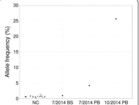
For patient 31, both allele-specific PCR and PCR-RFLP analysis of the initial sample (July 2014) showed JAK2
V617F weakly positive result (Fig. 1). No such mutation was reported by the NGS panel on the same sample. We asked whether the mutation was actually detected by
NGS but not reported based on the reportable minimum variant AF (10 %). To obtain the AF of JAK2 V617 re-gardless of variant callers, we checked aligned reads at the corresponding genomic position chr9:5073770G > T. V617F AF was 4.1 % in the initial sample, 0.9 % in matched buccal swab sample and 25.6 % in the second peripheral blood sample (October 2014) (Fig. 1). The mean noise level was 0.6 % based on 18 negative con-trols (0.2–1.3 %, 99 % confidence interval 0.45–0.79 %; Fig. 1). The slight but significant elevation of JAK2
V617F AF in the initial sample showed the mutation was indeed detectable by NGS and confirmed the weakly positive results by conventional molecular analysis.
Known FLT3 ITD mutations detected by Sanger se-quencing in patients 3, 36, 37 and 41 were originally not reported by the NGS panel. However, lengths of the ITD mutations (30–189 bp, n= 4) fell within the range re-ported to be detectable by Pindel (17–185 bp) [29], which was one of the variant callers used in this study. In the study by Spencer et al. employed sonication-based DNA fragmentation during sequencing library prepar-ation, start positions of aligned reads were randomly dis-tributed [29]. In contrast, start positions of aligned reads in this study were fixed by primers of corresponding PCR amplicons. We hypothesized that the ITD detection sensitivity also depended on the relative positions of ITD within corresponding amplicons.
Comprehensive bioinformatics evaluation ofFLT3ITD detection methods
We evaluated the performance of variant callers for ITD detection when the mutation position varied along the amplicon. A simulation approach was chosen to encom-pass a wide range of possible ITD mutations regardless of actual sample availability. A read simulator ITDsim was developed to simulate 2 × 275 bp sequencing data for ITD mutations at various starting positions, based on the amplicon with highest reported FLT3 ITD mutation rate (chr13:28,608,112–28,608,312) in the region. Given that the length of amplified region was 201 bp, starting positions included all these 201 positions and mutation length ranged from 1 to 201 bp (total 40401 combina-tions). For each combination of starting position and length, a pair of FASTQ files was simulated with sequen-cing depth 2000X and ITD AF 50 %. To determine the best possible performance of ITD detection, the simu-lated FLT3 sequencing reads were perfect-matching se-quences derived from the GRCh37/hg19 reference genome without errors and all other amplicons were not simulated. Bioinformatics analysis of the simulated data was performed in the same way as the clinical samples.
Pindel, GATK HaplotypeCaller and Samtools detected 58.9, 45.5 and 23.0 % of simulated ITD mutations, re-spectively (Fig. 2a). Although Pindel detected most ITD
mutations among the three callers, the maximum length of detected ITD mutation decreased from 201 bp to 45 bp at the 41stnucleotide and 201st nucleotide of the amplicon, respectively (Fig. 2a). ITD detection sensitivity was demonstrated to depend on not only length but also its relative position in the amplicon.
A novelFLT3ITD detection algorithm ITDseek
We therefore closely examined the BWA-MEM align-ments and observed two major differences between wild-type and ITD sequencing reads: (1) longer se-quences became soft-clipped (ignored for analysis in-cluding variant calling), compared with consistently short soft-clipped sequences representing universal adapters, and (2) there was additional alignment of part of the soft-clipped sequences to the same FLT3 locus, but marked as supplementary alignment that was ig-nored for analysis in a similar manner as soft-clipping. Based on these observations, a novel ITD mutation algo-rithm ITDseek was developed and detected 73.1 % of the simulated ITD mutations (Fig. 2a). For most starting positions in the amplicon (1 to 198), although the mini-mum length of detected ITD mutation ranged from 15 to 83 bp, the maximum detection length was consist-ently 201 bp (the longest simulated length). ITDseek was insensitive to the remaining 3 starting positions (199–201 bp).
More importantly, ITDseek detected 96.8 % (10152 of 10491) of combined false negatives by the other three callers (Fig. 2b). ITDseek increased overall detection rate from 74.0 % (other three callers) to 99.2 % (all four cal-lers). Computation requirement of ITDseek was minimal that it took <20 s and <2GB RAM to analyze a full MiSeq V3 run of 8 samples by 8 CPU cores (5-year-old Intel Xeon 2.8GHz processors).
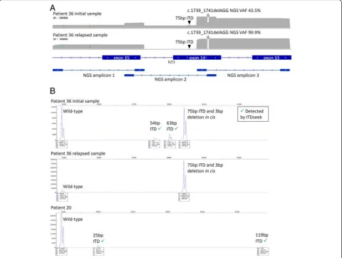
analysis of the original and relapsed samples supported these observations (Fig. 3b).
In patient 20, which was reported ITD negative by Sanger sequencing, ITDseek detected two ITD muta-tions (119 and 25 bp). PCR fragment analysis confirmed
both mutations (Fig. 3b). The allelic burden (1.4 and 3.2 %, respectively) was below the detection limit of Sanger sequencing. Taken together, after further reso-lution and application of ITDseek, there were still 2 samples (patients 20 and 36) with discrepancies and
concordance was 98.2 % (109 of 111).FLT3mutation tection was more sensitive by NGS since ITDseek de-tected 2 additional ITD alleles with low VAF in both patients 20 and 36, notwithstanding the one complex lele in patient 36 that was undetected by NGS due to al-lele drop-out.
NGS mutation profile
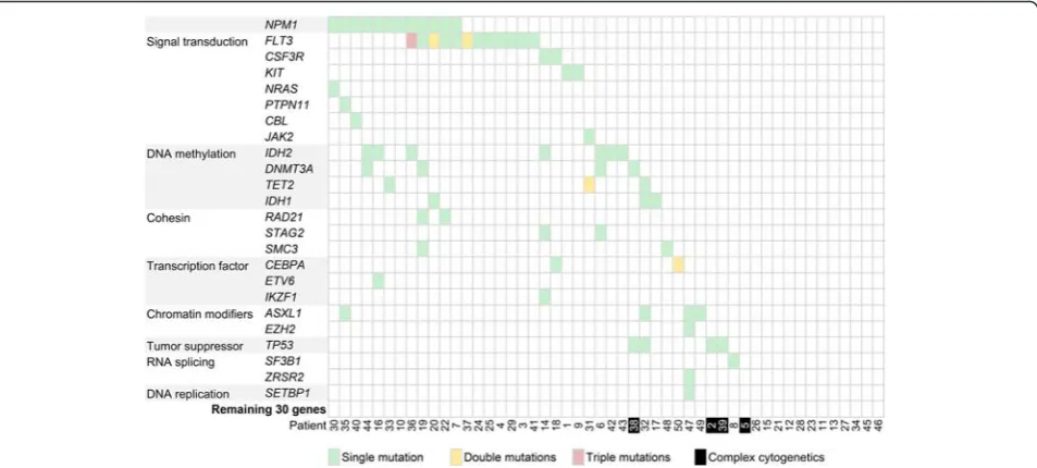
The mean count of sequencing reads obtained per sam-ple was 3.81 million (range 2.02–8.51 million) and the mean sequencing depth was over 3000X. Seventy-seven mutations in 24 genes (Table 1) were detected in 37 of 50 patient samples (74 %). On average 2 mutations (range 1–5) were detected per positive sample. Muta-tions were detected in the following genes: FLT3 (n= 16), NPM1 (n= 12), IDH2 (n= 7), ASXL1, DNMT3A,
TET2, TP53 (all n= 4), CEBPA, IDH1 (both n= 3),
CSF3R, KIT, RAD21, SMC3, STAG2 (all n= 2), CBL,
ETV6, EZH2, IKZF1, JAK2, NRAS, PTPN11, SETBP1,
SF3B1 and ZRSR2 (all n= 1). The two most frequently mutated genes in our patient cohort, FLT3 and NPM1, were in keeping with the mutational frequency of genes in AML as reported in the literature (Fig. 4) [30]. Based on mechanism of action, genes involved in signal trans-duction (FLT3, CSF3R, KIT, NRAS, PTPN11, CBL and
JAK2) and DNA methylation (DNMT3A, TET2, IDH1
and IDH2) were the most frequent mutated groups, ac-counting for 24 (31 %) and 18 (23 %) out of 77 detect-able mutations [31].
The small patient number in our cohort precluded meaningful correlation between gene mutations with clinical features and outcome or survival analysis. This
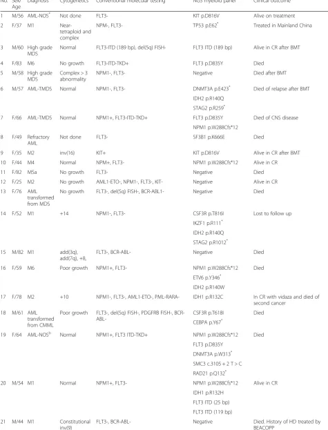
Table 1Summary of 50 patients in this study.
No. Sex/ Age
Diagnosis Cytogenetics Conventional molecular testing NGS myeloid panel Clinical outcome
1 M/56 AML-NOS* Not done FLT3- KIT p.D816V Alive on treatment
2 F/37 M1
Near-tetraploid and complex
NPM-, FLT3- TP53 p.E62* Treated in Mainland China
3 M/60 High grade MDS
Normal FLT3-ITD (189 bp), del(5q) FISH- FLT3 ITD (189 bp) Alive in CR after BMT
4 F/83 M6 No growth FLT3-ITD-TKD+ FLT3 p.D835Y Died
5 M/58 High grade MDS
Complex > 3 abnormality
NPM1-, FLT3- Negative Died after BMT
6 M/57 AML-TMDS Normal NPM1-, FLT3- DNMT3A p.E423* Died of relapse after BMT
IDH2 p.R140Q STAG2 p.R259*
7 F/66 AML-TMDS Normal NPM1+, FLT3-ITD-TKD+ FLT3 p.D835Y Died of CNS disease
NPM1 p.W288Cfs*12 8 F/49 Refractory
AML
Not done FLT3- SF3B1 p.K666E Died
9 F/35 M2 inv(16) KIT+ KIT p.D816V Alive in CR after BMT
10 F/44 M4 Normal NPM+, FLT3- NPM1 p.W288Cfs*12 Alive in CR
11 F/82 M5a No growth FLT3- Negative Died
12 F/25 M2 No growth AML1-ETO-, NPM1-, FLT3-, KIT- Negative Alive in CR
13 F/76 AML transformed from MDS
No growth FLT3-, del(5q) FISH-, BCR-ABL1- Negative Died
14 F/52 M1 +14 NPM1-, FLT3- CSF3R p.T816I Lost to follow up
IKZF1 p.R111*
IDH2 p.R140Q STAG2 p.R1012*
15 M/82 M1 add(3q),
add(7q), +8,
FLT3-, BCR-ABL- Negative Died
16 F/59 M6 Poor growth NPM1+, FLT3- NPM1 p.W288Cfs*12 Died
ETV6 p.Y346*
IDH2 p.R140W
17 F/78 M2 +10 NPM1-, FLT3-, AML1-ETO-, PML-RARA- IDH1 p.R132C In CR with vidaza and died of second cancer
18 M/61 AML transformed from CMML
Poor growth FLT3-, del(5q) FISH-, PDGFRB FISH-, BCR-
ABL-CSF3R p.T618I Died
CEBPA p.Y67*
19 F/64 AML-NOSb Normal NPM1+, FLT3 ITD-TKD+ NPM1 p.W288Cfs*12 Died
FLT3 p.D835Y DNMT3A p.W313* SMC3 c.3105 + 2 T > C RAD21 p.Q132*
20 M/54 M1 Normal NPM1+, FLT3- NPM1 p.W288Cfs*12 Alive in CR
IDH1 p.R132H FLT3 ITD (25 bp) FLT3 ITD (119 bp)
21 M/44 M1 Constitutional
inv(9)
FLT3-, BCR-ABL- Negative Died. History of HD treated by
Table 1Summary of 50 patients in this study.(Continued)
22 F/44 M2 Not done FLT3 ITD + TKD- NPM1 p.W288Cfs*12 Died
RAD21 c.1161 + 2 T > A FLT3 ITD (72 bp) 23 F/33 M2
therapy-related
Poor growth NPM1-, FLT3- Negative Died. History of treated breast
cancer
24 F/26 M3 No growth PML-RARA(s)+, FLT3 ITD + TKD- FLT3 ITD (33 bp) Alive in CR
25 M/65 M5a +8 NPM1-, FLT3 ITD + TKD- FLT3 ITD (54 bp) Died
26 F/74 M0 +22 CBFB-MYH11- NPM1- FLT3- BCR-ABL1- Negative Alive in CR and maintained on
monthly vidaza
27 F/77 AML-TMDS +8 Not done Negative Lost to follow up. History of lung
cancer
28 M/18 M2 t(8;21),-Y AML1-ETO+, KIT-, NPM1-, FLT3- Negative Alive in CR
29 F/61 AML-NOSa Poor growth NPM1-, FLT3 ITD-TKD+, BCR-ABL-, AML1-
ETO-FLT3 p.D835Y Alive in CR
30 F/39 M2 +8 AML1-ETO-, NPM+, FLT3- NPM1 p.W288Cfs*12 Alive in CR
NRAS p.G12D 31 M/70 M1 Normal NPM-, FLT3-, PDGFRB FISH-,
FIP1L1-PDGFRA-, BCR-ABL-,
TET2 p.S1848* NR to vidaza. Further treatment in
Mainland China TET2 p.G1152E
JAK2 V617F weak+ JAK2 p.V617F (VAF 4.1 %,
below original reportable threshold)
32 F/80 M2 del(5q), +8 Not done IDH1 p.R132C Alive and on vidaza
TET2 p.K693Nfs*18 TP53 p.R249S ASXL1 p.W960*
33 F/68 M4 Normal FLT3- NPM1 p.W288Cfs*12 Difficult CR and on vidaza
TET2 p.L346Rfs*2 34 F/24 M2 t(8;21) AML1-ETO+, BCR-ABL-, PML-RARA-,
NPM1-,
FLT3-Negative Alive in CR
35 M/60 M5a +8 Not done NPM1 p.W288Cfs*12 Alive on treatment
PTPN11 p.G503A ASXL1 p.G646Wfs*12 36 F/62 M1 Normal AML1-ETO-, BCR-ABL1-, PML-RARA-, NPM1
+, FLT3 ITD+in ciswith 3 bp deletion (c.1739_1741delAGG)
NPM1 p.W288Cfs*12 CR for 1 year. Relapsed on treatment
FLT3 p.Q580_V581delinsL (c.1739_1741delAGG) FLT3 ITD (54 bp) FLT3 ITD (63 bp) IDH2 p.R140Q
37 F/46 M4
Near-tetraploid
AML-ETO-, BCR-ABL1-, FLT3 p.D835Y Responded to sorafenib and HHT.
Received BMT from sibling donor and on treatment
FLT3 ITD+ (30 bp), FLT3 TKD+ FLT3 ITD (30 bp)
38 M/66 AML-TMDS Complex > 3 abnormalities
FLT3-, BCR-ABL1-, JAK2 V617F- DNMT3A p.M801Nfs*11 Alive on vidaza. History of lung cancer
TP53 p.R175H 39 F/62 AML-M5
post-BMT relapse
Complex > 3 abnormalities
Not done TP53 p.Y220C Died
40 M/62 M5a transformed from MDS
Normal NPM1+, FLT3-, MLL FISH- NPM1 p.W288Cfs*12 Died
CBL c.1096-1G > T
notwithstanding, TP53 mutations were found in three out of four patients (75 %) with complex and unfavor-able cytogenetics. The mutational spectrum of the cyto-genetic unfavorable group was rather different from that of the cytogenetic normal or intermediate group in our patient cohort. Interestingly, both ASXL1p.G646Wfs*12 and NPM1 p.W288Cfs*12 mutations were detected in the initial sample of patient 35 but not detected in the post-treatment sample. Although whether the two muta-tions occurred in the same or independent clones is un-known, they represented one of the first documented exceptions to mutual exclusion nature of ASXL1 and
NPM1mutations in AML [32].
Discussion
While infrequent in CN-AML, mutations affectedTP53
is associated with a complex karyotype [33], as con-firmed by our patient cohort. This may define a distinct subgroup of AML that displays primary resistance to therapy and a very dismal prognosis [34, 35]. It is un-clear whether TP53 mutations cause and promote in-creasing cytogenetic instability, or whether these are secondary mutations occurring after the onset of chromosomal instability. One study showed that a subset of patients with complex karyotype did not have TP53
mutations, whilst all TP53-mutated AML were found in complex karyotype, suggesting that complex karyotype preceded TP53 mutations [36]. Also most mutations of
TP53 were associated with del(17p) [36], supporting the contention that TP53 mutations were the second hit in
leukemogenesis. More recently,TP53 mutations associ-ated with therapy-relassoci-ated AML were shown to be present before the exposure to chemotherapy, suggesting that a pre-leukemic clone harboring TP53 mutation gained survival advantage after chemotherapy, rather than induced by chemotherapy [37]. Hence TP53-mutated AML and therapy-related AML may have more in common than previously recognized.
Detection of FLT3 ITD is an important test for CN-AML due to its impact on prognosis and treatment [38]. Highly variable length, allelic burden and num-ber of the ITD mutations were observed [39]. These characteristics pose a challenge in detection by next-generation sequencing, specifically not the sequencing process per se, but the bioinformatic analysis of the short sequences obtained (<300 bp). Pindel was shown to be the state-of-the-art ITD variant caller, particularly its detection of ITD alleles with length up to 185 bp [29]. However, our study showed that Pin-del detected 3 ITD alleles of length 33 bp (patient 24) to 72 bp (patient 22) but not 4 ITD alleles of length 30 to 189 bp. Similarly, a recent study showed that Pindel detected 14 ITD alleles of length up to approximately 60 bp but not 3 ITD alleles of length approximately 50, 90 and 110 bp [40].
By comprehensive evaluation based on 40401 simu-lated ITD alleles with length up to 201 bp, we demon-strated that detection from amplicon-based NGS data was dependent on relative ITD position within amplicon, in addition to ITD length. Other laboratories may also Table 1Summary of 50 patients in this study.(Continued)
42 M/88 AML-NOS* Not done Not done IDH2 p.R140Q Alive on palliative care
43 M/63 M2 Normal Not done IDH2 p.R172K Alive on vidaza
44 F/60 M2 and bone marrow fibrosis
Normal 5/7/del(20q) FISH-, BCR-ABL-, FLT3-, JAK2
V617F/CALR/MPL-DNMT3A c.855 + 1G > T Alive on treatment NPM1 p.W288Cfs*12
IDH2 p.R140Q
45 F/28 M4Eo inv(16) CBFB FISH+, CBFB-MYH11 PCR+ Negative Alive in CR
46 F/84 M4Eo Not done CBFB-MYH11 PCR+, FLT3-, NPM1-, KIT- Negative Died
47 M/51 Atypical CML
Normal CSF3R-, BCR-ABL-, JAK2 V617F-, PDGFRB , PDGFRA
FISH-EZH2 c.1852-2A > G Died SETBP1 p.D868N
ASXL1 p.G646Wfs*12 ZRSR2 p.Q103*
48 F/53 M2 t(8;21) KIT- SMC3 p.S674_R675insL Alive in CR
49 M/65 High-grade MDS
Normal Not done ASXL1 p.G646Wfs*12 Alive on vidaza + eltrombopag trial
50 M/51 M2 Normal NPM1-, FLT3-, AML1-ETO- CEBPA p.R343Afs*79 Alive on treatment
CEBPA p.K313dup
CRcomplete remission,NRnon-remission,FISHfluorescence in-situ hybridization,vidaza5-azacytadine,HHThomoharringtonine,HDHodgkin lymphoma,
PML-RARA(s) short isoform of fusion transcript from PML bcr3 breakpoint,VAFvariant allele frequency,FLT3testing included detection of both ITD and TKD *
Diagnosis on PB only a
Aparticulate aspirate and diagnosis by immunophenotyping only b
use the simulator ITDsim to evaluate the performance of their own bioinformatics pipeline. We also developed a novel detection algorithm ITDseek because recently developed tools including BreaKmer [41] and Genomon ITDetector [42] were developed for sequencing library prepared with random DNA fragmentation and similar to Pindel. Amplicon Indel Hunter [43] was developed for amplicon-based sequencing data but the actual im-plementation was not available for parallel evaluation. ITDseek detected most false negatives (97 % of simu-lated samples) of Pindel, GATK and Samtools. For the actual samples in this study, ITDseek detected ITD al-leles up to 189 bp missed by Pindel. ITDseek was de-signed to process the de facto standard BAM alignment file with minimal computation time (<20 s for a whole MiSeq run by 8 CPU cores) and is expected to be easily incorporated in various bioinformatics pipelines of other laboratories.
Conclusions
We show that gene panel testing by NGS approach in a diagnostic molecular pathology laboratory allows sensitive and accurate detection of actionable AML gene mutations to individualize patient management. The diagnostic approach to AML is facing a paradigm shift in the genomics era [44]. As more targeted ther-apy become available, the greater is the clinical de-mand for comprehensive molecular profiling. The results of genomic study hold promise for better un-derstanding disease pathogenesis and classification, re-fining prognostic stratification and uncovering new drug targets. Further studies should focus on the
clinical utility of the genomics to document whether this approach translated into improvements in AML patient outcome and survival [45]. The novel algo-rithm ITDseek presented in this paper improves the detection of FLT3-ITD in the laboratory setting of amplicon-based next-generation sequencing.
Competing interests
The authors declare that they have no competing interests.
Authors’contributions
CHA developed the bioinformatics algorithm and wrote the paper. ESKM and TLC conceived the study. AW and ESKM collected samples and curated the information on diagnosis, conventional testing results and clinical outcome. AW and DNH performed the experiments. CHA, TLC and ESKM analyzed the data. All authors read and approved the final manuscript.
Acknowledgements
The authors thank all clinicians who provided the patient samples for study and the information on clinical outcome.
Received: 18 December 2015 Accepted: 14 January 2016
References
1. Grimwade D, Hills RK, Moorman AV, Walker H, Chatters S, Goldstone AH, et al. Refinement of cytogenetic classification in acute myeloid leukemia: determination of prognostic significance of rare recurring chromosomal abnormalities among 5876 younger adult patients treated in the United Kingdom Medical Research Council trials. Blood. 2010;116(3):354–65. 2. Renneville A, Roumier C, Biggio V, Nibourel O, Boissel N, Fenaux P, et al.
Cooperating gene mutations in acute myeloid leukemia: a review of the literature. Leukemia. 2008;22(5):915–31.
3. Dohner H, Estey EH, Amadori S, Appelbaum FR, Buchner T, Burnett AK, et al. Diagnosis and management of acute myeloid leukemia in adults: recommendations from an international expert panel, on behalf of the European LeukemiaNet. Blood. 2010;115(3):453–74.
cytogenetically normal AML patients: further evidence for CEBPA double mutant AML as a distinctive disease entity. Blood. 2011;117(8):2469–75. 5. Wouters BJ, Lowenberg B, Erpelinck-Verschueren CA, van Putten WL, Valk PJ,
Delwel R. Double CEBPA mutations, but not single CEBPA mutations, define a subgroup of acute myeloid leukemia with a distinctive gene expression profile that is uniquely associated with a favorable outcome. Blood. 2009; 113(13):3088–91.
6. Dufour A, Schneider F, Metzeler KH, Hoster E, Schneider S, Zellmeier E, et al. Acute myeloid leukemia with biallelic CEBPA gene mutations and normal karyotype represents a distinct genetic entity associated with a favorable clinical outcome. J Clin Oncol. 2010;28(4):570–7.
7. Green CL, Koo KK, Hills RK, Burnett AK, Linch DC, Gale RE. Prognostic significance of CEBPA mutations in a large cohort of younger adult patients with acute myeloid leukemia: impact of double CEBPA mutations and the interaction with FLT3 and NPM1 mutations. J Clin Oncol. 2010;28(16):2739–47. 8. O’Donnell MR, Abboud CN, Altman J, Appelbaum FR, Arber DA, Attar E, et al.
Acute myeloid leukemia. J Natl Compr Canc Netw. 2012;10(8):984–1021. 9. Mardis ER, Ding L, Dooling DJ, Larson DE, McLellan MD, Chen K, et al. Recurring mutations found by sequencing an acute myeloid leukemia genome. N Engl J Med. 2009;361(11):1058–66.
10. Cancer Genome Atlas Research Network. Genomic and epigenomic landscapes of adult de novo acute myeloid leukemia. N Engl J Med. 2013;368(22):2059–74. 11. Walter MJ, Shen D, Ding L, Shao J, Koboldt DC, Chen K, et al. Clonal
architecture of secondary acute myeloid leukemia. N Engl J Med. 2012; 366(12):1090–8.
12. Patel JP, Levine RL. How do novel molecular genetic markers influence treatment decisions in acute myeloid leukemia? Hematology Am Soc Hematol Educ Program. 2012;2012:28–34.
13. Chung SS. Genetic mutations in acute myeloid leukemia that influence clinical decisions. Curr Opin Hematol. 2014;21(2):87–94.
14. Schlenk RF, Dohner K, Krauter J, Frohling S, Corbacioglu A, Bullinger L, et al. Mutations and treatment outcome in cytogenetically normal acute myeloid leukemia. N Engl J Med. 2008;358(18):1909–18.
15. Patel JP, Gonen M, Figueroa ME, Fernandez H, Sun Z, Racevskis J, et al. Prognostic relevance of integrated genetic profiling in acute myeloid leukemia. N Engl J Med. 2012;366(12):1079–89.
16. Metzeler KH, Walker A, Geyer S, Garzon R, Klisovic RB, Bloomfield CD, et al. DNMT3A mutations and response to the hypomethylating agent decitabine in acute myeloid leukemia. Leukemia. 2012;26(5):1106–7.
17. Voso MT, Santini V, Fabiani E, Fianchi L, Criscuolo M, Falconi G, et al. Why methylation is not a marker predictive of response to hypomethylating agents. Haematologica. 2014;99(4):613–9.
18. Cortes JE, Kantarjian H, Foran JM, Ghirdaladze D, Zodelava M, Borthakur G, et al. Phase I study of quizartinib administered daily to patients with relapsed or refractory acute myeloid leukemia irrespective of FMS-like tyrosine kinase 3-internal tandem duplication status. J Clin Oncol. 2013; 31(29):3681–7.
19. Wang F, Travins J, DeLaBarre B, Penard-Lacronique V, Schalm S, Hansen E, et al. Targeted inhibition of mutant IDH2 in leukemia cells induces cellular differentiation. Science. 2013;340(6132):622–6.
20. Li H. Toward better understanding of artifacts in variant calling from high-coverage samples. Bioinformatics. 2014;30(20):2843–51.
21. Li H, Handsaker B, Wysoker A, Fennell T, Ruan J, Homer N, et al. The sequence alignment/map format and samtools. Bioinformatics. 2009;25(16): 2078–9.
22. DePristo MA, Banks E, Poplin R, Garimella KV, Maguire JR, Hartl C, et al. A framework for variation discovery and genotyping using next-generation DNA sequencing data. Nat Genet. 2011;43(5):491–8.
23. Koboldt DC, Zhang Q, Larson DE, Shen D, McLellan MD, Lin L, et al. VarScan 2: somatic mutation and copy number alteration discovery in cancer by exome sequencing. Genome Res. 2012;22(3):568–76.
24. Ye K, Schulz MH, Long Q, Apweiler R, Ning Z. Pindel: a pattern growth approach to detect break points of large deletions and medium sized insertions from paired-end short reads. Bioinformatics. 2009;25(21):2865–71. 25. McLaren W, Pritchard B, Rios D, Chen Y, Flicek P, Cunningham F. Deriving
the consequences of genomic variants with the Ensembl API and SNP Effect Predictor. Bioinformatics. 2010;26(16):2069–70.
26. Robinson JT, Thorvaldsdottir H, Winckler W, Guttman M, Lander ES, Getz G, et al. Integrative genomics viewer. Nat Biotechnol. 2011;29(1):24–6. 27. Quinlan AR, Hall IM. BEDTools: a flexible suite of utilities for comparing
genomic features. Bioinformatics. 2010;26(6):841–2.
28. Stajich JE, Block D, Boulez K, Brenner SE, Chervitz SA, Dagdigian C, et al. The Bioperl toolkit: Perl modules for the life sciences. Genome Res.
2002;12(10):1611–8.
29. Spencer DH, Abel HJ, Lockwood CM, Payton JE, Szankasi P, Kelley TW, et al. Detection of FLT3 internal tandem duplication in targeted, short-read-length, next-generation sequencing data. J Mol Diagn. 2013;15(1):81–93. 30. Ohgami RS, Arber DA. The diagnostic and clinical impact of genetics and
epigenetics in acute myeloid leukemia. Int J Lab Hematol. 2015;37 Suppl 1:122–32.
31. Roboz GJ. Epigenetic targeting and personalized approaches for AML. Hematology Am Soc Hematol Educ Program. 2014;2014(1):44–51. 32. Carbuccia N, Trouplin V, Gelsi-Boyer V, Murati A, Rocquain J, Adelaide J, et
al. Mutual exclusion of ASXL1 and NPM1 mutations in a series of acute myeloid leukemias. Leukemia. 2010;24(2):469–73.
33. Haferlach C, Dicker F, Herholz H, Schnittger S, Kern W, Haferlach T. Mutations of the TP53 gene in acute myeloid leukemia are strongly associated with a complex aberrant karyotype. Leukemia. 2008;22(8):1539–41.
34. Rucker FG, Schlenk RF, Bullinger L, Kayser S, Teleanu V, Kett H, et al. TP53 alterations in acute myeloid leukemia with complex karyotype correlate with specific copy number alterations, monosomal karyotype, and dismal outcome. Blood. 2012;119(9):2114–21.
35. Kihara R, Nagata Y, Kiyoi H, Kato T, Yamamoto E, Suzuki K, et al.
Comprehensive analysis of genetic alterations and their prognostic impacts in adult acute myeloid leukemia patients. Leukemia. 2014;28(8):1586–95. 36. Ohgami RS, Ma L, Merker JD, Gotlib JR, Schrijver I, Zehnder JL, et al. Next-generation sequencing of acute myeloid leukemia identifies the significance of TP53, U2AF1, ASXL1, and TET2 mutations. Mod Pathol. 2015;28(5):706–14.
37. Wong TN, Ramsingh G, Young AL, Miller CA, Touma W, Welch JS, et al. Role of TP53 mutations in the origin and evolution of therapy-related acute myeloid leukaemia. Nature. 2015;518(7540):552–5.
38. Leung AY, Man CH, Kwong YL. FLT3 inhibition: a moving and evolving target in acute myeloid leukaemia. Leukemia. 2013;27(2):260–8.
39. Levis M. FLT3 mutations in acute myeloid leukemia: what is the best approach in 2013? Hematology Am Soc Hematol Educ Program. 2013;2013: 220–6.
40. Bolli N, Manes N, McKerrell T, Chi J, Park N, Gundem G, et al. Characterization of gene mutations and copy number changes in acute myeloid leukemia using a rapid target enrichment protocol. Haematologica. 2015;100(2):214–22.
41. Abo RP, Ducar M, Garcia EP, Thorner AR, Rojas-Rudilla V, Lin L, et al. BreaKmer: detection of structural variation in targeted massively parallel sequencing data using kmers. Nucleic Acids Res. 2015;43(3), e19. 42. Chiba K, Shiraishi Y, Nagata Y, Yoshida K, Imoto S, Ogawa S, et al. Genomon
ITDetector: a tool for somatic internal tandem duplication detection from cancer genome sequencing data. Bioinformatics. 2015;31(1):116–8. 43. Kadri S, Zhen CJ, Wurst MN, Long BC, Jiang ZF, Wang YL, et al. Amplicon
indel hunter is a novel bioinformatics tool to detect large somatic insertion/ deletion mutations in amplicon-based next-generation sequencing data. J Mol Diagn. 2015;17(6):635–43.
44. Bene MC, Grimwade D, Haferlach C, Haferlach T, Zini G. Leukemia diagnosis: today and tomorrow. Eur J Haematol. 2015;95(4):365–73.
45. Wang ML, Bailey NG. Acute myeloid leukemia genetics: risk stratification and implications for therapy. Arch Pathol Lab Med. 2015;139(10):1215–23.
• We accept pre-submission inquiries
• Our selector tool helps you to find the most relevant journal
• We provide round the clock customer support
• Convenient online submission
• Thorough peer review
• Inclusion in PubMed and all major indexing services
• Maximum visibility for your research
Submit your manuscript at www.biomedcentral.com/submit