Original Research Article.
Histological Analysis of Primary Brain Tumors in a Tertiary Care Hospital:
A Retrospective Study of 5 Years
Jayanti Mehta
1, Bhawna Bansal
2*, Alka Mittal
3, Kusum Mathur
4, Ramita Vijay
21Professor, 2PG Resident, 3Senior Demonstrator, 4Senior Professor,
Department of Pathology, SMS Medical College & Hospital, Jaipur, Rajasthan, India.
ABSTRACT
Background: Central nervous system neoplasms represent a
unique heterogeneous population of neoplasms and include both benign and malignant tumors. The objective of this article is to provide an overview of the descriptive epidemiology of CNS tumors.
Materials and Methods: A total of 1967 cases of CNS tumors
were retrieved from the archives of department of pathology, SMS medical college, Jaipur for a period of 5 years. The diagnosis in all the cases was made on histological examination. All cases were confirmed applying revised WHO classification 2007. The relative frequency of tumors and distribution of age and sex were analyzed. IHC was done as and when required.
Results: In our study astrocytic tumor was the most common
lesion followed by meningioma. Meningioma was second most common tumor. On the basis of origin of cell type glial cell tumors were most common followed by meningeal tumors. Age distribution in our study showed that tumors were more common in age group of 31 – 40 years followed by 41 – 50 years. According to WHO classification majority of lesions belonged to grade 1 and grade 4.
Conclusion: The most frequent type of CNS tumors in this
study was astrocytic tumor followed by meningioma. This study may provide the representative incidence of various types of CNS tumors. A retrospective epidemiological review of brain tumors is particularly important because it can demonstrate the changes in tumor spectrum of a population. Further multicentric studies should be conducted to have substantial data for use in future.
Keywords: Central Nervous System, Neoplasm, Astrocytic
Tumor.
*Correspondence to:
Dr. Bhawna Bansal,
E – 77, Murlipura Scheme,
Bank Colony, Jaipur, Rajasthan, India.
Article History:
Received: 20-05-2017, Revised: 24-06-2017, Accepted: 21-08-2017
Access this article online
Website:
www.ijmrp.com
Quick Response code
DOI:
10.21276/ijmrp.2017.3.5.003
INTRODUCTION
Central nervous system (CNS) neoplasms represent a unique heterogeneous population of neoplasms and include both benign and malignant tumors. CNS tumors comprise 2% to 5% of all tumors.1 The annual incidence of tumors of CNS ranges from 10 to 17 per 100,000 people for intracranial tumors and 1 to 2 per 100,000 people for intraspinal tumors; the majority of these are primary tumors and only 1/4th to ½ are metastatic.2 The majority of patients die within 1st year of diagnosis of malignant lesion and less than 3% survive more than 3 years.3
Site of lesion is important because any CNS neoplasm regardless of histologic grade or classification may have fatal consequences if situated in a critical brain region. Seventy percent of childhood CNS tumors arise in the posterior fossa; a comparable number of tumors in adults arise within the cerebral hemispheres above the tentorium.2
The age distribution of CNS tumors is said to be bimodal, one peak in children, the second peak in 45-70 yrs of age.4
Benign tumors of brain tend to grow slowly and some of them may
be cured by surgery with or without radiation therapy. The malignant tumors grow more rapidly and are associated with shorter survival. Some of these highly lethal tumors such as medulloblastoma and ependymoblastoma have a tendency to disseminate throughout the CNS.
The objective of this article is to provide a current overview of the descriptive epidemiology of central nervous system tumors in our hospital setup. Our target was to study incidence of various lesions in light of WHO classification (2007)5 and study relevant statistics.
MATERIALS ND METHODS
wax. The wax blocks were cut in 5-6 micron sections and stained by hematoxylin and eosin stain. All cases were confirmed applying revised WHO classification (2007). The relative frequency of
tumors and distribution of age and sex were analyzed.IHC is done as and when required. Following Data was collected: Path no, Name, Age, Sex, Site of lesion.
Table 1: Relative frequencies of various tumorsaccording to histological types
HISTOLOGICAL SUBTYPE NUMBER PERCENT
TUMORS OF NEUROEPITHELIAL ORIGIN
Astrocytic Tumors 752 38.2
Oligodendroglioma 156 7.9
Ependymoma 83 4.2
Choroid Plexus Tumors 2 0.1
Astroblastoma 2 0.1
Ganglioglioma 8 0.4
Neurocytoma 10 0.5
Paraganglioma 17 0.8
Pineocytoma 1 0.05
Medulloblastoma 88 4.4
PNET 3 0.1
Atypical Teratoid 1 0.05
TUMORS OF CRANIAL AND PARASPINAL NERVES
Schwannoma 246 12.5
Neurofibroma 17 0.9
MPNST 2 0.1
TUMORS OF MENINGES
Meningioma 489 24.9
Solitary Fibrous Tumor 1 0.05
Hemangiopericytoma 6 0.3
Hemangioblastoma 10 0.5
LYMPHOMAS AND HEMATOPOIETIC NEOPLASMS
Plasmacytoma 10 0.5
Lymphoma 16 0.8
GERM CELL TUMOR
Germionoma 1 0.05
Teratoma 1 0.05
TUMORS OF SELLAR REGION
Craniopharyngioma 45 2.3
TOTAL 1967 100
RESULTS
A total of 1967 cases of CNS tumors were retrieved from the archives of department of pathology, SMS medical college, Jaipur from January 2012 to December 2016 during period of 5 years. In our study most common lesion was astrocytoma followed by meningioma. We observed 752 cases of astrocytoma out of total 1967 cases of CNS tumors. Meningioma was second most common tumor (489 cases of all CNS tumors). 83 cases of ependymoma were seen. 246 cases of schwannoma and 156 cases of oligodendroglioma were seen.
According to origin of cell type tumors of neuroepithelial origin (n = 1123) were most common followed by meningeal tumors (n = 506). Tumors of cranial and paraspinal nerves were 265 cases (schwannoma + neurofibroma + MPNST) were seen and 88 cases of medulloblastoma were seen.
In our study we found that astrocytic tumors were more common in males whereas meningeal tumors were more common in females. Ependymoma was also commonly seen in males. Craniopharyngioma and hemangioblastoma were also seen more in males. Overall CNS tumors showed predilection for males (n = 1084) in comparison to females (n = 883). Male to female ratio was 1.23: 1.
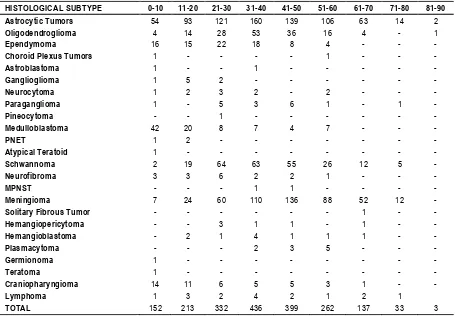
Age distribution in our study showed that tumors were more common in age group of 31 – 40 years (n = 436) followed by 41 –
craniopharyngioma were present. 213 lesions were noticed in 11 –
20 yrs of age group and 330 lesions were noticed in 21 – 30 yrs of age group. 3 lesions were noticed in 81 – 90 yrs of age group. Our
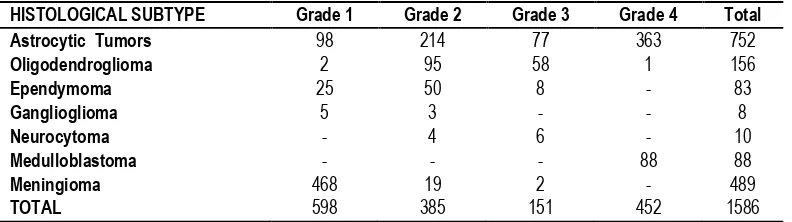
study showed frontal lobe to be the commonest site for primary brain tumors. According to WHO classification 1586 cases were graded, in which majority of lesions belonged to grade 1.
Table 2: Sex wise distribution of various tumors
HISTOLOGICAL SUBTYPE MALES FEMALES
Astrocytic Tumors 472 280
Oligodendroglioma 104 52
Ependymoma 46 37
Choroid Plexus Tumors 1 1
Astroblastoma 1 1
Ganglioglioma 5 3
Neurocytoma 8 2
Paraganglioma 10 7
Pineocytoma 1 -
Medulloblastoma 60 28
PNET 3 -
Atypical Teratoid 1
Schwannoma 137 109
Neurofibroma 5 12
MPNST 2 0
Meningioma 164 325
Solitary Fibrous Tumor 1 -
Hemangiopericytoma 2 4
Hemangioblastoma 7 3
Plasmacytoma 10 0
Germionoma - 1
Teratoma - 1
Craniopharyngioma 32 13
Lymphoma 12 4
TOTAL 1084 883
Table 3: Age wise distribution of CNS tumors
HISTOLOGICAL SUBTYPE 0-10 11-20 21-30 31-40 41-50 51-60 61-70 71-80 81-90
Astrocytic Tumors 54 93 121 160 139 106 63 14 2
Oligodendroglioma 4 14 28 53 36 16 4 - 1
Ependymoma 16 15 22 18 8 4 - - -
Choroid Plexus Tumors 1 - - - - 1 - - -
Astroblastoma 1 - - 1 - - - - -
Ganglioglioma 1 5 2 - - - -
Neurocytoma 1 2 3 2 - 2 - - -
Paraganglioma 1 - 5 3 6 1 - 1 -
Pineocytoma - - 1 - - - -
Medulloblastoma 42 20 8 7 4 7 - - -
PNET 1 2 - - - -
Atypical Teratoid 1 - - - -
Schwannoma 2 19 64 63 55 26 12 5 -
Neurofibroma 3 3 6 2 2 1 - - -
MPNST - - - 1 1 - - - -
Meningioma 7 24 60 110 136 88 52 12 -
Solitary Fibrous Tumor - - - 1 - -
Hemangiopericytoma - - 3 1 1 - 1 - -
Hemangioblastoma - 2 1 4 1 1 1 - -
Plasmacytoma - - - 2 3 5 - - -
Germionoma 1 - - - -
Teratoma 1 - - - -
Craniopharyngioma 14 11 6 5 5 3 1 - -
Lymphoma 1 3 2 4 2 1 2 1
Table 4: CNS tumors distribution and grading according to WHO classification
DISCUSSION
Brain tumors appear to show an increasing trend over the past 30 yrs, but the rise probably results mostly from the use of new neuroimaging techniques. Treatments have not improved prognosis for the most rapidly fatal brain tumors. Established brain tumor risk factors (exposure to therapeutic ionizing radiations, rare mutations of parental genes and family history) explain only a small proportion of brain tumors. Among associations currently being investigated, those of interest include reproductive and menstrual factors for glioma and meningioma, cell phone use for glioma and acoustic neuroma, familial aggregation for meningioma, allergic conditions for glioma, and a variety of inherited polymorphisms potentially associated with glioma. A prior hypothesis will be needed for these studies and for studies involving genetic polymorphisms that, in conjunction with environmental carcinogens or behavioral factors, may increase brain tumor risk. In addition to these promising leads, new hypotheses should consider previous findings from well-established risk factors, such as gender, race and ethnicity. New concepts in brain tumor etiology and clinical management are the goal of such research, with an aim at eradicating this devastating disease.
In our study the distribution of all CNS tumors according to age showed a gradual increase in tumor cases with increasing age, highest in the age group 31-50 years and decreasing thereafter. Fan et al reported proportionally low frequencies of CNS tumors at both ends of age spectrum (below 10 years old and greater than 70 years). The highest frequency was noted in the 50-59 year age group.6 The rise in incidence of brain tumors is consistent with virtually all other adult tumors.7
Mean age of patients having brain tumors vary between different populations. A possible explanation for this is aging populations in developed countries.8
The mean age of patients diagnosed with meningiomas in one European report was 57.6 years, in an American report 59 years and in an Asian report 58.1 years.8-10 Barker et al found a peak incidence of meningiomas in the age group 60-69 years.11 The peak age group for this tumor type in our study was considerably younger (41-50 years). The meningioma inour study showed increased in incidence in the 3rd and4th decade and with the most cases occurring in the range of 41-50 years.
According to balkishan B yeole et al3 too, brain - nervous system cancer were more common in males than females but according to lee et al9 CNS tumors occurred in females more often than in males (female: male ratio 1.43:1)
Rachet et al proposed that brain tumors are 20-50% more common in men in western nations.12 The life time risk of being
diagnosed with a CNS malignancy is estimated to be 0.67% for men and 0.52% for women.13 In separate studies performed on two continents, Mckinney et al, and Fan et al found comparable results suggesting a male to female ratio of 1.5:1.6,7. In our study male to female ratio was 1.23:1.
Ganghoria S et al found that astrocytoma was commoner in males than females. 68.75% of astrocytoma was seen in males and male to female ratio was 1:0.86.14 According to Surawicz et al gliomas affect about 40% more males than females.15 Barker et al found theincidence of malignant gliomas to beespeciallyhigher among male patients.11 The male to female ratio noticed in our study was 1.685 :1. The male predominance was seen in all ages.
Tumors of neuroepithelial origin were more frequently seen in male patients.11,15,16. In Ourstudy we found male to female ratio as 1.73:1 in patients with tumors of neuroepithelial origin.
A review of data obtained from SEER program suggested a higher incidence of meningiomas in females across all ages, while Wiemels et al found that the previously described two fold predominance of female meningioma cases may be inverted in the rare cases of prepubertal meningiomas.17 In our study, tumors of meningeal origin (comprised predominantly of meningiomas) were found more in female patients, with male: female ratio of 0.52:1.
Medulloblastomas and PNET had male predominance in the CBTRUS study.15 In our study, from a total of 88 cases of medulloblastoma 60 were males and all 3 cases of PNET were males.
In our study we found astrocytoma to be the most common tumor (38.34%) followed by meningioma (24.93%). The same was found by Aryan G. et al in Nepal who noticed that astrocytomas were most common tumors of CNS followed by meningiomas.18 In contrast to our study meningioma was most common tumor as noted by Ganghoria S. et.al.14 The same was in favor with surawicz et al15 in USA. Lee et al9 in korea also found that most common tumor was meningioma (31.2%).
The second most common type of primary CNS tumor in our study was meningioma (24.93%) followed by peripheral nerve sheath tumors (13.41%) and oligodendrogliomas (7.7%). There were 88 cases of medulloblastoma (4.48%) and 83 cases of ependymoma (4.23%) and 45 cases of craniopharyngioma (2.29%).
Frontal lobe was the most common site of involvement in brain tumors. This is in favor to the findings of Torres et al19, Andrews et al20, Jalali and Dutta21, Jamal et al.22
Histological grading is a means of predicting the biological behavior of neoplasm. The grading factors tremendously affect the choice of therapy. The commonest type of astrocytoma in our
HISTOLOGICAL SUBTYPE Grade 1 Grade 2 Grade 3 Grade 4 Total
Astrocytic Tumors 98 214 77 363 752
Oligodendroglioma 2 95 58 1 156
Ependymoma 25 50 8 - 83
Ganglioglioma 5 3 - - 8
Neurocytoma - 4 6 - 10
Medulloblastoma - - - 88 88
Meningioma 468 19 2 - 489
study was WHO grade 4 type. A possible cause of this may be linked to their late presentation to hospital. According to WHO classification 1586 cases were graded, in which majority of lesions belonged to Grade I.
It is hard to compare the different studies due to differences in the case material and study techniques. This study may not represent an accurate incidence of CNS tumors in West India (Rajasthan) due to limited number of cases. Furthermore the study was based on a single tertiary care hospitalanalysis
CONCLUSION
The most common type of CNS tumors in this study was astrocytoma followed by meningioma. This study may provide the representative incidence of various types of CNS tumors. A retrospective histopathological analysis of brain tumors is definitely important for future research because it can reveal the changes in tumor spectrum of a population. As the geographic area changes, histopathology of tumors change which affect the management. Further nationwide multicenter studies should be conducted to have substantial data for purpose of research in future.
REFERENCES
1. Masoodi T, Gupta RK, Singh JP, Khajuria A. Pattern of central nervous system neoplasms: A study of 106 cases JK-practitioner vol. 17,no.4 october-december 2012.
2. Frosch MP, Anthony DC, Girolami DU. The central nervous system in: Robbins and cotran, editor. Text book of pathologic basis of disease. 9th edition, Newyork, Elsevier; 2010 : 1251-151-1391.
3. Yeole BB. Trends in brain cancer incidence in India. Asian Pac J cancer Prev.2008;9:267-70.
4. Lantos PL, Louis DN, Rosenblum M.K et.al. Tumors of nervous
system. In: Graham DI, Lantos PL, Eds, Greenfield’s
neuropathology, 7th edition, Arnold, London 2002.
5. Louis DN, Ohgaki H, Wiestler OD, Cavenee WK. WHO classification of tumors of central nervous system, 4th Edi. Lyon, FR: IARC press; 2007.
6. Fan K, Pezeshkpour GH. Ethnic Distribution of Primary Central Nervous System Tumors in Washington, DC, 1971 to 1985. J NatI Med Assoc. 1992;84(10):858-63.
7. McKinney PA. Brain Tumors: Incidence, Survival and Aetiology J Neurol Neurosurg Psychiatry. 2004, 75 (Suppl II):ii12-7.
8. Provost D, Cantagrel A, Lebailly P, Jaffre A, Loyant V, Loiseau H, et al. Brain tumors and exposure to pesticides: a case–control study in southwestern France. Occup Environ Med. 2007; 64: 509-14.
9. Lee C, Jung K, Yoo H, Park S, Lee SH. Epidemiology of Primary Brain and Central Nervous System Tumors in Korea J Korean Neurosurg Soc. 2010;48:145-52.
10. Flowers A. Brain Tumors in the Older Person. Cancer Control. 2000;7(6):523-38.
11. Barker DJP, Weller RO, Garfield JS. Epidemiology of primary tumors of the brain and spinal cord: a regional survey in southern England J Neurol Neurosurg Psychiatry. 1976;39:290-6.
12. Rachet B, Mitry E, Quinn MJ, Cooper N, Coleman NP. Survival from brain tumors in England and Wales up to 2001 British Journal of Cancer. 2008;99: S98-101.
13. Gurney J, Kadan-Lottick N. Brain and other central nervous system tumors: rates, trends, and epidemiology Current Opinion in Oncology. 2001;13:160-6.
14. Ghanghoria S, Mehar R, Kulkarni CV, Mittal M, Yadav A, Patidar H. Retrospective histological analysis of CNS tumors - A 5 year study. Int J Med Sci Public Health. 2014;3:1205-7.
15. Surawicz TS, McCarthy BJ, Kupelian V, Jukich PJ, Bruner JM, Davis FG. Descriptive epidemiology of primary brain and CNS tumors: Results from the Central Brain Tumor Registry of the United States, 1990–1994. Neuro-Oncology. 1999;1:14-25. 16. Materljan E, Materljan B, Sepèiæ J, Tuškan-Mohar L, Zamolo G, Erman-Baldini I. Epidemiology of Central Nervous System Tumors in Labin Area, Croatia, 1974-2001. Croat Med J. 2004;45:206-12.
17. Wiemels J, Wrensch M, Claus EB. Epidemiology and etiology of meningioma. J Neurooncol. 2010;99: 307-14.
18. Aryal G. Histopathological pattern of central nervous system tumor: A three year retrospective study. Journal of Pathology of Nepal 2011;1:22 -5.
19. Torres LF, Almeida R, Avilla S et al. Brain tumors in south brazil: A retrospective study of 438 cases, Arq neuropsiquiatr 1990 sep; 48(3): 279 – 85.
20. Andrews NB, Ramesh R, Odjidja T.A. Preliminary survey of CNS tumors in tema, ghana. West Africa J med 2003 Jun;22[2]; 167 – 72.
21. Jalali R, Dutta D. Prospective analysis of incidence of central nervous system tumors presenting in a tertiary cancer hospital from India. J. neurooncol 2008; 87: 111-114.
22. Jamal S, Mamoon N, Mushtaq S. et al. Pattern of central nervous system tumors: A study of 430 cases. Pak J. Pathol 2005; 16 [4]: 106 – 109.
[
Source of Support: Nil. Conflict of Interest: None Declared.
Copyright: © the author(s) and publisher. IJMRP is an official publication of Ibn Sina Academy of Medieval Medicine & Sciences, registered in 2001 under Indian Trusts Act, 1882. This is an open access article distributed under the terms of the Creative Commons Attribution Non-commercial License, which permits unrestricted non-commercial use, distribution, and reproduction in any medium, provided the original work is properly cited.
Cite this article as: Jayanti Mehta, Bhawna Bansal, Alka Mittal, Kusum Mathur, Ramita Vijay. Histological Analysis of Primary Brain Tumors in a Tertiary Care Hospital: A Retrospective Study of 5 Years. Int J Med Res Prof. 2017 Sept; 3(5):14-18.