ORIGINAL ARTICLE
Studies of a rice sterile mutant
sstl
from the TRIM collection
Chia‑Ling Chang
1,2, Jerry C. Serapion
2, Han‑Hui Hung
3, Yan‑Cheng Lin
4, Yuan‑Ching Tsai
5, Wann‑Neng Jane
2,
Men‑Chi Chang
1, Ming‑Hsin Lai
3and Yue‑ie C. Hsing
1,2*Abstract
Background: Rice (Oryza sativa) is one of the main crops in the world, and more than 3.9 billion people will con‑ sume rice by 2025. Sterility significantly affects rice production and leads to yield defects. The undeveloped anthers or abnormal pollen represent serious defects in rice male sterility. Therefore, understanding the mechanism of male sterility is an important task. Here, we investigated a rice sterile mutant according to its developmental morphology and transcriptional profiles.
Results: An untagged T‑DNA insertional mutant showed defective pollen and abnormal anthers as compared with its semi‑sterile mutant (sstl) progeny segregates. Transcriptomic analysis of sterile sstl‑s revealed several biosynthesis pathways, such as downregulated cell wall, lipids, secondary metabolism, and starch synthesis. This downregula‑ tion is consistent with the morphological characterization of sstl‑s anthers with irregular exine, absence of intine, no starch accumulation in pollen grains and no accumulated flavonoids in anthers. Moreover, defective microsporangia development led to abnormal anther locule and aborted microspores. The downregulated lipids, starch, and cell wall synthesis‑related genes resulted in loss of fertility.
Conclusions: We illustrate the importance of microsporangia in the development of anthers and functional micro‑ spores. Abnormal development of pollen grains, pollen wall, anther locule, etc. result in severe yield reduction.
Keywords: Anther development, Microspores, Transcriptomic analysis, Semi‑sterile, Sterility
© The Author(s) 2019. This article is distributed under the terms of the Creative Commons Attribution 4.0 International License (http://creat iveco mmons .org/licen ses/by/4.0/), which permits unrestricted use, distribution, and reproduction in any medium, provided you give appropriate credit to the original author(s) and the source, provide a link to the Creative Commons license, and indicate if changes were made.
Background
Rice (Oryza sativa) is one of the most important staple crops in the world and is also an economical and culti-vated food in Asia. One of the major limitations in rice yield production is sterility. Several studies provided strong evidence that male sterility was a serious defect in rice production and led to significantly decreased rice production (Thangasamy et al. 2011; Wang et al. 2013; Guo et al. 2013; Pu et al. 2017) or even almost complete loss (Liu et al. 2010; Ko et al. 2014; Yang et al. 2014; Li et al. 2016).
Male sterility is associated with a series of anther development steps and/or pollen maturation at the rice reproductive stage. Studies of the developmental mor-phology of pollen revealed several important steps,
such as cell differentiation into microspores and cir-cular anther locules, the formation of four specialized cell layers in the anther, meiosis of the pollen mother cell, microspores with a well-developed pollen wall, microspore mitosis, and intine establishment of mature pollen (Raghavan 1988; Zhang et al. 2011; Walbot and Egger 2016). Previous studies concluded that the pro-cess of pollen maturation in rice was initiated in the L1, L2 and L3 layers of the stamen primordia, and cen-tral archesporial (AR) cells were generated from the mitotic division in the L2 layer to develop the primary parietal cells (PPCs) and primary sporogenous cells (PSCs) (Zhang et al. 2011; Kelliher et al. 2014; Yang et al. 2018). After AR cells divide and differentiate to PSCs and PPCs, PSCs form as sporogenous cells and later generate microspores; PPCs divide and develop into secondary parietal layers (SPCs). The outer layer of SPCs becomes the endothecium and middle layer,
Open Access
*Correspondence: bohsing@gate.sinica.edu.tw
and the inner layer becomes the tapetal cell (Zhang and Wilson 2009).
Receptor protein kinase (RPK) is important in regulating tapetal cells and microsporocytes dur-ing sporophytic generation. In Arabidopsis, a putative leucine-rich repeat receptor protein kinase (LRR-RPK)
gene, EXCESS MICROSPOROCYTES1 (EMS1)/EXTRA
SPOROGENOUS CELLS (EXS), participates in early division of the anther and microsporocyte cells to con-trol tapetum and microsporocyte development (Cai and Zhang 2018; Verma 2019). TAPETUM DETERMI-NANT1 (TPD1) was found as a cysteine-rich protein ligand of EMS1. TPD1 interacts with the EMS1 LRR domain to generate precursors of tapetal cells by pro-moting cell division of inner SPCs; the developmental tapetum layer determines the number of sporogenous cells by suppressing proliferation (Huang et al. 2016). A rice protein binding cassette with the LRR-RLK pro-tein MULTIPLE SPOROCYTE1 and its ligand TPD1-like protein (OsTDL1A) is involved in PPC division to contribute SPCs for the middle layer and establish the
tapetum. The anther with double mutated OSTDL1A
and MULTIPLE SPOROCYTE1 genes illustrated a lack of middle layers and tapetum and increased the num-ber of microsporocytes in the early development of pollen (Yang et al. 2016). On mitosis, primary sporog-enous cells divide into secondary sporogsporog-enous cells and become pollen mother cells (PMCs). PMCs pass through meiosis to generate microspores. At the same time, programmed cell death (PCD) of the tapetum layer occurs during meiosis. Several regulators respond to microspore generation, and PCD of tapetal cells such as a rice basic helix-loop-helix (bHLH) protein, TAPE-TUM DEGENERATION RETARDATION (TDR), con-trols tapetum degeneration to contribute to microspore development (Li et al. 2006). Gibberellic acid-inducible transcription factor GAMYB upregulates TDR1 expres-sion, and a frame shift with a C deletion of the allele gamyb-4 did not go through normal meiosis or PCD of tapetum in rice (Liu et al. 2010). The bHLH142 that conjugates with TDR1 turned on the downstream gene ETERNAL TAPETUM1 (EAT1) to organize microspore development. With T-DNA insertion into bHLH142, the tapetal PCD and microspore development were defective in anthers of rice loss-of-function bhlh142 (Ko et al. 2014).
Microspores are produced after PMC meiosis, and young microspores are released from the tetrads later. The rice Wax-deficient anther1 (Wda1) defective mutant showed a significant defect of very-long-chain fatty acids in the anther epidermis and pollen wall that led to a severe absence of pollen (Jung et al. 2006).
Thus, the fatty acid of pollen wall conformation plays an important role in microspore maturation.
The pollen wall is composed of the outer pollen wall exine and inner wall intine. Transmission electron micro-scope (TEM) observation revealed that exine consists of the tectum, bacula, and nexine together as a bridge-like structure of the pollen outer wall (Shi et al. 2015). The pollen walls of microspores are constructed during tape-tum layer degradation and again indicate the importance of lipid metabolism (Li et al. 2011). Transcriptomic anal-ysis revealed that transcripts encoding cytochrome P450, acyltransferases, and lipid transfer proteins are produced from the tapetum secretion stage to support fatty acid accumulation and transportation to build exine (Huang et al. 2009). Without PCD, tapetum did not degener-ate to generdegener-ate fatty acid components such as sporopol-lenin for exine formation. Rice PERSISTENT TAPETAL CELL1 (PTC1), which affects fatty acid synthesis in the exine structure, promotes tapetum degradation to gener-ate sporopollenin for pollen wall development (Li et al. 2011). Scanning electron microscopy (SEM) revealed that the tapetal cell also produces orbicules during tapetum degeneration, which surround the surface of the tape-tum layer, and orbicules provided the fatty acid prod-uct for assembly of sporopollenin for exine (Daku et al. 2016; Ruggiero and Bedini 2018). A rice lipid transfer protein OsC6 involved in lipid transfer from tapetum to exine is regulated by bHLH, TDR, and GAMYB. The truncated osc6 mutation resulted in defective orbicules and disordered exine structure in the pollen wall (Zhang et al. 2010). Thus, a well-developed exine is an indica-tor of maturation of microspore fertility during anther development.
transfers UDP-glucose for polysaccharide composition requirement, and also regulates starch and protein syn-thesis in pollen grain maturation to determine rice fertil-ity (Moon et al. 2013). The polysaccharides of intine also regulate water content in pollen dehydration and disperse pressure when mature pollen spores expand with internal content accumulation (Xu et al. 2016).
Taiwan Rice Insertional Mutagenesis (TRIM) was ini-tiated in 2002. In this mutant population, 93,000 rice mutated lines were created by T-DNA insertion by using a japonica variety, Tainung 67 (Wu et al. 2017). The reverse genetics approach provided a large-scale popu-lation of T-DNA mutagenesis screening for comparing phenotypic characterization and rice genomic function. The TRIM population was used in two studies on male-sterile mutants. T-DNA insertion into bHLH142 led to no response to PCD of tapetum, and this gene was reg-ulated by the early tapetum developing-related genes
UNDEVELOPED TAPETUM1 (UDT1) and GAMYB,
then was responsible for downstream TDR1 and EAT1 regulation (Ko et al. 2014). Another TRIM mutant study indicated that a rice SUMO E3 ligase SIZ1 participated in controlling endothecium development during anther dehiscence to manipulate the fertility of rice. Mutant siz1 showed the inability of anther dehiscence and no cavity of two adjacent locules; thus, it released no pollen grains during anthesis. In addition, this mutant had a shorter plant with few tillers and low seed set rate (Thangasamy et al. 2011).
Here we report a morphological characterization of a male sterile mutant from the TRIM collection and inves-tigate the mechanism of sterility. We illustrate the impor-tance of microspore fertility in anther development and the functional biosynthesis of microspores. We also per-formed a comparative transcriptomic analysis between fertile and sterile anthers to prove that fatty acid biosyn-thesis pathways participate in the morphology of pollen wall formation.
Methods
Plant materials and growth condition
T-DNA insertional mutant line M0037841 with 30 T2 seeds were obtained from the TRIM collection and grown at the genetically modified plant isolation field (GM field) in the Taiwan Agricultural Research Institute (TARI) for agronomic trait evaluation. Leaf samples from individual plants of the T2 population were harvested and frozen under liquid nitrogen for genomic DNA extrac-tion experiments. Progeny from the semi-sterile line of M0037841 were both grown in the TARI GM qualified field or a growth chamber under day 28 °C/night 25 °C, 16 h of light and 80% humidity at Academia Sinica, Tai-pei. Yield component analysis including panicle number,
grain number, fertility rate, and grain weight was per-formed by the semi-sterile segregates in the TARI GM field. The anther samples for floral morphology obser-vation, pollen viability assay, gene expression analysis, phenotypic characterization of cross-sections, TEM and SEM were collected from plants grown in the growth chamber.
Genomic DNA extraction and PCR‑based genotyping
Genomic DNA was extracted from leaves by using the Wizard Genomic DNA purification kit (Promega Cor-poration, Madison, WI, USA). All T-DNA mutagenesis plants from T2 were identified by T-DNA tagging regions, a T-DNA construct pTAG8, such as hygromycin phos-photransferase gene (HPT), GUS gene, and CaMV35S enhancer with PCR-based genotyping assay. The PCR reaction in 20 μL volume involved 50 ng genomic DNA, 0.2 μm primers and Taq DNA Polymerase 2× Master Mix RED (Ampliqon, Herlev, Denmark) for amplifica-tion. The sequences of primers are in Additional file 1: Table S1.
Pollen viability and germination analysis
Mature pollen grains were collected before anthesis in the fertile, semi-sterile and sterile types from semi-sterile progeny segregates and stained with iodine–potassium iodide (I2–KI) solution for pollen viability. Pollen viabil-ity was evaluated by using pollen shape and staining for classification into four grades as dark black (+++), par-tial black (++), orange (+) and yellow (−). More than 2000 individual pollen grains were counted under a Zeiss AxioImager Z1 microscope. More than 1000 pol-len grains per sample were co-cultured with 15% sucrose Aldrich, St. Louis, MO, USA), 5% starch (Sigma-Aldrich, St. Louis, MO, USA), and 0.005% orthoboric acid (Merck, Darmstadt, Germany) at 28 °C for 30 min for detecting pollen tube germination rate.
Morphological analysis of anther characterization
for 24 h. Then, after three rinses with 0.1 M K-phosphate buffer (pH 7.0) for 20 min each, samples were trans-ferred to 1% OsO4 in 0.1 M K-phosphate (pH 7.0) for 4 h at room temperature. The samples were washed again with 0.1 M K-phosphate buffer (pH 7.0) as for the previ-ous rinsed steps. Samples were dehydrated with an etha-nol series and propylene oxide, and embedded in Spurr resin. The cross sections were prepared in 1-μm sections and stained with toluidine blue for anther transverse sec-tion detecsec-tion and anther locule area investigasec-tion under a Zeiss AxioImager Z1 microscope. The ultrathin sec-tions in 70–90 nm were stained with uranyl acetate and lead citrate for TEM imaging by using a Philips CM 100 TEM system at 80 kV. For cryo-SEM, anthers and pol-len grains at Mp stage were frozen in liquid nitrogen and placed into the sample preparation chamber at − 160 °C. After the temperature reached − 85 °C and sublimation/ etching proceeded for 15 min, samples were coated with platinum particles at − 130 °C. Samples were moved to a cryo-stage SEM chamber and examined by using a Cryo-SEM system at temperature − 190 °C (Quanta 200 SEM/ cryo system Quorum PP2000TR FEI).
RNA extraction and transcriptomic analysis
Anther samples were collected from YM or LpM stages for total RNA isolation by using TRIzol reagent (Thermo Fisher Scientific, Wilmington, DE, USA), and 5 μg total RNA was prepared for RNA sequencing. For RNA sequencing, single-end libraries were created and sequenced on an Illumina HiSeq 2000 system in 90-bp read length for 10 million reads per sample. Sequence data were deposited in the NCBI gene expression omni-bus (accession no. GSE129579). RNA sequencing data were evaluated by RNA-seq analysis, differential expres-sion of genes and heat map drawing with the CLC Genomics Workbench 11 (CLC Bio, QIAGEN Bioin-formatics, Aarhus, Denmark). The RNA-seq transcripts were annotated by using the RAP-DB database (https :// rapdb .dna.affrc .go.jp/index .html) and compared with the Rice Expression Profile Database (http://ricex pro.dna. affrc .go.jp/). Anther-specific transcriptomic genes were also confirmed by Rice Anther Expression Plots (https ://www.cpib.ac.uk/anthe r/ricei ndex.html). EXPath Tool (http://expat htool .itps.ncku.edu.tw/), Kyoto Encyclope-dia of Genes and Genomes (KEGG) (http://www.genom e.jp/kegg/), and MapMen (https ://mapma n.gabip d.org/) were used for gene function prediction and analysis.
Gene expression analysis
Total RNA from YM and LpM were converted to cDNA with use of oligo (dT) primers by the SuperScript III First-Strand Synthesis System (Thermo Fisher Scientific, Wilmington, DE, USA). Real-time quantitative RT-PCR
was performed with the SYBR Green-based quantitative PCR method by mixing 20 ng cDNA, 0.25 μm primers and fluorescence Power SYBR Green PCR Master Mix (Applied Biosystems, Foster City, CA, USA) for gene expression detection. The gene expression was calculated by the 2−ΔΔCt method with OsUBI, OsACT11, OsUBQ5, and eEF-1α used as internal control genes. All primer sequences are in Additional file 1: Table S1.
Results
A semi‑sterile mutant from TRIM collection
Sterility is one of the important regulators of the repro-ductive stage in rice yield production. To study this phe-nomenon of sterility, we selected the T-DNA insertional mutant line M0037841 from the TRIM collection. Thirty T2 seeds of M0037841 were grown in a GM field in TARI. The T2 population in M0037841 was identified without co-segregation between the phenotype of sterility and the T-DNA insertional event of the genome (Additional file 2: Table S2).
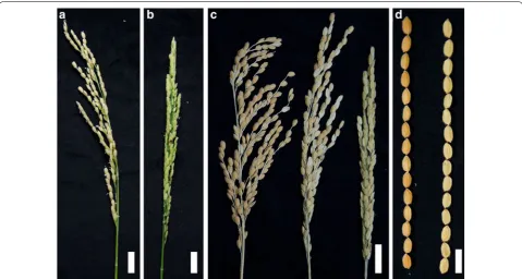
From the developmental morphology of the T2 popu-lation in M0037841, we noticed two plants were semi-sterile and thus designated them semi-semi-sterile (sstl) for the rest of the study (Additional file 2: Table S2). From the progeny segregates of the mutant sstl, characteristic distributions of fertility were classified as fertile pani-cle plants (SSTL-F), semi-sterile panicle plants (sstl-ss), and sterile panicle plants (sstl-s) (Fig. 1a–c). In sstl prog-eny segregates, SSTL-F and sstl-s lines did not differ in plant height or tiller number during the vegetative stage (Additional file 3: Figure S1). In the reproductive stage, yield component traits were analyzed as fertility rate, number of panicles, grain number and grain weight in SSTL-F, sstl-ss, and sstl-s. The fertility rate of sstl-ss was 49.2% (Table 1), showing half fertile seeds per panicle (Fig. 1c). SSTL-F, sstl-ss, and sstl-s all had 15 panicles per plant, but the grain number of each panicle gradu-ally increased from SSTL-F and sstl-ss to sstl-s. The grain size from fertile and sterile spikelets did not differ. The kernel shape, along with palea and lemma, did not differ (Fig. 1d).
Thus, the sterile characterization of sstl-s was not caused by a T-DNA insertion, and sstl-s still inherited the sterility trait from the progenitor semi-sterile sstl.
Sterility from sstl‑s pollen viability
stuck to anther locules that they may not be exposed eas-ily as mature pollen in SSTL-F after cutting the anthers (Fig. 2c). During anthesis in SSTL-F, the mature pol-len grains were released from anther dehiscence and the filaments elongated. However, only elongated fila-ments were found in sstl-s (Fig. 2d). The anther locules of sstl-s were juicy and sticky and thus the pollen grains were barely released after filament elongation. This find-ing was consistent with the anther locules of sstl-s after chopping (Fig. 2c, d). By SEM observations of SSTL-F and sstl-s anthers, we confirmed that sstl-s had a shrinking anther and smaller pollen sac as compared with SSTL-F
(Fig. 2e–h). Additionally, the pollen grains of sstl-s did not fill up with content and formed a stomatocyte-like shape during maturation (Fig. 2g–j). The pollen sacs of sstl-s had no surrounding pollen grains on anther locules because pollen grains of sstl-s did not form as spherical as com-pared with mature pollen of SSTL-F (Fig. 2g, h).
Pollen viability and pollen tube germination analyses were performed to determine the severity of defective pollen in sstl-s. Because mature pollen grains contained abundant starch to provide energy for supporting pol-len maturation and polpol-len tube germination (Lee et al. 2016), we studied pollen viability by using I2–KI stain-ing. Figure 2k showed a significant difference between mature pollen grains of SSTL-F (left) and sstl-s (right) in pollen viability, as indicated by staining color and pollen spherical shape. The starch accumulation of these pol-len grains was illustrated as dark black (+++), partial
black (++), orange (+) and yellow (−) after I2–KI stain-ing (Fig. 2l). The proportion of dark-black mature pollen in SSTL-F, sstl-ss, and sstl-s was 96%, 64%, and 0.35%, respectively, revealing low pollen viability in the sterile mutant (Fig. 2l). Besides, sstl-ss showed 18% partial black pollen, with none in SSTL-F and sstl-s. The proportion of dark-black pollen in semi-sterile sstl-ss was about 60% and agreed well with half fertile seeds per panicle in sstl -ss (Table 1 and Fig. 2l). In addition, examination of pol-len-tube germination of fertile and sterile pollen grains Fig. 1 Phenotypic characterization of segregants of the semi‑sterile mutant (sstl). a Plants with fertile panicles; b completely sterile panicles; c three types of fertility in the sstl progeny segregates: from left to right as fertile, semi‑sterile, and sterile. Bar = 2 cm. d Seed size between fertile and sterile indicated. Bar = 1 cm
Table 1 Yield component analysis of sstl progeny segregates
Yield component analysis of three types in sstl progeny segregates. F fertile, ss
semi-sterile, s sterile, AFRP average fertility rate per panicle, ANP average number of panicles per plant, ANGP average number of grains per panicle, AGWP average grain weight per panicle. The statistical analysis was performed by one-way ANOVA with Tukey test post hoc analysis. Data were collected from 7 individual plants in each type and presented as mean ± SEM
Values followed by different letters indicated a significant difference for each variable (P ≤ 0.05)
Phenotypes AFRP (%) ANP ANGP AGWP (g)
SSTL‑F 92.80 a 15.71 a 148.43 ab 4.01 a
sstl‑ss 49.20 b 15.43 a 163.67 b 2.57 b
confirmed an 81.7% germination rate in SSTL-F but only 0.26% in sstl-s (data not shown).
The defects in sstl-s pollen reveal that its anther loc-ules did not undergo anther dehiscence because the pol-len sacs did not expand and polpol-len grains did not form a spherical shape. Almost zero percentage of pollen viabil-ity and pollen germination rates were consistent with the sterility of the mutant sstl-s as well as pollen-defective morphology.
Anther morphology of sstl‑s
Cross sections of anthers in SSTL-F and sstl-s at spe-cific anther development stage provided tools to under-stand male sterility. At YM stage, both SSTL-F and sstl-s had four cell layers (i.e., epidermis, endothecium, middle layer, and tapetum) (Fig. 3a, f, k). Microspores were released from tetrads by callose degradation in SSTL-F (Fig. 3a) but few from sstl-s anthers (Fig. 3f, k). The tapetum layer started degenerating and the micro-spores formed vacuolated as an oval shape in SSTL-F anthers (Fig. 3b). Tapetum layers of sterile sstl-s still degraded (Fig. 3g, l), similar to the fertile line SSTL-F anther at VP stage. Later on, microspores went through the first mitosis and became bicellular pollen. The micro-spores were smaller from the EpM than VP stage and the starch began to accumulate in fertile microspores (Fig. 3c). Meanwhile, the tapetal cells were almost com-pletely degenerated and left only a thin layer on the internal surface of the anther locule in SSTL-F (Fig. 3c). In contrast, the sstl-s anther showed a delay of tapetum degradation (Fig. 3h, m) and a larger size of tapetal cell at the EpM stage. In the LpM stage, microspores differenti-ated into two sperm cells by the second mitosis division. SSTL-F microspores contained a high amount of starch and lipid accumulation (Fig. 3d). The endothecium and epidermal cells of SSTL-F degenerated in the LpM stage (Fig. 3d), but sstl-s anthers contained defective pollen grains, and anther locules maintained a larger epider-mis and endothecium cell as compared with fertile ones (Fig. 3i, n). The tapetum layer of SSTL-F degenerated and disappeared at this stage (Fig. 3d) but remained par-tially in the sstl-s anther because of delayed degradation (Fig. 3n). The final stage of microspore maturation was the formation of plump round mature pollen in SSTL-F
(Fig. 3e), but only defective pollen was produced in sstl -s anthers (Fig. 3j, o). The epidermis degenerated and became smaller than those from previous stages in the SSTL-F anther. However, as compared with sstl-s anthers, the epidermal cells of sstl-s were three times larger than SSTL-F cells (Fig. 3j, o). The mutant sstl-s contained defective pollen and its anthers also contained almost no microspores in the developmental process. Microspores were barely generated in sstl-s anthers, and sstl-s showed undeveloped anther locules without PMCs in early-stage cross sections (Additional file 4: Figure S2). This abnor-mal anther locule illustrated that these anthers had no specialized four layers (i.e., epidermis, endothecium, middle layer, and tapetum) to form a circular anther loc-ule. The functional PMCs were not produced to generate microspores in sterile sstl-s (Additional file 4: Figure S2). From TEM observations, only epidermal cells remained in these slender twig-like undeveloped anther locules (Additional file 4: Figure S2f, i).
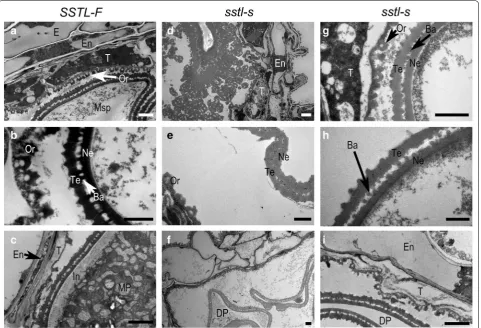
To better understand the defective pollen of ster-ile sstl-s during pollen development, we used TEM analysis for the structure of anthers and microspores in the wild type and mutant. During VP stage, con-sistent with the previous studies, both tapetal cells of fertile SSTL-F and sterile sstl-s degenerated and thus promoted orbicule formation (Fig. 4a, d, g). The orbi-cules were constructed as a cone shape with the high electronic denseness of the tapetum inner side layer to provide a lipid component for the pollen exine (Zhang et al. 2010). The orbicule and pollen exine (at tectum, bacular and nexine) were darker under TEM, which indicates concentrated lipid accumulation. The orbi-cule and exine were much darker in fertile SSTL-F than sterile sstl-s, which suggests less lipid content in the sstl-s anther (Fig. 4b, e, h). At LpM stage, the struc-ture of exine was complete, shown as bridge-like, with ordered tectum, bacular and nexine (Fig. 4b), whereas the exine of sstl-s contained no space between the tectum and nexine, so it became a messy aggregation in exine (Fig. 4e). The other type of abnormal exine in sterile sstl-s was the defective bridge-like shape in the pollen wall (Fig. 4h). Mature pollen of fertile SSTL-F contained conspicuous intine (Fig. 4c), whereas intine was absent in sstl-s (Fig. 4i). Intine contains a high
(See figure on next page.)
amount of polysaccharides (Caffall and Mohnen 2009). In addition, intine formation is controlled by glycosyl-transferase, and complete intine would determine the accumulation of starch, protein, and other important contents for mature pollen grain (Moon et al. 2013). Sterile sstl-s exhibited completely defective intine, lead-ing to defective pollen grain accumulation at Mp stage (Fig. 4f, i). Besides, the endothecium and tapetum lay-ers of fertile SSTL-F decreased entirely to a thin surface at Mp stage (Fig. 4c) rather than the slight reduction in sstl-s anthers (Fig. 4i).
We measured the area of anther locule with more than 20 individual anthers for SSTL-F and sstl-s. The defec-tive anther locules in sstl-s were less than half of those in SSTL-F (Fig. 5). The minimum anther locule area of sstl-s was about 0.5 × 1000 μm2 at LpM and Mp stages and was
Fig. 4 TEM analysis of anther development in SSTL‑F and sstl‑s. a At vacuolated pollen stage (VP), when the tapetal cell was decomposed to generate orbicules in SSTL‑F and d, gsstl‑s. Bars = 2 μm. b At late pollen mitosis stage (LpM), when the tapetum was degraded, and the pollen wall of microspores integrated completely in SSTL‑F and e, hsstl‑s. Bars = 1 μm. c At mature pollen stage (Mp), when endothecium degenerated and microspores were in the plump round shape with puffy intine of SSTL‑F and f, isstl‑s. Bars = 2 μm. Ba Bacular, DP defective pollen, E epidermis, En
endothecium, In intine, MP mature pollen, Msp microspore, Ne nexine, Or orbicule, T tapetum, Te tectum
Fig. 5 Area of anther locules in SSTL‑F and sstl‑s. More than 20 individual anthers were calculated from each sample stage. VP
consistent with the cross section of sstl-s anthers (Fig. 3j, o). The statistical analysis of the anther locule area from sstl-s also gave similar findings of a small anther locule or unexpanded anther in sstl-s (Fig. 2e–h). This informa-tion suggests that the abnormal anther locule results in a shrunken and un-developmental anther in sstl-s.
Transcriptomics analysis of the sstl‑s mutant
With the morphologic observations of the sstl-s mutant, we considered that processes such as starch accumula-tion, sporopollenin, and pollen wall structure as well as biosyntheses such as sucrose, secondary production, and lipid component metabolism must be involved in the
pollen maturation process. To discuss thoroughly the biosynthesis pathways, we compared transcriptomics of different anther stages between SSTL-F and sstl-s (Fig. 6). The heat map illustrated five clusters with specific differ-entially expressed genes. Clusters I and II were upregu-lated genes from sstl-s in LpM and YM stages, clusters III and IV were specifically upregulated genes from SSTL-F in LpM or YM stages individually, and cluster V genes were upregulated in both YM and LpM stages from SSTL -F (Fig. 6a). From these five clusters, several genes listed in Additional file 5: Table S3 were selected for validation with real-time q-PCR analysis (Fig. 6b, c). The expression pattern between RNA-seq and q-PCR was similar and had almost equal log2 fold-change. The q-PCR expres-sion of these selected genes also matched the heat map distribution in the five clusters; for example, genes in Os03g0236200, Os04g0486600, and Os10g0167300 were upregulated only in sstl-s; Os10g0524500, Os03g0429800, Os01g0854800 and Os08g0131100 of SSTL-F were sig-nificantly upregulated in YM with more than two-fold change on both RNA-seq and q-PCR; Os11g0210100, Os03g0234900, Os05g0209600, and Os05g0518300 of SSTL-F were particularly highly expressed in LpM; and Os02g0626100, Os02g0106100, Os05g0542800, and Os06g0156600 of SSTL-F had two-fold higher expres-sion in both YM and LpM (Fig. 6b, c). Meanwhile, two late pollen preference genes, Os09g0381400 (similar to Ervatamin C) and Os09g0388400 (HAD-superfamily hydrolase-like), were also expressed in fertile pollen grains (Fig. 6b, c) (Moon et al. 2018). The expression of Os09g0388400 was 16-fold higher in fertile SSTL-F than sstl-s, which indicated that the sterile sstl-s anther con-tained a high amount of defective pollen grains.
The transcriptomics analysis also revealed that the sterile sstl-s anther genes contained low expression of biosynthesis pathway genes such as xanthine dehydro-genase (Os03g0429800), which participates in purine metabolism; the catalytic cycle of cytochrome P450 (Os01g0854800 and Os08g0131100), involved in sec-ondary metabolites synthesis for exine formation; class III peroxidase (Os11g0210100 and Os03g0234900), which uses electrons in ox-reduction reaction to manipulate phenolic compound synthesis and sec-ondary metabolites; GDSL esterase (Os05g0209600, Os05g0518300, and Os06g0156600), related to lipolytic enzymes and thus controlling pollen wall structure; fructosyl transferase (Os02g0106100), participating in starch composition of mature pollen grains; and the polygalacturonases-like 9 (Os05g0542800), involved in carbohydrate metabolism of polysaccharides for intine utilization. We used Rice Anther Expression Plots analysis to confirm that these significant transcripts of Fig. 6 Gene expression pattern in different anther stages. a
Heat‑map analysis of RNA‑seq data in YM and LpM stage for fertile and sterile anthers. b Relative contribution of fold changes between RNA‑seq and qPCR data in YM and c LpM. Gene expression of q‑PCR data normalized to that of OsUBI, OsACT11, OsUBQ5, and eEF‑1α. YM
pollen development were expressed during anther-spe-cific stages (Additional file 6: Figure S3). For instance, genes that appear from later meiosis to uninucleate microspore stage were consistent with our data in the YM stage. No Pollen 1 (Os10g0524500), xanthine dehydrogenase (Os03g0429800), and cytochrome P450 86A7-2 (Os01g0854800) were specifically upregu-lated in YM only, which was identical to the pattern in Rice Anther Expression Plots analysis. Os02g0106100, Os05g0542800, Os11g0210100, and Os09g0388400, significant genes in LpM stage, also responded to the same stage of bicellular pollen and mature pollen stage as in Rice Anther Expression Plots.
Discussion
Somaclonal variation might cause the sstl trait
On cosegregation of the T-DNA insertion and ster-ile phenotype of two TRIM mutants M0024091 and M0017896, SUMO E3 ligase SIZ1 and bHLH142 were found to control male sterility via anther dehiscence and PCD of tapetum (Thangasamy et al. 2011; Ko et al. 2014). However, in the current study, the phenotype (sterility) and genotype (T-DNA insertion) did not cosegregate in the TRIM mutant M0037841. Previ-ous studies have shown that rice T-DNA insertional mutants, such as Tos17 and TRIM, show low tagging efficiency (Droc et al. 2013; Wei et al. 2016b; Wu et al. 2017). The transformation was frequently used to study both forward and reverse genetics in knock-down/knockout mutants, overexpression lines, and T-DNA activation tagging lines, etc. In rice transfor-mation, the procedure included repetitive tissue cul-ture steps such as rice embryogenic callus generation, antibiotic selection of callus, and seedling regenera-tion. This long-term cullus growth period may lead to somaclonal variation including single nucleotide poly-morphism (SNP), indels, chromosome doubling, and chromosome translocations (Wei et al. 2016a). From 600 to 2000 SNPs/indels per plant may occur during rice regeneration and transformation in the TRIM mutants (Wei et al. 2016a). Thus, this M0037841 semi-sterile sstl mutation might be also caused by soma-clonal variations during transformation.
From the developmental morphology of sstl prog-eny segregates, the completely sterile sstl-s line inher-ited the sterility and showed defects in anther locule and pollen grain. This kind of somaclonal variation might be applied to crop improvement with stable heritability and also may speed up the breeding pro-cess and replace traditional crossbreeding (Jain 2001). Additional file 2: Table S2 illustrates that 6 T-DNA insertional mutant lines have normal fertility. From
TRIM database information, we already know that the T-DNA insertion site of M0037841 locates in the exon of a Mei2-like protein (Os02g0517531). Mei2-like of Arabidopsis is a regulator of meiosis and expressed in gametes of pollens (Kaur et al. 2006). The rice genome contains 6 OsMei2-like genes (Anderson et al. 2004). Thus, M0037841 might maintain fertility because of the gene abundance.
Defective events start in microsporangia in the sstl‑s
mutant
sstl-s imply that microsporangia development is defec-tive; thus, no sporogenous cells are produced in abnor-mal anther locules during early anther development.
Several biosynthetic pathways are affected in sstl‑s
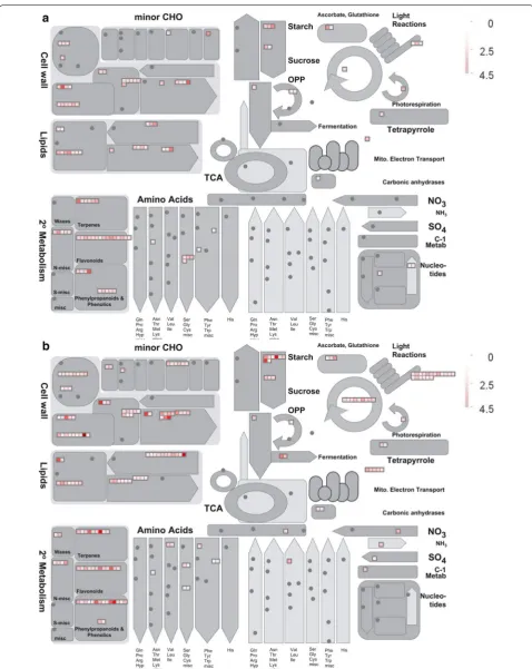
Our studies indicate the pollen in the mutant sstl-s is severely reduced, with abnormal pollen wall in exine and intine, shrunken pollen grains, low pollen viabil-ity and no pollen grain germination. From comparative transcription analysis, the RNA-seq data was substanti-ated by q-PCR assay illustrating that our silicon data are as reliable as the actual expression on real-time q-PCR (Fig. 6b, c). We evaluated the difference in transcrip-tomics between sstl-s and SSTL-F as compared with reference homologs from the KEGG database and tried to understand the multiple impacts on sterility. From the 5 significant transcript heat map clusters (Fig. 6a) and EXPath Tool analysis (Additional file 7: Table S4), clusters I and II revealed that transcripts of sstl-s are involved in degradation of carbon metabolism and amino acid metabolism, such as glyceraldehyde-3-phosphate dehydrogenase (GAPDH) (Os04g0486600), enolase 2 gene (Os10g0167300), and glutamate decarboxylase 3 (OsGAD3) (Os03g0236200). In gluconeogenesis, GAPDH participates in glycolysis at the 6th step to break down glucose for energy contribution and enolase 2 catalyzes 2-phosphoglycerate (2-PG) to phosphoenolpyruvate from the 9th or second to the last step of glycolysis (Yang et al. 2015; Swain et al. 2017). The expression of OsGAD3, which catalyzes glutamate to γ-aminobutyric acid for glutamate degradation (Zhang et al. 2007; El-Kereamy et al. 2012), was increased in sstl-s. In clusters III, IV, and V, the upregulated genes of fertile SSTL-F share simi-lar functions, such as phenylalanine metabolism, cutin/ suberine/wax biosynthesis, secondary metabolites, fla-vone/flavonol biosynthesis, and starch/sucrose metabo-lism (Additional file 7: Table S4). We also performed MapMen analysis and illustrated that biosynthetic path-ways in cell wall, lipids, secondary metabolism, and starch are affected in sstl-s (Fig. 7). Integral pollen exine was controlled by genes related to fatty acid compo-nents, secondary products, and cutin/wax biosynthesis for pollen wall conformation (Quilichini et al. 2015; Shi et al. 2015). Therefore, metabolic events are important in sporophytic development. For instance, a well-devel-oped exine of pollen has a tightly bound fatty acid-like sporopollenin that organizes a bridge-like structure and then the fatty acid assembles aliphatic derivatives such as cutin and wax to form the pollen wall and anther cuticle (Li et al. 2010; Li-Beisson et al. 2013). These functional metabolisms in lipids and fatty acid are consistent with the observation of a rich lipid structure in SSTL-F exine
TEM ultrathin section characterizations (Fig. 4). Mean-while, lipid participates in phenolic secondary metabo-lites in flavonoid production. The flavonoids function as antioxidants to protect the plant against abiotic stress and biotic stress to clean superoxide anions O2−, hydro-gen peroxide (H2O2), hydroxyl radicals (OH−), and sin-glet oxygen (1O2), etc. when these oxygen species (ROS) accumulate under stress (Hernández et al. 2009). In rice flowering stage, flavonoids protect anthers and pollens against ultraviolet damage and increase the bright yellow color to attract pollinators (Thompson et al. 2010; Ning et al. 2018). Our data illustrate that sterile sstl-s shows downregulated flavonoid synthesis-related genes (Fig. 7 and Additional file 7: Table S4). Figure 2b, c also show white anthers and pollen grains, indicating no flavonoid accumulation. A male-sterile maize mutant of irregular pollen exine 1 (ipe1) also had defective pollen grains and abnormal pollen exine with low wax and flavonoid accu-mulation (Chen et al. 2017). With the secondary metabo-lomics essential for pollen development, any defect of these controlling genes may cause sterility and loss of yield production. The oxidative phosphorylation path-way may regulate secondary metabolites production and Arabidopsis soluble pyrophosphatases (PPa3) specifically present in root hairs and pollen tissues (Gutiérrez-Luna et al. 2018; Segami et al. 2018). Our transcriptional data also demonstrate that pyrophosphatase-related genes are upregulated only in fertile anthers and they might participate in a secondary metabolic pathway for pol-len wall structure. The starch accumulation of mature pollen is initiated from glucose-6-phosphate (Glc-6-P), which converts to Glc-1-P, and Glc-1-P then converts to ADP-glucose in the amyloplasts that depend on plastidic ADP-Glc synthesis (Lee et al. 2016). The starch in pollen grains may offer energy for pollen grains in maturation and germination. The I2KI staining (Fig. 2k) of wild-type and mutant pollen agrees with the expression difference in starch synthesis genes (Fig. 7 and Additional file 7: Table S4).
was ZmMs30, a maize GDSL, which was found essential for male fertility because the knockout ZmMs30 mutant showed a disordered pollen wall and defective pollen grains (Ji et al. 2017; An et al. 2019).
Conclusion
Here we report an untagged T-DNA insertional mutant M0037841 that has defective pollen and abnormal anthers from its sstl progeny segregaters. The abnormal anther locule is related to the damaged microsporangia and results in no microspore generation. The defective anther locule of sstl-s also illustrates the importance of the anther tapetum, which provides lipid-like sporopol-lenin for pollen maturation. Transcripts of sterile sstl-s are downregulated in several secondary-metabolism bio-synthesis pathways and lead to irregular exine and absent intine in sstl-s. This information reveals the defective pol-len grains of sstl-s and coincides with the irregular pollen wall and loss of pollen viability. Comparative transcrip-tomics analysis illustrates that the mechanism of male sterility is related to biosynthetic pathways for cell wall, lipids, secondary metabolism, and starch accumulation. We illustrate the importance of microsporangia devel-opment and male fertility of microspores. A series of morphological observations of mutant sstl-s anther dem-onstrated that the defective pollen grains are caused by unproduced intine and unfunctional exine of the pollen wall, then the defective pollen of sstl-s do not germinate and become sterile.
Additional files
Additional file 1: Table S1. Primers list.
Additional file 2: Table S2. Genotyping and phenotyping analysis of M0037841 segregants.
Additional file 3: Figure S1. Panicle development of sstl progeny seg‑ regates. a, c, fSSTL-F plants show the fertile panicles. b, d, esstl-s mutant shows sterile spikelets during reproductive stage.
Additional file 4: Figure S2. Structure analysis of the undeveloped anther sstl-s. a‑c Cross section and d‑i TEM analysis showing abnormal anthers of sstl-s in the early stage. (a‑c) Bar=40 μm. (d‑f) Bar=2 μm. (g‑i)
Bar=1 μm.
Additional file 5: Table S3. Genes selected from RNA‑seq heat‑map analysis.
Additional file 6: Figure S3. The differential expressed genes of SSTL-F
and sstl-s during anther developing stages using Rice Anther Expression Plots (https ://www.cpib.ac.uk/anthe r/ricei ndex.html). PM=Premeiosis; M=Meiosis; UM=Uninucleate Microspore; BP=Bicellular Pollen; TP=Tricellular pollen; MP=Mature Pollen.
Additional file 7: Table S4. Prediction of gene function in RNA‑seq heat‑ map analysis result.
Abbreviations
bHLH: basic helix‑loop‑helix; EAT1: ETERNAL TAPETUM1; EMS1: EXCESS MICROSPOROCYTES1; EpM: early pollen mitosis stage; GDSL: Gly‑Asp‑Ser‑Leu; GM filed: genetically modified plants isolation field; I2–KI: iodine–potassium iodide; LpM: late pollen mitosis stage; Mp: mature pollen stage; NP1: Rice No Pollen 1; PCD: programmed cell death; PMC: pollen mother cells; PPC: primary parietal cells; PS: primary sporogenous; SEM: scanning electron microscopy; SNP: single nucleotide polymorphism; SPC: secondary parietal layers; SPL: SQUAMOSA PROMOTER‑BINDING PROTEIN‑LIKE; sstl: semi‑sterile mutant; SSTL‑ F: fertile plants of sstl progeny segregates; sstl‑s: sterile plants of sstl progeny segregates; sstl‑ss: semi‑sterile plants of sstl progeny segregates; TARI: Taiwan Agricultural Research Institute; TDR: TAPETUM DEGENERATION RETARDATION; TEM: transmission electron microscope; TPD1: TAPETUM DETERMINANT1; TRIM: Taiwan Rice Insertional Mutagenesis; VP: vacuolated pollen stage; YM: young microspore stage.
Acknowledgements
We thank Ms. Lie‑Hong Wu for maintenance of greenhouse plants, and Laura Smales (BioMedEditing, Toronto, Canada) for English editing.
Authors’ contributions
CLC and YICH are responsible for the experimental design. All sample col‑ lection was prepared by CLC. JCS and YCT were in charge of T2 population sample management. HHH and MHL performed the rice cultivation in TARI. CLC performed molecular analyses of PCR‑genotyping, gene expression and bioinformatics analysis of transcriptomic data. YCL performed pollen staining analysis. WNJ prepared TEM samples for observation. CLC observed samples and took the photographs. CLC consolidated results and wrote the manu‑ script. YIH and MCC edited the manuscript. All authors read and approved the final manuscript.
Funding
This work was supported by Academia Sinica, Taiwan to YICH.
Availability of data and materials
Not applicable.
Ethics approval and consent to participate
Not applicable.
Consent for publication
Not applicable.
Competing interests
The authors declare that they have no competing interests.
Author details
1 Department of Agronomy, National Taiwan University, Taipei 106, Taiwan. 2 Institute of Plant and Microbial Biology, Academia Sinica, Nankang, Tai‑ pei 115, Taiwan. 3 Crop Science Division, Taiwan Agricultural Research Institute, Taichung 413, Taiwan. 4 Department of Life Science, National Taiwan Univer‑ sity, Taipei 106, Taiwan. 5 Department of Agronomy, National Chiayi University, Chiayi 600, Taiwan.
Received: 17 April 2019 Accepted: 30 June 2019
References
An X, Dong Z, Tian Y, Xie K, Wu S, Zhu T, Zhang D, Zhou Y, Niu C, Ma B, Hou Q, Bao J, Zhang S, Li Z, Wang Y, Yan T, Sun X, Zhang Y, Li J, Wan X (2019)
ZmMs30 encoding a novel GDSL lipase is essential for male fertility and valuable for hybrid breeding in maize. Mol Plant 12:343–359. https ://doi. org/10.1016/j.molp.2019.01.011
Caffall KH, Mohnen D (2009) The structure, function, and biosynthesis of plant cell wall pectic polysaccharides. Carbohydr Res 344:1879–1900. https :// doi.org/10.1016/j.carre s.2009.05.021
Cai W, Zhang D (2018) The role of receptor‑like kinases in regulating plant male reproduction. Plant Reprod 31:77–87. https ://doi.org/10.1007/s0049 7‑018‑0332‑7
Chang Z, Jin M, Yan W, Chen H, Qiu S, Fu S, Xia J, Liu Y, Chen Z, Wu J, Tang X (2018) The ATP‑binding cassette (ABC) transporter OsABCG3 is essential for pollen development in rice. Rice 11(58):1–15. https ://doi.org/10.1186/ s1228 4‑018‑0248‑8
Chao LM, Liu YQ, Chen DY, Xue XY, Mao YB, Chen XY (2017) Arabidopsis
transcription factors SPL1 and SPL12 confer plant thermotolerance at reproductive stage. Mol Plant 10:735–748. https ://doi.org/10.1016/j. molp.2017.03.010
Chen X, Zhang H, Sun H, Luo H, Zhao L, Dong Z, Yan S, Zhao C, Liu R, Xu C, Li S, Chen H, Jin W (2017) IRREGULAR POLLEN EXINE1 is a novel factor in anther cuticle and pollen exine formation. Plant Physiol 173:307–325.
https ://doi.org/10.1104/pp.16.00629
Choi H, Jin JY, Choi S, Hwang JU, Kim YY, Suh MC, Lee Y (2011) An ABCG/WBC‑ type ABC transporter is essential for transport of sporopollenin precursors for exine formation in developing pollen. Plant J 65:181–193. https ://doi. org/10.1111/j.1365‑313x.2010.04412 .x
Daku RM, Rabbi F, Buttigieg J, Coulson IM, Horne D, Martens G, Ashton NW, Suh DY (2016) PpASCL, the Physcomitrella patens anther‑specific chalcone synthase‑like enzyme implicated in sporopollenin biosynthesis, is needed for integrity of the moss spore wall and spore viability. PLoS ONE 11(1):e0146817. https ://doi.org/10.1371/journ al.pone.01468 17
Droc G, An G, Hsing YI, Hirochika H, Pereira A, Undaresan CS, Han CD, Upad‑ hyaya N, Ramachandran S, Comai L, Leung H, Guiderdoni E (2013) Mutant resources for the functional analysis of the rice genome. In: Wing R, Zhang Q (eds) Genetics and genomics of rice, vol 5. Springer, Berlin, pp 81–115
El‑Kereamy A, Bi YM, Ranathunge K, Beatty PH, Good AG, Rothstein SJ (2012) The Rice R2R3‑MYB transcription factor OsMYB55 is involved in the toler‑ ance to high temperature and modulates amino acid metabolism. PLoS ONE 7(12):e52030. https ://doi.org/10.1371/journ al.pone.00520 30
Guo C, Ge X, Ma H (2013) The rice OsDIL gene plays a role in drought tolerance at vegetative and reproductive stages. Plant Mol Biol 82:239–253. https :// doi.org/10.1007/s1110 3‑013‑0057‑9
Gutiérrez‑Luna FM, Hernández‑Domínguez EE, Valencia‑Turcotte LG, Rod‑ ríguez‑Sotres R (2018) Review: “Pyrophosphate and pyrophosphatases in plants, their involvement in stress responses and their possible relationship to secondary metabolism”. Plant Sci 267:11–19. https ://doi. org/10.1016/j.plant sci.2017.10.016
Hernández I, Alegre L, Breusegem FV, Munné‑Bosch S (2009) How relevant are flavonoids as antioxidants in plants? Trends Plant Sci 14:125–132. https :// doi.org/10.1016/j.tplan ts.2008.12.003
Huang MD, Wei FJ, Wu CC, Hsing YI, Huang AH (2009) Analyses of advanced rice anther transcriptomes reveal global tapetum secretory functions and potential proteins for lipid exine formation. Plant Physiol 149:694–707.
https ://doi.org/10.1104/pp.108.13112 8
Huang J, Zhang T, Linstroth L, Tillman Z, Otegui MS, Owen HA, Zhao D (2016) Control of anther cell differentiation by the small protein ligand TPD1 and its receptor EMS1 in Arabidopsis. PLoS Genet 12(8):e1006147. https ://doi. org/10.1371/journ al.pgen.10061 47
Jain SM (2001) Tissue culture‑derived variation in crop improvement. Euphyt‑ ica 118:153–166. https ://doi.org/10.1023/a:10041 24519 479
Ji R, Wang H, Xin X, Peng S, Hur Y, Li Z, Feng H (2017) BrEXL6, a GDSL lipase gene of Brassica rapa, functions in pollen development. Biol Plant 61:685–692. https ://doi.org/10.1007/s1053 5‑017‑0735‑6
Jung KH, Han MJ, Lee DY, Lee YS, Schreiber L, Franke R, Faust A, Yephremov A, Saedler H, Kim YW, Hwang I, An G (2006) Wax‑deficient anther1 is involved in cuticle and wax production in rice anther walls and is required for pollen development. Plant Cell 18:3015–3032. https ://doi.org/10.1105/ tpc.106.04204 4
Kaur J, Sebastian J, Siddiqi I (2006) The Arabidopsis‑mei2‑Like genes play a role in meiosis and vegetative growth in Arabidopsis. Plant Cell 18:545–559.
https ://doi.org/10.1105/tpc.105.03915 6
Kelliher T, Egger RL, Zhang H, Walbot V (2014) Unresolved issues in pre‑meiotic anther development. Front Plant Sci 5(347):1–9. https ://doi.org/10.3389/ fpls.2014.00347
Ko SS, Li MJ, Sun‑Ben KuM, Ho YC, Lin YJ, Chuang MH, Hsing HX, Lien YC, Yang HT, Chang HC, Chan MT (2014) The bHLH142 transcription factor coordi‑ nates with TDR1 to modulate the expression of EAT1 and regulate pollen development in rice. Plant Cell 26:2486–2504. https ://doi.org/10.1105/ tpc.114.12629 2
Lee S‑K, Eom J‑S, Hwang S‑K, Shin D, An G, Okita TW, Jeon J‑S (2016) Plastidic phosphoglucomutase and ADP‑glucose pyrophosphorylase mutants impair starch synthesis in rice pollen grains and cause male sterility. J Exp Bot 67:5557–5569. https ://doi.org/10.1093/jxb/erw32 4
Li N, Zhang DS, Liu HS, Yin CS, Li XX, Liang WQ, Yuan Z, Xu B, Chu HW, Wang J, Wen TQ, Huang H, Luo D, Ma H, Zhang DB (2006) The Rice Tapetum Degeneration Retardation gene is required for tapetum degradation and anther development. Plant Cell 18:2999–3014. https ://doi.org/10.1105/ tpc.106.04410 7
Li H, Pinot F, Sauveplane V, Werck‑Reichhart D, Diehl P, Schreiber L, Franke R, Zhang P, Chen L, Gao Y, Liang W, Zhang D (2010) Cytochrome P450 family member CYP704B2 catalyzes the & #x03C9;‑hydroxylation of fatty acids and is required for anther cutin biosynthesis and pollen exine formation in rice. Plant Cell 22:173–190. https ://doi.org/10.1105/tpc.109.07032 6
Li H, Yuan Z, Vizcay‑Barrena G, Yang C, Liang W, Zong J, Wilson ZA, Zhang D (2011) PERSISTENT TAPETAL CELL1 encodes a PHD‑finger protein that is required for tapetal cell death and pollen development in rice. Plant Physiol 156:615–630. https ://doi.org/10.1104/pp.111.17576 0
Li Y, Li D, Guo Z, Shi Q, Xiong S, Zhang C, Zhu J, Yang Z (2016) OsACOS12, an orthologue of Arabidopsis acyl‑CoA synthetase5, plays an important role in pollen exine formation and anther development in rice. BMC Plant Biol 16:256. https ://doi.org/10.1186/s1287 0‑016‑0943‑9
Li‑Beisson Y, Shorrosh B, Beisson F, Andersson MX, Arondel V, Bates PD, Baud S, Bird D, Debono A, Durrett TP, Franke RB, Graham IA, Katayama K, Kelly AA, Larson T, Markham JE, Miquel M, Molina I, Nishida I, Rowland O, Samuels L, Schmid KM, Wada H, Welti R, Xu C, Zallot R, Ohlrogge J (2013) Acyl‑lipid metabolism. The Arabidopsis book, vol 11. American Society of Plant Biologists, Washington, p e0161. https ://doi.org/10.1199/tab.0161
Liu Z, Bao W, Liang W, Yin J, Zhang D (2010) Identification of gamyb‑4 and anal‑ ysis of the regulatory role of GAMYB in rice anther development. J Integr Plant Biol 52:670–678. https ://doi.org/10.1111/j.1744‑7909.2010.00959 .x
Liu Z, Lin S, Shi J, Yu J, Zhu L, Yang X, Zhang D, Liang W (2017) Rice No Pollen 1 (NP 1) is required for anther cuticle formation and pollen exine pattern‑ ing. Plant J 91:263–277
Moon S, Kim SR, Zhao G, Yi J, Yoo Y, Jin P, Lee SW, Jung KH, Zhang D, An G (2013) Rice GLYCOSYLTRANSFERASE1 encodes a glycosyltransferase essential for pollen wall formation. Plant Physiol 161:663–675. https ://doi. org/10.1104/pp.112.21094 8
Moon S, Oo MM, Kim B, Koh H‑J, Oh SA, Yi G, An G, Park SK, Jung KH (2018) Genome‑wide analyses of late pollen‑preferred genes conserved in various rice cultivars and functional identification of a gene involved in the key processes of late pollen development. Rice 11:28. https ://doi. org/10.1186/s1228 4‑018‑0219‑0
Ning L, Lin Z, Gu J, Gan L, Li Y, Wang H, Miao L, Zhang L, Wang B, Li M (2018) The initial deficiency of protein processing and flavonoids biosynthe‑ sis were the main mechanisms for the male sterility induced by SX‑1 in Brassica napus. BMC Genom 19:806. https ://doi.org/10.1186/s1286 4‑018‑5203‑y
Pu CX, Han YF, Zhu S, Song FY, Zhao Y, Wang CY, Zhang YC, Yang Q, Wang J, Bu SL, Sun LJ, Zhang SW, Zhang SQ, Sun DY, Sun Y (2017) The rice receptor‑ like kinases DWARF AND RUNTISH SPIKELET1 and 2 repress cell death and affect sugar utilization during reproductive development. Plant Cell 29:70–89. https ://doi.org/10.1105/tpc.16.00218
Quilichini TD, Grienenberger E, Douglas CJ (2015) The biosynthesis, composi‑ tion and assembly of the outer pollen wall: a tough case to crack. Phyto‑ chemistry 113:170–182. https ://doi.org/10.1016/j.phyto chem.2014.05.002
Raghavan V (1988) Anther and pollen development in rice (Oryza sativa). Am J Bot 75:183–196
Ruggiero F, Bedini G (2018) Systematic and morphologic survey of orbi‑ cules in allergenic angiosperms. Aerobiologia 34:405–422. https ://doi. org/10.1007/s1045 3‑018‑9522‑x
Segami S, Tomoyama T, Sakamoto S, Gunji S, Fukuda M, Kinoshita S, Mitsuda N, Ferjani A, Maeshima M (2018) Vacuolar H+‑pyrophosphatase and
Shi J, Cui M, Yang L, Kim Y‑J, Zhang D (2015) Genetic and biochemical mecha‑ nisms of pollen wall development. Trends Plant Sci 20:741–753. https :// doi.org/10.1016/j.tplan ts.2015.07.010
Swain DM, Sahoo RK, Srivastava VK, Tripathy BC, Tuteja R, Tuteja N (2017) Function of heterotrimeric G‑protein γ subunit RGG1 in providing salinity stress tolerance in rice by elevating detoxification of ROS. Planta 245:367–383. https ://doi.org/10.1007/s0042 5‑016‑2614‑3
Thangasamy S, Guo CL, Chuang MH, Lai MH, Chen J, Jauh GY (2011) Rice
SIZ1, a SUMO E3 ligase, controls spikelet fertility through regulation of anther dehiscence. New Phytol 189:869–882. https ://doi.org/10.111 1/j.1469‑8137.2010.03538 .x
Thompson EP, Wilkins C, Demidchik V, Davies JM, Glover BJ (2010) An Arabidop-sis flavonoid transporter is required for anther dehiscence and pollen development. J Exp Bot 61:439–451. https ://doi.org/10.1093/jxb/erp31 2
Verma N (2019) Transcriptional regulation of anther development in Arabidop-sis. Gene 689:202–209. https ://doi.org/10.1016/j.gene.2018.12.022
Walbot V, Egger RL (2016) Pre‑meiotic anther development: cell fate specifica‑ tion and differentiation. Annu Rev Plant Biol 67:365–395. https ://doi. org/10.1146/annur ev‑arpla nt‑04301 5‑11180 4
Wang Y, Lin YC, So J, Du Y, Lo C (2013) Conserved metabolic steps for sporopol‑ lenin precursor formation in tobacco and rice. Physiol Plant 149:13–24.
https ://doi.org/10.1111/ppl.12018
Wang QL, Sun AZ, Chen ST, Chen LS, Guo FQ (2018) SPL6 represses signalling outputs of ER stress in control of panicle cell death in rice. Nat Plants 4:280–288. https ://doi.org/10.1038/s4147 7‑018‑0131‑z
Wei FJ, Kuang LY, Oung HM, Cheng SY, Wu HP, Huang LT, Tseng YT, Chiou WY, Hsieh‑Feng V, Chung CH, Yu SM, Lee LY, Gelvin SB, Hsing YIC (2016a) Somaclonal variation does not preclude using rice transformants for genetic screening. Plant J 85:648–659. https ://doi.org/10.1111/tpj.13132
Wei FJ, Tsai YC, Hsu YM, Chen YA, Huang CT, Wu HP, Huang LT, Lai MH, Kuang LY, Lo SF, Yu SM, Lin YR, Hsing YIC (2016b) Lack of genotype and phenotype correlation in a rice T‑DNA tagged line is likely caused by introgression in the seed source. PLoS ONE 11(5):e0155768. https ://doi.org/10.1371/journ al.pone.01557 68
Wu HP, Wei FJ, Wu CC, Lo SF, Chen LJ, Fan MJ, Chen S, Wen IC, Yu SM, Ho THD, Lai MH, Hsing YIC (2017) Large‑scale phenomics analysis of a T‑DNA tagged mutant population. GigaScience 6:1–7. https ://doi.org/10.1093/ gigas cienc e/gix05 5
Xu T, Zhang C, Zhou Q, Yang ZN (2016) Pollen wall pattern in Arabidopsis. Sci Bull 61:832–837. https ://doi.org/10.1007/s1143 4‑016‑1062‑6
Yang X, Wu D, Shi J, He Y, Pinot F, Grausem B, Yin C, Zhu L, Chen M, Luo Z, Liang W, Zhang D (2014) Rice CYP703A3, a cytochrome P450 hydroxylase, is essential for development of anther cuticle and pollen exine. J Integr Plant Biol 56:979–994. https ://doi.org/10.1111/jipb.12212
Yang T, Wang L, Li C, Liu Y, Zhu S, Qi Y, Liu X, Lin Q, Luan S, Yu F (2015) Receptor protein kinase FERONIA controls leaf starch accumulation by interacting with glyceraldehyde‑3‑phosphate dehydrogenase. Biochem Biophys Res Commun 465:77–82. https ://doi.org/10.1016/j.bbrc.2015.07.132
Yang L, Qian X, Chen M, Fei Q, Meyers BC, Liang W, Zhang D (2016) Regula‑ tory role of a receptor‑like kinase in specifying anther cell identity. Plant Physiol 171:2085–2100. https ://doi.org/10.1104/pp.16.00016
Yang X, Li G, Tian Y, Song Y, Liang W, Zhang D (2018) A rice glutamyl‑tRNA syn‑ thetase modulates early anther cell division and patterning. Plant Physiol 177:728–744. https ://doi.org/10.1104/pp.18.00110
Zhang D, Wilson ZA (2009) Stamen specification and anther development in rice. Chin Sci Bull 54:2342–2353. https ://doi.org/10.1007/s1143 4‑009‑0348‑3
Zhang H, Yao H, Chen F, Wang X (2007) Purification and characterization of glutamate decarboxylase from rice germ. Food Chem 101:1670–1676.
https ://doi.org/10.1016/j.foodc hem.2006.04.027
Zhang D, Liang W, Yin C, Zong J, Gu F, Zhang D (2010) OsC6, encoding a lipid transfer protein, is required for postmeiotic anther development in rice. Plant Physiol 154:149–162. https ://doi.org/10.1104/pp.110.15886 5
Zhang D, Luo X, Zhu L (2011) Cytological analysis and genetic control of rice anther development. J Genet Genom 38:379–390. https ://doi. org/10.1016/j.jgg.2011.08.001
Publisher’s Note