The Effect of Pathological Concentrations of Phytanic Acid on
the Fatty Acid Composition, Vitamin E Concentration and
Function of Membranes of Cultured Mammalian
Retinal Cells
by
Sarah Phyllis Young
A thesis submitted for the degree of Doctor of Philosophy
in the University of London
Unit of Biochemistry, Endocrinology and Metabolism
Institute of Child Health
ProQuest Number: 10015807
All rights reserved
INFORMATION TO ALL USERS
The quality of this reproduction is dependent upon the quality of the copy submitted.
In the unlikely event that the author did not send a complete manuscript and there are missing pages, these will be noted. Also, if material had to be removed,
a note will indicate the deletion.
uest.
ProQuest 10015807
Published by ProQuest LLC(2016). Copyright of the Dissertation is held by the Author.
All rights reserved.
This work is protected against unauthorized copying under Title 17, United States Code. Microform Edition © ProQuest LLC.
ProQuest LLC
789 East Eisenhower Parkway P.O. Box 1346
ACKNOWLEDGEMENTS
I would especially like to express my gratitude and appreciation to my supervisor, Dr.
David Muller for his constant advice, support and encouragement throughout this
project.
I would like to thank Dr. Andrew Johnson for his help and advice. I would also like to
thank Mr Geoff Lynes o f the Mass Spectrometry Department at Great Ormond Street
Hospital for Sick Children for his assistance with the synthesis of the phytanic acid,
without which this project could not have proceeded, and with the fatty acid analysis by
GC-MS. I am very grateful to Dr. John Greenwood for his generous gift of the
immortalised rat retinal pigment epithelial cell lines. I would like to thank Dr. Peter
Clayton for allowing me the time to complete my thesis.
Thanks are also due to Mr Ian Merryweather for teaching me the vitamin E assay and to
Mr David Stanton especially for his help with the maintenance of equipment and for his
advice on analytical procedures.
I would like to acknowledge all the friendship, help and moral support I have received
from everyone in the Biochemistry Unit at the Institute o f Child Health.
I am grateful to Kenny and Alexandra for their support and understanding.
Finally I would like to thank the Child Health Research Appeal Trust for funding this
ABSTRACT
The purpose o f this study was to investigate possible mechanisms for the retinal
degeneration seen in adult Refsum disease. This is an inherited disorder in which
phytanic acid, a dietary branched chain fatty acid, caimot be catabolised and as a result it
accumulates in tissues and serum. Phytanic acid has the same structure as the side chain
of the tocopherols (vitamin E), and some of the signs and symptoms seen in patients
with severe and chronic vitamin E deficiency are similar to those seen in adult Refsum
disease. For these reasons it has been suggested that an accumulation o f phytanic acid in
membranes may interfere with vitamin E fimction. Alternatively phytanic acid may
exert its pathological effect through alteration of membrane composition and structure,
thereby affecting membrane functions. These hypotheses were investigated by studying
the effect o f modulating phytanic acid and a-tocopherol concentrations on the lipid
composition and certain functional parameters o f retinal cell lines in culture. Methods
were established and validated for (i) the supplementation o f the cell lines with
reproducible concentrations of phytanic acid and a-tocopherol, (ii) the quantification of
various membrane components of interest, (iii) the measurement o f membrane fluidity
and the membrane transport o f choline, and (iv) the determination o f the susceptibility
of altered membranes to in vitro lipid peroxidation. Results showed a) the phospholipid fraction o f retinal cells readily incorporated phytanic acid resulting in an altered
membrane fatty acid composition b) there was no competition in uptake between
phytanic acid and a-tocopherol, c) the incorporation of phytanic acid increased
membrane fluidity and the transport of choline, and d) the incorporation of phytanic acid
did not appear to affect the susceptibility of membranes to in vitro lipid peroxidation. Taken together these results suggested that phytanic acid did not interfere with the
incorporation and function o f a-tocopherol in retinal membranes but directly affected
CONTENTS
Acknowledgements
2
Abstract
3
Table o f Contents
4
List o f Figures
7
List o f Tables
10
List o f Abbreviations
11
Bibliography
250
CHAPTER 1 : INTRODUCTION 12
1.1 Introduction 13
1.2 Peroxisomes 13
1.3 Peroxisomal Disorders 14
1.4 Adult Refsum Disease (ARD) 17
1.5 Phytanic Acid 21
1.6 Possible Mechanisms for the Pathogenesis in Adult Refsum Disease 30
1.7 Retinal Degeneration in Adult Refsum Disease 33
1.8 Plan o f Investigation 46
CHAPTER 2: THE CULTURE OF RETINAL DERIVED CELL LINES 48
2.1 Introduction 49
2.2 Materials 50
2.3 Bovine Retinal Pigment Epithelial (RPE) Cells 51
2.4 Immortalised Rat Retinal Pigment Epithelial Cell Line 53
2.5 Y79 Retinoblastoma Cell Line 54
2.6 Storage o f Cells in Liquid Nitrogen 55
CHAPTER 3: FATTY ACID ANALYSIS AND SYNTHESIS 64
3.1 Introduction 65
3.2 Materials 65
3.3 Preparation of Fatty Acid Methyl Esters (FAMEs) from Culture Medium,
Crude Membrane Preparations and Lipid Fractions 66
3.4 Analysis o f FAMEs by Gas Chromatography 68
3.5 Standards and Quantitation 74
3.6 Reproducibility of FAME analysis o f Red Cell Ghosts 84
3.7 Synthesis of Phytanic Acid 84
CHAPTER 4: UPTAKE OF PHYTANIC ACID BY RETINAL CELL
LINES 89
4.1 Introduction 90
4.2 The Uptake of Phytanic Acid by Retinal Cell Lines 90
4.3 The Distribution of Phytanic Acid in Cellular Lipid Fractions 100
4.4 Neutral Lipid Staining 107
4.5 Uptake o f Phytanic Acid into the Phospholipid Fraction of RPE Cells and Comparison to Uptake into the Total Cellular Lipid Pool 108
4.6 Discussion 115
CHAPTER 5: THE ANALYSIS AND UPTAKE OF a-TOCOPHEROL BY
RETINAL CELL LINES 119
5.1 Introduction 120
5.2 Materials 120
5.3 Analysis o f a-Tocopherol 120
5.4 Uptake o f a-Tocopherol by Retinal Cell Lines 126
CHAPTER 6: THE ANTIMETABOLITE HYPOTHESIS 134
6.1 Introduction 135
6.2 Vitamin E 135
6.3 Antioxidant Function o f Vitamin E 139
6.4 Other Functions o f Vitamin E 143
6.5 Vitamin E Deficiency in Man 146
6.6 Comparison o f Severe Vitamin E Deficiency to Adult Refsum Disease 149
CHAPTER 7: TH E EFFECT OF PHYTANIC ACID ON TH E UPTAKE OF a-TO C O PH ER O L (AND VICE VERSA) BY RAT RPE CELLS 155
7.1 Introduction 156
7.2 A Pilot Study on the Effect o f Phytanic Acid on the Uptake of
a-T ocopherol 156
7.3 The Effect o f a Range of Phytanic Acid Concentrations on a-Tocopherol
Uptake by Rat RPE Cells 158
7.4 The Effect o f Phytanic Acid on the a-Tocopherol Concentration of Rat
RPE Cells Presupplemented with a-Tocopherol 161
7.5 The Effect of a-Tocopherol on the Uptake of Phytanic Acid 163
7.6 Discussion 168
CH APTER 8: TH E EFFECT OF PHYTANIC ACID ON THE
ANTIOXIDANT FUNCTION OF a-TO C O PH ER O L 170
8.1 Introduction 171
PARTI
8.2 Supplementation o f Y79 Cells with Linolenic Acid (18:3n3) 174
PART II
8.3 The Effect o f Phytanic Acid and a-Tocopherol on In Vitro Lipid
Peroxidation of 18:3n3 Supplemented Y79 Cells 181
CH A PTER 9: TH E M OLECULAR DISTORTION HYPOTHESIS 204
9.1 Introduction 206
PA RTI
9.2 The Effect o f Phytanic Acid Supplementation on the Fluidity o f Retinal
Cell Membranes 217
PART II
9.3 The Effect o f Phytanic Acid On The Uptake o f [Methyl-^H]Choline by
Y79 Cells 234
9.4 Summary 245
LIST OF FIGURES
Chapter 1
Figure 1-1 : Structure of an isoprene unit and some examples o f isoprene
compounds derived from this unit. 22
Figure 1-2: One possible pathway for the a-oxidation o f phytanic acid based on
current evidence. 24
Figure 1-3: Structure of the eye and retina. 35
Figure 1-4: Structure of a rod photoreceptor cell. 37
Chapter 2
Figure 2-1 : Dispersion of a-tocopherol in medium. 59 Figure 2-2: a-Tocopherol supplemented FBS and medium. 60 Figure 2-3: Washing efficiency of cells exposed to a-tocopherol. 61 Figure 2-4: Washing efficiency of cells exposed to phytanic acid. 63
Chapter 3
Figure 3-1 : Mass spectrum of 16:0 methyl ester. 70
Figure 3-2: Mass spectrum o f 20:4n6 methyl ester. 71 Figure 3-3: Mass spectrum o f phytanic acid methyl ester. 72 Figure 3-4: Examples of the fragmentation of fatty acids methyl esters. 73 Figure 3-5: Fatty acid standards analysed by GC-FID. 77 Figure 3-6: Fatty acid profile of RPE cells cultured in control medium, analysed
by GC-FID. 78
Figure 3-7 : Fatty acid profile of rat RPE cells supplemented with 100 pmol/1
phytanic acid for 7 days, analysed by GC-FID. 79 Figure 3-8: Fatty acid profile o f Y79 cells cultured in control medium, analysed
by GC-FID 80
Figure 3-9: Fatty acid profile o f Y79 cell crude membranes supplemented with
50 pmol/118:3n3 for 7 days, analysed by GC-FID. 81 Figure 3-10: Standard curves o f phytanic acid, 18:0 and 22:6n3. 83 Figure 3-11 : Response factors of fatty acid standards. 85 Figure 3-12: Coefficient o f variation (c.v) o f replicate measurements of fatty acids
in red blood cell ghosts. 86
Chapter 4
Figure 4-1 : Incorporation of phytanic acid into confluent bovine retinal cells. 91 Figure 4-2: Incorporation of phytanic acid into non-confluent bovine retinal cells 93 Figure 4-3 : Uptake of phytanic acid with time by bovine retinal cells, 94 Figure 4-4: Uptake of phytanic acid with time by rat RPE cells. 96 Figure 4-5: Effect of concentration on the uptake of phytanic acid by rat RPE
cells. 98
Figure 4-6: Effect of concentration on the uptake of phytanic acid by Y79 cells. 99 Figure 4-7 : Representative separation profiles of lipid standards using
hexane:diethyl ether:acetic acid. 103
Figure 4-10: Uptake o f phytanic acid into whole cell and phospholipid fractions. Figure 4-11 : Total lipid and phospholipid fatty acid profiles of RPE cells.
Figure 4-12: Total lipid and phospholipid fatty acid profiles of Y79 cells.
Chapter 5 Figure 5-1: Figure 5-2: Figure 5-3: Figure 5-4: Figure 5-5:
a-Tocopherol standard curve.
Stability o f a-tocopherol in red blood cells stored at -20°C Uptake o f a-tocopherol by bovine retinal cells.
Uptake o f a-tocopherol by RPE cells with time.
The uptake o f a-tocopherol by RPE cells with increasing concentration.
Figure 5-6: The uptake o f a-tocopherol by the Y79 cells with increasing concentration.
I l l 113 114 124 125 127 128 130 131 Chapter 6 Figure 6-1: Figure 6-2: Chapter 7 Figure 7-1: Figure 7-2: Figure 7-3: Figure 7-4: Figure 7-5: Figure 7-6: Figure 7-7: Figure 7-8:
The structures of the tocopherol and tocotrienol forms o f vitamin E. 136
Lipid peroxidation. 141
Uptake o f a-tocopherol by RPE cells in the presence of phytanic
acid. 157
Uptake o f phytanic acid by RPE cells in the presence o f a -
tocopherol. 157
Effect o f phytanic acid on a-tocopherol uptake by RPE cells. 159 Effect o f phytanic acid on a-tocopherol uptake by RPE cells. 160 Effect o f phytanic acid on RPE cell a-tocopherol concentration. 162 Effect o f a-tocopherol on phytanic acid uptake by RPE cells. 164 Effect o f a-tocopherol on phytanic acid uptake by RPE cells. 165 Effect o f a-tocopherol on the uptake of phytanic acid into lipid
fractions. 167
Chapter 8
Figure 8-1 : Formation of malondialdehyde from a lipid peroxyl radical via a
cyclic endoperoxide. 173
Figure 8-2: Supplementation o f Y79 cells with 18:3n3 (effect on n-3 fatty acid
profile). 176
Figure 8-3: Supplementation of Y79 cells with 18:3n3 (effect on 16:1, 18:1 and
18:0 relative concentrations). 178
Figure 8-4: Metabolism o f linolenic acid to docosahexaenoic acid. 179
Figure 8-5: MDA standard curve. 184
Figure 8-6: Free MDA production with increasing substrate concentration (Y79
cells). 187
Figure 8-7: In vitro peroxidation o f Y79 cell sonicates. 188 Figure 8-8: The effect o f a-tocopherol on in vitro peroxidation o f Y79 cell
sonicates. 190
Figure 8-9: The effect o f a-tocopherol on in vitro peroxidation o f Y79 cell
sonicates. 191
Figure 8-10: The effect o f ± phytanic acid and ± a-tocopherol on free MDA
Figure 811 : The effect of a range o f phytanic acid concentrations and ± a
-tocopherol on free MDA production. 195
Figure 8-12: The effect of ± phytanic acid ± a-tocopherol on free MDA
production. 196
Figure 8-13: The difference in free MDA produced between the ± a-tocopherol supplemented Y79 cells over a range o f phytanic acid
concentrations. 198
Chapter 9
Figure 9-1 : Cross-section of a biological membrane showing some o f the main
components. 208
Figure 9-2: Phospholipid structures. 209
Figure 9-3: Measurement o f steady state fluorescence anisotropy using the
fluorescence probe diphenylhexatriene (DPH). 219
Figure 9-4: Structure o f DPH and TMA-DPH. 221
Figure 9-5: Positioning of DPH in membranes. 222
Figure 9-6: Steady state fluorescence anisotropy measurements o f RPE cell
membranes. 227
Figure 9-7: Steady state fluorescence anisotropy measurements o f RPE cell
membranes. 229
Figure 9-8: Steady state fluorescence anisotropy measurements o f Y79 cell
membranes. 230
Figure 9-9: The uptake o f [methyl-^HJcholine by untreated Y79 cells. 238 Figure 9-10: The effect o f phytanic acid on [methyl-^HJcholine uptake by Y79
cells. 239
Figure 9-11 : The effect o f phytanic acid on [methyl-^H]choline uptake by Y79
cells. 241
Figure 9-12: The effect o f phytanic acid on [methyl-^H] choline uptake by Y79
LIST OF TABLES
Chapter 1
Table 1-1: Summary of the peroxisomal disorders. 15
Chapter 3
Table 3-1 : Fatty acid standards used for the identification of fatty acids from red
blood cell and retinal cell extracts. 75
Chapter 4
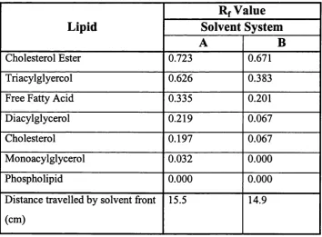
Table 4-1 : TLC standards used for the identification o f fractions separated from
whole cell and crude membrane lipid extracts. 102 Table 4-2: Comparison of Rf values of lipid standards. 102 Table 4-3: Distribution o f phytanic acid in lipid fractions from whole cell and
crude membrane lipid extracts of phytanic acid supplemented RPE
cells. 105
Chapter 6
Table 6-1 : Biological activity of different forms of vitamin E relative to
RRR-a-tocopherol as determined by the rat resorption assay. 138
Chapter 8
Table 8-1 : Reproducibility o f the determination and extraction of free MDA following oxidative stress using a H2 0 2/Cu^^ in vitro lipid
peroxidation system. 185
Chapter 9
Table 9-1 : The effect of increasing membrane suspension concentration on
steady state fluorescence anisotropy measurements. 226 Table 9-2: Reproducibility of steady state fluorescence anisotropy ( r j
LIST OF ABBREVIATIONS
AA Arachidonic acid
ARD Adult Refsum disease
BHT Butylated hydroxytoluene
CM Chylomicrons
c.v. Coefficient o f variation
DMA Docosahexaenoic acid
DPH Diphenylhexatriene
DSC Differential scanning calorimetry
EPA Eicosapentaenoic acid
ES External standard
BSA Bovine serum albumin
faf- BSA Fatty acid free-bovine serum albumin
FAME Fatty acid methyl ester
FBS Foetal bovine serum
FID Flame ionisation detection
GC Gas chromatography
HDL High density lipoproteins
HPLC High performance liquid chromatography
IDE Intermediate density lipoproteins
IS Internal standard
LDL Low density lipoproteins
LPL Lipoprotein lipase
MDA Malondialdehyde
MS Mass spectrometry
PBS Phosphate buffered saline
PVC Packed cell volume
PTS Peroxisomal targeting sequences
PUFA Polyunsaturated fatty acid
rbc Red blood cell
RCDP Rhizomelic chondrodysplasia punctata
RPE Retinal pigment epithelial
TLC Thin layer chromatography
UV Ultraviolet
VLCFA Very long chain fatty acids
1. INTRODUCTION
1.1 In t r o d u c t io n... 13
1.2 Pe r o x is o m e s...13
1.3 Pe r o x is o m a l Dis o r d e r s...14
1.4 Ad u l t Re f s u m Dis e a s e(A RD )...17
1.4.1 Clinical Findings... 17
1.4.2 Neuropathology in Adult Refsum Disease... 19
1.4.3 Diagnosis o f Adult Refsum Disease...20
1.4.4 Occurrence and Genetics o f Adult Refsum Disease...20
1.4.5 Treatment...21
1.5 Ph y t a n ic Ac i d...21
1.5.1 Dietary Origins O f Phytanic Acid...21
1.5.2 Metabolism o f Phytanic Acid - The a-Oxidation Pathway...23
1.5.3 Subcellular Localisation o f Phytanic Acid a-Oxidation...27
1.6 Po s s ib l e Me c h a n is m s f o rt h e Pa t h o g e n e s isin Ad u l t Re f s u m Dis e a s e...30
1.6.1 The Antimetabolite Hypothesis...31
1.6.2 The Molecular Distortion Hypothesis...33
1.6.3 Induced Essential Fatty Acid Deficiency...33
1.7 Re t in a l De g e n e r a t io n in Ad u l t Re f s u m Di s e a s e... 33
1.7.1 The Structure o f the Retina...34
1.7.2 Retinitis Pigmentosa...38
1.7.3 Fatty Acid Composition o f Rod Outer Segment Membranes...40
1.7.4 The Effect o f a Dietary Deficiency o f n-3 Fatty Acids on Visual Function 41 1.7.5 Deficiency o f Docosahexaenoic Acid in Retinitis Pigmentosa...42
1.7.6 Function o f Docosahexaenoic Acid in the Retina...44
1.7.7 Phytanic Acid and Retinal Degeneration...45
L I Introduction
Classical or adult Refsum disease (ARD) is an autosomal recessive metabolic disorder
in which degenerative changes in the retina, and central and peripheral nervous systems
are the major features. The disease is caused by an isolated deficiency o f the enzyme,
phytanic acid a-hydroxylase, which catalyses the first step in the degradative pathway
of the exogenous fatty acid, phytanic acid. ARD has been classified as a peroxisomal
disorder as the pathway o f phytanic acid degradation is generally believed to occur in
the peroxisome. The resulting accumulation o f phytanic acid in the serum and tissues of
adult Refsum patients is thought to be responsible for the clinical features o f this
condition. However, the underlying mechanism(s) as to how this occurs has not been
elucidated. Several hypotheses have been proposed, but relatively little progress towards
our understanding o f the mechanism(s) of this disease has been made. The following
study was designed to investigate the pathogenesis o f the retinal abnormalities in ARD.
Cultured mammalian retinal cell lines supplemented with phytanic acid in the medium
were used as an in vitro model for ARD. The effect o f modulating phytanic acid concentrations on membrane fatty acid composition and function were studied.
In this introduction I will briefly consider peroxisomal disorders including ARD,
phytanic acid metabolism and possible mechanisms for the pathogenesis of the retinal
abnormalities in ARD.
1,2 Peroxisomes
Peroxisomes are subcellular organelles that perform a variety o f metabolic functions and
were first characterised by De Duve and Baudhuin in 1966. These organelles are
thought to be ubiquitous amongst cell types except the mature erythrocyte and are
particularly abundant in the liver and kidney. They are bounded by a single membrane
and vary in morphology, size and abundance. Peroxisomes are usually spherical but may
in some cell types be interconnected forming an intracellular compartment referred to as
a peroxisomal reticulum. They are formed by division o f pre-existing peroxisomes and
have a half life o f about 1.5 to 2 days. Peroxisomes were so named due to the peroxide
L-amino acids, oxalate and polyamines are oxidised by peroxisomal enzymes with the
concomitant production o f hydrogen peroxide. Catalase then converts hydrogen
peroxide to water and molecular oxygen or the hydrogen peroxide is used to peroxidise
substrates including ethanol, methanol, nitrites, quinone, and formate.
Both anabolic and catabolic functions are carried out by peroxisomes (Lazarow and
Moser, 1995). For example, peroxisomes are involved in the biosynthesis o f the
plasmalogens (phospholipids which have a 1,2 unsaturated alcohol in ether linkage at
the Snl position on the glycerol backbone), cholesterol (in addition to the cholesterol
biosynthesis that occurs in the endoplasmic reticulum) and bile acids. They also
contribute to the gluconeogenic pathway by serving as the site o f deamination of some
amino acids. Peroxisomes possess a p-oxidation system which shortens the cholesterol
side chain during the biosynthesis of bile acids and is thought to be involved in the
biosynthesis o f the polyunsaturated fatty acids, docosahexaenoic acid (22:6n3) and
docosapentaenoic acid (22:5n6) (Sprecher et al., 1995). Peroxisomal P-oxidation also
contributes to the degradation of fatty acids and eicosanoids, overlapping in its
specificity for fatty acids with the mitochondrial p-oxidation system. However very
long chain fatty acids (VLCFA) (> 22 carbons in length) and long-chain dicarboxylic
acids are preferentially catabolised by the peroxisomal system. There is also evidence
that «-substituted branched chain fatty acids such as pristanic acid are degraded
principally by this p-oxidation system (ten Brink et al., 1992a; Singh et al., 1992). The
peroxisome is most probably the site o f a-oxidation of the p-substituted fatty acid,
phytanic acid. The exact location of the a-oxidation pathway has been a matter of some
debate and is discussed in more detail in section 1.5.3.
1.3 Peroxisomal Disorders
The important metabolic role played by peroxisomes is evident from the devastating
effects that occur when one or more peroxisomal functions are impaired. The
peroxisomal disorders are a heterogeneous group o f inherited disorders and are
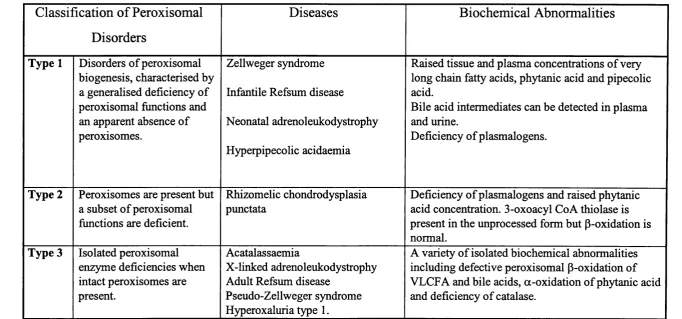
classically divided into three categories (see table 1.1). For a review see Lazarow and
Moser (1995). The first group comprises the disorders o f peroxisomal biogenesis. The
Classification o f Peroxisomal
Disorders
Diseases
Biochemical Abnormalities
Type 1 Disorders o f peroxisomal biogenesis, characterised by a generalised deficiency of peroxisomal fimctions and an apparent absence o f peroxisomes.
Zellweger syndrome
Infantile Refsum disease
Neonatal adrenoleukodystrophy
Hyperpipecolic acidaemia
Raised tissue and plasma concentrations o f very long chain fatty acids, phytanic acid and pipecolic acid.
Bile acid intermediates can be detected in plasma and urine.
Deficiency o f plasmalogens.
Type 2 Peroxisomes are present but a subset o f peroxisomal fimctions are deficient.
Rhizomelic chondrodysplasia punctata
Deficiency o f plasmalogens and raised phytanic acid concentration. 3-oxoacyl Co A thiolase is present in the unprocessed form but P-oxidation is normal.
Type 3 Isolated peroxisomal enzyme deficiencies when intact peroxisomes are present.
Acatalassaemia
X-linked adrenoleukodystrophy Adult Refsum disease
Pseudo-Zellweger syndrome Hyperoxaluria type 1.
A variety o f isolated biochemical abnormalities including defective peroxisomal p-oxidation of VLCFA and bile acids, a-oxidation o f phytanic acid and deficiency o f catalase.
Included in this group are Zellweger syndrome, infantile Refsum disease, neonatal
adrenoleukodystrophy and hyperpipecolic acidaemia. Zellweger syndrome represents
the most severe form of peroxisomal disorders and patients with this disease usually die
within the first year of life. Symptoms of this syndrome include severe hypotonia,
severe retardation, characteristic dysmorphic features, neonatal seizures, hepatomegaly,
renal cysts and visual abnormalities including retinitis pigmentosa-like symptoms.
Biochemical abnormalities include an accumulation of VLCFA, intermediates of bile
acid biosynthesis, phytanic acid and pipecolic acid, the latter being an intermediate in
lysine catabolism. Plasmalogens are deficient and there is a mislocalisation of catalase
to the cytoplasm. The spectrum of symptoms and biochemical defects is similar in the
other diseases within this group, although they tend to be milder.
In recent years insights into the mechanisms involved in the disorders of peroxisomal
biogenesis have been made (reviewed in Subramani, 1997). The demonstration of
peroxisomal membrane ’’ghosts” in cells from patients suggested that there may be a
defect in the targeting of peroxisomal proteins, all of which are encoded by the nucleus,
to the peroxisomal matrix. Peroxisomal proteins are imported into the peroxisome post-
translationally and are targeted by integral signals within the protein structure that are
not cleaved after translocation. Two peroxisomal targeting sequences (PTS) that are
responsible for directing proteins into the peroxisomal matrix have been identified and
both are highly conserved (Subramani, 1997). PTSl is the tripeptide serine-lysine-
leucine and is found at the C-terminal of proteins destined for the peroxisome. The
majority o f peroxisomal matrix proteins carry this signal (or variants o f this). PTS2, the
polypeptide (arginine/lysine)-(leucine/valine/isoleucine)-X5-(histidine /glutamine)-
(leucine/alanine) is found at the N-terminal end of a small number of peroxisomal
matrix proteins. It seems that the import of PTSl carrying proteins alone or in
combination with PTS2 proteins are affected in type 1 peroxisomal disorders. The genes
involved are those that encode the peroxisomal membrane receptors for these signals.
Defective import of PTS2 proteins alone occurs in rhizomelic chondrodysplasia
punctata (RCDP). This disease constitutes the second class o f peroxisomal disorders in
including plasmalogen biosynthesis and phytanic acid degradation. RCDP is
phenotypically different from the type one disorders most noticeably in the shortening
of proximal limbs.
The third type o f peroxisomal disorders are those caused by the deficiency o f a single
peroxisomal enzyme. Acyl Co A oxidase, bifimctional enzyme and 3-oxoacyl Co A
thiolase are enzymes in the peroxisomal p-oxidation pathway. Isolated deficiency of any
of these enzymes has been found to lead to disorders that are clinically similar to
Zellweger syndrome and have thus been previously designated as “pseudo-Zellweger
syndrome”. Other diseases assigned to this category are acatalassaemia, X-linked
adrenoleukodystrophy, hyperoxaluria type 1 and most probably ARD (see below).
1.4 Adult Refsum Disease (ARD)
1.4.1 Clinical Findings
Adult or classical Refsum disease, originally termed heredopathia atactica
polyneuritiformis, is a rare autosomal recessive metabolic disorder first identified by
Sigvald Refsum in 1946 and has been reviewed by Steinberg (1995). The metabolic
defect is an isolated deficiency of phytanic acid a-hydroxylase activity (ten Brink et al.,
1992b; Pahan et al., 1996) (see 1.5.2). As a consequence of this enzymatic deficiency
there is a gradual accumulation of phytanic acid in the tissues and blood o f patients,
leading to the onset o f symptoms and a progressive deterioration o f their condition. The
age o f onset is variable and may occur in childhood or much later in the fourth or fifth
decades of life. Night blindness is often the first manifestation of the disease which goes
on to develop into retinitis pigmentosa-like symptoms (see section 1.7.2). Retinitis
pigmentosa along with peripheral neuropathy, cerebellar ataxia and an elevated
cerebrospinal fluid protein concentration in the absence o f pleocytosis (abnormal cell
count), are the defining clinical features of this disease. The neuropathy is usually
symmetrical with distal lower limb regions being mainly affected. Muscles o f the lower
limbs become weak and atrophied. Motor and sensory nerve conduction velocities are
reduced and evidence o f denervation has been shown by electromyography. Other
features o f the peripheral neuropathy include a reduction or loss o f vibration sense,
(Refsum et al., 1984). The pigmentary retinopathy is atypical with the pigmentation
having a ‘salt and pepper’ appearance rather than the more usual ‘bony spicule’
appearance seen in other forms o f retinitis pigmentosa (Refsum et al., 1984).
Pigmentation usually occurs in the periphery of the retina. Other ophthalmologic
changes have also been observed such as optic atrophy resulting in blindness, cataracts
and vitreous opacities. Electroretinographic findings have demonstrated a loss of rod
and cone responses (Refsum et al., 1984).
The pathological abnormalities in the eyes of a patient with ARD have been described
(Toussaint and Danis, 1971). An accumulation of lipid was noted in the sclera,
trabecular meshwork, iris muscles and in the RPE. There was an absence of rods and
cones, the outer nuclear and plexiform layers were atrophied, the inner nuclear layer was
thinned and the ganglion cells were reduced in number. Retinal vessels were narrowed
and occluded. In many areas of the posterior pole, the RPE was absent. The optic nerve
showed mild demyelination.
A range o f other symptoms may also occur in ARD including anosmia (loss of the sense
o f smell), an ichthyosis-like dry scaling skin condition, nerve deafness, pupillary
abnormalities, nystagmus (a rapid involuntary oscillation of the eyeballs) and
electrocardiographic abnormalities. Skeletal abnormalities are also observed and include
a shortening of the fourth metatarsal, syndactyly (a union o f two or more digits),
hammer toes, pes cavus (an exaggeration o f the normal arch of the foot), and
osteochondritis dissecans (displaced fi*agment o f cartilage and bone in a joint cavity,
usually in the knee). The range o f symptoms seen at the time of diagnosis of the disease
is variable and children often present with an incomplete syndrome (Steinberg, 1993).
The clinical course can be that o f a gradual or a rapid progressive deterioration of the
patient’s condition. Sudden death has occurred in a number o f cases, thought to have
been caused by cardiac complications or respiratory failure, although renal failure was
reported in one patient (Refsum et al., 1984). A marked worsening o f the condition of
some patients has been known to occur under conditions of stress such as during a
serious illness, pregnancy, following rapid weight loss or after surgery. Mobilisation of
and causes the clinical deterioration. Very high concentrations of phytanic acid in the
serum ( >100 mg/dl compared to normal concentrations o f < 0.5 mg/dl) can result in
toxic symptoms including weight loss, failure to thrive, and fatigue in both humans and
experimental animals (Refsum et al., 1984). Serum concentrations o f phytanic acid of
200-250 mg/dl have been observed to have fatal consequences (Refsum et al., 1984).
Periods o f exacerbation are usually followed by remission where the progress o f the
disease appears to be interrupted for some time (Refsum et al., 1984; Steinberg, 1995).
1.4.2 Neuropathology in Adult Refsum Disease
The neuropathy seen in ARD is characterised by a chronic distal symmetrical
sensorimotor neuropathy. Histopathological and electron microscopic studies have
revealed interstitial hypertrophic changes in the peripheral nerves of Refsum patients
(Refsum et al., 1984). The nerve hypertrophy is maximal proximally and most evident
in the limb girdle plexuses. Spinal nerve roots and sensory ganglia may also be affected
(Thomas et al., 1997). Nerve hypertrophy is caused by repeated demyelination and
remyelination o f nerve axons. A proliferation o f Schwann cells occurs in response to
demyelination and those cells not involved in remyelinating the axon form a concentric
arrangement around the nerve fibre. Such ‘onion bulb’ formations in Refsum disease
have been noted to contain many unmyelinated axons and collagen bundles are densely
packed both inside and around the whorls (Refsum et al., 1984). A reduction in the
number of myelinated nerve fibres and a segmental demyelination o f teased-fibre
preparations are also found. These changes are suggestive o f a primary defect in
Schwann cell fimction. Schwann cells have been found to contain osmiophilic bodies
that are probably lipid granules and these are sometimes associated with typical
lipofuscin granules (Refsum et al., 1984). Fat deposits in the central nervous system, the
choroid plexuses and in the soft membranes enveloping the brain and spinal cord
(leptomeninges) have also been found. Large crystalline inclusions have been observed
in Schwann cell cytoplasmic processes with a structure suggestive of an
intramitochondrial origin (Refsum et al., 1984). Axonal and hepatic liver mitochondrial
abnormalities have been reported (Refsum et al., 1984).
Kuntzer et al (1993) performed a longitudinal study o f nerve conduction with
Motor and sensory conduction velocities were reduced in a non-uniform manner which
supports the histopathological evidence of a sensorimotor demyelinating neuropathy
with segmental demyelination.
1.4.3 Diagnosis of Adult Refsum Disease
Confirmation o f the diagnosis o f ARD may be made by measuring the concentration of
phytanic acid in plasma, which in normal subjects is usually below 0.5 mg/dl (16
pmol/1) but can be elevated to 5-100 mg/dl (160-3200 pmol/1) in untreated patients. In
one o f the original studies, phytanic acid accounted for 5-30 % o f the total serum lipid
fatty acids in nine patients (Refsum et al., 1984). In addition to measuring the plasma
phytanic acid concentration, the phytanic acid oxidative capacity o f cultured skin
fibroblasts may be measured. Fibroblasts from patients with ARD oxidise phytanic acid
at about 5 % the rate o f fibroblasts fi"om unaffected individuals and heterozygotic
fibroblasts oxidise at approximately 50 % the normal rate (Steinberg, 1995).
Heterozygotes may sometimes show a slight accumulation o f phytanic acid in their
serum, but remain asymptomatic (Steinberg, 1995).
1.4.4 Occurrence and Genetics of Adult Refsum Disease
The occurrence o f ARD is very rare; Steinberg has estimated that the number of
confirmed cases in the literature is about 150 (Steinberg, 1995). Most cases have been
reported from Northern Europe. Very recently the gene encoding an enzyme with
phytanoyl-CoA a-hydroxylase activity was identified (Jansen et al., 1997; Mihalik et
al., 1997). In the ARD patients which have been studied, a number of inactivating
mutations in this gene have been found which confirms the biochemical evidence for a
deficiency o f phytanoyl-CoA a-hydroxylase activity as the metabolic defect in ARD
(Jansen et al., 1997; Mihalik et al., 1997). This gene has been localised to chromosome
lOp (Mihalik et al., 1997) which is the same region where the gene responsible for an
atypical form o f the disease, in which phytanic acid accumulation occurs alongside an
1.4.5 Treatment
ARD can be treated by dietary management. Phytanic acid is believed to be entirely of
exogenous origin as studies investigating whether endogenous synthesis takes place
have yielded negative results (Steinberg, 1995). Thus by limiting the intake o f phytanic
acid and its precursor phytol, phytanic acid accumulation can be halted. There is
evidence that patients either retain some residual phytanic acid oxidation activity or are
able to excrete the unchanged acid. There is often a delay in the response to treatment,
probably due to mobilisation of phytanic acid from fat stores, but eventually a much
reduced plasma concentration can be achieved. This may be aided in the initial stages of
the treatment by plasmapheresis. Circulating concentrations very rarely return to
normal, but remain slightly elevated at levels of between 10-30 mg/dl (Steinberg, 1995).
Treatment in this way halts the progression of the disease and appears to prevent
relapses. An improvement in many of the symptoms such as the peripheral neuropathy,
ichthyosis, muscle strength and gait is seen (Refsum et al., 1984; Steinberg, 1995),
whereas retinal and auditory fimction, while stabilised do not appear to regress
(Steinberg, 1995).
1.5 Phytanic Acid
1.5.1 Dietary Origins Of Phytanic Acid
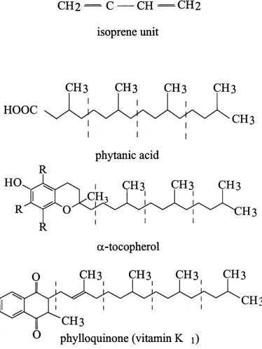
Phytanic acid (3,7,11,15-tetramethylhexadecanoic acid) is a saturated, multi-branched
fatty acid composed o f four isoprenoid units (figure 1-1). It is derived fi*om the phytol
side chain of chlorophyll and is present in our diet in a number of food sources (see
below). Degradation o f chlorophyll releases phytol which, as the fi*ee alcohol, can be
converted to phytanic acid (Hansen, 1980). This process can occur in animals including
man after free phytol has been absorbed from the diet (Steinberg, 1995). The enzymatic
conversion of phytol to phytanic acid has been demonstrated in rat liver mitochondrial
and microsomal fractions (Muralidharan and Muralidharan, 1986). The conversion of
free phytol to phytanic acid is also thought to occur in the intestinal tract of ruminants
through microbial action (Hansen, 1980). This results in an increase in the amount of
preformed phytanic acid absorbed by these animals. Hence the major sources of
CH3
CH2 —
C — CH — CH2
isoprene unit
CH3
CH3
HOOC
phytanic acid
a-tocopherol
CH3
CH3
CH3
O
phylloquinone (vitamin K i)
meat, and dairy products. The plasma concentration o f phytanic acid in cattle is also
influenced by their method of feeding. Cattle fed grass-silage have a higher plasma
phytanic acid concentration than pasture fed animals. Hansen (1980) suggested this
could be due to an increase in the amount of liberated phytol as a result of the mildly
acidic environment of silage. In non-ruminant animals, intestinal conversion of phytol to
phytanic acid is thought to be of little significance. Phytanic acid and phytenic acid, an
intermediate in the conversion of phytol to phytanic acid, have been shown to
accumulate in the liver o f germ free rats fed phytol (Steinberg et al., 1966). Intake of
preformed phytanic acid in man has been estimated to be between 50 to 100 mg/day.
The amount of total phytol (esterified to chlorophyll or as the free alcohol) in the diet
represents only 10% o f the amount of preformed phytanic acid. In its esterified form
phytol is poorly absorbed and is lost in the faeces, but both free phytol and phytanic acid
are efficiently absorbed (Hansen, 1980; Steinberg, 1995).
1.5.2 Metabolism of Phytanic Acid - The a-Oxidation Pathway
Normally only trace amounts of phytanic acid are detected in the plasma of humans,
indicating that it is efficiently catabolised. Phytanic acid has a methyl group in the p
position which prevents it from being degraded by the p-oxidation pathways in
mitochondria and peroxisomes. Instead it undergoes an initial a-oxidation step,
resulting in the removal o f a 1-carbon unit and the formation o f pristanic acid
(Steinberg, 1995). Pristanic acid is then further degraded by cycles o f p-oxidation. The
exact sequence of the a-oxidation pathway and its intracellular location have been
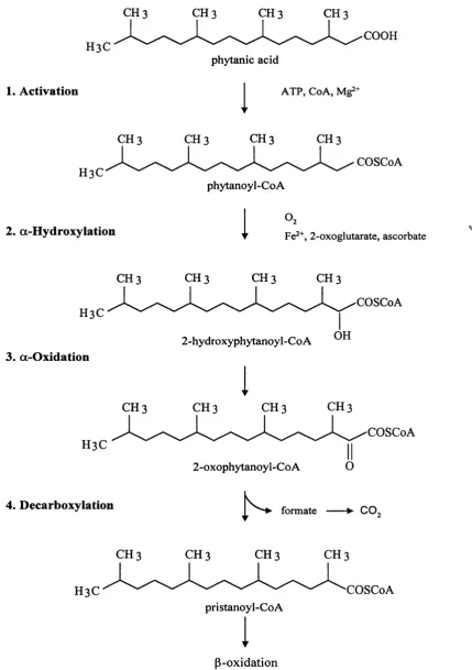
subjects o f much debate and controversy. A proposed scheme o f the pathway based on
the evidence to date and involving at least four steps is shown in figure 1-2.
1) Activation to Phytanoyl-CoA
Evidence to date suggests that the conversion of phytanic acid to its coenzyme A
derivative is the initial step in the pathway. Phytanoyl-CoA ligase activity has been
detected in peroxisomal, mitochondrial, and endoplasmic reticulum fractions of human
and rat liver and skin fibroblasts (Pahan and Singh, 1993; Singh et al., 1994). In
peroxisomes this activity was found to be distinct from lignoceroyl and palmitoyl CoA
CH 3 CH 3 CH 3 CH 3
H3C
1. Activation
COOH phytanic acid
1
ATP, CoA, Mg^+H3C
CH 3 CH 3 CH 3 CH 3
phytanoyl-CoA
2. a-Hydroxylation
COSCoA
1
“ ■t Fe2+, 2 - oxoglutarate, ascorbate
CH 3 CH 3 CH 3 CH 3
H3C
3. a-Oxidation
H3C
4. Décarboxylation
COSCoA
2-hydroxyphytanoyl-CoA
1
CH 3 CH 3 CH 3 CH 3
COSCoA
2-oxophytanoyl-CoA
formate --- ► CO„
C H 3 C H 3 C H 3 C H 3
H3C COSCoA
pristanoyl-CoA
1
P-oxidation
o f the peroxisomal membrane (Pahan and Singh, 1995a). A requirement for the
activation o f phytanic acid to phytanoyl-CoA in the a-oxidation pathway has been
implicated by the results from a number o f studies (for example Muralidharan and
Muralidharan, 1987; Singh et al., 1990; Watkins et al., 1994; Mihalik et al., 1995).
Watkins and co-workers (1994) demonstrated using isolated rat liver peroxisomes that
the release o f into the aqueous phase from [2,3-^H]phytanic acid required the
presence o f phytanoyl-CoA synthetase activity. Croes et al (1996) demonstrated that the
a-oxidation of 3-methyl-substituted fatty acids in permeabilised rat hepatocytes (where
intracellular organelles and membranes remained intact) required the presence of CoA,
ATP, and Mg^^ indicating the involvement o f an acyl-CoA synthetase in this pathway.
However there are conflicting reports concerning the subsequent fate o f phytanoyl-CoA.
Both phytanoyl-CoA and a-hydroxyphytanoyl-CoA (thought to be the next intermediate
in the pathway) were detected by high performance liquid chromatographic (HPLC)
analysis o f the products formed following a-oxidation o f phytanic acid in rat liver
peroxisomes (Mihalik et al., 1995). Also 3-methylpalmitoyl-CoA and 2-hydroxy-3-
methylpalmitoyl-CoA were identified as intermediates of the a-oxidation of 3-
methylpalmitate (as a substitute substrate for phytanic acid) by permeabilised rat
hepatocytes, rat liver homogenates and isolated peroxisomes (Croes et al., 1996). This
suggests that phytanoyl-CoA is the substrate for the second step in the pathway (a-
hydroxylation) which would result in the production o f a-hydroxyphytanoyl-CoA.
However Pahan and Singh (1993) have reported that it is the free acid and not
phytanoyl-CoA that is the substrate for this second step. They showed using an isolated
peroxisomal matrix or permeabilised peroxisomes that a-oxidation activity did not
require cofactors for the activation o f fatty acids. In addition activity was unaffected by
an acyl-CoA ligase inhibitor, naproxen. They also showed that phytanoyl-CoA is
quickly hydrolysed after transportation into the peroxisomal matrix.
2) a-Hydroxylation
The second step in the a-oxidation pathway, which is the step that is impaired in ARD,
is the a-hydroxylation of phytanoyl-CoA to 2-hydroxyphytanoyl-CoA in the matrix of
the peroxisome (Pahan and Singh, 1993). The evidence for this step comes from a
humans and in patients suffering from certain types of peroxisomal disorders in which
defective phytanic acid degradation occurs has been demonstrated (ten Brink et ah,
1992a). Formation o f 2-hydroxyphytanic acid from phytanic acid has also been shown
in vivo in human subjects, ten Brink et al (1992b) detected [l-^^C]2-hydroxyphytanic acid in the plasma of control subjects administered a dose of [l-^^C]phytanic. In the
plasma o f ARD patients taking part in the same study this metabolite was not detected.
Muralidharan and Muralidharan (1987) have characterised the a-hydroxylation of [1-
^"^Cjphytanic acid in rat liver and showed there was a time dependent increase in the
amount o f [l-*'*C]2-hydroxyphytanic acid formed. Mihalik et al (1995) have
demonstrated the enzyme that catalysed the a-hydroxylation o f phytanic acid exhibits
co-factor requirements (2-oxoglutarate, Fe^^ and ascorbate) which they reported as
being consistent with a dioxygenase reaction mechanism. These co-factors were also
shown to be required for the a-oxidation of 3-methyl-substituted fatty acids in rat liver
homogenates (Croes et al., 1996). Pahan et al (1995b) showed that the a-hydroxylation
of phytanic acid in peroxisomes isolated from human liver and fibroblasts and Hep G2
cells could be inhibited by imidazole antimycotics suggesting that this step is mediated
by a cytochrome P-450 containing enzyme.
3) a-Oxidation
The next step in the a-oxidation of phytanic acid is an oxidative decarboxylation of 2-
hydroxyphytanic acid resulting in the formation of pristanic acid and CO2. Evidence that
this step involves more than one reaction was provided by Draye et al (1987) who
demonstrated the production o f 2-oxophytanic acid from L-2-hydroxyphytanic acid in
rat kidney cortex. 2-hydroxyphytanic acid oxidase activity has also been detected in rat
and human liver (Wanders et al., 1994), and an H2O2 producing enzyme with this
activity has been localised to the peroxisome in human liver (Wanders et al., 1994;
Wanders et al., 1995). The conversion o f an a-hydroxyl group to an a-keto group is
analogous to the mechanism o f degradation of other a-hydroxyl acids (Wanders and van
Roermund, 1993), for instance the conversion of lactate to pyruvate and acetyl-CoA.
However ten Brink et al (1992a) failed to detect 2-oxophytanic acid in the plasma of
healthy humans and patients with peroxisomal disorders, even in those conditions where
o f peroxisomal biogenesis). It is possible that 2-oxophytanic acid is rapidly
decarboxylated as it is formed, thus preventing its detection.
4) Decarboxylation
Carbon dioxide and pristanic acid are usually cited as the products o f phytanic acid a -
oxidation. However a number o f studies have provided evidence that the
decarboxylation o f 2-oxophytanic acid occurs via the formation of formic acid which is
then converted to CO2. Poulos et al (1993) first identified formic acid as the major
water soluble product formed from [l-^"^C]phytanic acid and [1-^"^C]3-
methylhexadecanoic acid degradation by human skin fibroblasts. The authors speculated
that CO2 was produced from formic acid and not vice versa, as formic acid production
was in excess o f CO2 production. Mihalik et al (1995) reported similar results in a-
oxidation experiments with [l-^"^C]phytanic acid in rat liver peroxisomes. These
findings are supported by the work of Croes and colleagues who demonstrated that
formic acid was a primary product o f a-oxidation in intact rat hepatocytes (Croes et al.,
1996) and in peroxisomal enriched fractions from human liver (Casteels et al., 1997).
Croes et al (1997) have also reported that in addition to formate, a-oxidation of 3-
methyl substituted acids results in the production o f an aldehyde which can be converted
to an acid. This suggests that pristanal would be the major product o f a-oxidation of
phytanic acid which would then be converted to I pristanic acid.
In summary, phytanic acid is converted to pristanic acid by an a-oxidation pathway in
the first stage o f its degradation. Research into this pathway has revealed that at least 4
steps may be involved; activation o f phytanic acid to phytanoyl-CoA (possibly followed
by post-transport hydrolysis to the free acid), a-hydroxylation to the 2-hydroxyl-
derivative, oxidation to the 2-oxo-derivative, and decarboxylation to pristanoyl-CoA (or
pristanic acid), possibly via the formation of pristanal, with the concomitant production
o f formate which is then converted to carbon dioxide.
1.5.3 Subcellular Localisation of Phytanic Acid a-Oxidation
Phytanic acid accumulation occurs in a number o f the peroxisomal disorders (Lazarow
located in the peroxisome. However studies on the subcellular localisation of the a -
oxidation pathway have produced conflicting results. The presence o f a-oxidation
activity has been demonstrated in peroxisomes (for example Singh et al., 1993a; Singh
et al., 1993b; Wanders et al., 1994; Croes et al., 1996; Casteels et al., 1997), in
mitochondria (Tsai et al., 1969; Muralidharan and Kishimoto, 1984; Skjeldal and
Stokke, 1987; Watkins and Mihalik, 1990; Wanders et al., 1991; Singh et al., 1993b;
Wanders and van Roermund, 1993) and in the endoplasmic reticulum (Huang et al.,
1992; Singh et al., 1993a). In type 1 peroxisomal disorders, mitochondria with abnormal
structure and function have been observed in some cases, and thus a mitochondrial
localisation is feasible.
Most investigations have employed rat and human tissues, usually skin fibroblasts and
liver, using methods that either measured individual steps or the entire a-oxidation
pathway. For example, ^H release from [2,3-^H]phytanic acid was used to measure the
a-hydroxylation step and this activity was found to be localised to peroxisomes isolated
from rat liver (Watkins et al., 1994; Mihalik et al., 1995). The formation o f 2-
ketophytanic acid from 2-hydroxyphytanic acid has been used as a measure of 2-
hydroxyphytanic acid oxidase activity. This was found to be associated with the
distribution o f the peroxisomal enzyme, catalase, in fractions of rat kidney cortex
(Draye et al., 1987) and it was localised to peroxisomes in rat liver (Wanders et al.,
1994). The release o f ^"^C02 from [l-^"^C]phytanic acid is a measure o f the entire a -
oxidation pathway. For example, using this technique Singh et al (1993b) demonstrated
that in humans the subcellular location o f this pathway was predominantly peroxisomal
whereas in rats it was predominantly mitochondrial. When release was measured
in control human skin fibroblast fractions, activity was found in peroxisomal,
mitochondrial and ER fractions, with predominant activity in the peroxisomal fraction
(Singh et al., 1993a). In subcellular fractions obtained from fibroblasts from ARD
patients, activities in the mitochondrial and ER fractions were comparable to controls,
whereas peroxisomal activity was deficient (Singh et al., 1993a). The low levels of a-
oxidation activity normally found in the endoplasmic reticulum and mitochondria may
explain the low levels o f residual activity in Refsum patients (Steinberg, 1995).
converted to CO2 (Poulos et al., 1993; Mihalik et al., 1995; Croes et al., 1996; Casteels
et al., 1997) suggests that using '^C02 release from [l-^"^C]phytanic acid as a measure of
a-oxidation activity may give misleading results. This is because formate may be
degraded to CO2 by at least two pathways; a folate dependent pathway in the cytosol
and mitochondria, and a catalase dependent peroxidative pathway in peroxisomes
(Mihalik et al., 1995) and this may explain why activity has been detected in different
organelles. In addition, Casteels et al (1997) suggested that the low levels o f activity
reported in many studies could be due to the fact that CO2 production is not a direct
measure o f a-oxidation.
Other evidence for the subcellular localisation o f the a-oxidation pathway comes from
the concentrations o f intermediates o f this pathway in the serum o f patients with
peroxisomal disorders. Serum concentrations o f 2-hydroxyphytanic acid are raised in
patients with disorders o f peroxisomal biogenesis who suffer from global peroxisomal
function deficiencies. This suggests that the conversion o f 2-hydroxyphytanic acid to
pristanic acid is a peroxisomal function (ten Brink et al., 1992a). It also suggests that a -
hydroxylation of phytanic acid could, however, occur at a site other than the
peroxisome. Thus it has been suggested that the a-oxidation pathway may be split
between the mitochondria and the peroxisome rather than being exclusive to one
organelle (ten Brink et al., 1992a). This concept is supported by the observation of
Wanders and van Roermund (1993) that intact rat hepatocytes had higher a-oxidation
activity than whole homogenates and postnuclear supernatant.
The studies mentioned above are a few examples of the investigations on the subcellular
location o f a-oxidation o f phytanic acid that have been reported in the literature.
Overall, it is difficult to draw a general consensus from these studies and a number of
factors may have contributed to the differences in results. For instance, where enzyme
markers were not used the purity o f the cellular fractions are unknown (Tsai et al., 1969;
Muralidharan and Kishimoto, 1984). The recoveries o f activity after cell fractionation
have not been reported in some studies (Tsai et al., 1969; Muralidharan and Kishimoto,
1984; Watkins and Mihalik, 1990). Nycodenz, a substance commonly used for the
activity (Singh et al., 1993b), although activity can be recovered following its removal
by dialysis. Therefore, in studies where Nycodenz had not been removed, peroxisomal
activity may have been inhibited (Watkins and Mihalik, 1990; Wanders et al., 1991).
The necessary co-factors for a-oxidation activity (as determined by Mihalik et al (1995)
and Croes et al (1996)) were not included in many studies.
In summary, although the site of subcellular localisation o f phytanic acid a-oxidation
has not been unequivocally proven, it seems very likely that at least part o f the pathway
occurs in peroxisomes and that activity is not probably confined to a single organelle.
Two papers describing the identification of the gene for phytanoyl-CoA hydroxylase
(Jansen et al., 1997; Mihalik et al., 1997) have recently been published. These two
studies have shown that the protein encoded by this gene contains a type- 2 peroxisomal
targeting sequence in its N-terminus (see section 1.3) (Jansen et al., 1997; Mihalik et al.,
1997) and that it interacts with a peroxisomal receptor for this sequence (Mihalik et al.,
1997). Thus ARD can now be unequivocally classified as a type 3 peroxisomal disorder
and the a-hydroxylation step o f the a-oxidation pathway as a peroxisomal function.
1.6 Possible Mechanisms for the Pathogenesis in Adult Refsum
Disease
It is evident from the clinical observations of ARD and results from animal studies that
a build up o f phytanic acid in tissues and serum has toxic consequences. In humans it
has been demonstrated that phytanic acid accumulates in the peripheral and central
nervous system (MacBrinn and O’Brien, 1968) and it could therefore be responsible for
the observed neurological symptoms. However, the neurological symptoms have not
been reproduced in animals fed large doses o f phytol or phytanic acid. Steinberg et al
(1966) attempted to create an animal model o f ARD using the mouse, rat, rabbit and
chinchilla. In order to cause an accumulation o f phytanic acid in tissues, it was
necessary to feed the animals phytol or phytanic acid in amounts that exceeded the
capacity o f the animal to metabolise them. Between 2 - 5 % by weight in the diet of
phytol or phytanic acid were administered over a period o f up to 2 months. Excessive
amounts o f phytanic acid accumulated in the liver and serum and fatty deposits were
caused death. The retina and peripheral and central nervous systems appeared to be
unaffected. However due to the necessity o f feeding large doses o f phytol or phytanic
acid, which proved to be toxic, only short term studies could be carried out. The amount
o f phytanic acid which accumulated in the lumbar plexes and sciatic nerves o f rats in
this study was 0.1 mg/g. This is much lower than that reported in the sciatic nerve of an
ARD patient (approximately 8 mg/g). The polecat is an animal with a low a-oxidation
capacity. It has been attempted to reproduce the neurological symptoms of ARD in this
animal in a long term feeding experiment over two generations in an attempt to mimic
the chronic nature o f ARD (Refsum et al., 1984). Again large amounts o f phytanic acid
accumulated in peripheral tissues, but only trace amounts were detected in brain lipids
and neurological lesions did not develop. The results from these studies suggest that
phytanic acid accumulates in the nervous system of humans more easily than it does in
other animals, but do not rule out the idea that phytanic acid could cause the
neurological symptoms seen in ARD.
The biochemical mechanism(s) whereby phytanic acid accumulation leads to the
spectrum o f signs and symptoms observed in ARD has not been elucidated. A number
o f hypotheses have been suggested and are discussed by Steinberg (1993; 1995) and by
Stokke and Eldjam (in Refsum et al., 1984). A brief outline o f the main hypotheses is
given below.
1.6.1 The Antimetabolite Hypothesis
Phytanic acid is structurally similar to a number of molecules which, like phytanic acid,
are derived from the isoprenoid unit, a five carbon structure. Examples of such
molecules include the fat soluble vitamins E, K and A (see figure 1-1), ubiquinone, an
electron carrier in the inner mitochondrial membrane, dolichol, a lipid found o f the
endoplasmic reticulum membrane involved in the glycosylation o f proteins and the
prenyl groups that serve as lipid anchors, positioning proteins in membranes (see section
1.6.1.2). It has been suggested that an accumulation of phytanic acid may interfere with
the fimction o f structurally similar molecules (Steinberg, 1995) and the strongest
1.6,L1 Interference with the Function o f Vitamin E
Vitamin E is closely related to phytanic acid in structure as it possesses a phytyl side
chain which serves to anchor it in membranes. Vitamin E is considered to be the major
lipid soluble chain-breaking antioxidant (Burton and Traber, 1990) and thus plays an
important role in protecting cellular membranes from lipid peroxidation. A severe
deficiency o f this vitamin in humans occurs in a number o f disorders involving fat
absorption or transport (Harding, 1987; Sokol, 1993) and results in a spectrum of
symptoms similar to those observed in ARD. This includes peripheral and central
neuropathy, retinal degeneration and muscle weakness (Muller and Goss-Sampson,
1990). It is possible that an accumulation o f phytanic acid in tissues and cellular
membranes in some way perturbs the function of vitamin E and so induces a vitamin E
deficiency-like state. This hypothesis is further discussed in chapter 6.
1.6.1.2 Interference with Protein Prénylation
An interference with the function o f membrane proteins that are modified with famesyl
and geranylgeranyl (prenyl) groups has been suggested (Steinberg, 1995). An example
of such a protein is ras, a GTP-binding protein that plays a central role in receptor
tyrosine kinase signal transduction pathways which are stimulated by growth factors and
some hormones such as insulin. Prénylation targets ras to the plasma membrane and
without this association the protein does not function. High concentrations of phytanic
acid may interfere with the prénylation of proteins or the placement of these proteins in
membranes. In support o f this hypothesis is the discovery that choroideremia, a form of
retinal degeneration similar to that found in ARD, is caused by a defect in a gene
encoding a protein with homology to the component A o f rat geranylgeranyl transferase
(Seabra et al., 1993). A deficiency o f component A activity in lymphoblasts of patients
with choroideremia has been demonstrated (Seabra et al., 1993).
1.6.1.3 Interference with the Regeneration o f 11-cis Retinol in the RPE
An interference with the normal metabolism o f retinol has been suggested as a possible
mechanism for the retinitis pigmentosa seen in ARD (Refsum et al., 1984). Bernstein
and co-workers investigated the ability of cell membranes from cultured foetal bovine
retinal pigment epithelial cells treated with 2 0 0 pmol/ 1 phytanic acid to esterify retinol
and isomerise all-trans retinoids to 11-cis retinoids (Bernstein et al., 1992). These
phototransduction, by the retinal pigment epithelial cells. No difference between
phytanic acid treated cells and controls were observed.
1.6.2 The Molecular Distortion Hypothesis
The accumulation of phytanic acid in tissues in ARD is non-specific and widespread.
Infiltration o f cellular lipid pools by this exogenous fatty acid will result in its
incorporation into membrane lipid structures at the expense of other fatty acids. Such an
alteration o f membrane structure will affect the physicochemical properties o f those
membranes and in turn may have an effect on membrane associated fimctions. Thus it is
possible that a disturbance o f membrane fimctions underlies the pathogenesis of the
disease. It has been suggested that the demyelination that occurs in Refsum disease is a
result o f myelin destabilisation through incorporation of phytanic acid into its structure
(Steinberg, 1995). See chapter 9 for further discussion.
1.6.3 Induced Essential Fatty Acid Deficiency
It has been suggested that phytanic acid accumulation in membranes may displace other
fatty acids and thus induce an essential fatty acid deficiency state in Refsum patients. A
deficiency o f linoleic acid (18:2n6) in man is characterised by a number of symptoms
including ichthyosis (Gurr and Harwood, 1991) which is also a symptom of Refsum
disease. Low levels o f this fatty acid in tissues of Refsum patients have been observed
(refs, within Steinberg, 1995).
1.7 Retinal Degeneration in Adult Refsum Disease
The earliest clinical manifestation of Refsum disease is usually night blindness which
suggests that the retina is more sensitive to phytanic acid accumulation than other
tissues. It is possible that due to the high turnover o f photoreceptor outer segment
membranes (see section 1.7.1), phytanic acid is incorporated into these structures at a
higher rate than into other membranes, which may explain the relative vulnerability of
the retina. The vulnerability o f the photoreceptors within the retina to alterations or
deficits in structural components of photoreceptor membranes and in components of the
visual transduction cascade is evident fi*om the many different forms of photoreceptor
retinitis pigmentosa. During membrane turnover of the photoreceptors, components of
these membranes are recycled within the retina. Disturbances in membrane turnover and
recycling will therefore have a detrimental effect on photoreceptor function and may
affect their viability. Phytanic acid accumulation in the retina may directly interfere with
photoreceptor membrane function by displacing fatty acids from membrane lipids
(molecular distortion hypothesis) or by indirectly altering membrane fatty acid
composition by competing with vitamin E and rendering the photoreceptor membranes
more susceptible to lipid peroxidation (antimetabolite hypothesis). It may also be
interfering in some way with the retinal recycling of photoreceptor membrane
components.
In view o f the susceptibility o f the retina to phytanic acid accumulation, this tissue was
chosen to study the mechanism(s) of pathogenesis o f ARD. In the following section an
overview o f retinal structure, the various forms of retinitis pigmentosa and what is
known about the underlying causes of this group of retinal diseases is presented. The
unusual fatty acid composition of the photoreceptor membranes and how alterations in
composition may affect visual function is also described.
1.7.1 The Structure of the Retina
The retina is the thin layer o f nervous tissue that lines the posterior half o f the eye cup
which is responsible for the conversion of light stimulus into nervous excitations (see
figure l-3a). Partial processing of the sensory information occurs in the retina before it
is relayed to the visual cortex in the brain, via the optic nerve, resulting in the perception
of vision. The retina is composed of a number of cell types and can be divided into
several layers (see figure l-3b). The outer most layer is the retinal pigment epithelium
(RPE), a single layer o f hexagonal cells containing melanin pigments. The RPE forms
part o f the blood-retinal barrier lying in close proximity to the choriocapillaries which
provide the blood supply to the outer layers o f the retina. The RPE performs a number
of functions that are important for the maintenance of the health and normal functioning
of the photoreceptors. This layer of cells mediates the passage o f nutrients and
metabolites between the choriocapillaries and the photoreceptors and also acts as a store
sclera ins
choroid
pupil retinal
pigm ent epithelium
retina vitreous hum our
lens
optic nerve
cornea
aqu eou s hum our
b lo o d v e sse ls to retina
incident
light
ganglion axons (to brain)
neural layer o f retina (horizontal, bipolar, amacrine, muller cells)
photoreceptor retinal layer (rod and pigment
cone cells) epithelial cells
Figure 1-3: Diagramatic representation of; a) the human eye, b) the human retina.