University of Pennsylvania
ScholarlyCommons
Publicly Accessible Penn Dissertations
1-1-2013
Navigating the Extremes of Biological Datasets for
Reliable Structural Inference and Design
Brett Thomas Hannigan
University of Pennsylvania, [email protected]
Follow this and additional works at:
http://repository.upenn.edu/edissertations
Part of the
Bioinformatics Commons, and the
Biophysics Commons
This paper is posted at ScholarlyCommons.http://repository.upenn.edu/edissertations/871 For more information, please [email protected].
Recommended Citation
Hannigan, Brett Thomas, "Navigating the Extremes of Biological Datasets for Reliable Structural Inference and Design" (2013). Publicly Accessible Penn Dissertations. 871.
Navigating the Extremes of Biological Datasets for Reliable Structural
Inference and Design
Abstract
Structural biologists currently confront serious challenges in the effective interpretation of experimental data due to two contradictory situations: a severe lack of structural data for certain classes of proteins, and an incredible abundance of data for other classes. The challenge with small data sets is how to extract sufficient information to draw meaningful conclusions, while the challenge with large data sets is how to curate, categorize, and search the data to allow for its meaningful interpretation and application to scientific problems. Here, we develop computational strategies to address both sparse and abundant data sets. In the category of sparse data sets, we focus our attention on the problem of transmembrane (TM) protein structure determination. As X-ray crystallography and NMR data is notoriously difficult to obtain for TM proteins, we develop a novel algorithm which uses low-resolution data from protein cross-linking or scanning mutagenesis studies to produce models of TM helix oligomers and show that our method produces models with an accuracy on par with X-ray crystallography or NMR for a test set of known TM proteins. Turning to instances of data abundance, we examine how to mine the vast stores of protein structural data in the Protein Data Bank (PDB) to aid in the design of proteins with novel binding properties. We show how the identification of an anion binding motif in an antibody structure allowed us to develop a phosphate binding module that can be used to produce novel antibodies to phosphorylated peptides - creating antibodies to 7 novel phospho-peptides to illustrate the utility of our approach. We then describe a general strategy for designing binders to a target protein epitope based upon recapitulating protein interaction geometries which are over-represented in the PDB. We follow this by using data describing the transition probabilities of amino acids to develop a novel set of degenerate codons to create more efficient gene libraries. We conclude by describing a novel, real-time, all-atom structural search engine, giving researchers the ability to quickly search known protein structures for a motif of interest and providing a new interactive paradigm of protein design.
Degree Type Dissertation
Degree Name
Doctor of Philosophy (PhD)
Graduate Group
Genomics & Computational Biology
First Advisor William F. DeGrado
Second Advisor Jeff G. Saven
Keywords
computational biology, degenerate codons, gene libraries, protein design, protein engineering, structural search
Subject Categories
Bioinformatics | Biophysics
NAVIGATING THE EXTREMES OF BIOLOGICAL DATASETS
FOR RELIABLE
STRUCTURAL INFERENCE AND DESIGN
Brett T. Hannigan
A DISSERTATION
in
Genomics and Computational Biology
Presented to the Faculties of the University of Pennsylvania
In Partial Fulfillment of the Requirements for the Degree of
Doctor of Philosophy
2013
Supervisor of Dissertation
__________________________
William F. DeGrado
Professor of Pharmaceutical Chemistry, University of California, San Francisco
Graduate Group Chairperson
__________________________
Maja Bucan, Professor of Genetics, University of Pennsylvania
Dissertation Committee
William F. DeGrado, Professor of Pharmaceutical Chemistry, University of California San Francisco
Jeffery G. Saven, Associate Professor of Chemistry, University of Pennsylvania
Kathryn M. Ferguson, Associate Professor of Physiology, University of Pennsylvania
Shane T. Jensen, Associate Professor of Statistics, University of Pennsylvania
Roland L. Dunbrack, Jr., Professor, Institute for Cancer Research, Fox Chase Cancer Center
Ahmet Sacan, Assistant Professor, School of Biomedical Engineering, Science & Health Systems
NAVIGATING THE EXTREMES OF BIOLOGICAL DATASETSFOR RELIABLE STRUCTURAL
INFERENCE AND DESIGN
COPYRIGHT
2013
Brett Thomas Hannigan
This work is licensed under the Creative Commons Attribution- NonCommercial-ShareAlike 3.0 License
To view a copy of this license, visit
iii
iv
ACKNOWLEDGMENT
No man is an island,
Entire of itself,
Every man is a piece of the continent,
A part of the main.
-John Donne
I’d like to begin by thanking my mentor and advisor, Bill DeGrado. I came to graduate
school without a fully formed idea of what I’d like to study – only the somewhat vague notion that I
wanted to put my background in computers and computation to use studying problems of a
biological nature, and if possible, make the world a little better place. Bill showed me the exciting
ways computationalists contribute to the field of structural biology and had a million ideas for
projects I could pursue. He is a quintessential polymath, equally at home discussing chemical
synthesis, biologic pathways, and methods for structure minimization. Importantly for me, he was
always eager to explain the details of any topic in which my knowledge was deficient, and I have
the distinct impression that it is in these teaching moments that he is happiest. Moreover, the
work he chooses to focus on in his lab holds great promise to make the world a better place –
from designing new drugs to combat evolving flu strains, to developing a vaccine for HIV.
One way to recognize a great leader is to survey with whom he chooses to surround
himself. Bill’s lab was filled with such excellent scientists that even if you were not to know him,
you’d have to conclude that Bill must have serious talent. I’d like to especially thank Gevorg
Grigoryan and Jason Donald, two computational post-docs who were always eager to discuss
research in my first few years as a DeGrado lab member. Cinque Soto, another outstanding
post-doc, took a special interest in mentoring me, making sure I always thought about the big
picture of the science we were working on and was the driving force behind completing the paper
that makes up chapter 2 of this thesis. I’d also like to thank Ilan Samish for sharing his vast
v
PhD student when I joined the lab, got me started on my first project, and continued to provide
useful guidance long after he graduated. Kathleen Molnar and Chaim Schramm are both very
talented and provided great advice over the years, but more importantly, they were also great
running partners. I had the great fortune of working with Michelle McCully and Gözde Ulas on an
incredibly interesting HIV project. This project allowed Michelle and me to become “expert”
molecular biologists together and our experiments were some of the most fun I had during my
time in the lab. Both Jun Wang and Hyunil Jo were very generous with their time, teaching me all
that I know with regards to peptide synthesis and purification, and generally making sure I was
safe in the chemistry lab. Nate Joh and Jenny Hu were always willing to help me set up
experiments and Yibing Wu walked me through NMR experiments a number of times. Lisa Span
helped me with my first forays into protein expression. Paul Billings was always eager to help
troubleshoot protein expression issues. Bruk Mensa helped immensely whenever I had
questions about molecular biology, and also provided constant musical entertainment throughout
the lab. Zac Kornberg will someday make a great doctor, but until then, I’m glad he chose to
spend time in our lab. Gabriel Gonzalez had an infectiously positive attitude in the lab and was a
generous collaborator. Shaoqing Zhang was a great friend and motivator. I’ll never forget having
the feeling of someone standing over me, turning around, and being greeted with an enthusiastic
“Read anything interesting?” His devotion to science is inspiring. Thanks also to Jessica
Thomaston, Manasi Bhate, Mimi Nick, and Leo Gendelev for listening to my presentations and
always providing useful feedback.
I’d also like to take the time to thank the Jim Wells lab for allowing me to spend time in
their lab learning phage display techniques. J.T. Koerber graciously devoted significant time to
helping me put together the phage display experiments for my hemagglutinin binder project and
was a fantastic teacher.
In 2011 Bill moved our lab from Penn to UCSF. When I was unable to make the move
vi
lab, Casim took the time to meet regularly with me, talk science, and offer advice. He treated me
with the same care as a full member of his lab, and for this I am very grateful.
I’d like to thank my graduate group, GCB, and especially our chair, Maja Bucan, for giving
me the opportunity to pursue my studies at Penn. I’d also like to thank Dr. Harold Riethman for
taking the time to talk with me and providing me with very useful advice upon my acceptance to
the program, Dr. Junhyong Kim for allowing me to T.A. his Introduction to Computational Biology
class, and Dr. Carlo Maley for his friendship.
Graduate school can be very stressful and therefore I think it is important to have an
outside interest that can act as a release. For me, that is running. I’d like to thank the folks at
Pretzel City Sports who put on excellent races throughout the year all over the Delaware Valley.
I’d be remiss if I failed to mention the incredible debt of gratitude I owe to my parents
Tom and Ann and my sister Bonnie. No person could ask for a more loving and encouraging
family.
Thanks to my two sons. Patrick, it’s been the most joyous experience of my life to watch
you grow up over the past two and a half years. I’m so excited to see what a curious, generous,
and funny personality you have. And Wesley, you’re only 3 weeks so I’ll excuse the excessive
crying, but I can tell that you will be a fun and precocious child.
And finally thanks so much to my wife, Monique, for supporting me throughout graduate
school. First by agreeing to move clear across the country just so I could go back to school and
make almost no money for 6+ years and then for quitting your awesome job to move back across
the country when our lab moved. Thanks for keeping me upbeat when I was feeling low. Thanks
vii ABSTRACT
NAVIGATING THE EXTREMES OF BIOLOGICAL DATASETS FOR RELIABLE STRUCTURAL
INFERENCE AND DESIGN
Brett T. Hannigan
William F. DeGrado
Structural biologists currently confront serious challenges in the effective interpretation of
experimental data due to two contradictory situations: a severe lack of structural data for
certain classes of proteins, and an incredible abundance of data for other classes. The challenge
with small data sets is how to extract sufficient information to draw meaningful conclusions,
while the challenge with large data sets is how to curate, categorize, and search the data to
allow for its meaningful interpretation and application to scientific problems. Here, we develop
computational strategies to address both sparse and abundant data sets. In the category of
sparse data sets, we focus our attention on the problem of transmembrane (TM) protein
structure determination. As X-ray crystallography and NMR data is notoriously difficult to
obtain for TM proteins, we develop a novel algorithm which uses low-resolution data from
protein cross-linking or scanning mutagenesis studies to produce models of TM helix oligomers
and show that our method produces models with an accuracy on par with X-ray crystallography
or NMR for a test set of known TM proteins. Turning to instances of data abundance, we
examine how to mine the vast stores of protein structural data in the Protein Data Bank (PDB) to
aid in the design of proteins with novel binding properties. We show how the identification of
an anion binding motif in an antibody structure allowed us to develop a phosphate binding
viii
antibodies to 7 novel phospho-peptides to illustrate the utility of our approach. We then
describe a general strategy for designing binders to a target protein epitope based upon
recapitulating protein interaction geometries which are over-represented in the PDB. We follow
this by using data describing the transition probabilities of amino acids to develop a novel set of
degenerate codons to create more efficient gene libraries. We conclude by describing a novel,
real-time, all-atom structural search engine, giving researchers the ability to quickly search
known protein structures for a motif of interest and providing a new interactive paradigm of
ix
TABLE OF CONTENTS
ACKNOWLEDGMENT
... IV
ABSTRACT
... VII
LIST OF TABLES ... XIII
LIST OF ILLUSTRATIONS ... XV
CHAPTER 1 ... 1
1.1 Introduction ... 1
1.2 References ... 6
CHAPTER 2 A PHOTON-FREE APPROACH TO TRANSMEMBRANE PROTEIN
STRUCTURE DETERMINATION ... 8
2.1 Abstract ... 8
2.2 Introduction ... 9
2.3 Results ... 12
Overview of modeling protocol ... 12
Idealized hide-and-seek test ... 14
Hide-and-seek test using TM structures from the PDB... 14
Restrained sampling using low resolution experimental data ... 15
Refinement using XPLOR-NIH ... 17
2.4 Discussion ... 19
Modeling TM homo-dimers ... 20
Modeling larger TM homo-oligomeric complexes ... 21
2.5 Conclusion ... 25
2.6 Materials and Methods ... 26
Details regarding the low-resolution experimental data ... 26
Generation of the homo-oligomeric models ... 28
Clustering ... 29
Side chain placement ... 29
Refinement with XPLOR-NIH ... 29
x
Root mean square distance (RMSD) calculations ... 30
2.7 Acknowledgments ... 31
2.8 Figures ... 32
2.9 Supplementary Figures ... 42
2.10 References ... 52
CHAPTER 3 NATURE-INSPIRED DESIGN OF MOTIF-SPECIFIC ANTIBODY
SCAFFOLDS ... 54
3.1 Abstract ... 54
3.2 Introduction ... 54
3.3 Results ... 57
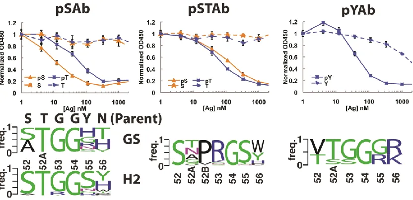
Design of PS Ab scaffolds ... 57
Characterization of PS Ab scaffolds ... 59
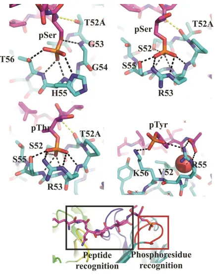
Structural analysis of phosphopeptide recognition ... 60
Generation of novel PS Abs using the pSer and pSer/pThr scaffolds ... 61
3.4 Discussion ... 62
3.5 Acknowledgements ... 64
3.6 Methods ... 65
Vector construction ... 65
Generation of Phage Libraries ... 65
Phage Display Selections, ELISAs, and Western blots ... 66
Protein expression and purification ... 66
Biacore analysis ... 67
Crystalization of peptide:Fab complexes ... 67
Accession codes ... 69
3.7 Figures ... 69
3.8 Supplementary Figures ... 78
3.9 References ... 90
CHAPTER 4 USING DESIGNABILITY TO DESIGN A PROTEIN BINDER TO
HEMAGGLUTININ ... 95
4.1 Introduction ... 95
4.2 Results ... 97
xi
Identifying peptide scaffolds for a hemagglutinin binder ... 99
Peptide synthesis of designed binders... 100
Phage display of peptide binders ... 101
Affinity maturation using phage libraries ... 104
Placing helical designs onto protein scaffolds ... 106
4.3 Conclusions ... 107
4.4 Materials and Methods ... 109
Initial scaffold search ... 109
Peptide synthesis ... 111
Addition of cross-linkers ... 112
Peptide purification ... 112
Circular dichroism spectroscopy ... 112
Bio-layer interferometry ... 113
Creation of phage display constructs ... 113
Creation of phage libraries ... 113
Phage selection procedures ... 114
ELISA assays ... 115
4.5 Acknowledgements ... 116
4.6 Figures ... 117
4.7 References ... 134
CHAPTER 5 SUPER CODONS: CREATING OPTIMAL SETS OF NUCLEOTIDE
MIXTURES FOR USE IN GENE LIBRARY PRODUCTION ... 137
5.1 Abstract ... 137
5.2 Introduction ... 138
5.3 Results and Discussion ... 141
Development of target amino acid distributions ... 141
Creating libraries with derived target distributions ... 145
Extending Super Codons to target alternative distributions... 151
BLOSUM-based distributions ... 151
Antibody mutagenesis ... 151
Custom distributions and multiple-alignments... 152
5.4 Conclusions ... 152
5.5 Methods ... 154
Target distribution derivation ... 154
Testing significance of correlations ... 156
5.6 Figures ... 157
xii
5.8 References ... 168
CHAPTER 6 A REAL-TIME ALL-ATOM STRUCTURAL SEARCH ENGINE FOR
PROTEINS ... 170
6.1 Abstract ... 170
6.2 Introduction ... 171
6.3 Design and Implementation ... 173
Overview ... 173
Forward Index ... 173
Structural words... 174
Database ... 175
Alignment and RMSD ... 176
Streaming results ... 176
Data set ... 177
6.4 Results ... 177
Building motifs ... 177
Discovering motifs ... 178
Assembling larger fragments ... 179
Connecting hot-spot residues ... 179
6.5 Availability and Future Directions ... 180
6.6 Figures ... 182
6.7 Supplementary Figures ... 187
6.8 References ... 192
CHAPTER 7 CONCLUSIONS AND DISCUSSION ... 194
7.1 Conclusions and discussion ... 194
7.2 Figures ... 198
xiii
LIST OF TABLES
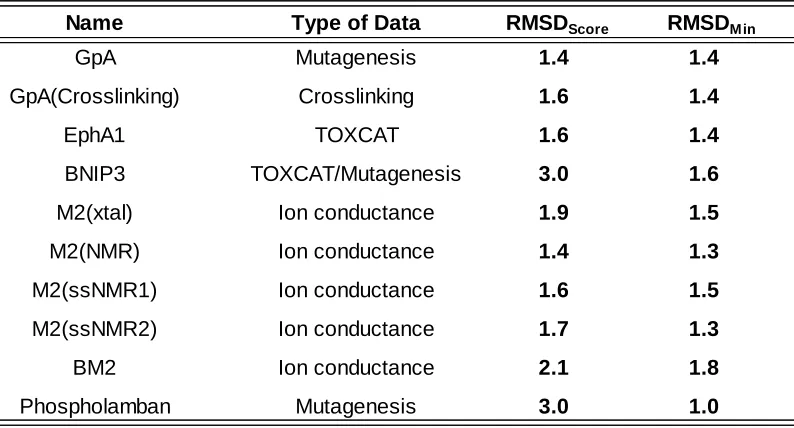
2.1 Application of the sampling method using inter-helical Cβ distances
from experimentally determined helical TM structures 32
2.2 Application of the sampling method using low-resolution experimental data 32
S2.1 Hide-and-seek test using inter-subunit Cβ distance as simulated experimental
Data 42
S2.2 GpA disruption data 43
S2.3 GpA disulfide cross-linking data 44
S2.4 Pentamer disruption data 45
S2.5 EphA1 TOXR data 46
S2.6 BNIP3 unified mutagenesis score values 47
S2.7 M2 perturbility index (PI) data 48
S2.8 BM2 perturbility index (PI) data 49
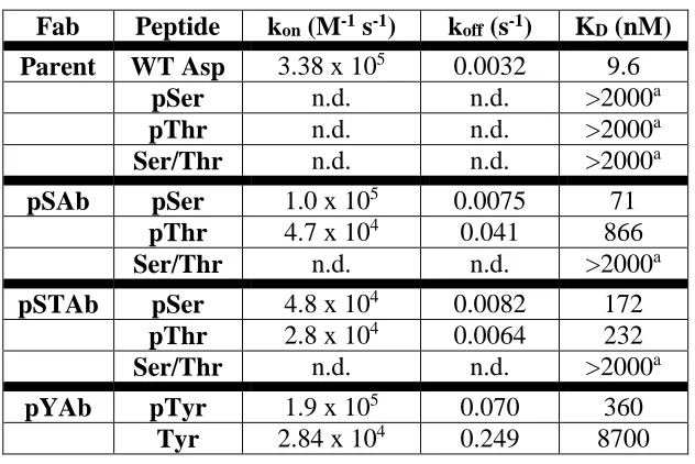
3.1 Affinity measurements of Ab scaffolds 69
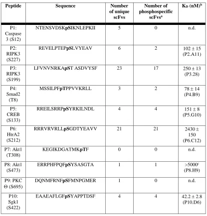
3.2 Summary of scFv hits versus ten new phophopeptide targets 70
S3.1 Functional description of H2 loop residues 78
xiv
S3.3 Crystallization and cryoprotection conditions for Fab complexes 79
S3.4 Data collection and refinement statistics (molecular replacement) 80-81
4.1 A list of the five hemagglutinin binder designs 117
5.1 Common degenerate codons and a brief description 157
5.2 Optimal Super Nucleotide mixtures found to fit our 20 target amino acid
distributions 157
5.3 Summary of realized amino acid distributions 158
S5.1 The twenty target distributions we propose 164
S5.2 The amino acid distributions which best match the desired distributions
given in Supplemental Table 1 as found by our algorithm 165
S5.3 The best calculated distributions when each distribution was treated
separately 166
S5.4 Natural abundance of each amino acid in the complete
UniProt/Swiss-Prot database 167
S6.1 Default Motif Set 187-188
xv
LIST OF ILLUSTRATIONS
2.1 Helix sampling scheme 33
2.2 RMSD Distributions using native inter-subunit Cβ distances. 35
2.3 PI versus inter-subunit Cβ distance profiles and superimposition of
best-model and NMR structure 36
2.4 RMSD distributions for models generated using low-resolution
experimental data. 37
2.5 A comparison between the backbone of the native structure and best
scoring model after refinement 39
2.6 Energy profiles versus RMSD to native after refinement with XPLOR-NIH. 40
S2.1 Inter-subunit Cβ distance versus percentage pentamer formation for
native phospholamban 50
S2.2 Structural heterogeneity between different structures of M2 51
3.1 Design of phospho-specific Ab scaffold 71
3.2 Selection and characterization of pSer-, pSer/pThr-, and pTyr-specific
scaffolds 73
xvi
3.4 Generation of novel recombinant phospho-specific (PS) Abs using the
pSAb and pSTAb scaffolds 76
S3.1 Structure of nest motif in non-antibody and antibody scaffolds 82
S3.2 Biacore traces of phospho-specific Fabs binding to phosphorylated peptides. 83
S3.3 Density maps of Fab structures 84
S3.4 Structural comparison between the mouse and humanized Fab
and the bound and unbound Fab 85
S3.5 Electrostatic surface representations of parent Fab, pSAb, pSTAb, and pYAb 86
S3.6 Comparison between the natural PS chicken scFv and designed pSTAb
structures 87
S3.7 Phosphoresidue-binding pocket from natural phosphopeptide-binding
domains 88
4.1 An overview of MaDCaT-based scaffold search 118
4.2 An overview of the helix-dimer scaffold search 120
4.3 Our five initial designs for peptide binders to hemagglutinin 121
4.4 Circular Dichroism spectra of four of our synthesized peptides with
xvii
4.5 Circular dichroism of our three peptides without chemical cross-linkers 125
4.6 Bio-layer interferometry data examining binding between our helical
peptide designs and hemagglutinin 126
4.7 ELISA results for our initial helix designs placed in phage 128
4.8 ELISA results obtained for designs bth_1 and bth_2 as well as phage
displaying the wild-type pVIII gene 129
4.9 Diversification of initial peptide designs for phage display 130
4.10 ELISA results after one, two, and three rounds of selection against
hemagglutinin. 131
4.11 Threading protein scaffolds onto helical peptide designs 132
4.12 Gene layout for protein scaffold designs in pVIII gene 133
5.1 Theoretical amino acid distributions of six commonly used degenerate
codons 159
5.2 The expected number of mutations from an initial sequence when
randomizing 10 positions 160
5.3 The twenty target and delivered amino acid distributions 151-162
xviii
degenerate codon and Super Codons 162
5.5 Example of Super Codons used for antibody design 163
6.1 Subdivision of protein structures 182
6.2 Incremental assembly of a motif 183
6.3 Identifying alternative “nest”-like motifs 184
6.4 Building a tertiary interaction 185
6.5 Finding backbones compatible with hot spot residues 186
1
Chapter 1
1.1 Introduction
Knowledge of protein structure is essential in our quest to understand the complex
signaling and interaction networks that make life possible. The past two decades have seen an
explosion in the number of protein structures solved to atomic level, with only around 500 such
structures deposited in the Protein Data Bank (PDB) in 1990 to nearly 100,000 today (1). This
amazing growth belies the fact that not all categories of proteins have been solved with equal
success. In particular, membrane proteins make up fewer than 2% of solved structures in the
PDB (2) despite comprising an estimated 25% of the human proteome (3). This discrepancy is
largely due to the difficulty in obtaining atomic level data for proteins embedded in their native
lipid environment. To compensate for this difficulty, researchers have developed a number of
experimental techniques to obtain low-resolution data on membrane protein structure,
including the TOXCAT assay (4), the ToxR assay (5), reversal potential assay (6), cysteine
cross-linking (7), and scanning mutagenesis studies (8). The data obtained from these experiments is
extremely sparse, typically on the order of a single data point per residue. Despite this paucity,
in chapter 2, we describe a computational approach which uses this sparse data to infer the
structure of homo-oligomeric, transmembrane proteins to an accuracy rivaling that of X-ray
crystallography and NMR.
The wealth of protein structural data that is now available carries with it the potential to
inform efforts in designing novel protein-protein interactions. Through extensive mining of
protein structures, countless groups have identified and categorized structural motifs that are
frequently involved in various protein interactions, from the GxxxG motif involved in
2
binding proteins (10). In chapter 3, we leverage this data to engineer an antibody which binds
to novel phosphorylated peptides. Whereas, traditionally, phospho-specific antibodies have
been generated through immunization (11), we developed a rational, structure-based approach
paired with high-throughput screening. We exploited a previously identified structural motif
called a “nest” (12) which forms a cationic hole and reasoned that the phosphate group of a
phosphorylated peptide might be a perfect anion to fill such a hole. Scanning through the set of
solved antibody/peptide structures, we found an example of an antibody in which one of the
complementarity determining regions (CDR) involved in peptide binding formed a perfect nest.
In this structure, the nest was involved in an interaction with the carboxyl group of an aspartic
acid residue on the peptide. Using a technique called phage display (13), we selected mutants
based on the original antibody that preferentially bound peptides whose aspartic acid was
replaced with a phosphorylated residue (serine, threonine, or
phospho-tyrosine.) More impressively, we were then able to use this modified, phospho-specific motif as
a “phosphate-binder” module and isolated 51 phospho-specific antibodies against seven
different phosphorylated peptides unrelated to the original peptide. This technique offers the
promise of a much more efficient means of generating antibodies to recognize specific
post-translationally modified peptides and highlights the power of leveraging our knowledge of
known structural binding motifs to engineer new interactions.
In chapter 4 we extend the idea of mining structural data for protein interacting motifs
in order to design a binder to the influenza fusion protein hemagglutinin. A recent report details
the successful design of two protein binders to hemagglutinin with nanomolar affinity (14). In
this work, the designers first docked individual amino acids to a solved structure of
3
overall binding energy. Once two or three of such residues were identified, the authors
searched through a database of small, easily expressible proteins which they could use as
scaffolds to hold these hot-spot residues. These proteins were in turn redesigned to
accommodate the “hot-spot” residues and provide a level of complementarity to the
hemagglutinin molecule. Eighty-eight designs were expressed and tested for binding activity.
Two designs were found to bind, and after a round of affinity maturation, both designs
produced variants with dissociation constants in the single-digit nanomolar range. We
hypothesize that an approach which explicitly attempts to create binding interfaces that mimic
those found frequently in nature would have a higher success rate. To that end, we developed a
design methodology based upon the concept of “designability” (15), the idea that out of the vast
ensemble of possible packing arrangements of protein secondary structure, only a limited
subset is ever observed, and some of those arrangements can accommodate a wide variety of
amino acid sequences. We used computational techniques to search a non-redundant database
of known protein packing arrangements to identify designable motifs which were a good match
to our hemagglutinin epitope of interest. These designable motifs provide a scaffold upon
which we can then computationally design amino acids to drive binding to hemagglutinin. We
apply our approach to design short helical peptides to bind to hemagglutinin, experimentally
characterize their behavior, and attempt to isolate modified versions with enhanced binding
characteristics through phage display. Although our first-generation peptide designs do not
bind at levels detectable by our assays, we are able to draw valuable conclusions regarding the
difficulty of designing peptide binders and propose a modified protocol to provide a protein
4
As shown in chapters 3 and 4, while the use of large sets of structural data can help in
the design of novel protein interactions, to date our computational models are not robust
enough to produce high-affinity designs directly. Typically, designs with lower binding affinity
are produced and then used as starting points for affinity maturation in gene library
experiments such as phage display. In these experiments, a library is produced by introducing
mutations to the initial design, and variants are screened for enhanced binding activity through
high-throughput assays. One popular method of introducing mutations is through the use of
degenerate codons (16), essentially mixtures of nucleic acids which form defined amino acid
distributions. However, while widely used, traditional degenerate codons have a number of
shortcomings. First, many experiments will mutate more residues than can be exhaustively
sampled in the library. Consequently, shaping what regions of sequence space are explored by
the library can dramatically affect how likely it is that improved designs will be found.
Traditional degenerate codons sample space without regards to what amino acid was present in
the initial, albeit weak, design. A more efficient approach would sample the original amino acid
with a higher frequency, as it is already known that that amino acid is at least compatible with
binding. Second, sequencing projects have provided an incredible source of data on the amino
acid transition probabilities seen during evolution. It would be useful to use these probabilities
to direct the mutations introduced in gene libraries rather than settle for an entirely random
approach. Finally, many degenerate codons introduce stop codons at significant levels. Any
decrease in the probability of introducing a stop codon has the potential to greatly increase the
effective size of the gene library, and thus significantly improve the likelihood of finding
improved designs. In chapter 5 we describe the development of a novel algorithm that
5
deficiencies. The use of these mixtures in gene library experiments should focus the sampling of
sequence space to regions closer to the original design, increasing the percentage of mutants
that still fold properly and retain some of the initial binding interactions. Additionally, sequence
space will be explored more intelligently by explicitly making use of our knowledge of amino
acid substitution rates seen through evolution and by decreasing the introduction of stop
codons compared to the most popular degenerate codon. Gene libraries offer the incredible
potential to find enhanced binders from a pool of billions, but will only be successful if enhanced
binders make it into the pool in the first place. By intelligently directing how the pool samples
sequence space around the initial design, we will greatly improve our chances of success.
Finally, one of the difficulties that comes with an incredible abundance of data is how to
efficiently search it and retrieve only that information which is relevant to the query of interest.
Imagine the internet without Google. A vast sea of data would be present with almost every
fact known to humankind, and yet there would be no practical way to make sense of it or to
search through it. The utility of the internet would be dramatically curtailed. In many ways, this
is the present situation regarding the immense store of protein structural data we have
amassed. As shown in chapters 3 and 4 we are able to mine structural data to improve our
efforts in protein design, but the search tools currently available have severe limitations. First,
the search algorithms focus only on the backbone structure of proteins, while neglecting the
details of side-chain interactions. As it is the side-chains which are typically involved in protein
packing, binding interactions, and catalysis, this lack of searchability is a significant oversight.
Secondly, the structural search tools developed to date lack the ability to return results in
real-time, severely impeding the natural search-design-repeat feedback cycle and slowing design
6
structural search algorithm called Suns. Suns uses the popular molecular-visualization package
PyMOL (17) to allow the user to select structural motifs to use as a query to our database of
non-redundant protein structures, and immediately returns results matching the given query to
an arbitrary tolerance. We show how Suns can be used to design novel protein structures, find
scaffolds which can accommodate given side-chain motifs, and quickly discover secondary
structures which would be good candidate scaffolds for the hemagglutinin hot-spot residues
discussed in chapter 4. With its near-instantaneous search capability, Suns promises to
dramatically increase the utility of the structural data available, and open up new avenues for
assessing a protein structure’s designability.
1.2 References
1. Yearly Growth of Total Structures. RCSB PDB. [Online] [Cited: 11 6, 2013.]
http://www.rcsb.org/pdb/statistics/contentGrowthChart.do?content=total&seqid=100. 2. Membrane Proteins of Known 3D Structure. [Online] [Cited: 11 5, 2013.]
http://blanco.biomol.uci.edu/mpstruc/.
3. Mapping the human membrane proteome: a majority of the human membrane proteins can be classified according to function and evolutionary origin. Almén, M.S., et al. 50: BMC Biology, 2009, Vol. 7. doi:10.1186/1741-7007-7-50
4. TOXCAT: A measure of transmembrane helix association in a biological membrane. Russ, W.P. and Engelman, D.M. 3: Proceedings of the National Academy of Sciences, 1999, Vol. 96. pp 863-868.
5. Computer simulations and modeling-assisted ToxR screening in deciphering 3D structures of transmembrane α-helical dimers: ephrin receptor A1. Volynsky, P.E., et al. s.l. : Physical Biology, 2010, Vol. 7. doi: 10.1088/1478-3975/7/1/016014
6. A functionally defined model for the M2 proton channel of influenza A virus suggests a mechanism for its ion selectivity. Pinto, L.H., et al. 21: Proceedings of the National Academy of Sciences, 1997, Vol. 94. pp 11301-11306.
7. Detecting the conformational change of transmembrane signaling in a bacterial chemoreceptor by measuring effects on disulfide cross-linking in vivo. Hughson, A.G. and
7
8. Combinatorial alanine-scanning. Morrison, K.L. and Weiss, G.A. 3: Current Opinion in Chemical Biology, 2001, Vol. 5. pp 302-307.
9. The GxxxG motif: a framework for transmembrane helix-helix association. Russ, W.P. and Engelman, D.M. 3: Journal of Molecular Biology, 2000, Vol. 296. pp 911-919.
10. The leucine zipper: a hypothetical structure common to a new class of DNA binding proteins.
Landschulz, W.H., Johnson, P.F. and McKnight, S.L. 4860: Science, 1988, Vol. 240. pp 1759-1764.
11. Overview of the generation, validation, and application of phosphosite-specific antibodies.
Brumbaugh, K., et al. s.l. : Methods in Molecular Biology, 2011, Vol. 717. pp 3-43.
12. A novel main-chain anion-binding site in proteins: the nest. A particular combination of φ,ψ values in successive residues gives rise to anion-binding sites that occur commonly and are found often at functionally important regions. Watson, J.D. and Milner-White, J. 2: Journal of
Molecular Biology, 2002, Vol. 315. pp 171-182.
13. Filamentous fusion phage: novel expression vectors that display cloned antigens on the virion surface. Smith, G.P. 4705: Science, 1985, Vol. 228. pp 1315-1317.
14. Computational Design of Proteins Targeting the Conserved Stem Region of Influenza Hemagglutinin.Fleishman, S.J., et al. 6031: Science, 2011, Vol. 332. pp 816-821.
15. The designability of protein structures. Helling, R., et al. : Journal of Molecular Graphics and Modelling, 2001, Vol. 19. pp 157-167.
16.Nomenclature for incompletely specified bases in nucleic acid sequences: recommendations 1984. Cornish-Bowden, A. 9: Nucleic Acids Research, 1985, Vol. 13. pp 3021-3030.
8
Chapter 2
A photon-free approach to transmembrane protein structure determination
2.1 Abstract
The structures of membrane proteins are generally solved using samples dissolved in micelles,
bicelles, or occasionally phospholipid bilayers using X-ray diffraction or magnetic resonance.
Because these are less than perfect mimics of true biological membranes, the structures are
often confirmed by evaluating the effects of mutations on the properties of the protein in their
native cellular environments. Low-resolution structures are also sometimes generated from the
results of site-directed mutagenesis when other structural data are incomplete or not available.
Here we describe a rapid and automated approach to determine structures from data on
site-directed mutants for the special case of homo-oligomeric helical bundles. The method uses as
input an experimental profile of the effects of mutations on some property of the protein. This
profile is then interpreted by assuming that positions that have large effects on
structure/function when mutated project towards the center of the oligomeric bundle. Model
bundles are generated and correlation analysis is used to score which structures have
inter-subunit Cβ distances between adjoining monomers that best correlate with the experimental
profile. These structures are then clustered and refined using energy-based minimization
methods. For a set of 10 homo-oligomeric TM protein structures ranging from dimers to
pentamers, we show that our method predicts structures to within 1 to 2 Å backbone RMSD
relative to X-ray and NMR structures. This level of agreement approaches the precision of NMR
structures solved in different membrane mimetics.
9
2.2 Introduction
Helical transmembrane protein structure determination represents a significant
challenge. Fewer than 2% of all experimentally determined structures deposited in the Protein
Data Bank (1) (PDB) are membrane proteins, yet 20-25% of open reading frame (ORFs) from
recently sequenced genomes encode for proteins that embed in the membrane(2)(3). Even with
advances in conventional methods for protein structure determination such as X-ray
crystallography and NMR spectroscopy, the fundamental problems of obtaining
diffraction-quality crystals, protein expression and purification, and protein-size limitations still remain.
Computational methods for modeling transmembrane protein structure are becoming
increasingly more important if we hope to decrease the discrepancy in structural information
between globular and membrane proteins.
Depending on the scientific question being asked, the laborious (and sometimes
insurmountable) task of experimentally determining the structure of a membrane protein using
conventional methods may not be necessary. For example, Zhu et al.(4) recently used disulfide
crosslinking information to build models for the helical transmembrane (TM) dimers glycophorin
A (GpA) and integrin αIIβ3. The resulting models for GpA had a root mean square distance
(RMSD) over the backbone atoms of 1 to 1.5 Å with the NMR structures. Metcalf et al.(5) used
mutagenesis data and protein sequence variation to build models for the TM homo dimers GpA
and BNIP3 apoptosis factor. The RMSD for the GpA model was 1.3 Å. We hypothesize that other
forms of low-resolution experimental data can potentially provide sufficient information to
accurately model other transmembrane protein structures. Experimental data from a variety of
10
The earliest structural modelsby Brünger and coworkersfor the TM region of GpA were
based solely on the energetics of interaction between helices (6)(7). The resulting models were
compared against mutagenesis data showing the disruptive effects that non-polar mutations
had on GpA’s ability to dimerize (8). The structural models agreed with the mutagenesis data
and showed that key residues oriented toward the helical interface were sensitive to nonpolar
mutations.
The approach of modeling helical TM regions using the energetics of interaction
between helices has been extended to larger oligomers. Phospholamban is a TM
homo-pentamer that is important in calcium storage and release in cardiocytes. Mutagenesis studies
(7)(9) showed that mutations of key hydrophobic residues disrupted pentamer oligomerization.
A global search of conformational space revealed five low-energy helical bundles (7), only one of
which was found to be in agreement with an extensive set of mutagenesis data (7)(9), and
ultimately the experimentally determined structures (10)(11). This five-fold symmetrical
structure has a left-handed twist; most critical residues lie at the helix/helix interface and show
large interaction energies. Interestingly, the lowest energy conformer did not agree with the
experimental results, indicating that energy is a necessary -- but insufficient -- criterion for
assessing models.
Herezyk and Hubbard (12) used a different approach to model helical TM
homo-olgimers. Using a combination of Monte Carlo/Simulated Annealing (MCSA) and Molecular
Dynamics/Simulated Annealing (MDSA) along with a set of orientational restraints derived from
published mutagenesis data (7)(9), Heryzk and Hubbard constructed models for GpA and
phospholamban. Unlike the modeling approach of Brünger and coworkers, which made use of
11
derived from mutagenesis data in their modeling procedure. The resulting model for GpA had an
RMSD to native of 0.9 Å over the backbone atoms. A comparison of the profile between
interaction energy and mutagenesis data revealed an excellent level of agreement for
phospholamban.
More recent approaches for modeling helix TM homo-oligomers fall into one of these
two categories: modeling methods based purely on energetics (13)(14)(15)(16) and those that
use some combination of energetics and low-resolution experimental data (4)(5)(17)(18). The
incorporation of experimental data directly into the modeling process provides two obvious
benefits. First, the experimental data corrects for inaccuracies in the force field and for
approximations regarding the environmental conditions. Second, by using experimental data
directly in the modeling process, the conformational space that needs to be sampled can be
greatly reduced.
We have developed a novel approach for modeling helical TM homo-oligomers that
incorporates a variety of low-resolution mutagenesis data directly into the modeling process.
Our modeling approach consists of two phases. In the first phase we use a symmetric rigid-body
search to generate an ensemble of models that is consistent with a given set of low-resolution
data. In the second phase we cluster and then refine only the centroid models using the
CHARMM22 force field. At the heart of our rigid-body search is a simple scoring function that
restrains the conformational search by maximizing the correlation between inter-subunit Cβ
distance and experimental data while minimizing steric clashes between helices. Our
correlation term allows us to use a variety of low-resolution mutagenesis data without the need
for scaling the data or converting the data into distance restraints4 or angular restraints (12).
12
experimental data such as mutagenesis, ToxR, TOXCAT, ion channel and crosslinking data to
model the TM regions of GpA, phospholamban, M2, BM2, BNIP3 and the ephrin receptor
tyrosine kinase (EphA1). The final models ranged in RMSD from 0.6 Å to 2.1 Å when compared
to the native structures. This approach to modeling helical TM protein structure can be of
enormous benefit when conventional methods of protein structure determination fall short.
2.3 Results
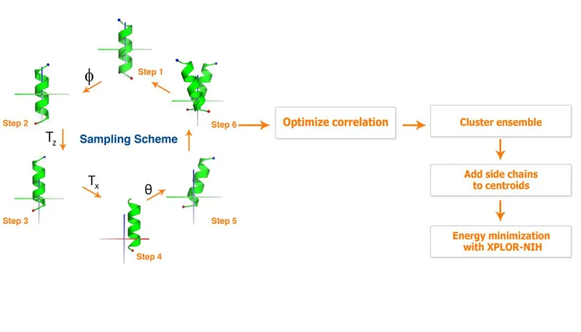
Overview of modeling protocol
Our modeling protocol can be broken down into two phases. The first phase involves
rigid-body sampling using an ideal or experimentally-determined helix. The second phase
involves side chain placement, clustering and refinement of the models with a molecular
mechanics force field. We briefly describe the first phase here. A detailed description of the
second phase can be found in the Methods. Rigid-body sampling (RBS) begins with a helix that is
transformed to the global frame of reference so that the axis of the helix is coincident with the
global Z-axis and its geometric center is at the origin.Four degrees of freedom are required to
define the relationship between monomers in a structure with exact rotational symmetry. Here
we apply two rotations and two translations to define the location of the helix in the unit cell.
The individual steps in our modeling protocol are illustrated in Figure 1.
At the heart of our RBS method is the use of the correlation coefficient (r) to evaluate
the degree to which experimental data correlates with the projection of the side-chains in the
oligomer, as defined by the inter-subunit contact distance for each residue in the structure. The
inter-subunit contact distance is defined here as the distance between Cβ atoms on identical
residues of a homo-dimer, and this provides a quantitative measure that can be correlated with
13
correlation coefficient is a measure of the linear relationship between two variables and ranges
from a value of 1 for two perfectly correlated variables to a value of -1 for two perfectly
anti-correlated variables. The correlation coefficient is used to restrain the RBS protocol by
incorporating it directly into a scoring function that is used to optimize each pose (see
Methods). In this study, we correlate inter-subunit Cβ distance with the degree of experimental
perturbation associated with mutations or the extent of cross-linking in a Cys-scanning
experiment to determine how well a given hypothetical model agrees with experimental data.
The extent of Cys crosslinking and the perturbational effects of mutations generally increase
with decreasing inter-subunit distance (negative correlation). However, for simplicity, we refer
to all correlations as positive for structures that are in agreement with the expected
experimental outcome.
We demonstrate the utility of our RBS protocol by using it in three tests. In the first test
we use it to search for a set of idealized helical conformations using native inter-subunit Cβ
distances as “experimental data.” The second test is similar to the first test but uses a set of
nine symmetric helical TM structures obtained from the PDB instead of idealized helical
arrangements. It should be noted that the first and second tests are used to determine how
well our search strategy works under the most ideal conditions (i.e., where experimental data
correlates perfectly with inter-subunit Cβ distances). In the third and final test, we model these
same nine structures using low resolution experimental data to restrain the search. The
resulting ensembles of models from this test are clustered using a k-medoid clustering algorithm
(19). Side chains are then added to each of the centroid models using SCAP (20) followed by
all-atom refinement using the CHARMM22 force field implemented in the XPLOR-NIH package
14
Idealized hide-and-seek test
To test the RBS protocol, we constructed a set of ten helix dimer conformations by
randomly choosing values for the four search parameters (Tx, Tz, θ, and φ). Each set of four
parameters is then used to position a 16 residue ideal poly-alanine helix in space. The symmetry
mate is generated by rotating a copy of the helix 180° about the global Z axis. After construction
of the ten dimers, we determined the inter-subunit Cβ distances along the length of the helices.
These distances were used as simulated experimental data to restrain the rigid-body search with
the goal of recapitulating the original dimer conformation. In all ten cases, the simple scoring
function selects a model with an RMSD of 0.6 Å or less to the starting conformation (see
Supplementary Table 1).
Hide-and-seek test using TM structures from the PDB
The RBS protocol can generate the native pose with high accuracy for idealized cases. A
more challenging test would entail modeling actual helical structures from the PDB which may
not contain idealized geometry. We repeated the hide-and-seek test on a set of nine symmetric
helical TM structures from the PDB. Three of these structures are dimers, five are tetramers and
one is a pentamer. For each test case, we determined the inter-subunit Cβ distances from the
first two chains of the native structure. If a glycine is present along the protein sequence we
computed the distance between Cα atoms. For structures solved using NMR, we use the
average structure (see Methods) to obtain the native distances.
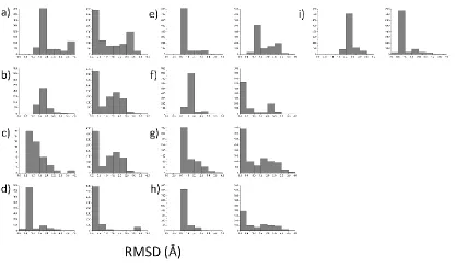
We use three separate measures of RMSD in assessing the performance of the RBS
protocol on experimentally determined structures. The first measure, RMSDScore, denotes the
RMSD between the best scoring model in the ensemble and the native structure. The second
15
denotes the RMSD of the best scoring model when the native helix is used in place of the ideal
helix in the rigid-body search. As shown in Table 1, all nine cases have a RMSDScore of 2.9 Å or
less. The dimer BNIP3 gives the best results with a RMSDScore of 0.9 Å. The worst performing
case, phospholamban, gives a RMSDScore of 2.9 Å. The remaining cases yield RMSDScore values
between 1.2 and 2.0 Å. While our scoring function does not select the lowest RMSD model in
the ensemble, it does perform reasonably well at generating low RMSD models (Figure 2). With
the exception of the BM2 case, a sizable population of models with RMSDs below 1.5 Å is always
generated. Producing an ensemble of models with relatively low RMSD to native is critical for
two reasons. First, models that are near-native will generally yield more favorable scores in the
refinement stage. Second, clustering will be more effective at assigning near-native models as
centroids.
We suspected that our sampling algorithm could generate a larger population of
near-native models if we introduced natural curvature into the starting helix. Superimposing an ideal
helix onto the corresponding native helix gives an RMSD that is larger than 1.0 Å for GpA, BM2,
and all of the M2 structures. To better assess how this deviation from ideality influences the
final result, we carried out the same search using the native helix in place of the ideal helix. The
resulting RMSDNative values are 0.6 Å or less for all cases with the exception of the M2(xtal) case
(Table 1). However, we note the existence of models with RMSDNative values of 0.6 Å or less for
all of the ensembles generated using a native helix (Figure 2).
Restrained sampling using low resolution experimental data
The first two tests show that when sufficient information between monomers is given in
the form of native distances, our RBS protocol can generate models with RMSDMin values
16
unavailable. Therefore, to assess the ability of the sampling protocol to perform similarly in a
practical situation, we used low-resolution experimental data to restrain the search. Besides
being the most stringent test thus far, given the inherent noise present in low-resolution
experimental data, this test will provide a meaningful benchmark in terms of the practicality of
our method. A description of the low-resolution experimental data is provided in the Methods
section.
Before carrying out the search, we wanted to test our hypothesis that inter-subunit Cβ
distance correlates with low-resolution experimental data. To do this we determined the
correlation coefficient and the associated p values between the inter-subunit Cβ distance data
obtained from each native structure and the corresponding set of experimental data (see
Supplementary Tables 2-8 for the experimental data). Phospholamban has the strongest
correlation with |r|=0.91 (p=4.6E-7). The dimer GpA has roughly the same |r| value of 0.78
(with an approximate p value of 5.0-E-6) for both the crosslinking and mutagenesis data. The
dimer EphA1 has |r|= 0.76 (p=3.2E-3). The M2 cases have roughly the same |r|=0.72 (with a p
value of about 3.7E-4). BNIP3 and BM2 have the weakest correlations with |r|=0.44 (p=5.7E-2)
and |r|=0.58 (p=4.7E-3) respectively. For all but one of the cases, the p-value for the correlation
between experimental data and inter-subunit Cβ distance is less than 0.05, indicating that the
correlation is unlikely due to chance. Based on the |r| values and associated p-values obtained
for the native structures, it would seem that correlating inter-subunit Cβ distance with
low-resolution experimental data can provide a useful filter when modeling TM homo oligomers
(Figure 3).
Using the low-resolution experimental data to generate TM bundles, we obtainedan
17
largest outliers with RMSDScore values of 3.0 Å. For BNIP3 it is not surprising that the RMSDScore is
so large given the weak correlation between inter-subunit Cβ distance and the experimental
data. For phospholamban, we noticed that the bundle radius for the top scoring model is about
1.0 Å smaller than in the native structure. A more important measure of performance of the
sampling protocol is how close to native conformation our sampling can reach. Clearly, if the
sampling protocol cannot generate a sufficient number of models that are close to native, it is
likely that all-atom refinement will be of little value in generating good models. The RMSDMin
value is 1.6 Å or less for 9 out of 10 cases (Table 2). With the exception of BM2, the sampling
protocol generates ensembles with a significant fraction of models less than 2.0 Å RMSD to
native (Figure 4). Based on these results it appears that when inter-subunit Cβ distance data
correlates strongly with mutational data, rigid-body sampling alone can be used to generate
reasonable starting conformations that can be further refined. However, since our scoring
function is designed as a filter it may not select the most energetically favorable conformation in
the ensemble of models. For this, we use a more detailed all-atom scoring function.
Refinement using XPLOR-NIH
The resolution of our simple scoring function does not capture detailed energetic
interactions such as van der Waals packing and Coulombic interactions. These interactions are
important for obtaining optimal packing between helices. To capture these important
interactions, we first cluster the ensemble of models generated using our RBS protocol, add side
chains to all centroids and then subject them to all-atom refinement using the CHARMM22 force
field in XPLOR-NIH (21). The most favorable scoring model according to XPLOR is deemed our
18
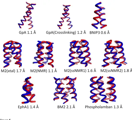
Refinement of the centroids gives an RMSDScore of 2.1 Å or better for all ten cases (Figure
5). For the dimers GpA, GpA(Crosslinking), EphA1 and BNIP3, the RMSDScore is 1.4 Å or less.
Results for larger homo oligomeric states are equally as impressive with RMSDScore ranging in
value from 1.1-2.1 Å. Given the spread in RMSD values between individual models in the native
NMR ensembles, which can be as large as 0.9 Å for some of the structures considered here, our
results would indicate that the RBS protocol coupled to clustering and refinement with
XPLOR-NIH has the potential to generate models comparable in accuracy to those obtained using
medium-resolution NMR. The importance of using a detailed all-atom scoring function is clearly
illustrated for the case of BNIP3. Using our simple scoring function to select a model from the
ensemble will give an RMSD to native of 3.0 Å. If we refine all of the models in the ensemble and
then select the most favorable scoring model according to CHARMM22, we obtain an RMSD to
native of 0.6 Å. Clearly, refining the entire ensemble of 1,000 models would be a time
consuming task and so we cluster the ensemble of models first and then refine only the
centroids. Using this approach, we also obtain a model with an RMSD to native of 0.6 Å but do
so in a fraction of the time it would take to refine the entire ensemble of models. The r value
between the experimental data and the inter-subunit Cβ distance for the refined models either
remained the same or improved when compared with the corresponding value for native.
As a control, we applied the same XPLOR-NIH refinement protocol to all the native
structures. This involved refinement of all the individual models in each NMR ensemble and not
the average model. We expect the experimentally determined structures after refinement to
have scores that are similar to or more favorable than the scores of our centroid models. We
observe this trend for all cases with exception of BM2 (Figure 6). We find that the refined native
19
This seems to imply that the native BM2 bundle may not be tightly packed which ultimately
leads to a less favorable van der Waals score. For most cases, refinement with XPLOR-NIH does
not significantly perturb the native structure. The RMSD between the unrefined and refined
native models is on average less than 1.0 Å (represented as blue circles Figure 6). For
phospholamban and BM2, refinement perturbs the native conformation to a larger extent. In
particular, the RMSD after refinement of the native BM2 ensemble resulted in two models
having RMSDs larger than 1.7 Å.
2.4 Discussion
We have presented a method for modeling helical TM homo-oligomers that uses a
rotationally symmetric rigid-body search followed by clustering and energy refinement using the
CHARMM22 force field in XPLOR-NIH. At the heart of our modeling procedure is a simple
scoring function composed of a VDW clash term and a correlation coefficient between
mutational data and inter-subunit Cβ distance. The simple scoring function is optimized to
obtain maximal agreement with experimental data while avoiding clashes between helices. The
novelty of our method is in its ability to directly restrain the search using low-resolution
experimental data. This prevents the search from needlessly meandering through space and
focuses the sampling to give the best agreement with experimental data.
Our method performs best when the experimental data correlate with |r| > 0.5 with the
native inter-subunit Cβ distance. In these cases, the rigid-body search does a reasonable job at
generating near-native backbone conformations. As the correlation becomes weaker, so does
the structural similarity between the native structure and the best scoring model. The
20
improves the RMSD value to native. In seven of the ten cases, the RMSD to native is 1.6 Å or
less.
Modeling TM homo-dimers
As a prerequisite for addressing the general TM homo-oligomer problem, we first
applied our modeling approach to the homo-dimer GpA using two sets of mutational data. One
set of data is from a fairly recent study and is comprised of crosslinking efficiency (4). Another
data set consists of dimer disruption data and has been used extensively by others to propose
different methods for modeling the TM region of GpA (5)(12)(13)(14). Using either a
combination of energetics and restraints derived from mutational data or using energetics
alone, all of these methods generate models for the TM region of GpA with RMSDs to native in
the range of 0.7-1.5 Å. Using either set of low-resolution experimental data, our modeling
approach achieves a similar level of accuracy for GpA.
Earlier work in our group made use of a Monte Carlo-simulated annealing (MCSA)
protocolto propose a model for the TM region of BNIP3 (5). The MCSA method used two energy
terms that would penalize both neutral and disruptive mutations. The method we propose here
is different in two ways. First, we do not use a stochastic approach for sampling conformational
space. Second, the present method does not rely solely on the energy to decide on the
plausibility of a model, but instead also relies on how well the inter-subunit Cβ distance
correlates with mutational data. While both methods manage to accurately model the
backbone of BNIP3, only the MCSA protocol correctly models the hydrogen bond between Nε2
of HIS 173 and Oγ from SER 172 reported by Sulistijo and Mackenzie (22). Since our refinement
protocol in XPLOR-NIH does not incorporate side chain rotamer sampling, we could not optimize
21
model this hydrogen bond by simply changing the rotameric state of HIS 173 and SER 172 in our
best scoring model. Changing the rotameric states results in a new model that scores better
than our original model. This suggests that the hydrogen bond may not be absolutely necessary
for dimerization of the helices (our best refined model did not have this hydrogen bond), but if
formed produces a slightly more stable complex conferring specificity to the dimer as pointed
out in the recent work of Lawrie et al (23).
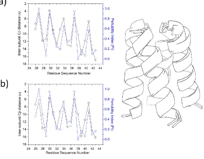
Modeling larger TM homo-oligomeric complexes
Our modeling protocol performed well on larger TM homo-oligomeric complexes. The
largest complex we considered is the pentamer phospholamban. Similar to the case for GpA,
the mutagenesis data for phospholamban has been used extensively in proposing a model for
the TM region (7)(12)(14). It is difficult to compare our results directly with earlier studies since
they were carried out before publication of the NMR structure for phospholamban. However, a
plot of the interhelical van der Waals energy per residue for phospholamban reveals a similar
periodic pattern observed in plots from earlier studies (Supplementary Figure 1). A salient
feature of using inter-subunit Cβ distance over interaction energy when constructing a profile is
that the former descriptor is less sensitive to force field effects. We note that our model for
phospholamban has a smaller radius than what is seen in the NMR structure. However, since
our modeling protocol does not account for the membrane environment or make use of
experimentally derived distance information (i.e., inter-monomer NOEs), the effect of the
non-bonded forces from the molecular mechanics force field dominate resulting in tightly packed
helices.
We also applied our modeling approach to the influenza proton transporters M2 and
high-22
resolution X-ray structure. When compared to the NMR model of M2, our protocol achieves an
RMSD of about 1.0 Å. Our automated method provides predictions for M2 that are better than
earlier predictions that relied heavily on the expertise and intuition of the investigators (24).
We also applied our modeling protocol to the recent solid state NMR structures of Sharma et al.
(25) and Cady et al. (26). Our best scoring models have an RMSD of 1.8 Å and 1.6 Å respectively
to these solid state structures. As a point of comparison, the NMR (solution and solid state) and
the high-resolution X-ray structures show a spread in RMSD between 0.8-1.6 Å.
In an earlier study, we also made use of correlation analysis in modeling the BM2 proton
transporter (18). In our previous approach we adopted a less efficient method that included the
generation of a large ensemble of sterically feasible helical bundles (both ideal and coiled
helices). The ensemble was scored using the correlation coefficient between the pertubility
index (PI) and an estimate of the phase angle for the helix. The surviving models were subjected
to refinement and then clustered. Two out of eight proposed models from our earlier study are
within 1.0 Å of our current best scoring model. It should be noted that all of the models from
our previous study exhibit a weaker correlation with the experimental data than the model we
propose here. The current study along with our earlier study show the generality of the use of
the correlation approach in modeling TM homo-oligomers; different geometric descriptors
between helices can be used in modeling TM homo-oligomers.
Two clear strengths with our modeling protocol are speed (~8 minutes on a single 2.40
GHz processor) and the ability to use data directly from experiments conducted in native cellular
membranes. This is in contrast to previous methods which often require the conversion of
experimental data into distance restraints (4)(27), angular restraints (12), or pseudo-energy