C h a r a c t e r i z a t i o n o f t h e M u r in e T C e l l S u b s e t s D e fin e d by CD45RA I s o f o r m s .
A thesis submitted for the degree of Doctor of Philosophy
to the Faculty of Immunology The University of London
by
Elizabeth Beatrice LIghtstone
Dedicated to my parents, Miriam and Richard Lightstone,
ProQuest Number: 10045725
All rights reserved
INFO RM A TIO N TO ALL U SER S
The quality of this reproduction is dependent upon the quality of the copy submitted.
In the unlikely event that the author did not send a complete manuscript
and there are missing pages, these will be noted. Also, if material had to be removed, a note will indicate the deletion.
uest.
ProQuest 10045725
Published by ProQuest LLC(2016). Copyright of the Dissertation is held by the Author.
All rights reserved.
This work is protected against unauthorized copying under Title 17, United States Code. Microform Edition © ProQuest LLC.
ProQuest LLC
789 East Eisenhower Parkway P.O. Box 1346
A b stra ct
T cells are functionally and phenotypically heterogenous. Correlation of phenotype with function allows the possibility to elucidate the contribution of T cell subsets in an immune response and opens an avenue towards more specific immunotherapy in autoimmunity and transplantation. A particular goal has been to phenotypically identify naive and memory T cells. In vitro studies in humans had suggested that expression of the heavy molecular weight isoforms (CD45RA) of CD45, a phosphotyrosine phosphatase, defined naive T cells. The studies in this thesis set out to determine if comparable subsets could be identified in the mouse with the aim of dissecting out the mechanisms involved in the generation of immunological memory. However, it became apparent that CD45RA did not simply define naive I cells. Three anti CD45RA antibodies were shown to split both CD4+ and CD8+ mouse T cell subsets into CD45RA+ and CD45RA- populations. Assessment of GD45RA as a marker of naive cells involved determining phenotype in vivo at all stages of maturation and differentiation as well as functional assessment in vitro and in vivo of purified CD4+ or CD8+ CD45RA+ and CD45RA- subsets. CD45RA defines a minority population of thymocytes which, particularly among CD4+ cells, is too small to account for all thymic emigrants. In the periphery, among CD4+ cells the CD45RA+ subset is maintained after thymectomy, with ageing and following transfer and expansion in irradiated hosts, again suggesting CD45RA is not a marker of naive cells. Indeed, in vivo CD4+CD45RA+ cells can provide help for secondary responses in an adoptive transfer assay, albeit not as well as the reciprocal subset. Among CD8+ cells there is a modest decline in the proportion of CD45RA+ cells with ageing, thymectomy and cell transfer suggesting that at least a fraction is naive. Functionally, in vitro, CD8+CD45RA+ cells behave as if naive when primary and secondary CTL responses to alloantigen are assayed. However, in vivo studies on T cell receptor transgenic mice show that CD45RA expression is not confined to naive cells. Lack of appropriate costimulation is postulated to account for the differences between the in
A cknow ledgem ents
1. My overwhelming thanks to Av Mitchison for taking me on in the first place, for his continued enthusiasm, encouragement, support, advice and not least for his practical skills, in particular at thymectomy! Above all, for having the forsight to arrange for me to work with Jacqueline Marvel.
2. None of the work in this thesis would have been undertaken without the guidance, advice and total support of Jacqueline Marvel, who didn't baulk at taking on someone who "had never got cells" and who made the whole passage so much smoother and simpler.
5. Particular thanks to the various people I had the good fortune to work with at the ICRF TIU and who made my life so much easier, especially Liz Thomson, Galia Rimon and Kathy Robertson.
4. I am grateful to the many people we collaborated with and who provided stimulation and enthusiasm as well as mice! In particular, Harald von Boehmer, Sophie Ezine, Dimitris Kioussis and Clio Mamalaki, Herman Waldmann and Shixin Quin, and closer to home, Hans Stauss and Nancy Samberg.
5. Grateful thanks to the Medical Research Council whose generosity funded my PhD.
6. Above all, eternal thanks to my husband Ian, whose constant love, support and encouragement have been my touchstones and without whom I would never have started, let alone finished. His patience has known no bounds.
TABLE OF CONTENTS
Page number
Title page 1
Abstract 2
Acknowledgements 3
table of contents 4
List of Figures 9
List of Tables 11
Abbreviations 12
Chapter one: General Introduction 14
1.1 T cell ontogeny 16
1.1.1 Bone marrow stem cells 17
1.1.2 Thymic precursors 18
1.1.3 Thymus T cell maturation 19
1.1.4 The role of the thymus - positive and negative 23 selection
1.1.5 Thymus - paradoxes and conclusions 28
1.2 Peripheral T cells 29
1.2.1 Adhesion molecules 31
1.2.2 Markers of T cell activation 34
1.3 Immunological memory 35
1.3.1 Definition of immunological memory 35
1.3.2 B cell memory 36
1.3.3 T cell memory 39
1.3.4 Measuring memory 41
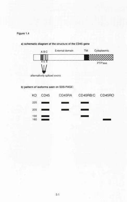
1.3.5 Markers of memory T cells (other than CD45) 43 1.4 T cell subsets defined by lymphokine production 45 1.5 The expression of CD45 on T cells 47 1.5.1 CD45 genomic structure - general structure 47 1.5.2 Alternative splicing of external domain exons 49 1.5.3 External domain - biochemistry 50
1.5.4 Cytoplasmic domain 52
1.5.5 Expression of CD45 isoforms 53
1.5.5.1 Lymphocyte expression of CD45 54 1.5.5.2 Expression of CD45R on murine lymphocytes 54 1.5.5.3 CD45R expression on rat lymphocytes 56 1.5.5.4 CD45 isoforms on human lymphocytes 56 1.6 Functional characteristics of CD45R+ and CD45R' T cells 57
1.6.1 CD45R subsets in the rat 57
1.6.2 CD45R subsets in the human 59
1.6.2.1 CD45R defines CD4+ suppressor inducers 59 1.6.2.2 CD45R distinguishes naive and memory T cells 61 1.6.2.3 CD45R and lymphokine production 66 1.6.3 Paradoxes and problems with CD45 isoforms 66
1.7 Aims of this thesis 6 8
Chapter two: Materials and methods 69
2.1 Tissue culture reagents and conditions 70
2.1.1 Media 70
2.2.1 Source 72
2.2.2 Strains used 72
2.2.3 Procedures 72
2.3 Antibodies 73
2.3.1 Antibodies used - specificities, isotype, sources &
references 73
2.3.2 Hybridomas 73
2.3.3 Antibody preparation and purification 75 2.4 Preparation of cells for staining 81
2.4.1 Spleen cells 81
2.4.2 Lymph node cells 81
2.4.3 Foetal thymocytes 81
2.4.4 Adult thymocytes 82
2.5 Surface staining of cells for FACS analysis 82
2.5.1 Staining protocol 82
2.5.2 Controls for staining 83
2.5.3 Fixation of cells 83
2.5.4 FACS analysis 83
2.5.5 Statistics for analysis for staining 84 2.6 Immunoprécipitation with anti 0045 antibodies 84 2.7 Cross blocking of anti CD45R antibodies 85 2.8 Bulk cultures with PHA +/- IL2 85 2.9 Adoptive transfer of Thy1.2 cells into Thy1.1 mice 85 2.10 Purification of CD4+ or CD8+ CD45RA+ and CD45RA' cells 86
2.10.1 Ficoll density centrifugation 86
2.10.2 Nylon wool columns 86
2.10.3 Complement killing of CD4+ or CD8+ cells 86 2.10.4 Panning to remove lgM+ and Class 11+ cells 88 2.10.5 Incubation with anti CD45RA antibodies 88 2.10.6 Magnetic separation of CD45RA+ and CD45RA- cells88 2.10.7 Yields following purification 89 2.11 Proliferation assays using purified CD4+ subsets 89
2.11.1 Cuture conditions 89
2.11.2 Preparation of feeder cells 90
2.11.3 Harvesting of proliferation assays 90 2.12 Assay of IL2 and IL4 in culture supernatants 90
2.12.1 IL2 assay 90
2.12.2 IL4 assay 91
2.13 The phenotype of naive, memory and effector CTL 92 2.13.1 Generation of H-2K^ specific CTL 92
2.13.2 Assay of CTL activity 94
2.14 Helper activity of primed purified CD4-^CD45RA+ and 95 CD4+CD45RA' in vivo
2.14.1 Priming of T and B cells 95
2.14.2 Purification and transfer of cells and antigen 95 2.14.3 Titration of helper activity by determining anti Thyl
antibody titres 97
2.15 In vivo depletion of CD45RA+ cells 98 2.16 Studies on transgenic mice 98 Chapter three: Anti CD45R antibodies stain mouse T cells 99
3.2 Results
3.2.1 Characterization of the three anti CD45R antibodies 100 3.2.2 CD45R antibodies stain mouse T cells 101 3.2.3 Expression of CD45R on T cells is not strain specific 105 3.2.4 Expression of CD45RB isoforms on spleen and
lymph node cells 110
3.2.5 Coexpression of CD45RA and CD45RB isoforms on spleen and lymph node cells 113 3.3 Summary of data in chapter 3 116 3.4 Conclusions and outline of studies undertaken 117 Chapter four: CD45RA expression In the thymus 119
4.1 Introduction 120
4.2 Results
4.2.1 CD45RA expression among thymocyte subsets in
young and adult B1OA mice 122
4.2.2 Thymocyte distribution among CD45RA+cells 122 4.2.3 CD45RA expression among 16 day
foetal thymocytes 127
4.2.4 The cells which survive cortisone treatment and
irradiation are enriched for CD45RA 131 4.2.5 Positively selected cells are not all CD45RA+ 132 4.3 Summary of findings in chapter 4 136 Chapter five: The maturation stage of the peripheral C D 4 + C D 4 5 R A + subset is different from that of the
CD8+CD45RA+ subset 137
5.1 Introduction 138
5.2 Results
5.2.1 Thymectomy leads to a fall in CD8+CD45RA+ T cells but not in CD4+CD45RA+ cells in the periphery 139 5.2.2 Maintenance of the CD45RA+ population among
spleen lymhocyte subsets with age in vivo 141 5.2.3 The percentage of CD44+ T cells increases with
age but anti CD44 also stains a population of CD45RA+
T cells 146
5.2.4 Incomplete loss of CD45RA on T cell subsets after
in vitro activation 148
5.2.5 Depletion of CD4 and CD8 does not alter the
expression of CD45RA on regenerating cells 148 5.2.6 Transfer of T cells in vivo into irradiated hosts leads to
expansion of donor cells and maintenance of expression
of CD45RA 150
5.3 Summary of findings in chapter 5 154 FUNCTIONAL STUDIES ON CD45RA T CELL SUBSETS 155 Chapter six :The CD4+CD45RA+ subset is hyporesponsive and
CD45RA does not define the Th1/Th2 split 156
6.1 introduction 157
6.2 Results
6.2.1 Staining profiles of purified CD4+CD45RA+ and
CD4+CD45RA' populations 157
6.2.2 Proliferation of the purified subsets in response to
PHA or anti CD3 159
6.2.3 IL2 and IL4 production by purified subsets 164
6.2.4 IL2 production 164
6.2.5 IL4 production 167
6.3 Summary of findings in chapter 6 171 Chapter seven : CD45RA as a marker of naive cells 172
7.1 Introduction 173
7.2 Results
7.2.1 CD45RA is expressed on naive murine CTL
precursors but absent in memory and effector CTL 173 7.2.2 Primary CTL responses are generated by
CD8+CD45RA+ and by CD8+CD45RA- T cells 174 7.2.3 In vivo primed CTL are CD8+CD45RA' 174 7.2.4 The specific cytolytic activity of bulk effector CTL is
contained in the CD8+CD45RA- subpopulation 178 7.2.5 Both CD4+CD45RA+ and CD4+CD45RA' cells can
respond to recall antigen in vivo 182 7.2.6 CD4+CD45RA+ cells can provide help for antibody
production in a secondary response to
antigen in vivo 186
7.2.7 CD45RA+ cells cannot be irreversibly depleted
in vivo 188
7.3 Summary of findings in chapter 7 197 Chapter eight : The phenotype of T cells responding to antigen
In vivo 198
8.1 Introduction 199
8.2 Results
8.2.1 Naive T cells are heterogenous in their expression
of CD45RA 202
8.2.2 Priming of TCR transgenic animals leads to massive expansion of the CD8+ population 202 8.2.3 Priming leads to an increase in the expression of
total CD45 204
8.2.4 Priming leads to a rise in expression of CD45RA and
CD44 on transgenic T cells 207
8.2.5 Continuous exposure to antigen in vivo results in
loss of CD45RA expression on the responding cells 210 8.2.6 Naive V|36+ T cells are not all CD45RA+ in vivo 213 8.2.7 Primed or tolerized Vp6+ cells do not lose expression
of CD45RA 213
8.3 Summary of findings in chapter 8 218
Chapter nine : Discussion 219
9.1 Introduction 220
9.3.2 Does CD45RA define a phenotypically continuous
thymic lineage? 227
9.3.3 Does expression of CD45RA in the thymus define
those cells which survive selection? 231
9.4 CD45RA in the periphery 233
9.4.1 Does the pattern of expression of CD45RA on T cell subsets in vivo follow that expected of naive T cells?234 9.4.2 Do purified CD45RA+ cells function as naive T cells?241
9.4.2.1 CD8+ cells 241
9.4.2.2 CD4+ cells 243
9.4.3 Correlating phenotype in vivo with antigen specific
responses 246
9.5 CD45RA*- cells are hyporesponsive rather than naive 248 9.6 Conclusions and speculations 254
Bibliography 260
List of Figures
1.1 Stages of T cell maturation in the thymus 20 1.2 Schematic diagram of the interaction between TCR & 30
MHO class II
1.3 Generation of memory cells from naive precursors 37 1.4a Schematic diagram of the structure of the CD45 gene 51 1.4b Pattern of isoforms seen on SDS-PAGE 51 2.1 SDS-PAGE gel of DEAE purified 14.8 ascites 78 2.2 Purification procedure used to isolate CD45RA+ &
CD45RA-T cell subsets 87
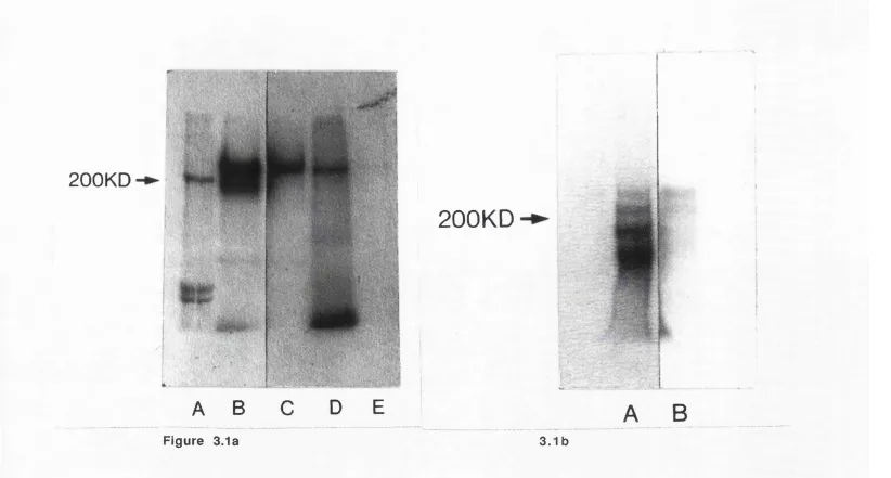
2.3 Titration of induction of class II by IL4 93 2.4 Experimental design to determine T cell help in vivo 96 3.1 Immunoprécipitation of spleen cells with anti CD45
& anti CD45R mAb 102
3.2 Staining of 0045+ spleen cells with three anti CD45R mAb 103 3.3 Cross inhibition of 3 anti CD45R mAb by each other
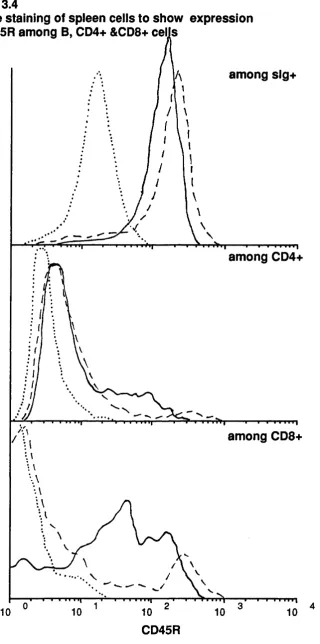
but not by anti CD45 nor by N rat Ig 104 3.4 Double staining of spleen cells to show expression of CD45R
among B, CD4+ and CD8+ cells 106
3.5 Double stain of spleen cells with anti CD45RA and
anti Thyl .2 107
3.6 Comparison of expression of 14.8 and RA3-3A1 among CD8+
spleen and lymph node cells 108
3.7 Comparison of expression of CD45R among CD4+ and CD8+
spleen cells in 3 strains 109
3.8 Expression of CD45RB on spleen and lymph node cells 111 3.9 Expression of CD45RB among spleen lymphocyte subsets 112 3.10 Double stain of spleen and lymph node cells with anti CD45RA
and anti CD45RB 114
3.11 Schematic diagram of expression of CD45RA and CD45RB 115 4.1 Expression of CD45RA among the thymocyte populations
defined by the expression of CD4 and CD8 123 4.2 Size of the cells within the thymocyte subsets defined by
expression of CD4 and CD8 124
4.3 Expression of CD45RA among CD4+CD8+ blasts 125 4.4 Expression of CD4 and CD8 among CD45RA+ thymocytes 129 4.5 Expression of CD45RA among positively selected 135
CD8 single positive cells expressing a transgenic TCR 5.1 Effect of thymectomy on numbers of splenic CD4+ and CD8+
cells expressing CD45RA 140
5.2 Comparison of expression of CD45RA among CD4+ and CD8+ cells in aged thymectomized and non thymectomized mice 142 5.3 Comparison of expression of CD44 among CD4+ and CD8+
cells in aged thymectomized and non thymectomized mice 143 5.4a Number of lymphocytes in spleens from B10A mice
of increasing age 144
5.4b Mean percentage of lg+, CD4+ and CD8+ cells in B10A spleens
5.5 Percentage of CD45RA+ cells within each lymphocyte subset in spleens from B1 OA mice of increasing age 145 5.6 Kinetics of expression of CD45RA on CD4+ and CD8+ cells in
bulk cultures stimulated with PHA and IL2 149 5.7 Expression of CD4, CD8 and of CD45RA within these subsets on
spleen cells from untreated or antibody treated mice 151 6.1 CD45RA expression on purified CD4+CD45RA- and
CD4+CD45RA+subsets 158
6.2 Kinetics of proliferation of purified CD4+ cells stimulated with PHA or cross linked anti CD3 and feeders 162 6.3 Kinetics of proliferation of purified CD4+CD45RA+ &
CD4+CD45RA' cells stimulated with PHA or cross linked anti
CD3 and feeders 163
6.4 Comparison of proliferation of unseparated CD4+ cells and CD4+CD45RA- cells stimulated with PHA or anti CD3 165 6.5 Titration of day 3 supernatants for IL4 activity 168 6.6 IL4 titration from CD4+CD45RA+ &CD45RA- cultures stimulated
for 4 days with PHA or anti CD3 ± feeders 169 7.1 Expression of CD45RA on purified subsets of naive CD8+ spleen
cells from DBA/2 mice 175
7.2 CD8+CD45RA+ & CD45RA' cells can respond to in vitro priming
against the Kh antigen 176
7.3 Expression of CD45RA on purified subsets of primed CD8+ spleen cells from C3H/He mice a) prior to and b) following
restimulation in vitro 177
7.4 Memory CTL are CD8+CD45RA' following in vivo priming 179 7.5 Expression of CD8, CD3 and CD45RA on T cells purified after
restimulation in vitro 181
7.6 Effector CTL are CD8+CD45RA- 183
7.7 Experimental design to determine T cell help in vivo 185 7.8 Titration of helper activity in CD4+CD45RA' & CD45RA+ subsets
after in vivo priming 187
7.9 Staining of spleen & lymph node cells 3 days after injection with 14.8 or an irrelevant antibody i.v. 189 7.10 Proliferation of spleen & lymph node cells to suboptimal
concentrations of PHA following in vivo antibody treatment 190 7.11 Staining of spleen & lymph node cells after different regimes of
depleting antibody treatment - day 2 and 4 193 7.12 Proliferation of spleen & lymph node cells in response to PHA or
LPS, from thymectomized injected mice or sham thymectomized
or unmanipulated mice 195
8.1 Transgenic mice used 200
8.2 Not all naive cells are CD45RA+ 203 8.3 Following priming, the level of total CD45 increases 208 8.4 Proliferating CD8+ transgenic T cells express increased
levels of CD45RA & CD44 209
8.6 Expression of CD45RA among the Mls-I^ responsive Vp6+ cells
from CBA mice 214
8.7 Outline of Mis system used to identify CD45RA phenotype of cells primed or tolerized to superantigen 215 9.1 Possible thymic pathways for CD45RA+cells 225 9.2 Model of expression of CD45RA on CD4+ and CD8+ T cells 256 List of tables
1.1 Thymocyte subset composition in H-Y TCR transgenic mice 26
1.2 Adhesion molecules 32
2.1 Antibodies used 74
4.1 Percentage of CD45RA+ cells among the thymocyte subsets defined by staining with anti CD4 & anti CD8 126 4.2 Percentage of cells belonging to CD4+ and CD8+ defined
subsets among CD45RA+ thymocytes 128 4.3 Percentage of CD45RA+ cells amongst 16 day foetal thymocyte
double negative and CD8 single positive subsets 130 4.4 Expression of CD45RA among the different thymocyte subsets
following hydrocortisone or irradiation 133 5.1 Expression of CD44 among CD4+CD45RA+,CD4+CD45RA',
CD8+CD45RA+,CD8+CD45RA- subsets in young & old mice147 5.2 Expression of CD45RA and CD44 among Thyl .2+ cells
transferred into irradiated A/Thyl.1 mice 152 6.1 Proliferation of CD4+CD45RA' and CD4+CD45RA+ cells in the
absence of feeder cells 160
6.2 Proliferation of GD4+GD45RA" and GD4+GD45RA+ cells in the
presence of feeder cells 161
6.3 IL2 levels in supernatants from GD4+GD45RA+ and
GD4+GD45RA' subsets stimulated with PHA or anti GD3 166 7.1 GTL activity of GD45RA T cell populations purified from primed
mice prior to in vitro restimulation 180 7.2 GTL activity of GD45RA T cell populations purified from primed
mice after in vitro restimulation 184 7.3 Number of cells in spleen and lymph node 2 & 4 days following
different regimes of antibody 192
7.4 Gell numbers and phenotype of spleen & lymph node cells from mice sham thymectomized or thymectomized and treated with
14.8 + MARI 8 194
8.1 Priming with antigen leads to massive proliferation of the GD8+
transgenic spleen cells 205
8.2 Kinetics of response of transgenic cells to varying doses of
peptide 206
8.3 Phenotype of spleen cells from GBA mice primed or tolerized
A bbreviations
aa amino acids
alio allogeneic
AMLR autologous mixed lymphocyte reaction ARC antigen presenting cells
ATCC American Type Culture Collection
BM bone marrow
Bq becquerel
BSA bovine serum albumin
C complement
CD cluster differentiation antigen
ODR complementarity determining region CFA complete Freund's adjuvant
cpm counts per minute CTL cytotoxic lymphocytes
CTLp cytotoxic lymphocytes precursors
D diversity
DEAE diethylaminoethyl cellulose DTI dithiothreitol
CIH2O distilled water
DMEM Dulbecco's modification of Eagle's medium
DN double negative
DP double positive
EDTA ethylene diamine tetra-acetic acid (tri- sodium salt) ELAM endothelial leucocyte adhesion molecule
FACS fluorescence activated cell sorter FCS foetal calf serum
F lic fluorescein isothiocyanate G protein GTR binding proteins
HERES N-2 hydroxyethylpiperazine - N'-2- ethanesulphonic acid MSA heat stable antigen
i.p. intraperitoneal
i.v. intravenous
ICAM intercellular adhesion molecule
Ig immunoglobulin
IL interleukin
IL2R IL2 receptor
INR influenza nuclear protein
J joining
kD kiloDalton
KLM keyhole limpet haemocyanin
LAR LCA related
LCA leukocyte common antigen
LFA lymphocyte function-associated antigen
Lin lineage
LN lymph node
LRS lipopolysaccharide 2 ME 2 mercaptoethanol
M molar
mAb monoclonal antibody
mCi milliCurie
mha minor histocompatibility antigens MHC major histocompatibility complex MLC mixed lymphocyte culture
MLR mixed lymphocyte reaction
mis minor lymphoc^e stimulating system
mM millimolar
MMTV mouse mammary tumour virus mRNA messenger ribonucleic acid
MTT 3,4,5-dimethylthiazole-2-yl-2,5,diphenyl tétrazolium bromide
N normal
OD optical density
PE phycoerythrin
pgp phosphoglycoprotein
PHA phytohaemaglutinin
PBS phosphate buffered saline
Pi propidium iodide
PTP'ase phosphotyrosine phosphatase
PWM pokeweed mitogen
rlL2 recombinant interleukin 2 Sea stem cell antigen
SDS-PAGE sodium dodecyl sulphate polyacrylamide gel electrophoresis
sig surface immunoglobulin SP single positive
TCR T cell receptor
TDL thoracic duct lymphocytes
IG transgenic
Th I helper
TM transmembrane
Tx thymectomy
V variable
CHAPTER ONE
7. General Introduction
It has been recognized for thousands of years that recovery from an episode of infectious disease is associated with long lasting protection, or immunity, against a second episode of the same illness (e.g. "Yet it was with those who had recovered from the disease that the sick and dying found most compassion. These knew what it was from experience, and had now no fear for themselves for the same man was never attacked twice - never at least fatally." - Thucydides,430 B.C.E.). Thus the concept of specific immunological memory was recognized well before the role of lymphocytes was known, and provided the basis for vaccination programmes (reviewed in Parrish, 1965). Studies on transplantation and recognition of transplantation tissue demonstrated that the immune system could discriminate self from non-self (Billingham, et al., 1953; Billingham, et a i, 1956; Gibson & Medawar, 1940). Abnormalities of the immune system can lead to a loss of self tolerance and autoimmunity. Furthermore, exposure to an antigen or tissue seen previously i.e. to which the host had been primed, leads to an accelerated immune response (Medawar, 1944; Medawar, 1945), taken as an indicator of memory for those particular antigens.
Whilst the importance of antibodies in immune responses was recognized early on, it has only been relatively recently that the cells of the immune system have been phenotypically and functionally characterized. Two main compartments have been identified i.e. B and T cells, the former produced in the bone marrow and providing the humoral arm of the immune response (Cooper, etal., 1965a; Cooper, et a!., 1965b; Cooper, et a!., 1966) whilst the latter are produced in the thymus and contribute cell mediated regulatory and effector functions (reviewed in Good, 1991 ; Miller, 1991).
as CD8) was present on CTL but not on T helper cells (Cantor & Boyse, 1975a; Cantor & Boyse, 1975b). The advent of monoclonal antibody technology allowed generation of monospecific antibodies which recognize particular cell surface molecules (Kohler & Milstein, 1975) and hence allowed further charaterization of functionally discrete subsets of B and T cells. Indeed, it was only with this technology that it was possible to raise an antibody which recognized the reciprocal subset to the Lyt2+ T cells, and identified the CD4 molecule (White, etal., 1978). CD4 and CD8 define mutually exclusive subsets of peripheral T cells and together account for over 95% of such cells. T cells provide the regulatory arm of the immune response as well as effector cells e.g. helper T cells are required to stimulate an appropriate B cell response (Mitchison, 1971).
With the recognition of the importance of T cells and their heterogeneity, a major therapeutic goal has been to generate increasingly specific agents to abrogate adverse immune responses without impairing an individual's ability to respond appropriately to infectious agents. Hence, a very important question in immunology has been to try to identify T cell molecules, using monoclonal antibodies, which distinguish naive or unprimed from memory or primed T cells. The studies presented in this thesis address this question, focusing on the isoforms of the leucocyte specific membrane glycoprotein, CD45. We will look at the generation of the mature peripheral T cell repertoire by the thymus, the activation of T cells, the lymphokines they produce and the nature of T cell memory.
This introduction aims to set the scene for the experiments undertaken in this thesis. The first section reviews the literature regarding T cell ontogeny and the role of the thymus in shaping the peripheral T cell repertoire. The second section is concerned with the phenotype and function of peripheral T cells together with a more general review of immunological memory. Finally, there is detailed review of the literature concerning CD45 at the time of initiating our studies. No introduction can be all inclusive and in particular I have omitted a detailed discussion of the biochemistry of T cell signalling and activation.
1,1 T cell ontogeny
immune system. However, this notion was rebutted by a series of elegant experiments undertaken in the early 1960's by two independent workers, Miller in the U.K. and Good in the U.S.A. Whilst studying the mechanisms involved in the development of a virally transmitted thymus dependent murine leukemia. Miller thymectomized neonatal rather than adult mice. Whilst studying clinical immunodeficiency. Good and his colleagues undertook similar experiments (reviewed in Good, 1991; Miller, 1991). Together, these experiments demonstrated that the cells produced by the thymus, later designated I cells, were necessary for the generation of an immunocompetent animal. Absence of the thymus led to a severe immunodeficiency syndrome, characterized by atrophic peripheral lymphoid organs and a deficiency of peripheral lymphocytes. Such animals only survived if maintained under germ free conditions. Furthermore, neonatally thymectomized mice were rendered tolerant to subsequent alio- and xeno grafts.
Subsequently, it was demonstrated that there are two lineages of lymphocytes, namely T cells produced by the thymus and B cells produced by the bone marrow (reviewed in Cooper, etal., 1968).
1.1.1 Bone marrow stem cells
Heimfeld, eta l., 1989), this same study emphasises just how few are necessary to reconstitute lethally irradiated mice. As few as 50-100 T h y l ioLin Sca-1 + cells are capable of radioprotection and long lasting reconstitution of such animals (Uchida & Weissman, 1992) and reviewed in Ikuta, etal., 1992). In percentage terms, this implies that 99.95% of bone marrow cells do not possess stem cell activity. These numbers highlight an important issue in immunology - subsets small enough to be overlooked may be of great functional consequence.
1.1.2 Thym ic pre curso rs
1.1.3 Thymus T cell maturation
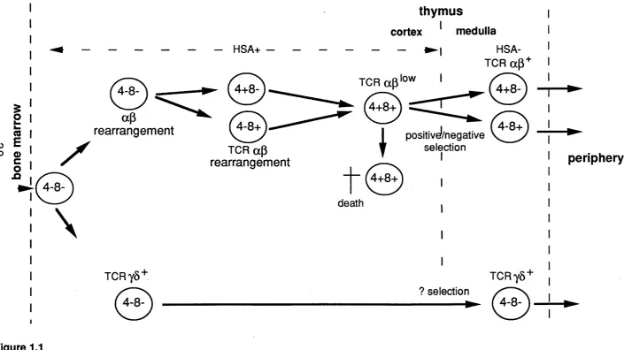
Only the most immature thymocytes are capable of homing to the thymus when injected i.v. More mature thymocytes can however regenerate the thymus if injected intrathymically (Scollay, etal., 1988). Once in the thymus immature thymocytes undergo a process of maturation which generates a population of self tolerant mature T cells expressing on their cell surface fully rearranged ap oryS TCR, with CD3 as well as CD4 or CD8. Different developmental steps can be characterized by the patterns of expression of TCR, CD3, CD4 and CD8 as well as several other surface antigens (illustrated in figure 1.1).
Although there is evidence to suggest functional and phenotypic differences between the foetal and adult thymus, the maturation pattern in the foetal thymus from seeding at day 13 to birth at day 20-21 shows a developmental pattern similar to that seen in adult with the advantage in early ontogeny of only having the least mature cells present and cells maturing in a relatively synchronous fashion. Thus, the ontogeny of foetal thymocytes provides clues to the ordering of differentiation events. It is now agreed that the least mature thymocytes, which are nevertheless committed to become T cells, do not express CD4 or CD8, based on a wide variety of in
wVo and in vitro studies e.g. (Fowlkes, et ai., 1985; Jenkinson, et ai., 1982) and reviewed in Fowlkes & Pardoll, 1989). This so called double negative subset is however heterogenous with respect to the expression of other surface markers. Careful kinetic and repopulation studies (reviewed in Shortman et ai., 1990) have suggested that the double negative cells which give rise to more mature thymocytes express the antigens HSA, CD44 (pgpl) and IL2R in the following sequence:
HSA+, IL2R-, CD44+ ^ HSA+, IL2R+, CD44+/- -> HSA+ IL2R- CD44-.
I
(0 ro EO Q) c o n
0
\
0
ap
rearrangement
TCRy8+
0 ,
TCR ap rearrangement
thymus
cortex ^ medulla
- ► !
HSA-TCR ap+
T ^ ' o w
IV V
0 0
^ positivel/negative
death
selection I
I
TCRy8+ ? selection / — X
periphery
Figure 1.1
inhibition of silencers of the a or y genes. Evidence exists to support both hypotheses but in general the latter now appears to be favoured (reviewed in Winoto, 1991).
Whereas the majority of day 13 and day 14 foetal thymocytes have TCR loci in germline configuration, thereafter progressive rearrangement of all TCR loci occurs. As implied above, y pairs with 6 and a pairs with p. For cells that go on to express aP TCR, the p gene is expressed first and rearranged prior to the a gene. By day 17 >80% of y, p and Ô loci are rearranged (reviewed in Robey, etal., 1990). Most rearrangement occurs during the double negative stage of thymocyte development as judged by Southern blot analysis of both whole populations of murine foetal thymocytes from different days of gestation and panels of cloned hybridomas produced by fusion of early stage thymocytes (Born, etal., 1986; Born, etal., 1988). Only fully rearranged TCR genes can pair, associate with the multichain complex CDS and be expressed at the cell surface (reviewed in Ashwell & Klausner, 1990). Hence, because of the difference in timing of gene rearrangement, in day 14 embryos most expressed TCR's comprise yÔ chains. However, with time this becomes a minority population and the majority express aP TCR's such that in an adult thymus y5 TCR's are expressed on 10% of double negative cells or 1% of total thymocytes whereas ap TCR is expressed on more than 65% of thymocytes, although the level of expression depends on the thymocyte subset and the stage of maturation. Although most rearrangement of TCR genes occurs during the double negative stage, surface expression is not detected until CD4 and CD8 are expressed i.e. the double positive stage
The next developmental stage among thymocytes is marked by the expression of the accessory molecules, CD4 and CD8. Recent data have shown that prior to expressing both these markers, double negative cells pass through a stage of expressing either a low level of CD4 (Hugo, et al., 1991; Matsumoto, eta l., 1989) or of CD8 (Macdonald & Howe, 1988; Paterson & Williams, 1987; Shortman, etal., 1988). Such immature single positives can be distinguished from mature single positives as they do not express TCR but do express HSA. Furthermore, in vitro they rapidly give rise to double positive cells (Nikolic-Zugic, etal., 1989).
Double positive cells constitute the majority of thymocytes. By this stage the TCR is fully rearranged, expressed at the cell surface and non- covalently associated with CD3, expression of which varies from dull to bright. The CD3bhght subset is thought to contain the most mature double
positive cells which then go on to become mature single positive cells. Due to the difficulty of culturing purified double positive cells in vitro, and prior to studies with TCR transgenic mice, the evidence that double positive cells were the immediate precursors of mature single positive thymocytes came from in wVo studies (Guidos, etal., 1989; MacDonald, etal., 1988; Smith, 1987) as well as manipulation of foetal thymic organ cultures (Zuniga- Pflucker, et al., 1989). Most double positive cells die in vivo and cannot be maintained in culture in vitro (Guidos etal., 1989; Petrie, etal., 1990; Scollay &K., 1984).
few peripheral thymocytes and an enormous increase in thymic single positive cells suggesting that inhibition of a G protein by the toxin prevents egress of mature thymocytes (Chaffin & Perlmutter, 1991).
1.1.4 The role of the thymus - positive and negative selection
The role of the thymus is not simply generation of thymocytes but generation of a peripheral I cell repertoire that expresses clonally diverse TCR's which are tolerant of self and which recognize non-self antigens presented by self MHC molecules. Diversity arises from random rearrangement of the TCR gene segments at the a (VaJa) and (3 (VpDpJp) loci, nucleotide additions at the joining sites, imprecise joining of the gene segments, together with recombination of the different chains. These processes could give rise to a large T cell repertoire of at least 10^ different specificities, the germline or unselected repertoire (reviewed in Kronen berg, et al., 1986). Two thymic selection mechanisms have been proposed to account for shaping the germline repertoire into the peripheral T cell repertoire. Tolerance to self results from thymic deletion of T cells which recognize self antigens. Such clonal deletion or negative selection was originally proposed by Burnet (Burnet, 1959). Recognizing foreign antigen presented by self MHC requires positive selection of TCR's which recognize self MHC (reviewed in Schwartz, 1989; Sprent, e ta l., 1988). Formal evidence for both processes now exists, but the mechanisms which allow apparently paradoxical events are less clear.
in Herman, et al., 1991). The main family of self superantigens in the mouse is the minor lymphocyte stimulating (Mis) system first described by Festenstein (Festenstein, 1973). In vitro, mixing cells from mice which are MHC identical but bear different Mis alleles generates potent proliferative responses (Festenstein, 1973). As with superantigens in general, the frequency of Mis reactive I cells is much higher than for "normal" antigens, (>10'1 versus 10-4-10-6) (Acha-Orbea & Palmer, 1991). Studies on the expression of TCR's bearing particular vp elements in the appropriate Mis strains provided further evidence for deletion of cells in the thymus which could react with self Mis e.g. Vp6+ and Vp8.1+ T cells are deleted in Mls-1^ mice (Macdonald, et al., 1988). It has now been shown that Mis antigens and related antigens are the products of by retroviral integrants that are scattered throughout the mouse genome. Such retroviruses include mouse mammary tumour viruses (MMTV) (Acha-Orbea, etal., 1991; Choi, etal., 1991; Dyson, etal., 1991 ; Frankel, etal., 1991 ; Marrack, e ta l., 1991 ; Woodland, etal., 1991) and a defective murine leukaemia virus (MuLV) (Janeway, 1991).
Recent work has shed light as to why these so called superantigens stimulate so many T cells. Kappler and Marrack's group (Choi, et al., 1990; Pullen, et al., 1990; Pullen, etal., 1991) have shown that superantigens bind MHC molecules outside the antigen binding groove on the lateral face of the Vp surface and interact with MHC molecules, largely as originally postulated by Janeway (Janeway Jr., et al., 1989). The relevance of superantigens in shaping the human peripheral T cell repertoire is less clear. Recent data on TCR Vp usage in the human autoimmune disease rheumatoid arthritis have suggested that superantigens may indeed play such a role (Paliard, et al.,
1991).
population of T cells expressing the same TCR to be followed in vivo. In a series of papers, von Boehmer's group describe just such a transgenic system (reviewed in von Boehmer, et a i, 1989). They generated transgenic mice expressing an ap TCR derived from a GTL clone which recognizes the male restricted H-Y antigen in the context of Dh. The transgenic (TG) TOR is expressed early in ontogeny. If it is expressed in a male mouse on a Dh background then the developing thymocytes encounter their nominal antigen in the context of self MHO and are deleted. This negative selection results in an absence of CDS single positive cells. Furthermore very few CD4 single positive cells are seen implying that negative selection in these mice occurs at the double positive stage. If the TG TCR is expressed in a female mouse on a background then the developing TG T cells fail to recognize self MHC and are not selected and again no single positive cells are seen. In contrast, if the TG TCR is expressed on T cells in female Dh mouse then these cells are positively selected since they recognize self MHC but they never encounter antigen i.e. H-Y expressed only in males, and hence are not negatively selected. Since the TG TCR is specific for class I the positively selected cells are CD8+CD4-. Overall, the original studies on these mice demonstrated that positive selection takes place in the absence of antigen but in the presence of self MHC (summarized in table 1.1). Such studies confirmed much earlier work on irradiated recipients which had suggested the importance of positive selection in shaping the peripheral T cell repertoire (Bevan, 1977; Zinkernagel, et a i, 1978).
The timing of positive and negative selection has been the source of debate. For negative selection, the most important factor would appear to be the timing of the presentation of antigen. Thus in von Boehmer's system antigen is present early in male mice and negative selection is seen to occur at the double positive stage. Self superantigens may be expressed later and deletion may therefore not occur until the single positive stage (Guidos, et al., 1990). However, in a study of the kinetics of mature T-cell development in the thymus, Egerton ef a/suggest that no extensive negative selection occurs after the mature cells are formed (Egerton, ef a/., 1990). Positive selection occurs at the double positive stage.
Table 1.1
The thymocyte subset composition seen in the presence of a transgenic TCR specific for male antigen H-Y presented by Db.
Class 1 H-Y Sex Double
positives
CD8 Single positives
Type of selection
Db + M Very few No negative
Db - F Yes Yes positive
transgenes either only on thymic cortical cells or only on medullary cells, demonstrated that whilst positive selection occurs on cortical thymic epithelial cells (Benoist & Mathis, 1989b), negative selection can occur both in the thymic cortex and medulla (Benoist & Mathis, 1989a) and reviewed in Davis, et ai, 1989).
Thus by the end of the thymic maturation process, clonally diverse mature I cells emerge which express fully rearranged ap (oryô) IC R 's, which recognize foreign antigen presented by self MHC, the majority of which do not react with self and which in general express either CD4 or CD8. The corollary of such stringent selective processes is that the thymus must be the site of not only the generation of millions of thymocytes, but also of the death of most of them. Early studies suggested that most thymocytes die in situ (Shortman & Jackson, 1974) and more recently it has been shown that most proliferation and death occurs at the double positive stage (reviewed in Shortman et a i, 1990). The proposed mechanism for intrathymic cell death is that of apoptosis or programmed cell death (Kerr, et a i, 1972). In vitro, cross linking of CD3 in foetal thymic organ cultures results in apoptosis of the developing thymocytes (Smith, et a i, 1989) and cultures of double positive thymocytes from TCR transgenic mice also show evidence of apoptosis after 24 hours in culture (Kisielow, et al., 1991). It was clear from in vivo studies on effects of irradiation or treatment with glucorticoid (Blomgren & Andersson, 1971) that the thymus does have the capacity to eliminate massive numbers of dead thymocytes in a very short time and that these treatments cause cell death by inducing apoptosis (Cohen & Duke, 1984; Sellins & Cohen, 1987). However, direct evidence for such cell death in vivo has only recently been demonstrated (Murphy, et ai, 1990).
using transgenic mice expressing the oncogene bcl-2 on thymic cortical cells, in contrast to normal mice where it is only expressed on medullary cells. Expression on cortical thymocytes blocks glucocorticoid depletion of thymocytes, inhibits irradiation induced cell death and confers resistance to anti CD3 induced apoptosis but does not block negative selection due to the presence of l-E and selfsuperantigen (Sentman, et ai, 1991). Thus, soluble anti CD3 treatment is not the experimental equivalent of negative selection. Furthermore, the bcl-2 transgenics show a rise in the proportion of single positive cells which are CD3high with a concomitant fall in the proportion of
CD3*o cells suggesting that the aberrant transgene expression allows maturation of cells which would normally die due to lack of positive selection.
1.1.5 Thymus - paradoxes and conclusions
effectiveness and outcome of ligation of the TCR (e.g Webb & Sprent, 1990 and reviewed in van Ewijk, 1991).
Furthermore, although there is good evidence for both positive and negative selection among immature thymocytes, it would be rash to claim that the resultant peripheral I cell repertoire is entirely deleted of T cells able to recognize self. Indeed, numerous studies have shown that self reactive I cells exist in the periphery. This may be due to a failure to have been presented with self antigens during ontogeny, but there is also strong evidence to suggest that some self reactive T cells escape or evade selective processes e.g. (Blackman, et al., 1991). Thus mechanisms must exist to maintain peripheral tolerance among mature T cells. A review of this complex topic is beyond the scope of this introduction. However, at least three mechanisms have been demonstrated: failure to present antigen in the periphery e.g (Cowing, 1985), peripheral anergy of self reactive T cells (reviewed in Schwartz, 1990), and peripheral deletion of self reactive T cells e.g. (Rocha & von Boehmer, 1991). Understanding such mechanisms will have important consequences for the manipulation of unwanted autoimmune responses.
Thus, developing thymocytes run a gauntlet of selective processes and most fail to survive. Along the way there is a highly organised sequence of expression of various cell surface molecules. There is now good evidence that the successful thymocyte progresses from a double negative progenitor, through the intermediate stages of immature single positive, double positive and finally emerges as a mature single positive T cell. The contribution of T cell surface molecules other than CD3, CD4, CDS and TCR is much less clear.
1.2 Peripheral T cells
More than 90% of peripheral T cells express either GD4 or CDS (Reinherz & Schlossman, 1980) and brightly express CD3 and most express the aP TCR. The antigen binding site of the TCR is formed primarily by the juxtaposition of six hypervariable polypeptide loops (complementarity determining regions, or CDRs 1-3) three of which are encoded within each NH2-terminal variable (V) domain of paired a and p chains (reviewed in
Figure 1.2
Schematic diagram of the Interaction between T cell receptor and MHC class II
T c e ll
TCR
Class
II-CDR CDR
a 1 1peptide j p i antigen
a helix # ■ d \ a helix
0 - 0
Although central to a specific T cell response, ligation of the TCR by antigen or antibody is not usually sufficient to induce I cell activation. The involvement of accessory molecules in adhesion and signalling is also required to provide the so-called second signal (reviewed in Schwartz, 1989). Furthermore, the presence of particular molecules is associated with particular T cell behaviour. Hence, in general CDS cells are cytolytic whilst CD4 cells provide help for T and B cell mediated responses. It has been evident for many years that the CD4 and CDS subsets are functionally heterogenous e.g. among CD4+ cells in their ability to provide help for Ig production, ability to induce suppression and production of lymphokines (reviewed inAbbas, et al., 1991). Following the advent of monoclonal antibodies there was an intense search for mAb's which recognized antigens defining subsets within the CD4 or CDS lineages. The markers identified thus far have been used to separate T cell subsets according to functional differences in adhesion, activation, memory and/or lymphokine production.
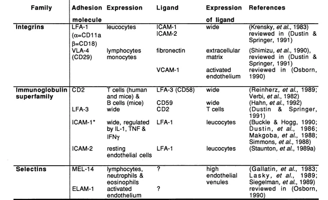
1.2.1 Adhesion molecules
Interactions between T cells and ligands on other cells are enhanced by adhesion and such adhesion has been demonstrated to be mediated by specific cell surface molecules. Initially the role of CD4 and CDS was regarded as facilitating adhesion between TCR and class II or class I respectively. These molecules do indeed have specific adhesive properties (Fames, at a!., 1990) and moreover, recent data have also suggested that adhesion leads to signal transduction via the associated phosphotyrosine phosphatase p56^^^ (reviewed in Rudd, eta!., 1989).
Several other adhesion molecules have been described. They structurally belong to three families - the integrins; the immunoglobulin superfamily; and the selectins (reviewed in Dustin & Springer, 1991). The major adhesion molecules involved in lymphocyte adhesion are summarized in table 1.2.
Table 1.2
Adhesion molecules
Family Adhesion Expression Ligand Expression References
molecule of ligand
Integrins
CO
LFA-1 leucocytes ICAM-1 wide (Krensky, ef a/., 1983)
(a=CD11a 1 CAM-2 reviewed in (Dustin &
P=CD18) Springer, 1991)
VLA-4 lymphocytes fibronectin extracellular (Shimizu, ef a/., 1990), (CD29) monocytes matrix reviewed in (Dustin &
Springer, 1991)
VCAM-1 activated reviewed in (Osborn, endothelium 1990)
Immunoglobulin superfamily
CD2 I cells (human LFA-3 (CD58) wide (Reinherz, af a/., 1989;
and mice) & Verbi, etal., 1982)
B cells (mice) CD59 wide (Hahn, etal., 1992) LFA-3 wide CD2 I cells (Dustin & Springer,
1991)
ICAM-1* wide, regulated LFA-1 leucocytes (Buckle & Hogg, 1990; by IL-1, TNF & D u s tin , e ta l., 1986;
IFNy Makgoba, e ta l., 1988;
Simmons, etal., 1988) 1 CAM-2 resting LFA-1 leucocytes (Staunton, ef a/., 1989a)
endothelial cells
Selectins MEL-14 lymphocytes, ? high (G allatin, af a/., 1983;
neutrophils & endothelial L a s k y , e ta l., 1989; eosinophils venules Siegelman, af a/., 1989)
ELAM-1 activated ? reviewed in (Osborn,
endothelium 1990)
independent adhesion events between T cells and other cells are part and parcel of lymphocyte trafficking. Such events are mediated by CD2/LFA-3 and LFA-1/ICAM-1 or -2 interactions. If no ligation of the TCR occurs then the cells de-adhere and pass on (reviewed in Dustin & Springer, 1991).
Once the TCR has been ligated, adhesion molecules serve to augment the interaction. One way of achieving this has been shown to be "inside-out" signalling whereby activation via the TCR results in an energy dependent increase in LFA-1 avidity for ICAM-1 which thus increases adhesion between the two cells (Dustin & Springer, 1989), prior to increased expression of the adhesion molecules. Increasing adhesiveness facilitates TCR-ligand interaction. Perhaps just as importantly, this increase in avidity is transient and thus allows for de-adhesion.
Cell migration has already been referred to. It is important to note that cell migration is directional and differing adhesion molecule interactions at the leading edge and training edge will be necessary to allow a cell to progress (Dustin & Springer, 1991).
surface carbohydrates may direct lymphocytes to diverse lymphoid organs (Lasky, etal., 1989; Siegelman, etal., 1989).
Cell localization will thus be partly influenced by the presence or absence of homing receptors and their ligands. Another important interaction of lymphocyte adhesion molecules is with extracellular matrix. V IA 2 interacts with collagen whilst VLA 3 and 4 bind fibronectin (Holzmann, et al., 1989; Takada, etal., 1988). Such interactions are important not only in localizing cells but also in the migration of cells in tissues and along endothelium (Dustin & Springer, 1991). GD44 or pgp-1 is another important adhesion molecule which has now been shown to bind an extracellular matrix component, hyaluronate (Aruffo, etal., 1990; Miyake, etal., 1990). This will be discussed further in section 1.2. 4.
Although such adhesion mechanisms are important in their own right in augmenting TCR-ligand interactions and directing where lymphocytes travel, the differing signals delivered by adhesion molecules has been the focus of recent studies. For example, some adhesion molecules, e.g. CD28, have been shown to function as alternative, antigen independent activation pathways for T cells (Moretta, etal., 1985). CD28 functions at several levels: by inducing increased transcription of IL2, by delaying degradation of the mRNA of several cytokines including IL2 but not IL4 and by activating PLC directly (Lindsten, et al., 1989). The ligand for CD28 is the B cell activation antigen, 87 or BB-1 (Linsley, etal., 1990).
1.2.3 Markers of T cell activation
important implications since expression of class II on T cells may allow antigen presentation by T cells to other T cells (Hewitt & Feldmann, 1989). Other antigenic differences have been identified with various mAb on activated T cells e.g. the 4F2 (Haynes, etaL, 1981) and 49.9 (Cotner, et al., 1983) antigens, but the significance of these remains unclear.
An important issue and one which is central to this thesis is the difference between activation and memory and is addressed in more detail in section 1.3. Suffice to point out at this stage that many of the markers associated with increased activation of T cells have also been used to differentiate memory from naive cells.
1.3 Im m unological memory
As mentioned in the introductory paragraph, the existence of immunological memory has been recognized for millenia. Recently, phenotypic identification of memory B and T cells has become an important goal in immunology. Before addressing the role of CD45 in T cell memory it is worth reviewing how memory is defined, how it is measured and the similarities and differences between B and T cell memory.
1.3.1 Definition of immunoiogicai memory
During the primary response to antigen, effector cells are generated. Some of these will be terminally differentiated e.g. antibody secreting plasma cells, but expanded clones of antigen specific memory cells will also be produced. It is not clear whether such cells go through an effector phase or are generated simultaneously with the effector cells. In B cells there is evidence for both possibilities (reviewed in Vitetta, et al., 1991) whilst the sequential pathway has been suggested for T cells on the basis of expression of various phenotypic markers (reviewed in Beverley, 1987). These possibilities are illustrated in figure 1.3. On re-exposure to antigen, such memory clones need to be able to rapidly expand and generate memory effectors. The process of immunological memory implies both qualitative and quantitative changes in the responding cells.
The obvious first choice mechanism for the generation and maintenance of memory would be production of long lived memory cells (Celada, 1971; Gowans & Uhr, 1966) but, as suggested by the rodent data on lymphocyte life span, this seem unlikely. Indeed, in humans, an early study on lymphocyte lifespan, which utilized the presence of acentric chromosome fragments following irradiation, suggested a mean lifespan of lymphocytes of 18 ± 2 months (Norman, etal., 1965). Yet, immunological memory in humans is maintained for many years. An alternative is that a memory cell is more easily activated than a naive cell due to alterations in its antigen receptor or expression of accessory molecules e.g. (Sanders et al.,
1988a; Sanders, e ta l., 1988b). Such altered reactivity allows easier periodic activation by cross reacting environmental antigens (Beverley, 1990) or anti-idiotypic interactions (Jerne, 1984) resulting in maintenance of the memory clones by intermittent proliferation. Thirdly, it has been proposed that it is the antigen itself which is needed to maintain memory e.g. by periodic réintroduction of the antigen by recurring infection (Mims, 1987) or by persistence of antigen in specialized reservoirs such as dendritic cells (Celada, 1971; Tew, etal., 1980). The contribution of these mechanisms in B and T cell memory is outlined below.
1.3.2 B cell memory
Figure 1.3
Generation of memory ceiis from naive precursors
Naive
Effector
Primary stimulation
t
death
Secondary stimulation
IgM and IgD, produced in primary responses (reviewed in Vitetta et al., 1991). The higher affinity of the antigen receptor on memory B cells is the result of two processes, namely selection of B cells expressing higher affinity receptors due to utilization of different V genes and somatic mutation of the antigen receptor V genes (reviewed in Berek & Milstein, 1988; French, etal., 1989).
Most memory B cell responses are dependent on help from CD4+ T cells and indeed depletion of I cells results in loss of the memory response (Jacobsen, e ta l., 1974). Certainly, isotype switching appears to be regulated by lymphokines secreted by T cells. IL4 induces B cells to switch to production of lgGi or IgE whilst suppressing the expression of IgM, IgGa, lgG2b and lgG2a- ylFN in contrast influences expression of lgG2a in LPS
stimulated B cells and suppresses alternative isotypes (reviewed in Coffman, etal., 1988). Thus the products of Th2 and Thi T cells (see section 1.4) respectively, act as non-competitive antagonists with respect to isotype expression (Stevens, etal., 1988).
Memory B cells may also be distinguishable by differential expression of surface markers e.g. they are thought to be J lld io (heat stable antigen) (Linton, et al., 1989). Furthermore, whilst most B cells express the heaviest molecular weight isoform of CD45, namely CD45RA, terminally differentiated effector B cells, plasma cells, express the lowest molecular weight isoform, CD45RO (Jensen, etal., 1989).
with a survival signal which prevents apoptosis without inducing cell division (Raff, 1992).
Memory B cells, once generated, are less T cell dependent and require less antigen to be activated than naive B cells (reviewed in Feldbush, et al., 1986). Naive and memory B cells also differ in their location and consequently in their exposure to and ability to interact with certain cells. In particular, only activated and memory B cells are able to interact with follicular dendritic cells which are known to retain antigen antibody complexes for prolonged time periods and may be the source of continued exposure to antigen necessary for the maintenance of B cell memory (Tew et a!., 1980). This leads to accumulation, differentiation and affinity maturation of memory B cells in germinal centres of lymphoid organs (Berek & Milstein, 1988).
Thus the essential difference between naive and memory B cells is the affinity and isotype of their antigen receptor. Somatic mutation allows not only the generation of an extraordinary diversity of antigen receptors but also flexibility to respond to a constantly changing environment. The obvious risk of such a flexible system is the generation of autoreactive B cells. Recent studies using transgenic mice have shown that B cell tolerance can occur both by elimination and anergy of auto reactive B cells (GoodnoW; et a!., 1990). However, there is also evidence that tolerance to many self antigens is maintained only at the level of T cells which results in failure of the provision of help to potentially autoreactive B cells (reviewed in Mitchison, 1990). Not surprisingly therefore, the T cell antigen receptor, the TCR, does not undergo somatic mutation (Kronenberg, etal., 1986).
1.3.3 T cell memory
also be explained by alterations in other surface molecules present on T cells. Increased expression of adhesion molecules following priming may then facilitate subsequent activation of such T cells without the necessity for any alteration in the affinity of the TCR (Sanders et al., 1988a). As described in section 1.2.1, adhesion molecules play an important role in T cell activation.
It has also been suggested that memory I cells are long lived. Comparison of the effects of adult thymectomy with those of in vivo administration of anti-mouse thymocyte serum (ATS) differentiated two subsets of peripheral T cells, termed Ti and T2 (Kappler, et al., 1974; Raff &
Cantor, 1971). Ti cells are enriched within thymus and spleen, express high levels of Thyl, are depleted within 10-12 weeks of adult thymectomy but are resistant to ATS. This is despite the fact that appropriate doses of ATS deplete about half the peripheral T cells (Araneo, etal., 1975). Isolated T1 cells show little functional activity in short term in vitro assays and
following thymectomy, animals thus depleted of Ti cells are unable to be primed to new antigens though they respond well to recall antigens. This led to the suggestion that Ti cells represent precursor naive T cells which are short lived and replenished by the thymus (Kappler et al., 1974). In contrast, T2 cells are enriched in lymph nodes and peripheral blood and
express lower levels of Thyl than T i cells and are unaffected by thymectomy. The T2 population is especially sensitive to ATS
administration. If the T2 population is depleted by ATS, mice can still be
primed against new antigens but fail to demonstrate antigen specific memory for antigens encountered prior to ATS treatment. Thus it was suggested that T2 cells were memory cells which had previously
encountered antigen. Indeed, it was shown that the cells remaining after ATS treatment, namely T i, could be driven into a T2 phenotype by priming
with antigen (Araneo etal., 1975; Araneo, etal., 1977). Studies on CD8+ CTL showed a similar split, with CTL precursors being insensitive to ATS and sensitive to adult thymectomy, and a reciprocal pattern for memory CTL (Duprez, etal., 1984; Zimmerman, etal., 1982).
on conversion to memory cells and those that define memory cells must be stably acquired (reviewed in Cerottini & MacDonald, 1989). Use of such markers has supported the view that virgin precursor T cells give rise to memory T cells following exposure to antigen (reviewed in Beverley, 1987).
Another way of addressing differences between naive and memory T cells is by the cells they interact with and the trafficking routes they take. For instance, it has been suggested that dendritic cells are uniquely competent to present antigen to unprimed T cells whereas only memory T cells can respond to antigen presented by B cells (Hayakawa & Hardy, 1988; Inaba & Steinman, 1984). This has important consequences for the efficiency of antigen presentation (reviewed in Lanzavecchia, 1990) as well as possibly reflecting different cell-cell interactions due to differential expression of adhesion molecules on the I cells and APC's. As suggested by the Ti /T2
data, memory cells are found predominantly in the recirculating pool of lymphocytes which will allow ready access to recall antigens (reviewed in Yednock & Rosen, 1989).
1.3.4 Measuring memory
Another approach used in vitro, has been that of limiting dilution assays. These determine the frequency of cells in a population which are able to respond to a particular antigen (reviewed in Lefkovits & Waldmann, 1984; Waldmann & Lefkovits, 1984). However, an assumption in any limiting dilution assay is that the cells being tested can grow under such conditions and again lack of response may not be equivalent to lack of memory. Another problem in both bulk restimulation assays or limiting dilution assays is that the response readout is usually proliferation. There is a school of thought which proposes that some memory cells may function by secreting lymphokines to direct a recall response and to provide help to other cells rather than by proliferating themselves. Assays which look at provision of help to B cells for antibody production largely overcome such limitations.