http://www.sciencepublishinggroup.com/j/ijcems doi: 10.11648/j.ijcems.20180403.13
ISSN: 2469-8024 (Print); ISSN: 2469-8032 (Online)
Analysis of Status and Risk Factors for Macrovascular
Complications in Type 2 Diabetes Mellitus
Rui Han
*, Wei Yang, Dan Yang, Xia Dong, Lin Song, Hua Liu
Department of Diabetes, the First Affliated Hospital of Kunming Medical University, Kunming, China
Email address:
*
Corresponding author
To cite this article:
Rui Han, Wei Yang, Dan Yang, Xia Dong, Lin Song, Hua Liu. Analysis of Status and Risk Factors for Macrovascular Complications in Type 2 Diabetes Mellitus. International Journal of Clinical and Experimental Medical Sciences. Vol. 4, No. 3, 2018, pp. 39-45.
doi: 10.11648/j.ijcems.20180403.13
Received: June 9, 2018; Accepted: June 26, 2018; Published: August 24, 2018
Abstract:
The status and risk factors for atherosclerosis (AS) were investigated and the sensitivity and accuracy of existing examine methods of AS were evaluated in patients with diabetes mellitus(T2DM). Ninety nine patients who diagnosed with the WHO 1999 criteria for T2DM were enrolled in this study, which were divided into atherosclerotic group(group A) and non-atherosclerosis group(group B). The clinical characteristics, including fasting plasma glucose(FPG), postprandial plasma glucose(PPG), total cholesterol(TC), triglyceride(TG), high-density lipoprotein cholesterol(HDL-C), low-density lipoprotein cholesterol(LDL-C), glycosylated hemoglobin A1c(HbAlc) and early examination methods of AS, including carotid intima-media thickness(CIMT), ankle brachial index(ABI) and pulse wave velocity (PWV) were recorded. The t-test, χ2 test or linear regression model were used for retrospective analyses then showed that the prevalence rate of carotid plaque was 29.3%( 29/99), there is no difference between the distribution of age and gender in the group with or without plaque (χ2 = 0.044, P>0.05; t = 0.850, P>0.05). It also demonstrated that systolic blood pressure(SBP), diastolic blood pressure (DBP), LDL-C, CIMT, history of smoke were significantly higher in plaques group than that of without plaques ( P<0.05). Logistic analysisshowed that LDL-C and CIMT were independent risk factors for incidence of cervical atheromatous plaques in type 2 diabetic patients (P<0.05). The risk of AS has increased by 1.7 times by every unit of LDL-C, as well as increased by 13.8 times by every
unit of CIMT. CIMT and PWV, but not ABI, are valuable for AS diagnosing. According to sensitivity and specificity, the best diagnostic value for CIMT is 0.9mm and for PWV is 1560.As a result, LDL-C and CIMT are independent risk factors for AS. Early intervention and controlling of risks factors will have clinical value for prevention and treatment of macrovascular diseases in T2DM. The corresponding cut-off points 1560 of PWV is better than 1400 for diagnosis of AS in this hospital.
Keywords:
T2DM, Atherosclerosis, CIMT, ABI, PWV1. Introduction
Atherosclerosis in type 2 diabetes mellitus (T2DM) patients is the major reason of cardiovascular disease and death. A previous study showed that the incidence of coronary atherosclerotic plaque in diabetic patients was significantly higher than patients without diabetes [1], and the increasing incidence of coronary artery disease resulted in more serious cardiovascular outcomes with the accelerated atherogenic processin in diabetes patients [2]. Arterial calcification and atheromatous plaque are common manifestations of atherosclerosis (AS) [3]. The damage starts from early lesion
of arterial wall hardening and closely related to cardiovascular disease [9, 10].
The aims of the present study were therefore to (i) analyze the incidence and risk factors of diabetic macrovascular diseases. (ii) evaluate the accuracy and sensitivity of existing methods for early detection of atherosclerosis, and to provide valuable reference for clinical diagnosis and to find out whether they are significant for early stage diagnosis of atherosclerosis in T2DM patients.
2. Materials and Methods
2.1. Study Participants
A total of 99 patients were enrolled in this study with T2DM who were hospitalized in the Department of Endocrinology at the first affiliated hospital of Kunming Medical University from 2014 to 2015, included 46 males, 53 females,( mean age±SD =55.59 +11.60). Eligible subjects were those who are in line with the WHO 1999 diagnostic criteria for T2DM without acute complications of diabetes and other primary diseases which may cause vascular damages of heart, brain and lower limbs. Based on carotid ultrasound test, participants were divided into atherosclerotic group (group A) and non-atherosclerosis group (group B). The study is approved by the hospital ethics committee, and all subjects signed written informed consent.
2.2. Collect Patients’ Clinical Data
The data were collected through questionnaire survey about the patients’ information on the general condition of patients (including smoking history), age, gender, duration of diabetes. The general clinical and laboratory data were recorded by researchers including fasting plasma glucose (FPG), postprandial plasma glucose (PPG), HbA1c, insulin, C peptide, lipid profile (TC, TG, HDL-C, LDL-C). During physical examination, height, weight and blood pressure were recorded. Blood pressure (BP) was measured with standard mercury sphygmomanometers and appropriately-sized cuffs. Smoking, drinking, caffeinated beverages and exercise were avoided at least 30 minutes before measuring blood pressure [11].
2.3. Atherosclerosis Check
2.3.1. Atherosclerosis Indices
The atherosclerosis indices of each subject were recorded. These indices include whether plaque is detected by neck vascular ultrasound, the extent of endovascular thickening, CIMT, PWV and ABI. The above examinations were completed by professional staffs of the hospital.
2.3.2. Atherosclerosis Measurement Methods and Diagnostic Criteria
(1) Measurement of carotid artery plaque. According to Wang Hongyu, Hu Dayi, e et al [12], the patient is placed in
the supine position with a pillow underneath his or her neck. Continues ultrasound scan in the longitudinal and transverse planes starts from the beginning of carotid artery, continues to internal and external carotid arteries. Intima was carefully examined in the carotid arteries and at the bifurcation. Diagnostic criterion of atherosclerosis is formation of atherosclerotic plaque or local uplift of carotid intima to the extent that endovascular thickness is equal to or greater than 1.3mm [13].
(2) Measurement of CIMT: The carotid intima-media thickness was measured using ultrasound as mentioned above. The carotid intima-media thickness was defined as the average of 3 highest values measured within 1 cm from bifurcation. According to 2003 European hypertension treatment guidelines [14], the condition that CIMT>0.9mm can be diagnosed as arterial intima-media thickening. (This diagnostic criteria was used in this study)
(3) Calculation of ABI: The ankle-brachial index is the ratio of the ankle to brachial systolic blood pressure. ABI is widely used as an indicator for arteriosclerosis obliterans (ASO). The diagnostic criteria for ABI was set by AHA (American Heart Association) in 1993. A value of ≤0.9 indicates the possibility of clogged arteries. The recommended normal range of ABI is 0.91 to 1.3 according to American Diabetes Association (ADA).
(4) The measurement of PWV: The height and weight parameters of each subjects have been inputted, then the PWV between the upper arm to the ankle was measured with double cuff using linear expansion technology by shock method.
2.4. Statistical Analyses
A database was established by using EpiData3.1 that can collect and enter the data. The central tendency of the data was described in form of mean±standard deviation (x±S). To compare the two groups, two independent samples t-test was used after test of normality. The qualitative variables were compared by using R×C table χ2 test and linear regression analysis. In addition, the logistic regression was adopted in multivariate analysis. All of the analysis and process were carried out by SPSS17.0. The predictive value index was analyzed through ROC analysis method by MedCalc. The level of significance was taken as a P value <0.05.
3. Results
3.1. Baseline Characteristics of T2DM (Table 1)
All patients with T2DM had 2w-14 years history of diabetes. The comparisons between the two groups show that the disease duration, BMI, SBP, DBP, HDL-C, TC, TG, FPG, PPG or HbA1c are not significantly different (P>0.05) while
the difference of LDL-C is statistically significant (P<0.05)
Table 1. Results of general clinical examination (x±s).
VAR Group A Group B t p
AGE 56.67±11.31 54.52±11.87 0.850 0.397
DISEASE DURATION 6.72±6.67 4.43±4.40 -1.702 0.097
BMI 27.10±12.50 24.25±4.43 -1.678 0.097
SBP 126.69±24.04 130.84±21.01 0.858 0.393
DBP 75.55±12.36 90.11±83.37 0.934 0.353
HDL-C 1.04±0.40 1.02±0.27 -0.272 0.786
LDL-C 3.27±1.33 2.87±0.77* -2.086 0.040
TC 4.76±0.93 4.65±0.96 -0.510 0.611
TG 2.14±1.58 2.05±2.22 -0.200 0.842
FPG 7.16±2.14 7.03±2.07 -2.70 0.788
PPG 10.23±4.18 10.61±3.53 0.469 0.640
HbA1c 9.36±2.54 9.64±2.82 0.473 0.637
Note: *P<0.05.
3.2. Variables Associated with Atherosclerosis
In the investigation there are 99 cases in which the number of atherosclerosis is 29 and of non-atherosclerosis is 70. The atherosclerotic percentage of diabetes is 29.3%.
There is no difference in gender (χ2= 0.044,P>0.05)and
age distribution (t=0.850, P>0.05), smoking history (P>0.05),
uric acid (P>0.05) or HOMA-IR ( linear regression analysis,
P=0.709). These data indicate these two groups are comparable.
3.3. The Risk Factors of Atherosclerosis
The multi-factor logistic regression equation was established with two unknowns, through setting the atherosclerosis of diabetes as dependent factor while the risk
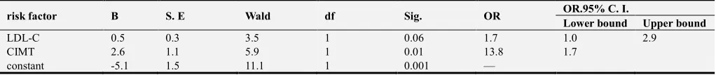
factors and the early results of examination CIMT, ABI, PWV as the independent factors. Then the equation is solved and two risk factors LDL-C and CIMT are incorporated with no protective factors. The final regression equation is as follows: AS =0.5LDL-C+2.6CIMT -5.1. The χ2 testing of the model is carried out as: χ2=10.295, P<0.05. The result of multiple-factor analysis shows that LDL-C and CIMT are the risk factors of atherosclerosis. The OR value of LDL-C is 1.7 while of CIMT is 13.8, which means that the possibility of atherosclerosis increases by 1.7 more times (1.0-2.9) as the LDL-C value increases by a unit while increases by 13.8 more times (1.7-114.4) as the CIMT value increases by a unit. The CIMT value of the atherosclerosis group is higher than that of non-atherosclerosis group (see Table 2).
Table 2. Risk factors of atherosclerosis.
risk factor B S. E Wald df Sig. OR OR.95% C. I.
Lower bound Upper bound
LDL-C 0.5 0.3 3.5 1 0.06 1.7 1.0 2.9
CIMT 2.6 1.1 5.9 1 0.01 13.8 1.7
constant -5.1 1.5 11.1 1 0.001 —
To further exploring the two risk factors between two groups, we regard the two risk factors as the independent variables and
the existence of atherosclerosis as the dependent variable. Then the results of two groups are compared (see Table 3).
Table 3. Results of LDL-C and CIMT for atherosclerosis and non-atherosclerosis (x±s).
index group n ((((x±s)))) t p
LDL-C non-atherosclerosis 70 2.95±0.83
-1.928 0.065
atherosclerosis 29 3.36±1.32
CIMT non-atherosclerosis 70 0.93±0.24
-2.536 0.027
atherosclerosis 29 1.12±0.33
Comparing LDL-C and CIMT between two groups, the results imply that the CIMT value of the atherosclerosis group is higher than that of non-atherosclerosis group (t=-2.5,
P<0.05), which is of statistical significance. It demonstrated that CIMT can be regarded as an early predictor of atherosclerosis in type 2 diabetic patients.
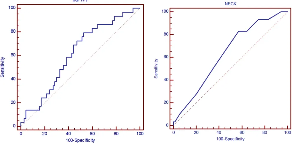
3.4. The Values of CIMT, PWV and ABI
CIMT and PWV contribute predictive value to the detection
of atherosclerosis, while ABI is barely satisfactory as a prediction. To evaluate the diagnostic value of CIMT, PWV and ABI, the area under the ROC curve (AUC) of the each index was computed.
Figure 1. ROC curves on PWV (left one) and CIMT (right one)prediction of atherosclerosis.
Figure 1 through analysis of ROC curves on CIMT and PWV, it turns out that CIMT (AUC=0.638) and PWV (AUC=0.625) contribute predictive value to the detection of atherosclerosis.
In terms of the results above, the sensitivity and specificity of the considerable CIMT and PWV index was further analyzed (see Table 4). The diagnostic boundary value was identified according to the point in the curve which is nearest to the optimal sensitivity and specificity. The results showed that the diagnostic boundary value of CIMT is 0.9mm which is consistent with the majority of reference while the diagnostic boundary value of PWV is 1560.
Table 4. diagnostic boundary value of CIMT and PWV index.
index diagnostic boundary value Sensitivity specificity
CIMT 0.9 82.76 42.86
PWV 1560 79.31 47.14
The ability of predicting atherosclerosis equals between the combinative index and the single ABI. To detect the ability of predicting atherosclerosis in the two indices of ABI and PWV, the linear regression analysis was conducted. Using linear regression analysis (F=0.083; P=0.921; R2=0.002), the results as above showed that the combinational indexes and ABI alone had the same ability to detect atherosclerosis, that is to say they had no statistical significance.
4. Discussion
Macrovascular complications are the leading cause of cardiovascular disease or even death in patients with T2DM. Compared with patients without atherosclerosis, risks of acute myocardial infarction is increase in patients with carotid atherosclerosis. In a long-term state of hyperglycemia,
non-enzymatic proteins are glycated to generate advanced glycation end products (AGEs), which act as an important part to the formation of atherosclerosis. AGEs, which are conducive to lipid deposition and esterification of macrophages and foam cells, can finally lead to impairment of vascular relaxation due to intima thickening, hypercoagulable state and prevention of the nitric oxide production in patients with high concentrations of AGEs. Nitric Oxide is needed as an important factor of vasodilation [15]. Carotid atherosclerotic plaque is regarded as an indicators of systemic atherosclerosis, since carotid arteries are the predilection sites of atherosclerotic plaques and often occur earlier than coronary and cerebral arteries, not only can they reflect systemic atherosclerotic lesions, but also they are considered as a major risk factor for stroke prediction [16]. There is no doubt that positive measures for prevention of atherosclerosis are necessary for patients with T2DM. Therefore, several relevant researches were carried out to explore the atherosclerotic risk factors for patients with T2DM and the indices for the early diagnose.
history, uric acid, insulin resistance.
As a result, it has been found that level of insulin resistance, blood pressure, blood glucose, high-density lipoprotein cholesterol, triglycerides, total cholesterol and body weight are not different between two groups. Smoking history is a risk factor for atherosclerosis. Nevertheless, the reason why these results are not consistent with other related findings may be related to the small sample size. The results show that uric acid doesn’t play a significant role in the development of atherosclerosis occurring in patients with T2DM, however, the role of uric acid in patients with different gender in the development of atherosclerosis is still controversial [17, 18]. Serum uric acid level is an independent risk factor for carotid atherosclerosis in patients with type 2 diabetes [19]. Our results are different from similar studies, the reason could be the regional difference. Particularly, the formation of soft plaque was set as the criterion in grouping patients of atherosclerosis in this study, while the selecting criteria for the above mentioned studies are color doppler.
The analysis of risk factors above indicated that LDL-C was statistically significant (P=0.040). LDL-C can damage endothelial cells and smooth muscle cells by oxidation, modification and glycation. The modified LDL-C finally transform macrophagocytes in intima into foam cells after they phagocytosed LDL-C. It plays an important role in the development of diabetic macrovascular disease, because accumulation and deposition of cholesteryl ester can increase the content of lipid plaque which is closely related to its stability [20]. Multivariate analysis method was used to analyze LDL-C, the result indicated that the possibility of atherosclerosis increased by 1.7 more times as the LDL-C value increased by a unit. This conclusion is also consistent with correlational studies by Yanhong W about analysis on risk factors of carotid atherosclerotic plaques in patients with T2DM who combine with cerebral infarction [21].
The indices like ABI, PWV, CIMT were used to detect the risk factors for atherosclerosis. CIMT was statistically significant in our study (P=0.0270). Combining with multivariate analysis method result, CIMT was regarded as an early predictor of atherosclerosis. It previously demonstrated that the possibility of acute myocardial infarction increased by 11% as the IMT value increased by a unit. Consistent with this conclusion, it suggested that the possibility of atherosclerosis increased by 13.8 more times as the CIMT value increased by 0. 1 mm. So it is necessary, in fact, we should keep a watchful eye on CIMT as an early predictor of atherosclerosis.
Then the indices and the approaches for the early diagnose of the atherosclerotic with T2DM were explored. Soft atherosclerotic plaques are the plaques which protrude in the lumen and their internal structure echos weakly while the hard ones which are fibrotic and calcific have an enhancement echo with acoustic shadow on the back [22]. Compared with hard plaques, soft ones are more instable, which contribute the major part to the causes of cardiovascular and cerebrovascular disease. In this experiment, CIMT was used to detect the formation of plaque and local uplift of artery intima. If the thickness of protruding inside the lumen is equal or greater
less than 1.3mm [21], atherosclerosis is definitely diagnosed. The detecting methods of atherosclerosis include CIMT, Flow-mediated vascular stretch Detection Technology (FMD), High-speed spiral CT, which are mainly used for detection of the advanced atherosclerosis. In the early detection methods, the examination for thickness of carotid intima-media is reliable due to the advantage that it can reflect the structural feature of arterial wall and lesion in the early period. Other noninvasive detections in early years mainly include ETRACKING and the examination of arterial elasticity containing ABI and PWV. Since atherosclerosis in diabetics can affect the whole arterial system and the atherosclerosis in carotid occurs earlier, carotid is used as the window to detect the macroangiopathy [23]. In current clinic, CIMT thickness is regarded as one of the reference index during diagnosing the early atherosclerosis.
Ankle Brachial Index (ABI), which measures the ratio of blood pressure of foot joints and wrist artery simultaneously, is an important index in evaluating peripheral artery disease (PAD). Pulse wave velocity (PWV) is an index of arterial wall hardening and closely related to cardiovascular disease. According to the theory, once atherosclerosis occurs, cardiac output fluctuations in the blood produced (pulse wave) conduction velocity will accelerate, the measurable fluctuations between two heartbeats (pulse wave) conduction velocity determine the degree of flexibility of blood vessels. As an equipment to detect early arteriosclerosis, due to the fact that its non-invasive, accurate, fast and inexpensive, Omron are gradually becoming the major tool used by medical workers and patients. Another study also shows that high ankle-brachial index (ABI) indicated the risk of cardiovascular disease (CVD) and peripheral arterial disease (PAD) in Chinese patients with type 2 diabetes mellitus [24]. Most people consider that the examination of arterial elasticity has a good correlation with risk factors of atherosclerosis due to its repeatability and noninvasive features, which plays a more and more important role for cardiovascular risk prediction in the future [25, 26].
current ABI results and previously reported studies may be due to the different detecting method, and possibly comparatively smaller sample size in the current study.
5. Conclusion
As described in above, in the investigation of 99 diabetic patients, the prevalence rate of atherosclerosis is 29.3%. The statistical analysis was used to probe the risk factors which can accelerate development of AS including clinical indices and to evaluate early examination methods of AS including PWV, ABI, CIMT in the two groups. It demonstrated that SBP, DBP, LDL-C, CIMT, history of smoke were significantly higher in plaques group than that of without plaques. The risk of AS has increased by 1.7 times by every unit of LDL-C, as well as increased by 13.8 times by every unit of CIMT. What’s more, CIMT and PWV, but not ABI, are valuable for AS diagnosing. The best diagnostic value for CIMT is 0.9mm and for PWV is 1560 in this hospital. Consequently, Early intervention and controlling of risks factors will have clinical value for prevention and treatment of macrovascular diseases in T2DM.
Financial Support
This work was supported by the Natural Science Foundation of Yunnan Province (2014FB029).
Acknowledgements
We thank the patients for their participation. We also thank Jing Chen, Ph. D who come from The University of Florida College of Medicine for helping edit the manuscript.
Conflict of Interest
There is no competing financial interests exist.
References
[1] Khazai B, Luo Y, Rosenberg S, et al. Coronary Atherosclerotic Plaque Detected by Computed Tomographic Angiography in Subjects with Diabetes Compared to Those without Diabetes. Plos One. 2015; 10:e0143187.
[2] Yahagi K, Kolodgie F D, Lutter C, et al. Pathology of Human Coronary and Carotid Artery Atherosclerosis and Vascular Calcification in Diabetes Mellitus. Arteriosclerosis Thrombosis & Vascular Biology. 2017; 37:191.
[3] Farrag A, Bakhoum S, Salem M A, et al. The association between extracoronary calcification and coronary artery disease in patients with type 2 diabetes mellitus. Heart & Vessels. 2013; 28:12-18.
[4] Madonna R, De Caterina R. Cellular and molecular mechanisms of vascular injury in diabetes--part II: cellular mechanisms and therapeutic targets. Vascular Pharmacology. 2011; 54:75-79.
[5] Rundek T, Gardener H, Della-Morte D, et al. The Relationship
between Carotid Intima-Media Thickness and Carotid Plaque in the Northern Manhattan Study. Atherosclerosis. 2015; 241:364-370.
[6] Steinl D C, Kaufmann B A. Ultrasound imaging for risk assessment in atherosclerosis. International Journal of Molecular Sciences. 2015; 16:9749-9769.
[7] Laurent S, Boutouyrie P. Arterial stiffness: a new surrogate end point for cardiovascular disease?. J Nephrol. 2007; 20 Suppl 12:S45-50.
[8] Katakami N, Osonoi T, Takahara M, et al. Clinical utility of brachial-ankle pulse wave velocity in the prediction of cardiovascular events in diabetic patients. Cardiovascular Diabetology. 2014; 13:1-11.
[9] Maeda Y, Inoguchi T, Etoh E, et al. Brachial-ankle pulse wave velocity predicts all-cause mortality and cardiovascular events in patients with diabetes: the Kyushu Prevention Study of Atherosclerosis. Diabetes Care. 2014; 37:2383.
[10] Ninomiya T, Kojima I, Doi Y, et al. Brachial-ankle pulse wave velocity predicts the development of cardiovascular disease in a general Japanese population: the Hisayama Study. Journal of Hypertension. 2013; 31:477-483.
[11] Youheng W, Wei Y, Junpeng F. Analysis of influence factors and the observation of carotid atherosclerosis in type 2 diabetic patients. Shaanxi Medical Journal. 2012; 41:451-453.
[12] Wang H Y, Da yi H U, Zhu T G. Study of Relationship between Coronary Lesion and the Large Artery Structure and Function. Chinese Journal of Medical Imaging Technology. 2002; 18:1230-1232.
[13] Yan-Li H U, Zhao-Ping L I, Zhang H X, et al. Evaluation of early arteriosclerosis in patients with diabetes mellitus by several non-invasive methods. Chinese Journal of Cardiovascular Medicine. 2010; 15:206-209.
[14] Salonen JT, Salonen R. Ultrasound B-mode imaging in observational studies of atherosclerotic progression. Circulation. 1993; 87:II56-65.
[15] Basta G, Schmidt A M, De C R. Advanced glycation end products and vascular inflammation: implications for accelerated atherosclerosis in diabetes. Cardiovascular Research. 2004; 63:582.
[16] Wu, Yan, He, et al. Carotid atherosclerosis and its relationship to coronary heart disease and stroke risk in patients with type 2 diabetes mellitus. Medicine. 2017; 96:e8151.
[17] Kawamoto R, Tomita H, Oka Y, Ohtsuka N. Relationship between serum uric acid concentration, metabolic syndrome and carotid atherosclerosis. Intern Med. 2006; 45:605-614.
[18] Nagahama K, Iseki K, Inoue T, Touma T, Ikemiya Y, Takishita S. Hyperuricemia and cardiovascular risk factor clustering in a screened cohort in Okinawa, Japan. Hypertens Res. 2004; 27:227-233.
[19] Li Q, Yang Z, Lu B, et al. Serum uric acid level and its association with metabolic syndrome and carotid atherosclerosis in patients with type 2 diabetes. Cardiovascular Diabetology. 2011; 10:72-72.
[21] Yanhong W, Pingping L, Jun W. Related risk analysis of factors for carotid atherosclerotic plaque in type 2 diabetes mellitus patients with cerebral infarction. Chinese Journal of Integrative Medicine on Cardio/Cerebrovascular Disease. 2012; 10:433-434.
[22] Hui C, Liming Y. Interventional treatment of acute occlusion of limb arteries after trauma. CONTEMPORARY MEDICINE. 2011; 17:57-58.
[23] Xiao-Li X U, Xiong J Q, Heng-Qing L I, et al. Echo-tracking technique for assessing carotid artery elasticity in patients with type 2 diabetes mellitus and hyperlipidemia. Journal of Practical Medicine. 2009; 25:1583-1585.
[24] Li Q, Zeng H, Liu F, et al. High Ankle-Brachial Index Indicates Cardiovascular and Peripheral Arterial Disease in Patients With Type 2 Diabetes. Angiology. 2015; 66:918.
[25] Ter A E, Stalenhoef A F, De G J. What is the role of non-invasive measurements of atherosclerosis in individual cardiovascular risk prediction? Clinical Science. 2007; 112:507-516.