R E S E A R C H
Open Access
Application of neural oscillators to study the
effects of walking speed on rhythmic activations
at the ankle
Sook-Yee Chong, Heiko Wagner
*and Arne Wulf
* Correspondence: [email protected] Department of Motion Science, University of Muenster, Horstmarer Landweg 62b, Muenster 48149, Germany
Abstract
Background:Spinal pattern generators (SPG) are neural networks in the spinal cord that do not require a central input from the brain to generate a motor output. We wanted to determine whether SPG can adapt to the changing motor demands from walking at different speeds, and performing silly walks.
Methods:An SPG model consisting of an oscillator made up of two neurons was utilised in this study; one neuron activates the soleus and the other activates the tibialis anterior. The outputs of the SPG model therefore represent the electromyographic measurements from each muscle. Seven healthy subjects were requested to perform silly walks, normal walking at self-selected speed (4.8 ± 0.5 km/h), 3.5 km/h, 4.0 km/h and 4.5 km/h on a treadmill. Loading and hip angles were used as inputs into the model.
Results:No significant differences in the model parameters were found between normal walking at self-selected speed and other walking speeds. Only the adaptation time constant for the ankle flexor during silly walks was significantly different from the other normal walking trials.
Conclusion:We showed that SPG in the spinal cord can interpret and respond accordingly to velocity-dependent afferent information. Changes in walking speed do not require a different motor control mechanism provided there is no disruption to the alternating muscular activations generated at the ankle.
Keywords:Locomotion, Walking speed, Silly walks, EMG, Spinal pattern generator, Neural network, Sensory afferents, Motor control
Background
Afferents interact continuously with different parts of the nervous system so as to enable a smooth and efficient gait. As the human nervous system should coordinate efficiently, responding and adapting to the immediate environment, it is important that the plethora of signals coming from the central, sensory and peripheral systems be selected and modulated, so that the motor output fulfils the demands of the locomotor task.
Different control networks at different levels of the nervous system contribute to human motor control. The lowest level of neural control, which is responsible for generating the basic patterns of locomotion, is believed to come from spinal pattern
generators (SPG) located in the spinal cord [1-3]. Brown [4] showed that decerebrate cats can produce locomotor-like electromyographic (EMG) patterns while walking on a treadmill. Similar results were also obtained from other vertebrates and invertebrates [1,5]. A study by Maegele et al. in 2002 [6] showed that clinically incompletely and completely spinal cord-injured patients can activate lower limb muscles after treadmill therapy. While these studies successfully showed the ability of the SPG to produce a motor output with no interference from the brain, they also demonstrated that the interactions between SPG and sensory inputs are important in generating a dynamic movement [7]. Taga [8] had shown that a real-time dynamic interaction between the neural and mechanical system, together with sensory information from the environ-ment, could influence the motor output of the lower limbs.
Walking at a slower or faster pace creates different motor demands on the neural system. A number of gait components such as stance and swing phase intervals and muscle activations change with increasing speed. However, in healthy humans, it is not known whether these changes result from sensory cues to the neural network in the spinal cord, since higher commands from the brain can intervene. The aim of our study was to determine whether neural networks in the spinal cord can adapt to changing sensory afferents, and directly influence muscular activity to meet the motor demands of walking at different speeds. We therefore studied the response of the SPG model in situations where gait components in a gait cycle will be different from normal walking: change in walking speed and in performing“silly walks”. Our study used an SPG model that is triggered only by sensory afferents with no interference from a cortical signal.
Methodology
Seven healthy male subjects (28.0 ± 4.4 years, 1.8 ± 0.1 m, 76.4 ± 9.5 kg) volunteered to participate in this study. They were thoroughly informed of the procedures and gave their consent. Each subject was requested to walk at his normal self-selected speed (4.8 ± 0.5 km/h), at 3.5 km/h, 4.0 km/h and 4.5 km/h on a treadmill (Kinetics s3, Kettler, Germany). In addition, they were asked to perform movements unlike their normal walking, i.e.“silly walks”, at a speed of their own choice (3.8 ± 0.4 km/h). Data from six consecutive strides were collected during steady-state walking. Three trials were recorded for each subject for each walking speed and silly walks, i.e. a total of 210 trials (for both right and left limbs). Trials were ignored if there were missing data in any one stride. Therefore, a total of only 176 trials was analysed in this study.
dorsiflexor muscles, respectively. Electrodes were placed according to recommendations by Hermens et al., 1999 [9]. The electromyographic (EMG) signals were centred, rectified and filtered using a fifth-order low-pass Butterworth filter with a cut-off frequency of 40Hz.
A simple Matsuoka oscillator [10,11] consisting of two neurons was used (Figure 1); one neuron activated the SOL and the other activated the TA. Thus, the outputs from the oscillator represented the corresponding activation of each muscle. The neurons were mutually inhibited, i.e. when one neuron was activated, the other was suppressed.
The oscillator was governed by the following equations (adapted from Matsuoka, 1985 [10]):
_
xiþxi¼
Xj
iaijyjþsibif ð1Þ
Tif_iþfi¼yi ð2Þ
yi¼ max 0ð ;xiÞ ð3Þ
wherefis the adaptation in the neuron, andTandb are the parameters that determine the time course of the adaptation. When b= 0 there is no adaptation and the output will increase and then remain at a constant value (Refer to Figure 1 in Matsuoka [10]).
xis the inner state of the neuron,yis the generated output of the neuron,sis the input signal, andais the strength of the connection between the two neurons;aij<0 fori≠j
(mutual inhibition) and >0 fori = j(self-excitation). We assume a symmetrical arrange-ment of neurons, i.e.aij= aji, aii= ajj.
The model is triggered by both the magnitude and the change in magnitude of loading and hip angles. Vertical force calculated from the insoles was first normalised to the subject’s weight. Normalised forceFand hip flexion/extension anglesHAof the ipsilateral limb (in radians) were used to determine the signal inputsiin equation (1):
si¼mi:pþni:p_þwi:qþvi:q_ ð4Þ with
_
p¼r1ðFpÞ ð5Þ
_
q¼r2ðHAqÞ ð6Þ
wherem, n, w and vrepresent the weights of each excitationp,p_,q,q_ respectively. The parametersa, b, m, n, r, T, v, wfrom the above equations determined the pattern and frequency of the output. A nonlinear least-squares fitting algorithm was used to determine a set of parameters that would fit the output to experimental data i.e. the output produced by the neuron representing the SOL would be fitted to measured EMG data of the SOL. This was done simultaneously for the TA. Initial values for each neuron were taken from the first value of the measured EMG data so as to solve the differential equations numerically. The fitting algorithm terminated once the relative deviation between two iterations fell below 0.001. A correlation coefficient R between the model output and experimental EMG data was calculated in each trial.
The following gait components were analysed; Maximum normalised force and maximum range of hip flexion-extension angles were calculated for each stride. Stance and swing phases determined from force profiles of each stride were also calculated. For these gait components, analysis of variance (ANOVA) and Tukey’s post-hoc test were performed to determine the significant differences between all the different walking types. In analysing the rectified EMG signals for different speeds, we adopted the method by Murray et al., 1984 [12]. Cumulative numerical integration (IEMG) for each EMG signal in each stride was calculated for all speeds. The maximum of the mean IEMG values was designated as 1.00, regardless of speed. The other mean values were normalised with respect to this maximum value [12]. To determine significant differences in the model parameters (p < 0.05), multivariate analysis of variance (MANOVA) along with analysis of variance (ANOVA) and Tukey’s post-hoc test were performed.
Results
No significant differences in R were found between normal walking at self-selected speeds and walking at other speeds. However, Rcalculated for silly walks (mean corre-lation Rmean= 0.70 ± 0.08) was significantly lower than for the other walking types
Figure 2Correlation coefficients (R) at different speeds and silly walks.The tops and bottoms of the boxes are the 25th and 75th percentiles ofRrespectively. Red lines indicate the median values. (*) denotes significant difference.
Significant differences were found in the gait components calculated (Table 1). As expected, an increase in walking speed is accompanied by a decrease in the relative stance phase duration, an increase in the relative swing phase duration, an increased range of hip flexion-extension angles, and increased peak activation values of the SOL and TA [12]. Since loading and hip angles were significantly different, this meant that inputs to the SPG model differed significantly for all walking types.
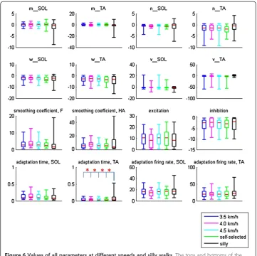
MANOVA revealed significant differences between the model parameters. To continue with the analysis, ANOVA followed by Tukey’s post-hoc test revealed no significant differences between the model parameters for normal walking at self-selected speeds and
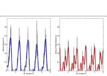
Figure 4Rave= 0.80.Muscle activation of the soleus (RSOL= 0.90) and the tibialis anterior (RTA= 0.71) of subject #1 walking at 4.0km/h with insole forces and hip angles as inputs (bold lines represent the output from the SPG model and thin lines the experimental EMG data).
other speeds (Figure 6). Only T2, the constant describing the time lag of the adaptation
effect in the TA, showed significant differences between the silly walks and the normal walking trials.
Discussion
The results of this study showed that neural networks in the spinal cord can activate muscles at the ankle to generate stepping motion during steady-state walking. In humans, it is difficult to determine whether the elevated EMG patterns during walking result from supraspinal control, activations from sensory inputs, or an interaction between supraspinal and spinal control. However, the outputs generated by our model, consisting only of spinal neurons, suggest that muscle activations can be generated by sensory inputs from loading and hip angles at the spinal level [13].
While there were significant differences in both the inputs (loading and hip angles) and outputs (IEMG) between the different walking speeds and silly walks (Table 1), significant differences were only found in T2 between silly walks and the other walking tests
(Figure 6). For normal walking at different speeds, this might imply that an insignificant change in a parameter is sufficient to cause a significant change in the output. Since the control of these parameters, which determine the neuronal properties of the SPG, could come from interneurons, presynaptic inhibition [11], or through descending pathways from supraspinal structures, the insignificant changes might imply that no regulation by the brain or inter-spinal circuitry is required to modulate the activation patterns during walking. Comparing our study to split-belt treadmill locomotion, Morton and Bastian (2006) found that subjects with cerebellar damage were able to perform rapid reactive adjustments to stride length and stance time when their legs were operating at different speeds [14]. Our study corroborates their findings that higher control is not needed to alter the motor output of moving limbs, but the corrections could instead be performed predominantly by spinal structures using available sensory information. It has also been suggested that the same neural circuitry is responsible for gait transitions between walking and running [15]. Thus, while an input from the cerebral cortex is required to initiate a movement, higher command centres need not be recruited to regulate motor output dur-ing locomotion regardless of speed.
Grasso et al. [16] suggested that the nervous system attempts to meet motor demands by controlling posture or limb joint motion rather than regulating muscle activations. We agree with their arguments, since we successfully used loading and hip angles as inputs to the SPG model to generate muscle activations. In addition, provided the gait patterns do not result in changes to equilibrium, the same neural network will be utilised [17]. Since the data were captured during steady-state walking, it might also be important that the alternating activations of the flexor and extensor are not disrupted. Perhaps, changes to gait components are secondary, and could result from changes in stride length rather than a different motor control mechanism.
activity increased significantly. Therefore, as in humans, no conscious effort is neces-sary during level walking regardless of walking pace, until an obstacle or a sudden change in the external environment is encountered, when corrective responses are required.
We expected differences in the silly walks as the subjects were intentionally requested to perform a movement unlike normal walking. Since the subjects were consciously aware that they had to perform‘something silly’, we postulated that the resulting muscle activations were due to a command from the brain. However, we found significant changes only inT2, the adaptation time constant for the TA. Persistent inward currents
(PIC) are known to be essential for the firing of motor neurons [20]. It was speculated that PIC are expressed in the extensors from birth, but less so in the flexors [21], be-cause while the extensors are mostly activated during walking, the flexors do not re-quire long-lasting bursts. It would therefore be more economical to modulate the flexors rather than the extensors. Nevertheless, it remains uncertain whether the TA requires more intervention from the brainstem or more neural circuitries than the
Table 1 Mean and standard deviation (std) of gait components at different speeds and during silly walks
3.5 km/h 4.0 km/h 4.5 km/h Self-selected 4.8 ± 0.5 km/h Silly walks 3.8 ± 0.4 km/h p
Mean std Mean std Mean std Mean std Mean std
Stance (%) 66.60 ±4.85 65.59 ±4.34 65.22 ±4.48 64.99 ±1.58 63.33 ±7.34 p < 0.05
Swing (%) 32.97 ±3.01 34.05 ±2.70 34.38 ±2.79 35.01 ±1.58 36.67 ±7.34 p < 0.05
Hip flexion-extension range (rad) 0.67 ±0.21 0.73 ±0.22 0.76 ±0.26 0.77 ±0.07 0.84 ±0.32 p < 0.05
Max F 1.11 ±0.19 1.17 ±0.22 1.23 ±0.25 1.26 ±0.20 1.25 ±0.29 p < 0.05
IEMG_SOL 0.47 ±0.26 0.51 ±0.27 0.53 ±0.31 0.53 ±0.20 0.69 ±0.20 p < 0.05
IEMG_TA 0.42 ±0.26 0.47 ±0.28 0.51 ±0.32 0.53 ±0.21 0.65 ±0.21 p < 0.05
Theoretical
Biology
and
Medical
Modelling
2013,
10
:9
Page
9
o
f
1
1
.com/content
SOL. The significant difference in T2 could also be due to the SPG model, which
requires a strong adaptation effect in generating stable oscillations (Additional file 1) (T2, silly walks= 0.12 ± 0.17 compared to T2, self-selected= 0.03 ± 0.04) [10]. The higher T2
value could therefore just be a way for the model to continue generating stable oscillations.
A limitation in this study was the restricted array of silly walks the subjects could perform while walking on a treadmill at a constant speed (an example is shown in ‘Additional file 1’). The movements performed by the subjects still involved an on-going, uninterrupted rhythmic pattern of activation between the antagonistic muscles at the ankle. Since we now know the same neural network is responsible for normal walking at different speeds, future studies can give the subjects a freer choice of the types of silly walks they would like to perform (like those seen in Monty Python’s sketch,The Ministry of Silly Walks). In such studies, significant differences in more model parameters might be found.
Conclusion
We proposed that SPG in the spinal cord can interpret and respond accordingly to velocity-dependent afferent information. Changes in walking speed do not require a dif-ferent motor control mechanism provided equilibrium is not affected and there is no disruption of the continuous rhythmic patterns produced at the ankle.
Additional file
Additional file 1:One subject performing a silly walk.
Abbreviations
SPG: Spinal pattern generators; EMG: Electromyographic; SOL: M. soleus; TA: M. tibialis anterior;a: Mutual inhibition (a < 0), self-excitation (a > 0);b: Determine time course of adaptation in neuron;f: Adaptation in neuron;m: Weight of hip angles input;n: Weight of change of hip angles;p: Smoothed force;q: Smoothed hip angles;r: Smoothing coefficient;s: Signal input;T: Determine time course of adaptation in neuron;v: Weight of change of force input; w: Weight of force input;x: Inner state of neuron;y: Neural output; F: Force; HA: Hip angles; Max F: Maximum force; IEMG: Cumulative numerical integration of EMG signals; IEMG_SOL: Cumulative numerical integration of soleus EMG signals; IEMG_TA: Cumulative numerical integration of tibialis anterior EMG signals; MANOVA: Multivariate analysis of variance; ANOVA: Analysis of variance; std: Standard deviation; ave: Average; PIC: Persistent inward currents.
Competing interest
The authors declare that they have no competing interests.
Authors' contributions
All the authors contributed significantly to the conception and design of the project. SY acquired, analysed and interpreted the data. All authors drafted and revised the manuscript. All authors read and approved the final manuscript.
Acknowledgements
This study was supported by the German Federal Ministry of Education and Research (BMBF), project no. 01EC1003A. Technical assistance was provided by Hanno Focke and David Schinowski. We acknowledge support by the German Research Foundation (DFG) and Open Access Publication Fund of University of Muenster.
Received: 15 May 2012 Accepted: 8 February 2013 Published: 13 February 2013
References
1. Grillner S, Wallen P:Central pattern generators for locomotion, with special reference to verterbrates. Ann Rev Neurosci1985,8:233–61.
2. Ijspeert A:Central pattern generators for locomotion control in animals and robots: a review. Neural Netw2008,21:642–53.
4. Brown TG:On the nature of the fundamental activity of the nervous centres; together with an analysis of the conditioning of rhythmic activity in progression, and a theory of the evolution of function in the nervous system.J Physiol-London1914,48:18–46.
5. Marder E, Bucher D:Central pattern generators and the control of rhythmic movements.Curr Biol2001, 11:R986–96.
6. Maegele M, Müller S, Wernig A, Edgerton VR, Harkema SJ:Recruitment of spinal motor pools during voluntary movements versus stepping after human spinal cord injury.J Neurotrauma2002,19(10):1217–29.
7. Baessler U:On the definition of central pattern generator and its sensory control.Biol Cybern1986,54:65–9. 8. Taga G:A model of the neuro-musculo-skeletal system for human locomotion. i. emergence of basic gait.
Biol Cybern1995,73:97–111.
9. Hermens HJ, Freriks B, Merletti R, Stegeman DF, Blok J, Rau G, Disselhorst-Klug C, Haegg G:European
Recommendations For Surface Electromyography, Results Of The SENIAM Project. Enschede: Roessingh Research and Development; 1999.
10. Matsuoka K:Sustained oscillations generated by mutually inhibiting neurons with adaptation.Biol Cybern 1985,52:367–76.
11. Matsuoka K:Mechanisms of frequency and pattern control in the neural rhythm generators.Biol Cybern1987, 56:345–53.
12. Murray MP, Mollinger LA, Gardner GM, Sepic SB:Kinematic and EMG patterns during slow, free, and fast walking.J Orthop Res1984,2:272–80.
13. Chong SY, Wagner H, Wulf A:Neural oscillators triggered by loading and hip orientation can generate activation patterns at the ankle during walking in humans.Med Biol Eng Comput2012,50(9):917–23. 14. Morton SM, Bastian AJ:Cerebellar contributions to locomotor adaptations during splitbelt treadmill walking.
J Neuroscience2006,26:9107–16.
15. Labini FS, Ivanenko YP, Cappellini G, Gravano S, Lacquaniti F:Smooth changes in the EMG patterns during gait transitions under body weight unloading.J Neurophysiol2011,106:1525–36.
16. Grasso R, Bianchi L, Lacquaniti F:Motor patterns for human gait: backward versus forward locomotion. J Neurophysiol1998,80:1868–85.
17. Lamb T, Yang JF:Could different directions of infant stepping be controlled by the same locomotor centre pattern generator?J Neurophysiol2000,83:2814–24.
18. Armstrong DM:The Supraspinal control of mammalian locomotion.J Physiol1988,405:1–37.
19. Armstrong DM, Drew TL:Discharges of pyramidal tract and other motor cortical neurones during locomotion in the cat.J Physiol1984,346:471–95.
20. Li Y, Gorassini MA, Bennett DJ:Role of persistent sodium and calcium currents in motoneuron firing and spasticity in chronic spinal rats.J Neurophysiol2004,91:767–83.
21. Cotel F, Antri M, Barthe JY, Orsal D:Identified ankle extensor and flexor motoneurons display different firing profiles in the neonatal rat.J Neurosci2009,29:2748–53.
doi:10.1186/1742-4682-10-9
Cite this article as:Chonget al.:Application of neural oscillators to study the effects of walking speed on rhythmic activations at the ankle.Theoretical Biology and Medical Modelling201310:9.
Submit your next manuscript to BioMed Central and take full advantage of:
• Convenient online submission
• Thorough peer review
• No space constraints or color figure charges
• Immediate publication on acceptance
• Inclusion in PubMed, CAS, Scopus and Google Scholar
• Research which is freely available for redistribution