BMC Medical Education 2001,
1 :5
Research article
Case-oriented computer-based-training in radiology: concept,
implementation and evaluation
Martin Dugas*
1
, Christoph Trumm
2
, Axel Stäbler
2
, Ernst Pander
2
,
Walter Hundt
2
, Jurgen Scheidler
2
, Roland Brüning
2
, Thomas Helmberger
2
,
Tobias Waggershauser
2
, Matthias Matzko
2
and Maximillian Reiser
2
Address: 1Department of Medical Informatics, Biometrics and Epidemiology (IBE) (Chairman: Prof. Dr. med. K. Überia), University of Munich, Marchioninistr. 15, D-81377 Munich, Germany and 2Department of Clinical Radiology (Chairman: Prof. Dr. med. M. Reiser), University of Munich, Germany
E-mail: Martin Dugas* - [email protected]; Christoph Trumm - [email protected]; Axel Stäbler - [email protected]; Ernst Pander - [email protected];
Walter Hundt - [email protected]; Jurgen Scheidler - [email protected]; Roland Brüning - [email protected]; Thomas Helmberger - [email protected]; Tobias Waggershauser - [email protected]; Matthias Matzko - [email protected]; Maximillian Reiser - [email protected]
*Corresponding author
Abstract
Background: Providing high-quality clinical cases is important for teaching radiology. We developed, implemented and evaluated a program for a university hospital to support this task.
Methods: The system was built with Intranet technology and connected to the Picture Archiving and Communications System (PACS). It contains cases for every user group from students to attendants and is structured according to the ACR-code (American College of Radiology) [2]. Each department member was given an individual account, could gather his teaching cases and put the completed cases into the common database.
Results: During 18 months 583 cases containing 4136 images involving all radiological techniques were compiled and 350 cases put into the common case repository. Workflow integration as well as individual interest influenced the personal efforts to participate but an increasing number of cases and minor modifications of the program improved user acceptance continuously. 101 students went through an evaluation which showed a high level of acceptance and a special interest in elaborate documentation.
Conclusion: Electronic access to reference cases for all department members anytime anywhere is feasible. Critical success factors are workflow integration, reliability, efficient retrieval strategies and incentives for case authoring.
Background
Access to radiological expert knowledge for a broad
spec-trum of users – from students to senior radiologists – is a continuous challenge in a routine clinical setting of a Published: 19 October 2001
BMC Medical Education 2001, 1:5
Received: 30 July 2001 Accepted: 19 October 2001
This article is available from: http://www.biomedcentral.com/1472-6920/1/5
university hospital. A balance between established and new teaching methods is needed to provide high quality, peer-reviewed content.
The knowledge transfer normally takes place during teaching sessions in small groups, lectures or in the con-text of a specific case during daily routine by personal in-teraction e.g. between attendant physicians and residents, house officers and students. For this reason all members of the radiologic department are involved in a general teaching file.
In this context we developed a new software tool to col-lect and document relevant cases – according to all dif-ferent levels of radiologic knowledge – from daily routine in an integrated manner by including all types of image sources. Access to this Intranet-based Teaching File – the "Lehrarchiv" – should be provided from any worksta-tion within the department.
The design of this teaching file should – in the long run – integrate all department members both as case authors and normal users. At the same time a high, homogeneous quality level should be guaranteed from the beginning.
By a differentiated authorization system two opposite prerequisites had to be fulfilled: On the one hand the necessary technical infrastructure to provide access to the system should be ensured, i.e. access to the Intranet should be possible anytime anywhere within the depart-ment. On the other hand clinical data with patient infor-mation and images must be protected against unauthorized use.
Another critical issue concerning the infrastructure is ac-quisition and archiving of image data, i.e. the pathway of the image from its generation to the teaching file. All ra-diological techniques should be included, especially CT, MR, conventional X-ray, angiography and ultrasound. In this context our objective was to answer the following questions:
• Is a teaching file with access anytime anywhere within the radiologic department and smooth integration into routine workflow feasible from a technical point of view?
• What format and data structure is appropriate for a ra-diologic case description?
• What kind of retrieval functions are required?
• Is an electronic radiologic teaching file accepted by stu-dents and physicians?
Materials and Methods
Image acquisition
In general there are three methods for image acquisition for a teaching file:
1. Secondary digitization of a primary analogous image e.g. a conventional radiographic film and subsequent electronic transfer transfer to the teaching file. This method is rather time-consuming and implies loss of im-age quality due to digitization, but is typically required when external radiographs or slides are included; the ad-ditional effort must be weighed against the relevance for the case presentation on an individual basis.
2. Transfer of a digital image from an external data source into the teaching file. This may be a digital image primarily stored in an electronic archive or digitized for other reasons.
3. Direct transfer of primary digitally generated, recent patient images (CT, MR etc.) into the teaching file.
All three methods have been implemented for our teach-ing file. The problems associated with different media are very common at present and characterize the current change from classical film-based to computerized radiol-ogy.
The direct digital transfer is – with respect to workflow integration and image quality – obviously the most at-tractive method and is the preferred way in the context of the establishment of PACS. This method becomes even more relevant, because the number of images per case is increasing continuously due to sophisticated radiological techniques. For this reason manual transfer of single im-ages becomes more and more impractical.
The technical implementation was realized with a DI-COM (Digital Imaging and Communications in Medi-cine) [1] interface – an international multi-vendor standard -, which enables the transfer of selected images to the teaching file in a very simplistic manner, similar to printing images. In order to preserve confidentiality the patient's identification is eliminated when a case is pub-lished.
Software concept
The teaching file system acts basically as a DICOM re-ceiver, i.e. the case repository behaves like a printing de-vice.
By means of a web frontend and individual user accounts each author can access and edit his own cases on the teaching file server. Authorized users can release cases for the public case repository.
We applied an iterative software design approach with rapid prototyping, i.e. we built prototypes, tested and continuously refined them; altogether about 10 iteration cycles were required. The data structure consists of two tables (cases and images) with altogether 23 items.
Hard- and Software
The technical concept is based on established Internet tools. An Apache [4] Webserver (version 1.4.2) on a Linux [5] machine (Distribution SuSE 6.3) provides PERL [6] (version 5.005) programs accessing a relation-al database [7]. The department network consists of a Gi-gabit-Backbone and NT-workstations with 100 MBit connections.
Evaluation Method
To quantify user acceptance we applied a paper based survey. Categorized items were recorded on a six point Lickert scale, from 1=absolutely correct to 6=completely wrong. Scores are given as mean +/- standard deviation.
Results
Case authoring
After a program development phase of approximately one year and 18 months of clinical routine use there are now approx. 350 released cases accessible (283 are work in progress); 31 users from our radiologic department are registered, but only eight of them contributed to more than 80% of cases.
The radiologist can select relevant images on a DICOM workstation during his routine work and send these to the teaching file through the DICOM-Send procedure in analogy to printing. At a later time he can connect to his teaching file account and edit the case. In addition he can upload secondary digitized images.
The textual description (free text) consists of four sec-tions:
• Diagnosis, which is mandatory,
• medical history / clinical presentation / laboratory, • radiologic findings and
• comment.
An efficient retrieval strategy is required to find "similar" cases with both high precision and recall. For this reason a systematic code is necessary. We developed a specific coding tool to enter the ACR-code in both dimensions (localization / pathology) for each case. If several find-ings are present, up to three ACR code-pairs can be stored. Due to the hierarchical design of the ACR code (e.g. localization Gastrointestinal System – Stomach – Pylorus) similar cases – both in terms of localization or pathology – can be found easily. In addition for each case administrative information (e.g. author ID, date of re-lease) is stored automatically.
Case retrieval
Retrieval of cases in the teaching file, which is shown in Fig. 1, is possible based on diagnosis, ACR-code, keyword or any combination. Due to the hierarchical structure of the ACR code similar pathologies or localizations can be retrieved.
Fig. 2 presents the browsing mode of the teaching file. All information on a particular case is presented. Using the navigation bar, any text information can be excluded; by this means the user can assess his radiological knowl-edge and diagnostic abilites.
Figure 1
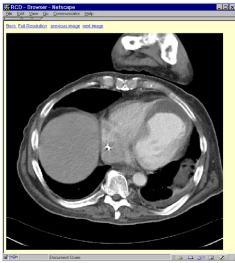
Fig. 3 provides a detailed view of a single image in the system. By means of navigation links all images associat-ed with a case can be viewassociat-ed in high resolution.
In addition to the retrieval function users can select spe-cific case series which are aggregated by authorized radi-ologists, typically on a specific teaching issue (e.g. selected lung cases). This provides added value for the teaching file: it can be used not only for retrieval of single cases, but also for presentations e.g. in scientific work-shops. Individual teaching sessions on specific topics, e.g. for students, are also provided with this tool.
Data analysis
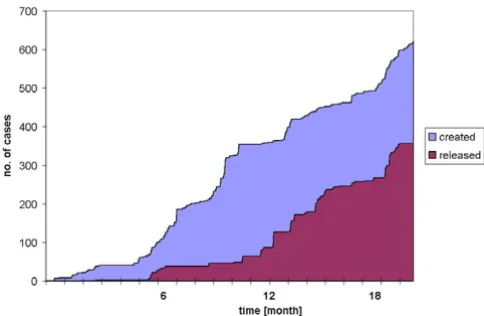
The system has now been in routine use for approximate-ly 18 months. Fig. 4 shows the development of the total number of cases in the radiologic teaching file over time differentiated by case status (created / released). The in-terval between creation and release of a case is 73 +/- 87 days (mean +/- 3.0.).
After 18 months there were 583 records in the table 'Cas-es' consisting of 19 items each, and 4136 records in the table 'Images' with 4 items each.
Evaluation
The system was evaluated by 101 medical students (3rd year) who studied two case series (conventional thorax and liver CT/MR) in small groups (max. 9 persons each, assisted by an instructor) during their mandatory radio-logical course. We included all students from a semester to avoid a selection bias with respect to individual
com-puter expertise. The learners themselves judged their computer knowledge as average (2,92 +/-1,06).
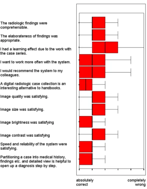
A student questionnaire showed a high level of accept-ance (see Fig. 5) according to the content of the system (comprehensible 2,51 +/- 0,96; appropriate description 2,50 +/- 1,07; subjective learning effect 2,88 +/- 1,33; recommendation of the system to colleagues 2,31 +/-1,24; alternative to handbooks 1,73 +/- 0,90) and image quality (brightness 2,05 +/- 0,86; contrast 2,32 +/- 0,95; size 2,24 +/- 1,10); however, we did not evaluate the gain in knowledge of the students. The software quality of the teaching file was honoured in terms of speed, reliability and case design.
We also asked the students for the optimal number of images to be contained in a "typical case": 10 +/- 5 (mean +/- S.D.). This is influenced by the selection of cases with a focus on CT and MR – probably less images were ap-propriate for conventional chest X-rays.
To improve the system the students suggested to apply interactive pointers or similar mechanisms to highlight specific radiological findings. From a technical point of view this can be implemented easily, but the authors
ar-Figure 2
Browsing mode of the teaching file [translated to English]. All information to a particular case is presented. Using the navi-gation bar, any text information can be excluded; by this means the user can assess his radiological knowledge and diagnostic abilites.
Figure 3
gued this would increase the amount of authoring work substantially.
The results from the questionnaire are consistent with the subjective impression gained during the teaching sessions.
We also did an evaluation with residents (n = 15) on a case series consisting of more complicated cases selected by a senior attendant radiologist. There was again a high level of acceptance (comprehensible cases 1,47 +/- 0,74; recommendation of the system to colleagues 1,60 +/-0,83).
Discussion
Computer-Based-Training (CBT) in Medicine has prov-en its clinical potprov-ential in several settings ([8],[9],[10],[11]). Compared with the situation 10 years ago [12], technological problems such as insufficient computer performance, lack of storage capacity or inad-equate display devices can now be solved.
Despite these advances electronic peer-reviewed and up-to-date teaching files in radiology which can be accessed anywhere in the department are still very rare. Recently, reports of successful CBT in the field of radiology have been published ([13],[14],[15],[16]), but typically the ra-diological content of the systems is very limited (both in terms of cases and in terms of authors).
Lessons learned
The first objective of this study concerns technical feasi-bility of a teaching file with access anytime anywhere within the radiologic department and smooth
integra-tion into routine workflow. From our experience – after 18 months of routine operation – an Intranet-based sys-tem with PACS-interface (i.e. DICOM-compatible) can fulfill this task; it is very important that images can be sent directly from the radiological workstation to the case repository.
There is a wealth of CD-ROMs and other resources ([17],[18],[19]) providing 'snapshots' of radiological knowledge, but a collection of routine clinical reference cases for a specific department and its particular devices is needed. Due to the sophistication of imaging methods a clinically relevant teaching file must be updated contin-uously. This is only feasible by minimizing the effort to create and maintain this database.
The second objective of this study concerns the ideal for-mat and data structure for a radiologic case description. Obviously from an academic point of view a detailed de-scription of the case using controlled vocabularies, in-cluding access to a complete electronic patient record would be desirable. In our setting this was not feasible because it is too time-intensive for the authors. There-fore we decided to apply a compact approach with free text data for medical history, radiological findings, diag-nosis and comment. Only location and pathology were entered in a coded manner, using the internationally es-tablished ACR code.
The third objective of this study addresses retrieval func-tions, which obviously depend on the data available in the system. From our experience both systematic and free text retrieval mechanisms are important. We ap-plied the ACR code, which is characterized by a high level of granularity, to provide a systematic access to the data-base. By this means 'similar' cases – concerning both lo-calization or pathology – can be found easily. We also considered to apply a controlled vocabulary for diagno-sis, but in our setting the effort for systematic coding would have been too high.
Case series arranged by senior radiologists are another method to organize the case collection and facilitate ac-cess.
The fourth objective concerns acceptance of the system by students and physicians. We did not measure the learning success of students, but the questionnaire indi-cates a high level of acceptance which is in line with ob-servations during the course. The constant growth of peer-reviewed cases over time provides good evidence for acceptance by the physicians.
From our experience, integration into daily routine work is the key success factor for a teaching file. Secondary
Figure 4
digitization of images is laborious and implies a loss of quality. Media conversions are such an obstacle that only in a filmless setting with PACS and smooth integration by DICOM-interfaces relevant cases can be collected during daily routine.
Knowledge management
To maintain a high level of quality in patient care is a ma-jor challenge in the context of labour turnover and conti-nous sophistication of radiological methods. The high degree of specialization makes knowledge transfer an important success factor for the department as a whole. There are many recent publications according to the rel-evance of knowledge management ([20],[21]) within or-ganizations.
A technical system like our teaching file is a small, but important building block of a department-wide knowl-edge management strategy, because it provides access to relevant teaching material anytime anywhere. Our eval-uation provides evidence that electronic teaching files are well accepted if quality of content and technical reli-ability is assured. From a medical informatics point of view, such case repositories offer the opportunity of
building decision support systems in the future. Whilst a PACS archive is a huge collection of images, a teaching file is a knowledge database. Due to the systematic cod-ing, an automatic search for similar cases is possible. In-telligent image analysis programs in the past mostly failed because a sufficient knowledge base could not be gathered. Meanwhile sophisticated methods for knowl-edge discovery and data mining ([22],[23]) have been developed, but these techniques require both high-vol-ume and high-quality reference datasets.
By national and international cooperation the vision of peer-reviewed, up-to-date and comprehensive teaching files in radiology could become reality.
Acknowledgements
Mr. Heilmann wrote important computer programs for the prototype sys-tem.
References
1. NEMA's OFFICIAL DICOM WEB Page [resource on World Wide Web]. 2001 [http://medical.nema.org/dicom.html]
2. The American College of Radiology [resource on World Wide Web]. 2000 [http://www.acr.org/]
3. Extensible Markup Language (XML) [resource on World Wide Web]. 2001 [http://www.w3.org/XML]
4. The Apache Software Foundation [resource on World Wide Web]. 2001 [http://www.apache.org]
5. SuSE Linux [resource on World Wide Web]. 2000 [http:// www.suse.de]
6. Wall L, Schwartz RL: Programming PERL.O'Reilly & Associates, Se-bastopol, CA, USA, 1992
7. PostgreSQL [resource on World Wide Web]. 2001 [http:// www.postgresql.org]
8. Evans RS, Pestotnik SL, Classen DC, et al: A Computer-Assisted Management Program for Antibiotics and Other Antiinfec-tive Agents.N EngI J Med 1998, 338:232-238
9. Schwid HA, Rooke GA, Ross BK, Sivarajan M: Use of a computer-ized advanced cardiac life support simulator improves reten-tion of advanced cardiac life support guidelines better than a textbook review.Crit Care Med 1999, 27:821-824
10. Dugas M, Batschkus MM, Lyon HC Jr: Mr. Lewis on the web – how to convert learning resources for intranet-technology.Med Educ 1999, 33:42-46
11. Lyon HC, et al: PlanAlyzer, an Interactive Computer-assisted Program to Teach Clinical Problem Solving in Diagnosing Anemia and Coronary Artery Disease.Acad Med 1992, 67 :821-828
12. Klar R, Bayer U: Computer-assisted Teaching and Learning in Medicine.Int J Biomed Comput 1990, 26:7-27
13. Hornof WJ, allance DW, Brenston PR, Self JA: A client server mod-el to facilitate creation of a medical image teaching library.J Digit Imaging 1999, 12:132-137
14. Zaidel M, Hopper K, lyriboz T: Interactive web-based radiology teaching file.Digit Imaging 1999, 12;2 Suppl 1:203-204
15. Khorasani R, Lester JM, Davis SD, et al: Web-based digital radiol-ogy teaching file: facilitating case input at time of interpreta-tion.AJR 1998, 170:1165-1167
16. Tran TH, Roach NA, O'Kane PL, Thune M: Creating a digital radi-ographic teaching file and database using a PC and common software.AJR 2000, 175:325-327
17. Chew FS, Smirniotopoulos JG: Educational Efficacy of Compu-ter-Assisted Instruction with Interactive Videodisc in Radiol-ogy. In: IMIA Yearbook 1994 (ed. v. Bemmel), Schattauer Verlag, Stuttgart, Germany, 1994468-474
18. Eurorad [resource on World Wide Web]. 2001 [http:// www.eurorad.org/]
19. Richardson ML: World-Wide Web Radiology Teaching File Server on the Internet.AJR 1995, 164:479-483
Figure 5
Publish with BioMed Central and every scientist can read your work free of charge
"BioMedcentral will be the most significant development for disseminating the results of biomedical research in our lifetime."
Paul Nurse, Director-General, Imperial Cancer Research Fund
Publish with BMC and your research papers will be:
available free of charge to the entire biomedical community
peer reviewed and published immediately upon acceptance
cited in PubMed and archived on PubMed Central
yours - you keep the copyright
[email protected] Submit your manuscript here:
http://www.biomedcentral.com/manuscript/
BioMedcentral.com 20. WWW Virtual Library on Knowledge Management
[re-source on World Wide Web]. 2000 [http://www.brint.com/km/]
21. Malhotra Y: Knowledge Management and Virtual Organiza-tions.Idea Group Publishing, Hershey, PA, USA 2000
22. Lavrac N: Selected techniques for data mining in medicine. Ar-tif Intell Med 1999, 16:3-23
23. Morik K, Imboff M, Brockhausen P, Joachims T, Gather U: Knowl-edge discovery and knowlKnowl-edge validation in intensive care.