R E S E A R C H
Open Access
Nanoparticle exposure driven circulating
bioactive peptidome causes systemic
inflammation and vascular dysfunction
Ekaterina Mostovenko
1, Tamara Young
2, Pretal P. Muldoon
1, Lindsey Bishop
3, Christopher G. Canal
1,
Aleksandar Vucetic
1, Patti C. Zeidler-Erdely
3, Aaron Erdely
3, Matthew J. Campen
2and Andrew K. Ottens
1*Abstract
Background:The mechanisms driving systemic effects consequent pulmonary nanoparticle exposure remain unclear. Recent work has established the existence of an indirect process by which factors released from the lung into the circulation promote systemic inflammation and cellular dysfunction, particularly on the vasculature. However, the composition of circulating contributing factors and how they are produced remains unknown. Evidence suggests matrix protease involvement; thus, here we used a well-characterized multi-walled carbon nanotube (MWCNT) oropharyngeal aspiration model with known vascular effects to assess the distinct contribution of nanoparticle-induced peptide fragments in driving systemic pathobiology.
Results:Data-independent mass spectrometry enabled the unbiased quantitative characterization of 841 significant MWCNT-responses within an enriched peptide fraction, with 567 of these factors demonstrating significant
correlation across animal-paired bronchoalveolar lavage and serum biofluids. A database search curated for known matrix protease substrates and predicted signaling motifs enabled identification of 73 MWCNT-responsive peptides, which were significantly associated with an abnormal cardiovascular phenotype, extracellular matrix organization, immune-inflammatory processes, cell receptor signaling, and a MWCNT-altered serum exosome population. Production of a diverse peptidomic response was supported by a wide number of upregulated matrix and lysosomal proteases in the lung after MWCNT exposure. The peptide fraction was then found bioactive, producing endothelial cell inflammation and vascular dysfunction ex vivo akin to that induced with whole serum. Results implicate receptor ligand functionality in driving systemic effects, exemplified by an identified 59-mer
thrombospondin fragment, replete with CD36 modulatory motifs, that when synthesized produced an anti-angiogenic response in vitro matching that of the peptide fraction. Other identified peptides point to integrin ligand functionality and more broadly to a diversity of receptor-mediated bioactivity induced by the peptidomic response to nanoparticle exposure.
Conclusion:The present study demonstrates that pulmonary-sequestered nanoparticles, such as multi-walled carbon nanotubes, acutely upregulate a diverse profile of matrix proteases, and induce a complex peptidomic response across lung and blood compartments. The serum peptide fraction, having cell-surface receptor ligand properties, conveys peripheral bioactivity in promoting endothelial cell inflammation, vasodilatory dysfunction and inhibiting angiogenesis. Results here establish peptide fragments as indirect, non-cytokine mediators and putative biomarkers of systemic health outcomes from nanoparticle exposure.
Keywords:MWCNT, Nanomaterial, Lung, Peptidome, MMP, Mass spectrometry, Carbon nanotubes, Inflammation, Vascular dysfunction, Thrombospondin, Nanoparticles
© The Author(s). 2019Open AccessThis article is distributed under the terms of the Creative Commons Attribution 4.0 International License (http://creativecommons.org/licenses/by/4.0/), which permits unrestricted use, distribution, and reproduction in any medium, provided you give appropriate credit to the original author(s) and the source, provide a link to the Creative Commons license, and indicate if changes were made. The Creative Commons Public Domain Dedication waiver (http://creativecommons.org/publicdomain/zero/1.0/) applies to the data made available in this article, unless otherwise stated.
* Correspondence:[email protected]
1Department of Anatomy and Neurobiology, Virginia Commonwealth
Background
Ultrafine (< 0.1μm) particulate represents a unique toxi-cological burden with the ability to circumvent or frus-trate normal mucociliary clearance, deposit deep into alveolar regions, and produce prolonged retention and enhanced toxicity [1–3]. Yet the health implications of environmental or occupational nanoscale particulate ex-tend well beyond the lung, with inflammation and dysfunction evident after exposure in the cardiovascula-ture and other organs like the brain [4–7]. Among engi-neered nanoparticles, multi-walled carbon nanotubes (MWCNT) stand out as particularly hazardous given their durability and fibrous shape akin to asbestos [8]. Systemic inflammation [9], atherosclerosis [10], vascular dysfunction [11], and even cognitive deficits [12] have been reported from MWCNT pulmonary exposure, impacting other organ systems such as the liver, kidneys [13] and brain [14]. Particle translocation is one possible explanation for the extensive systemic response to nano-scale particulate [13, 15, 16]. However, data on trans-location from the lung can be ambiguous for insoluble materials like MWCNT, with several studies showing no evidence for it [17, 18]. With detailed tracking of MWCNT following oropharyngeal aspiration, Mercer et al. showed only a modest 1% clearance to the lymphatics within 24 h, with less than 0.01% MWCNT transferred to other organs [19]. Implicated is an indirect mechan-ism underlying acute systemic effects that may extend to other partly soluble materials. Brook et al. proposed that proinflammatory mediators travel from the lung into cir-culation, the so-called “systemic spill-over” mechanism [6]. Yet cardiovascular deficits from nanoparticle exposure have been reported without pulmonary inflammation [20, 21]. This suggests that extra-pulmonary outcomes are me-diated by as-yet-unknown, non-cytokine, bioactive mole-cules released into the circulation [22,23].
Cardiovascular deficits are among the earliest and widest reported systemic effects of nanoparticle exposure [10,24]. Work from our group and others demonstrates impaired endothelium-dependent relaxation when naïve aortic ves-sels are treated with serum from MWCNT-exposed ani-mals [25, 26]. Yet, vessels from CD36 knockout animals were largely unaffected by serum from wild-type mice exposed to MWCNT [25]. In reverse, vasodilatory deficits in naïve aortas were largely muted when incubated with serum from thrombospondin (TSP) knockout animals exposed to MWCNT [26], pointing to a TSP-CD36 medi-ated effect. However, this does not explain how treatment with serum from MWCNT-exposed matrix metallopro-teinase 9 (MMP9)-null mice in part rescues vasodilation in naïve vessels [25]. The latter result speaks to the broader relevance of matrix protease activation within the lung and its potential involvement in transducing systemic effects after pulmonary insult [27, 28]. Thus, we posited that
protease-generated peptide fragments represented an untested bioactive fraction in promoting systemic pathobi-ology. MWCNT-induced lung responses may activate a di-verse set of matrix proteases to foster the release of peptide products across the lung-blood barrier, which may mediate at least part of the extra-pulmonary bur-den. Recent advances in data-independent quantitative mass spectrometry provides an ideal platform to assess endogenous unknown products [29]. Here, we used unbiased mass spectrometry to analyze paired broncho-alveolar lavage and serum biofluids to characterize Mitsui-7 MWCNT-aspiration induced peptidomic frag-ments introduced into circulation 4 h after insult with relevance to ongoing lung pathobiology. The bioactive contribution of the serum peptide fraction was function-ally interrogated ex vivo, with results supporting a newly proposed peptide-mediated indirect mechanism for the peripheral effects of nanoparticle lung exposure.
Methods
Animal model and sample collection
Specific pathogen-free male C57BL/6 J mice (Jackson La-boratory) were housed in an Association for Assessment and Accreditation of Lab Animal Care International ap-proved animal facility at the National Institute for Occupa-tional Safety and Health. Animal care and use procedures were conducted in accordance with the “Public Health Service Policy on Humane Care and Use of Laboratory An-imals”and the“Guide for the Care and Use of Laboratory Animals”. Food and water were provided ad libitum in ventilated cages in a temperature and humidity controlled environment with a 12-h light/dark cycle. Eight-week-old C57BL/6 J mice, were exposed to MWCNT (Mitsui-7, Hodogaya, Japan, > 99% carbon purity) via oropharyngeal aspiration at 0μg, 10μg, or 40μg (n = 6 per group) [30]. The MWCNT material was found negative for LPS using a ToxinSensor Chromagenic LAL Assay Kit (GenScript). MWCNT was prepared in dispersion media (DM) consist-ing of mouse serum albumin (0.6 mg/mL) and 1,2-dipalmi-toyl-sn-glycero-3-phosphocholine (10μg/mL). The average MWCNT was 49 nm in diameter and 3.86μm in length (geometric SD = 1.94) [30]. Matched serum and bronchoal-veolar lavage fluid (BALF) were collected 4 h following aspiration under anesthesia as previously described [23]. The 4 h collection time was selected here to remain consistent with prior published work using this model that demonstrated in vivo and ex vivo vascular out-comes of MWCNT exposure [14, 23, 25].
peptide concentration. Biofluids were clarified by centri-fugation through a 0.22μm Ultrafree-MC filtration unit (EMDMillipore, Billerica, MA) using manufacturer in-structions. Samples were then denatured for 30 min at room temperature (18 mM TCEP final concentration) in presence of HALT inhibitor cocktail (Thermo Scientific, Rockford, IL) and 20% final concentration acetonitrile. Reduced thiols were acetylated with iodoacetamide at a final concentration 30 mM with a 30 min incubation in the dark at room temperature. Samples were transferred onto pre-cleaned MicroCon YM-30 centrifugal filter units (EMDMillipore) and centrifuged per manufacturer instructions to isolate endogenous peptides from pro-teins and vesicles. The retentate was acidified using 0.4% formic acid to further disrupt peptide binding with col-lection via a second centrifugation of the filter unit. Re-sultant peptide-enriched filtrates were loaded (4.5μl) onto a Symmetry C18 reversed-phase column to remove lipids, reagents and salts. The peptidomic fraction for each serum sample was separated using a NanoAcquity UPLC (Waters, Milford, Massachusetts) online with a Waters Synapt G2 tandem mass spectrometer as de-scribed previously [31]. Briefly, the peptide fraction was separated on a 150 mm × 75μm HSS T3 reversed-phase capillary column at 55 °C for 65 min with an elution gradi-ent from 6 to 44% acetonitrile in water (0.1% formic-acid modified). The Synapt G2 was operated with ion mobility enabled data-independent acquisition (UDMSe) at a nom-inal 25,000 resolving power [32]. The precursor mass range was optimized between 400 and 1800 m/z to ac-count for larger endogenous peptides.
Mass spectral data processing and analysis
Spectra processing was performed employing PLGS v3.0.2 software (Waters) as described previously [31]. Ion tables for matched BALF and serum samples were clustered together in matching retention time (±2 min), drift time (±4 bins), and ion mass (±12 ppm) with Endo-geSeq. Results were filtered to include only reproducible ion events observed in two-thirds or more of the bio-logical replicates. For ions categorically falling below the limit of detection across all replicates in a group, a ran-domly generated set of values was imputed with a mean and coefficient of variance equating the limit of quantifi-cation observed across that group’s replicates [33]. The clustered ion matrix was then median centered and log2
transformed. Fold changes were calculated relative to the mean for the DM (0μg MWCNT) vehicle control group. Ions found significantly responsive to MWCNT treatment in serum and BALF biofluids were assessed to identify an overlap with known MMP and ADAM/TS substrates using the MEROPS database [34] and with proteins with predicted secretory domains using the Sig-nalP algorithm [35]. The search workflow included no
enzyme specificity for assessing endogenous measures with precursor and product ion match limits of 6 and 12 ppm, respectively. A random-decoy database method was used to control false peptide identification to under a 10% false discovery rate (FDR) using the peptide score, which is highly dependable given the high-resolution tandem mass spectral measures [36]. Matched product ion spectra were visualized using mMass software [37]. Identified pep-tides were further characterized using the enrichment ana-lysis tools in ToppGene [38] and STRING [39] online software suites, with results adjusted to a 5% FDR.
Matrix protease expression analysis in the lung
Four hours after DM vehicle, 10μg or 40μg MWCNT oropharyngeal aspiration, mice were euthanized and their left lung lobe was ligated while bronchoalveolar lavage was performed on the right lung lobe. BALF and whole lung homogenates were assessed for MMP9 pro-tein levels using an enzyme linked immunosorbent as-says according to manufacturer’s instructions (Boster, Pleasanton, CA).
Broader matrix protease and tissue inhibitors of metal-loproteinases were assessed in an existing lung tissue microarray dataset. Animals were exposed for 4 h to Mitsui-7 MWCNT by inhalation to deposit approxi-mately 4μg or 40μg as previously described [40]. Lung tissue was assessed for gene expression on an Illumina platform as previously described [40]. In summary, 375 ng of RNA was used to generate cRNA for hybridization to the arrays. MouseRef-8 BeadChips were analyzed on an Illumina BeadStation 500G reader (Illumina, San Diego, CA). Data processing and differential expression analysis was performed using the Limma R package as described elsewhere [41].
Serum cumulative inflammatory potential assay
Mouse endothelial cells were obtained from a commer-cial vendor (Cell Biologics, Chicago, IL) and maintained according to manufacturer’s recommendations at 37 °C and 5% CO2with complete endothelial cell medium
chemokine ligand 2 (Ccl2), C-C motif chemokine ligand 5 (Ccl5), vascular cell adhesion molecule 1 (Vcam1), (Icam1), tumor necrosis factor alpha (Tnfa), and trans-forming growth factor beta (Tgfb) (Applied Biosystems, Foster City, CA) were measured using the TaqmanR Gene Expression protocol (ThermoScientific, Waltham, MA) following the manufacturer’s instructions. Relative gene expression normalized to the endogenous control TATA-Box Binding Protein gene was analyzed using the 2−ΔΔCTmethod.
Myography vascular function assay
Analysis was performed as described previously [25]. Briefly, 2 mm segments of thoracic aorta rings were iso-lated from naïve C57BL/6 J mice, cleaned of connective tissue and mounted in a 4-chamber multi-wire myo-graph (610 M; Danish Myo Technology A/S, Aarhus, Denmark) submerged within solutions continuously maintained at 37 °C and bubbled with 21% O2–5% CO2
in nitrogen. Naïve vessels were initially submerged in physiological saline solution (PSS): 4.7 mM KCl, 119.0 mM NaCl, 25.0 mM NaHCO3, 5.5 mM glucose, 1.2 mM
MgSO4, 1.2 mM KH2PO4, 0.025 mM EDTA, 2.5 mM
CaCl2 and equilibrated for 30 min. Vessel viability was
then tested by assessing a contractile response to a high-concentration potassium chloride physiological sa-line solution (KPSS): 58.9 mM KCl, 64.9 mM NaCl, 25.0 mM NaHCO3, 5.5 mM glucose, 1.2 mM MgSO4, 1.2 mM
KH2PO4, 0.025 mM EDTA, 2.5 mM CaCl2. Afterwards,
all vessels were washed four times with PSS and equili-brated for 30 min. The naïve vessels were then treated and allowed to stabilize ex vivo in PSS spiked at 1% (v/v) with the serum peptide fraction derived from the animals previously exposed to MWCNT or DM in vivo. Then the cumulative concentration-response curve to acetylcholine (10−9to 10−4M) was acquired using LabChart software.
Electrical wound-healing angiogenesis assay
Mouse endothelial cells were plated at 2 × 10⁵cells/mL and grown to confluence on a 96-well 96W1E+ elec-trode plate coated with 0.01% poly-L-lysine for an Elec-tric Cell-Substrate Impedance System (ECIS; Applied Biophysics, Troy, NY). Transmembrane resistive imped-ance was recorded continuously (4 Hz) until establish-ment of tight intracellular contact. Synthetic TSP402–460
peptide or the serum peptide fraction derived from the animals previously exposed to MWCNT or DM in vivo were added to each well at 5% (v/v) in media. After a 4-h in vitro treatment, a 1 mA current pulse was passed through a central electrode on each well to instigate a wound. Angiogenesis regrowth towards the center of the well was then monitored as the recovery of transmem-brane impedance. The ECIS assay was preferred to the physical scratch method, as the protein coating on the
wells is unperturbed by the electrical stimulus. Results were reported as the average normalized impedance per hour as a percent of baseline impedance from the hour prior to treatment.
Exosome characterization
Serum exosomes were purified first by passing through a 0.22μm Ultrafree MC centrifugal unit to eliminated larger-sized debris and apoptotic bodies and then by size-exclusion chromatography to resolve from lipovesi-cles and serum protein using a 5 mm × 10 cm column packed with Sephacryl S-500 HR media (GE Bio-Sciences, Uppsala, Sweden) at 0.2 mL/min in PBS buffer. The opti-mized exosome fraction was collected from 3 to 5 min and assessed using a Zetasizer Nano S90 (Malvern Panaly-tical, Malvern, UK). Exosome samples were also fixed in 4% paraformaldehyde in 0.15 M sodium cacodylate buffer 1:1 (v:v), a 10μl drop was placed onto parafilm and cov-ered with a formvar-coated grid. Mounted samples were rinsed with PBS and fixed again with 1% glutaraldehyde in 0.1 M sodium cacodylate. The sample was then rinsed and negative stained using 0.5% aqueous uranyl acetate and air dried overnight. A JEOL JEM-1230 transmission electron microscope with Gatan Orius SC1000 side-mount CCD camera was used for imaging.
Gene expression analysis examined for c-Jun regulatory targets
RNA was extracted from naïve endothelial cells, as de-scribed earlier for the serum cumulative inflammatory potential assay, after a 4-h in vitro treatment with serum collected 4-h after exposing mice in vivo to DM vehicle, 10μg, and 40μg MWCNT. Extracted RNA was analyzed using Affymetrix GeneChip Mouse 430 2.0 arrays ac-cording to the manufacturer’s protocol and as described previously [14]. Briefly, 1μg of total RNA was used to produce biotinylated cRNA, synthesized by in vitro tran-scription using the GeneChip IVT Labeling Kit (Affyme-trix) and fragmented and hybridized to the Affymetrix GeneChip following the chip protocol. Arrays were scanned with an Affymetrix GeneChip Scanner 3000. Results meeting a minimal log change ratio of 0.5 from DM control andp< 0.01 were then selected if known as c-Jun regulatory targets.
Statistical analysis
Statistical analysis of log2-transformed mass spectral
ELISA and vesicular size results were assessed using one-way ANOVA with Holm-Sidak post hoc or one-way ANOVA on Ranks (for results with unequal variance) with Dunnett’s post hoc testing for multiple comparisons in SigmaPlot. Serum cumulative inflammatory potential assay results were assessed by two-way ANOVA with dose and denaturing as factors and Holm-Sidak post hoc testing in SigmaPlot. Myography and ECIS results were assessed by two-way ANOVA with dose as the first factor and acetylcholine concentration (myogra-phy) or time (ECIS) as the second factor followed by Holm-Sidak post hoc testing in SigmaPlot. Microarray results were analyzed using the Limma R package in Bioconductor v3.8, which uses linear regression models for each gene assessed for the Bayes moderated t-statistic and associated p-values, corrected for multiple testing using the Benjamini-Hockberg method. Data were plotted as the mean ± SE, with n denoting the number of bio-logical replicates described per figure legend.
Results
MWCNT-induced peptides overlap between the lung and circulation
Enriched-peptide fractions from animal-matched serum and BALF specimens were isolated by size-fractionation and hydrophobic solid-phase extraction from proteinaceous and lipid components and assessed by data-independent mass spectrometry to identify cross-talk between the lung and circulation. As expected, the depth of detection was more limited for BALF (3767 reproducible measures), at 29% of that for serum (12,972 reproducible measures). The lavage procedure involves the rapid partition of the bron-choalveolar surface molecular mass into the lavage buffer for a dilute representation of the airway epithelial lining fluid, which limits recovery and assessment [45]. That said, 1712, or nearly half of all reproducible measures assessed in BALF were detected within serum, demonstrating substan-tial overlap between the two compartments (Fig.1a). Inter-estingly, more than half of the BALF-serum common measures (872 of the 1712) were exclusively detected either among replicate specimens from MWCNT or DM vehicle treatment groups, 613 (70%) of those solely detected after MWCNT exposure (i.e., not in any specimens from DM treated animals). Statistically, 841 of 1712 species were responsive to MWCNT treatment at a 5% FDR (Fig. 1b). Of those, 567 species also demonstrated a significant abundance correlation between BALF and serum: 10μg exposure, R = 0.49, F = 84.7,p< 0.001; 40μg exposure R = 0.48, F = 126.5,p< 0.001 (Fig.1c). Thus, results here cor-roborated a robust molecular exchange between the lung and the circulation following MWCNT exposure allowing for further focused study of mediators of circulating sys-temic bioactivity in examining the identity of BALF-serum
common factors as having more direct relevance to MWCNT-induced pulmonary pathobiology.
MWCNT-responsive peptides identified from matrix protease substrates
The fold-change response for the serum enriched-peptide fraction is illustrated across chromatographic and mass di-mensions via 3D waterfall plots (Fig.2a). Evident is a pre-dominant increase in the assessed factors following 10μg or 40μg treatment (colored yellow to orange). The mass distribution fell between 1000 and 7000 Da, which was consistent for peptides, and generally exclusive of masses for small molecule metabolites below 1000 Da and teins above 7000 Da. The chromatographic retention pro-file reflected complete elution between 10 and 36% acetonitrile, which was confirmatory of an enriched-pep-tide fraction and effective lipid removal. Figure 1b illus-trated a greater number of increased peptide with the 40μg exposure relative to the 10μg MWCNT. Figure 2a illustrates that the majority of additional peptides with 40μg treatment are larger in mass, with greater density around 3 kDa and 6.5 kDa. As larger mass fragments are more likely to include complete binding motifs, they may convey broader bioactivity.
The diversity of the peptidomic response suggests exten-sive proteolytic processing within the lung and subse-quently in the circulation. Previously we demonstrated that MWCNT-induced vasodilatory deficits were dependent on MMP9 [25]. An MMP9 elevation in lung (F = 5.9, p = 0.013) and BALF (H = 11.6,p= 0.003) support its potential involvement in peptide fragment generation (Fig.2b). Yet MMP9 cleavage alone is insufficient to explain the shear multiplicity of peptides produced in response to MWCNT exposure. We thus looked farther afield in identifying pep-tides related to known substrates broadly across matrix proteases, including MMPs, ADAMs, and ADAMTSs. Re-sources available from the MEROPS peptidase project of the European Bioinformatics Institute provided a concise source of known substrates for these proteases that could then be used to significantly reduce the sequence search space [34], as beneficial to identifying endogenous peptide mass spectra [46]. Metalloproteases are further associated with extracellular signal-peptide release [47]. The SignalP resource from the Technical University of Denmark Bio-informatics unit extended our search capability to included predicted peptides of excreted proteins [35]. From these re-sources, 73 of the BALF-serum common measures found statistically responsive to MWCNT at one or both doses (10μg or 40μg) were identified (Fig. 2c
and Table 1), relating the genesis of MWCNT
14 14
6 6
8 8
12 10
12
10
BALF, Log2Intensity
Serum
, Log
2
Intensity
MWCNT
BALF, Log2Intensity
Serum
, Log
2
Intensity
6 6
8 8
10 14
12
14 12
10 MWCNT
B
C
-log
10
(p-v
alue)
2
0 4 6
3
1 5
log2Fold Change from DM
10 MWCNT
5% FDR
2
-4 -2 0 4 6
-6 2
0 4 6
3
1 5
-log
10
(p
-v
alue)
log2Fold Change from DM 40
5% FDR
Serum
BALF
11260 2055
1712
840 613
259 36%
15% 49%
Exclusive to MWCNT exposure
Absent with MWCNT exposure Altered with
MWCNT Exposure
A
2
-4 -2 0 4 6
-6
A
B
D
C
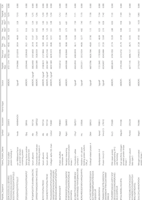
Table
1
Identified
sequences
within
the
MWCNT
-induced
pe
ptidome
(Continued)
Peptide_Sequence
Parent_Protein_Nam
e
Symbol
Accession
Homo-logue
Source
Peptide Mass Precursor Mass Prec. RT
a
Prec. DT
b
Prec. Merr
c
Prod. Merr
d
Peptide Score
FDR
PFPTFSSTAVMAKETTAF
EEGEG
STYTPSEGRLMTG
SERVPGLETT
PVGTSYPPGALTDQEV
E
Versican
core
protein
Vcan
Q62059
MEROPS
6606.0893
6625.0931
41.40
52.72
2.22
14.43
6.067
0.097
KLLLAFSLLLVLLLFQEQ
L
Protein
Vmn2r23
Vmn2r23
E9PXI5
SignalP
2195.3697
2214.3732
41.79
47.98
6.78
17.75
6.050
0.101
ELETSHLGKGCDRDTYS
EKSLH
RLCGAAAGTSELLPSP
SSSFN
WTVGLPTDNGHDSDQ
VFE
Calsyntenin-1
Clstn1
Q9EPL2
MEROPS,
SignalP
6644.0520
6663.0569
38.66
52.84
2.03
11.98
6.032
0.101
QATTQPSTTAGTSTTTT
TTTTAA
Ubiquilin-2
Ubqln2
Q9QZM0
MEROPS
2182.0237
2201.0355
39.22
47.21
3.03
13.97
6.025
0.101
PPASVVVGPVVVPR
Alpha-2-HS-glycoprot
ein
Ahsg
Q3UEK9
P29699
SignalP
1353.8132
1372.8346
32.85
63.44
2.21
11.64
5.985
0.101
EEAPQPALPFQPDSP
THFTP
Isoform
3
of
Seizure
protein
6
Sez6
Q7TSK2
–
3
SignalP
2187.0272
2206.0278
36.80
47.80
8.16
17.75
5.964
0.105
ATADAGSLSPRTCAAL
QKALD
DDNDEKVSGSSDDL
AEKMLLG
SGLEQEEHADETAERGGG
VPFD
DNA
repair
protein
complementing
XP-G
cells
homolog
Ercc5
P35689
MEROPS
6628.0364
6647.0762
38.70
52.58
3.22
17.40
5.952
0.105
aPrecursor
retention
time
bPrecursor
drift
time
cPrecursor
mass
error
in
ppm
dMaximum
product
mass
error
in
A
B
C
D
E
Fig. 3Functional relevance of the MWCNT-responsive peptidome.aVenn diagram reflecting functional associations among 73 identified peptides per ontology enrichment analysis. Groups denote the five most-enriched associations per biochemical, pathological, cellular and localization databases, with peptide-precursor protein symbols shown. Serum cumulative inflammatory potential assay results after treating endothelial cells for 4-h in vitro withbthe enriched-peptide serum fraction andcafter denaturing the peptide fraction. Presented as the mean ± SE,n= 6 serum peptide fractions from replicate mouse exposures per dose,#p< 0.05 for effect of denaturing; *p< 0.05 for effect of MWCNT.d
inhaled Mitsui-7 MWCNT with four-hour deposited doses on par with those modeled here. Array analysis in lung tissue showed significant upregulation for a broad array of other MMPs, ADAMs and ADAMTSs with MWCNT exposure (Fig. 2d). Furthermore, tissue inhibitors of metalloproteinases (TIMPs) were likewise modulated, with TIMP1 significantly increased at both doses, TIMP2 significantly decreased at both doses, and TIMP3 showing an opposing response between doses. Other proteases are also known to influence remodeling of the ECM after MWCNT exposure, to include lysosomal-released cathepsins [48]. Microarray data showed significant increased at both doses for cathepsins B, H, K, S and Z. Taken together, these data support involvement of multiple proteases, with likely promiscuous activation of one by another and tandem cleavage of common substrates, in generating the peptidomic response observed here.
MWCNT-induced circulating peptidome contributes to cardiovascular endothelial responses
Bioinformatic functional enrichment analysis was per-formed to inform on the pathobiological relevance of the circulating peptidomic response to MWCNT. Results clas-sified into five enriched associations (Fig. 3a and Table2). The largest group of 31 peptides were associated with an extracellular matrix localization and functionally to extracellular matrix (ECM) organization. With ECM reorganization being a principle factor in cardiovascular disease and inflammatory lesion development, it was
also fitting that many of the ECM-related measures were among the 27 peptides enriched in association with an abnormal cardiovascular phenotype and altered cardiovas-cular proliferative biological processes. The results also in-cluded 18 peptides reflecting relevance to cell-surface receptor interactions denoting signaling ligands within the peptidome. Peptide-receptor interactions are in-volved in numerous biological processes, including im-mune and associated inflammatory signaling, with 22 associated peptides. Lastly, 29 peptides were derived from known exosomal localized proteins, suggesting exosome involvement in systemic MWCNT responses. Collectively, the peptidomic fraction biochemically par-alleled our prior published inflammatory and vascular deficit outcomes induced in vivo andex vivo using the present MWCNT exposure model [14,23,25].
After establishing its pathobiological relevance, we then sought to assess the bioactive potential of the serum peptidomic fraction. The enriched peptide frac-tion was first assessed for its inflammatory potential on primary murine vascular endothelial cells, as previously reported for whole serum [14]. The assay assessed ex vivo endothelial induction across a battery of adhesion molecule and cytokine gene products linked with inflammatory-mediated immune responses. Naïve endo-thelial cells were treated in culture with the serum pep-tide fractions of MWCNT-exposed or DM control animals. As observed previously using whole serum from exposed animals [14], the peptide-fraction alone induced significant increases in canonical inflammatory markers
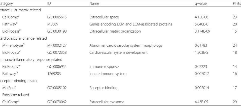
Table 2Enriched cellular, molecular and phenotypes associated with the MWCNT-induced peptidome
Category ID Name q-value #Hits
Extracellular matrix related
CellCompa GO:0005615 Extracellular space 4.15E-08 23
Pathwayb M5889 Genes encoding ECM and ECM-associated proteins 5.048E-6 20
BioProcessc GO:0030198 Extracellular matrix organization 3.174E-09 15
Cardiovascular change related
MPhenotyped MP:0002127 Abnormal cardiovascular system morphology 0.01783 24
BioProcessc GO:0072358 Cardiovascular system development 1.503E-5 18
Immuno-inflammatory response related
BioProcessc GO:0006955 Immune response 0.02223 14
Pathwayb 1269203 Innate immune system 0.007017 16
Receptor binding related
MolFune GO:0005102 Receptor binding 0.002014 17
Exosome related
CellCompa GO:0070062 Extracellular exosome 4.43E-05 29
Enrichment assessed in association with the following ontology database categories:
a
cellular component
b
molecular pathway
c
biological process
d
mouse phenotype
e
Ccl2,Vcam1, Icam1, and Tnfa, principally at the 40μg MWCNT dose relative to DM control (Fig. 3b). The magnitude of response was 189% (p < 0.001), 156% (p < 0.001), 136% (p < 0.001), and 150% (p < 0.033) of DM control levels for Ccl2,Vcam1, Icam1, and Tnfa, respectively. In contrast, Ccl5 showed no response to treatment. Tgfb and Il6 were additionally assessed in
this study, relevant to the MWCNT peptidome’s
enriched association with immune response processes. Application of the serum peptide fraction induced sig-nificant dose-dependent increases in Tgfb expression: 123%, p < 0.001 at 10μg; 140%, p < 0.001 at 40μg. In contrast, Il6 levels were absent significant effects of treatment. To support that the inflammatory response was driven by peptide components within the enriched fraction, an aliquot was denatured by reduction and alkyl-ation to disrupt peptide structure, buffer exchanged, and applied to naïve cerebrovascular endothelial cells. Notably, denaturing significantly reversed the peptide fraction’s prior inflammatory potential:Ccl2(40μg,p< 0.001),Ccl5 (40μg, p = 0.004), Vcam (40μg, p < 0.001), Icam1 (40μg, p < 0.001), Tnfa (40μg, p < 0.001), and Tgfb (10μg, p= 0.002; 40μg,p < 0.001) (Fig. 3c).
Next, the serum peptide fraction was assessed for its potential to induce vascular dysfunction as previously tested for whole serum using myography [25]. Naïve aor-tic rings treated with the serum peptide fraction from 10μg and 40μg dosed animals exhibited significant dys-function (F = 31.2,p< 0.001, main effect of MWCNT ex-posure) in acetylcholine-mediated dilation (Fig.3d). The effect was dose-independent, reaching only 57% the dila-tion achieved with DM control at either MWCNT dose. As a percent of DM control, the peptide fraction in-duced the same magnitude deficit as whole serum treat-ment, which reach just 56% of DM control relaxation at the 40μg dose [25]. However, the response to the pep-tide fraction lacked an added deficit previously reported with whole serum at the 10μg dose, which dilated to only 29% that reached with DM control serum.
MWCNT-responsive exosome-associated peptides coincide with serum exosomal shifts
As the MWCNT-responsive peptidome was significantly enriched in association with exosomes, we sought to next test an association of this change with a parallel in-crease in serum exosomes. An enriched exosomal frac-tion was purified by ultrafiltrafrac-tion followed by size-exclusion chromatography. The vesicle size distribution fell between 40 and 160 nm (Fig.3e) and excluded serum proteins that appear in later fractions between 6 and 20 nm. The measured vesicle distribution matched precisely that accepted for exosomes [49]. MWCNT exposure induced a dose-dependent shift to larger-sized serum-exosomal vesicles, reaching significance for the 40μg
dose (18.3% increase, p < 0.001): DM, 60.4 ± 1.0 nm; 10μg, 64.7 ± 0.9 nm; 71.4 ± 0.4 nm. Furthermore, the area under the hydrodynamic-size curve increased with MWCNT exposure, relative to DM control, purporting a greater number of exosome-sized serum vesicles, which reach significance at the 40μg dose (17.5% increase, p = 0.0284): DM, 924 ± 40.6; 10μg, 974 ± 8.3; 40μg, 1086 ± 26.1. The enriched vesicles were further assessed by transmission electron microscopy (Fig. 3e), with their size and morphology consistent with that previ-ously published for exosomes [50].
Identified thrombospondin fragment relates peptidome relevance to vascular dysfunction
The induction of endothelial-dependent vasodilatory defi-cits by serum factors from MWCNT exposed animals has recently been independently confirmed [25, 26]. The two studies offer complementary mechanistic insight demon-strating the co-dependence between MMP9 activity and TSP-mediated CD36 signaling; yet, a molecular intercon-nection was not readily apparent. Included within the MWCNT-responsive peptidome reported here was a 59-mer fragment from a TSP2 type-1 domain between resi-dues 402–460. Within TSP type-1 domains are three motifs that enable CD36 binding and modulation: R-x-R, W-xx-W-xx-W and CSVTCG motifs [51,52]. The identi-fied TSP402–460 peptide contains all three motifs (Fig. 4a),
which was well characterized by the product ion spectra (Fig. 4b). Significant increases in the TSP402–460 peptide
were measured dose-independently within serum at the 10μg (p= 0.002) and 40μg (p< 0.001) doses, but dose-de-pendently in BALF at the 40μg dose (p= 0.002) relative to DM control (Fig. 4c). The average serum TSP402–460
pep-tide concentration was then approximated at 24 nM and 20 nM in serum from 10μg and 40μg MWCNT-dosed ani-mals, respectively, using the mean mass spectrometer re-sponse to a known peptide concentration. The similar concentration between the two MWCNT doses paralleled the comparable vascular deficit induced by the serum pep-tide fraction per myography (Fig.3d). Furthermore, the in-creased serum amount of the 59-mer peptide at the two MWCNT doses closely profiled MMP9 protein levels within lung tissue (Fig.2b). Yet known MMP9 cleavage of TSP between residues 306–307 sits well away from the TSP402–460peptide identified here (Fig.4d), suggesting
complement of TSP cleavage events, particularly with se-quential processing by multiple proteases, cannot be known, we then looked to site prediction informatics. Using the feature-based tool PROSPER [61], we resolved that the c-terminal side of the TSP402–460peptide is a probable
neu-trophil elastase cleavage site (cleavage probability score 1.11 > 0.8 cutoff for positive prediction at an 82.9% accuracy for elastase-2). While the full complement of proteolytic processing needed to yield this peptide is expectedly
complex, its retained CD36 ligand characteristics exemplify the bioactive potential of the MWCNT-induced circulating peptidome.
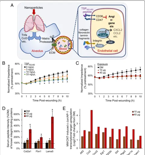
Peptide-mediated model of systemic effects following pulmonary nanoparticle exposure
Cumulatively, findings here establish a complex serum pep-tidomic response acutely following exposure to MWCNT that conveys bioactivity. Included among the responsive
A
B
D
C
Fig. 4MWCNT-responsive thrombospondin fragment identified with CD36-modulatory motifs.aAmong the identified peptides was a 59-mer thrombospondin fragment overlaping the second type-1 repeat domain (TSP402–460). Three established CD36 binding and modulatory motifs are
color-coded and shown in bold within the peptide sequence. Matched mass spectral ion fragmentation sites are shown along the 59-mer sequence with a line angled to the left denoting a b-ion (left-to-right) a line angled to the right denoting a y-ion (right-to-left).bThe TSP402–460peptide’s
corresponding fragment ion mass spectrum with peaks labeled for their reduced mass (MH+) and b/y-ion designation.cMass spectral ion intensity for the TSP402–460peptide plotted as mean ± SE,n= 6 matched serum and BALF specimens from replicate mouse exposures per dose, *p< 0.05.d
A
B
D
E
C
Fig. 5Peptide-mediated model of systemic bioactivation following pulmonary nanoparticle exposure. Results from this study support a proposed paradigm (a) by which nanoparticles in the lung activate matrix proteases, with a diversity of generated peptide products released into circulation with some acting as cell-surface receptor ligands that drive systemic vascular dysfunction and inflammation. The model depicts peptides products of thrombospondin and integrin-ligand proteins from the MWCNT-responsive peptidome triggering CD36 and integrin receptor signaling with downstream anti-angiogenic and inflammatory marker outcomes.bFollowing synthesis of the identified MWCNT-responsive TSP402–460peptide, its anti-angiogenic
factors are peptide fragments related to abnormal cardio-vascular and inflammatory responses, exemplified by the identification of a TSP402–460 peptide with CD36 binding
motifs. Results here support consideration of a new peptide-mediated model of systemic effects (Fig.5a) follow-ing nanoparticle exposure. Proposed is that a diverse assort-ment of matrix remodeling proteases activated following exposure induce peptide byproducts into circulation where they may be further modified yielding an expanded serum peptidomic complexity. The peptidomic response includes cell-surface receptor binding capacity that promotes extra-pulmonary effects. To validate the potential of this model, we synthesized the TSP402–460peptide and applied it
in a CD36/CD47-mediated endothelial cell in vitro wound healing angiogenesis assay (Fig. 5b). Addition of the TSP402–460peptide into the culture media produced a
sig-nificant anti-angiogenic effect (F = 43.5,p< 0.001), impair-ing cell regrowth as measured by recovered membrane impedance. Relative to vehicle control, a 6% deficit reestab-lishing baseline impedance 10 h after wounding was ob-served at a 3 ng/μL dose (p= 0.002), a 22 nM concentration in media similar to that estimated in the serum of exposed animals. A 3-fold or greater increase in TSP402–460
concen-tration saturated the deficit at 12% (p< 0.001). In compari-son, the serum peptide fraction from MWCNT exposed animals also significantly inhibited (F = 32.7,p< 0.001, main effect of dose) endothelial cell regrowth out to 10 h after wounding (Fig.5c). The induced deficit recovering baseline impedance was 9.5% (p < 0.001) relative to DM control, which fell between that induced at 3 and 10 ng/μL TSP402– 460peptide. These data establish the bioactive nature of the
circulating TSP402–460 peptide that, at least in part,
ac-counts for the anti-angiogenic capacity of the serum pep-tide fraction.
Looking beyond the TSP peptide, the MWCNT-respon-sive peptidome was enriched with other fragments of receptor-binding proteins (Fig. 3a). A number of them were derived from known integrin binding collagens, fi-bronectin and laminins, offering additional potential in mediating observed bioactivity. The MWCNT-response among these peptides was dose-dependent (Fig.5d) simi-lar to findings from the serum cumulative inflammatory potential assay (Fig. 3b), which contrasted with the dose-independent increase in the TSP402–460peptide and
CD-36 dependent myography (Fig.3d) and wound healing (Fig. 5c) findings. The MWCNT-responsive peptidome was also significantly enriched in association with interac-tions upstream of c-Jun transcription (FDR 0.0127) and integrins are known to induce c-Jun-mediated inflamma-tory factor transcription. Supporting the integrin-ligand potential of the MWCNT-responsive peptidome, in vitro treated endothelial cells exhibited significant dose-depen-dent upregulation of numerous c-Jun transcriptional products (Fig.5e) relative to DM control. Together, results
here implicate integrin-ligand functionality of the MWCNT-responsive peptidome as an additional part of a broader combinatorial molecular response mediating sys-temic bioactivity after nanoparticle exposure.
Discussion
The release of proinflammatory factors from the lung into the circulation, so-called “systemic spill-over”, has been posited as a causal link between respirable particle expos-ure and systemic toxicity, in lieu of, or in addition to par-ticle translocation [6]. Yet uncertainty exists as to the molecular nature and diversity of what is released and how bioactivity is induced. The present study establishes the paradigm that proteolytic byproduct peptides are released from the lung as circulating mediators and poten-tial biomarkers of exposure and/or extra-pulmonary health consequences. Peptide isolation by size fraction-ation and solid-phase extraction permitted the selective assessment of a peptidomic shift within the blood follow-ing MWCNT exposure, validated by unbiased liquid chro-matography data-independent mass spectrometry to exclude hydrophobic lipid moieties and proteinaceous ma-terial above 7000 Da. The enriched peptide fraction con-veyed inflammatory potential analogous to that previously demonstrated with whole serum from MWCNT-exposed animals when assessed ex vivo on naïve endothelial cells, despite mass-exclusion of typical cytokine/chemokine me-diators. The serum peptide fraction’s inflammatory poten-tial, however, was reversed when pretreated to denature the peptide content, confirming peptide involvement. The serum peptide fraction furthermore triggered vasodilatory and endothelial angiogenesis deficits in naïve vessels and cells ex vivo akin to that induced with whole serum. Col-lectively, these outcomes establish that the peptide frac-tion alone can convey much of the systemic bioactivity previously reported with full serum collected at the same 4 h time point after MWCNT exposure using the same model [14, 23, 25]. Moreover, the use of unbiased data-independent tandem mass spectrometry enabled par-tial identification of the MWCNT-responsive peptidome with a focus on correlated responses in the circulation and lung lavage to direct informatic inquiry towards biomolec-ular relationships with MWCNT-induced pulmonary pathobiology. The identified peptides were replete with fragments of known cell-surface receptor ligands, illus-trated most notably by a peptide from the type-1 repeat domain of TSP. Containing binding and modulating mo-tifs involved in promoting CD36-dependent endothelial dysfunction, this peptide exemplified a mechanistic role for circulating peptides triggering cell-surface receptor mediated systemic effects.
ADAMs and ADAMTS and associated metalloprotease inhibitors (TIMPs) in reaction to MWCNT pulmonary exposure. MWCNT can also promote lysosomal protease excretion [48], with several cathepsins involved in ECM re-modeling found upregulated in the lung after MWCNT exposure, enhancing the potential diversity of proteolytic processes involved in establishing the MWCNT-responsive peptidome. Sequential multi-protease processing helps to explicate the wide diversity of peptide products discovered responsive to MWCNT, with significant correlation be-tween lung lavage and serum fluid compartments denoting a pulmonary source for part of the circulating peptidomic response. TSP, for example, is an established substrate for at least five matrix metalloproteases and one cathepsin (MMPs 2, 9 and 14; ADAMTSs 1 and 7; cathepsin-G) [53–56]. TSP is also a known substrate for circulating pro-teases such as thrombin and Htra1 that also influence ECM reorganization [58, 59]. While a full decoding of sequential proteolytic processing of TSP falls beyond the scope of the present work, the identified TSP402–460
pep-tide points to additional, as yet unknown cleavage events. Site prediction software suggests that the c-terminal site represented by this peptide is a probable neutrophil elas-tase cleavage point, supported by the significant acute influx in lung neurophils using this model [23] and TSP as a known substrate of neutrophil elastase (exact sites unfound) [56,60]. Altogether, the peptidomic response re-vealed by these studies represents a post-exposure molecu-lar complexity as yet unappreciated, demarking an equally complex pathobiological response, with substantial diag-nostic potential.
Identification of the MWCNT-responsive peptides revealed here is challenged by the stochastic nature of their endogenous generation. Unlike bottom-up prote-omic mass spectrometry that involves in-tube diges-tion with a posidiges-tionally-restrictive enzyme like trypsin, unknown endogenous byproduct peptides have no stipulated restraints on their beginning or end resi-dues, obfuscating proteomic search engines. To allevi-ate some of this burden we employed target databases to significantly narrow the sequence search space [62]. We further utilized FDR-controlled peptide match scores rather than protein-level scoring, which is the default of most search engines that assume multiple peptides should match to a single protein in a confident identification. Focused here on peptides measured commonly between serum and BALF to provide selective relevance to pulmonary pathobiology, we were able to identify 73 in connection with known matrix protease substrates and signaling motifs. As a representative sampling of the peptide fraction, there was significant enrichment in matrikine-like fragments from ECM proteins with receptor-ligand functionality. For example, peptides of laminin 5 (Lama5), fibronectin 1 (Fn1) and collagen 3a
(Col3a1) were identified, all ECM proteins known to inter-act with integrin surface receptors. Furthermore, the MWCNT-responsive peptidome was significantly associ-ated with c-Jun signal transduction. Integrin signaling acti-vates a variety of cellular processes, to include c-Jun n-terminal kinase (JNK) mediated regulation of cell prolif-eration, migration, and cytokine/chemokine production. ECM-derived peptides acting as circulating integrin li-gands can moreover explain dose-dependent endothelial cytokine/chemokine induction observed ex vivo when treating with serum from MWCNT-exposed animals [14, 25]. In affirmation, microarray analysis of ex vivo treated naïve endothelial cells showed significant up-regulation of pathway targets downstream of JNK, reflecting inflammatory marker production and cell stress responses.
The serum peptide fraction alone induced much of the proinflammatory marker response previously reported with whole serum ex vivo treatment [14,23, 25]. Canonical in-flammatory markersCcl2,Vcam1,Icam1, andTnfawere all significantly elevated in a dose-dependent manner. Further-more, the magnitude of response observed here was on-par with that produced by whole serum. Given that the sample fractionation process excluded larger-mass cytokines and chemokines, bioactivity induced by the serum peptide frac-tion must reflect the presence of alternative proinflamma-tory ligands such as the dose-dependently increased fragments of integrin-binding proteins Col2a1, Fbn1, and
Lama5 or other cell-surface ligands such as gelactin 3
(Lgals3) or VG-1-related protein (Bmp6), with future stud-ies warranted to clarify details on specific peptide involve-ment. The ex vivo endothelial proinflammatory response to the peptide fraction, however, was not entirely the same, lacking the robust Ccl5upregulation induced with whole serum. We ascertained that the enriched peptide fraction lacked some components of the circulating bioactive pro-file. It is likely that larger proteolytic fragments, as well as other secreted mediators, were excluded from the peptide fraction. Non-peptide factors also surely contribute to the response diversity across doses and xenobiotic exposures. Yet a majority of the dose-dependent inflammatory re-sponse previously reported were reproduced with the serum peptide fraction studied here, substantiating proteo-lytic product involvement as an indirect mediator convey-ing a MWCNT response across the lung-blood barrier and acting as cell-surface receptor ligands to promote systemic effects (Fig.5a). The peptidomic findings here thus help ex-pound on the molecular “systemic spill-over” response as more substantially diverse than previously appreciated.
The identified TSP402–160fragment was studied here in
principal CD36 binding site, having the positively charged W-xx-W-xx-W and R-x-R motifs needed to interact with the negatively charged CD36 CLESH domain [51,63]. The fragment also contains the CSVTCG motif found to modu-late CD36 activation [52]. Studies show that synthetic pep-tides derived from TSP type-1 repeat domains exhibit anti-angiogenic properties in a CD36-dependent manner [64], and synthetic peptides as short as 18 amino acids con-taining the W-xx-W-xx-W motif [62] are potent inhibitors of cell proliferation and angiogenesis [65]. Thus the TSP402–460 peptide was synthesized and tested for
anti-angiogenic properties using a high-precision electrical wound-healing assay. Testing the TSP402–460 peptide at a
comparable nanomolar concentration in vitro induced a vascular regrowth deficit on par with the serum peptide
fraction from MWCNT-exposed animals (Fig. 5c),
confirming its bioactivity. We previously published that MWCNT-diminished vasodilatory responses were CD36-receptor mediated, with knockout animals exhibiting a muted response [25]. Mandler et al. also found that serum from TSP knockout animals treated with Mitsui-7 MWCNT lacked the ability to induce vasodilatory deficits in naïve aortas [26, 66]. They also showed that knocking out CD47 also offered vascular protection in ex vivo assays with serum from MWCNT exposed animals, which fits the dependence of TSP-CD36 driven nitric oxide responses on proximal localization of CD47 without the need for direct TSP-CD47 binding [67]. Yet circulating TSP increased by only 50% 4 h following in vivo MWCNT exposure, which is unlikely to dramatically perturb endothelial homeostasis [14]. Furthermore, testing MWCNT-induced vasodilatory dysfunction ex vivo was also largely abolished using serum
from MMP9-deficient mice [25], demonstrating a
co-dependence of these effects on MMP9 processing. Miss-ing was a factor(s) interlinkMiss-ing TSP, CD36 and MMP9. The identified TSP402–460fragment peptide offers a unifying
me-diator of CD36-related systemic pathobiology, increased in serum by over an order in magnitude, and well in excess of the modest 0.5-fold MWCNT-induced increase in circulat-ing TSP protein [14].
Notably, results from this study emphasize that the per-ipheral molecular response to nanoparticle exposure is con-siderably more diverse than previously appreciated. It then fits that systemic bioactivity occurs as a combinatorial re-sponse to several circulating factors. The peptide-mediated model in Fig. 5a proposes the involvement of multiple ligand-receptor triggered events. We surmise that this explains the produced dose-independent myography and wound-healing results that were consistent with a dose-independent magnitude of TSP402–460peptide in
cir-culation, which stands apart from the dose-dependent endothelial induction of pro-inflammatory cytokines and chemokines that are not downstream of CD36-receptor sig-naling. Inflammatory marker production is, however,
downstream of integrin-mediated c-Jun transcriptional ac-tivity. The MWCNT-responsive peptidome was enriched in peptides from receptor binding proteins, particularly integrin ligands upstream of c-Jun. The identified fragments of known integrin binding ligands exhibited dose-depen-dent increases, warranting follow-up investigation as to the combinatorial bioactivity across the responsive peptidome.
Unexpectedly, we found an enriched association be-tween the MWCNT-responsive peptidome and exoso-mal protein fragments. Exosomes play a pivotal role in cellular communication by delivering bioactive mole-cules such as mRNA, miRNA, proteins and perhaps peptides to distant tissues. Yet exosomal involvement in response to inhaled xenobiotics remains understudied; though, it was recently published that bronchial epithe-lial cells release exosomes when challenged with cigarette smoke extract [68]. It turns out that very few proteins, mainly surfactant-related, are specific enough to demark a vesicular lung origin. While the data here cannot affirm lung specificity, they do suggest it, with 28 of 29 exosome-associated peptides derived from proteins of known lung expression and 77.3% with a higher than average abundance (Tissue Atlas,www.proteinatlas.org). Presently, we found an increase in the size and abun-dance of serum exosomes following MWCNT exposure. An increase in diameter infers a greater volumetric increase in vesicle capacity, suggesting a quantitative shift in exosomal cargo as seen under other pathological conditions [69–71]. These findings demonstrate that MWCNT exposure alters the serum exosome popula-tion, implicating their potential involvement in systemic pathobiology. Exosomes also carry matrix proteases and thus may play a further role in peptidome processing [71]. Follow-up studies are needed to determine whether exosome-related peptides found here reflect induced surface protein cleavage or rather a shift in peptide exo-somal packaging with separate bioactive capacity.
between serum and BALF was not readily identified, and that the added peptidomic complexity observed exclu-sively in the circulation, relative to BALF, remains to be examined. As discussed earlier, endogenous peptides are particularly difficult to identify due to the vast sequence-search space involved. We used smaller targeted databases to facilitate identification; however, products outside those focused databases go unidentified. Informatic approaches to improve search selectivity are also needed to address identification of post-translational modified peptides. Blood polypeptides are often glycosylated, a particularly diverse modification that exacerbates the sequence search space issue and further complicates comprehensive identi-fication. Ultimately, results here establish existence of a diverse and bioactive circulating peptidomic response fol-lowing pulmonary nanoparticle insult, providing founda-tion for more through characterizafounda-tion of its mechanistic role in driving systemic pathobiology.
Conclusions
Findings here substantiate proteolytic peptide involvement in mediating systemic consequences of nanoparticle ex-posure. A wide variety of inhaled xenobiotics upregulate matrix proteases and produce various systemic effects. Agents from titanium dioxide to diesel exhaust have been shown to induce extra-pulmonary inflammation and car-diovascular impairments much as reported here for MWCNT [27,72–74]. Indeed, many insoluble metal and engineered nanoparticles (Au, Ag, titanium dioxide, ultra-fine carbon and other CNTs, etc.) induce pulmonary path-ology that may be accompanied by proteolytic generation and release of bioactive peptides. Once in circulation, frag-ments such as the TSP402–460peptide or those from
integ-rin ligands induce cell-surface receptor mediated systemic stress and inflammation, driving particulate exposure’s ad-juvant role in promoting everything from cardiovascular to neurodegenerative diseases. Furthermore, the revealed existence of a pathological peptidomic response to nano-particle exposure represents a highly diverse and dose-dependent molecular source of putative biomarkers that may provide greater specificity than previously available [9]. More thorough characterization of circulatory factors is warranted to assess the role of peptide byproducts me-diating systemic disease following pulmonary insult to a broader array of occupational and environmental xeno-biotic agents.
Abbreviations
BALF:Bronchoalveolar lavage fluid;Ccl2: C-C motif chemokine ligand 2;
Ccl5: C-C motif chemokine ligand 5; DM: Dispersion media; ECM: Extracellular matrix; FDR: False discovery rate; Il6: Interleukin 6; MMP: Matrix
metalloproteinase; MWCNT: Multi-walled carbon nanotubes;
Tgfb: Transforming growth factor beta;Tnfa: Tumor necrosis factor alpha; TSP: Thrombospondin;Vcam1: Vascular cell adhesion molecule 1
Acknowledgements
AKO and CGC thank Drs. Hu Yang and Juan Wang for access and assistance with the Zetasizer instrument and Judy Williams for assistance with electron microscopy. EM and AKO thank Pavel Lizhnyak and Pallavi Pilaka for assistance with informatics and sample processing. The authors thank Guy Herbert for performing the ECIS assays.
Funding
This study was funded by grants from the National Institute for Occupational Safety and Health (OH010495 to A.K.O. and M.J.C. and NTRC 939ZXFL to A.E.). The findings and conclusions in this report are those of the authors and do not necessarily represent the official position of the National Institute for Occupational Safety and Health, Centers for Disease Control and Prevention.
Availability of data and materials
The datasets used and/or analyzed during the current study are available from the corresponding author on reasonable request.
Authors’contributions
EM and AKO prepared the manuscript with further input from TY, AE and MJC. EM, TY, PPM, CGC, PCZ-E, AE and MJC contributed to the design of aspects of the study. LB and AE completed the animal modeling. PPM, EM and AKO performed the biofluid preparation and mass spectrometric analysis. EM and AKO completed the mass spectral data processing and informatic interrogation. AV provided informatic software support. TY performed the vascular-related functional assessments. CGC and EM carried out the exosome processing and analyses. PCZ-E completed the microarray experimentation.
Ethics approval
All animal procedures were approved by the Institutional Animal Care and Use Committee of the National Institute for Occupational Safety and Health and were conducted in accordance with the U.S. Public Health Service Policy on Humane Care and Use of Laboratory Animals and the National Institutes of Health Guide for the Care and Use of Laboratory Animals.
Consent for publication Not applicable.
Competing interests
The authors declare that they have no competing interests.
Publisher’s Note
Springer Nature remains neutral with regard to jurisdictional claims in published maps and institutional affiliations.
Author details
1
Department of Anatomy and Neurobiology, Virginia Commonwealth University, Box 980709, Richmond, VA 23298-0709, USA.2Department of
Pharmaceutical Sciences, University of New Mexico, Albuquerque, NM 87131, USA.3Pathology and Physiology Research Branch, National Institute for
Occupational Safety and Health, Morgantown, WV 26505, USA.
Received: 28 September 2018 Accepted: 10 May 2019
References
1. Hoet PH, Brüske-Hohlfeld I, Salata OV. Nanoparticles - known and unknown health risks. J Nanobiotechnology. 2004;2:12.
2. Daigle CC, Chalupa DC, Gibb FR, Morrow PE, Oberdörster G, Utell MJ, et al. Ultrafine particle deposition in humans during rest and exercise. Inhal Toxicol. 2003;15:539–52.
3. Chalupa DC, Morrow PE, Oberdorster G, Utell MJ, Frampton MW. Ultrafine particle deposition in subjects with asthma. Environ Health Perspect. 2004; 112:879–82.
4. Stone V, Miller MR, Clift MJD, Elder A, Mills NL, Møller P, et al. Nanomaterials versus ambient ultrafine particles: an opportunity to exchange toxicology knowledge. Environ Health Perspect. 2017;125:1–17.
6. Brook RD, Rajagopalan S, 3rd CAP, Brook JR, Bhatnagar A, Diez-Roux AV, et al. Particulate matter air pollution and cardiovascular disease: an update to the scientific statement from the American Heart Association. Circulation. 2010;121:2331–78.
7. Block ML, Elder A, Auten RL, Bilbo SD, Chen H, Chen JC, et al. The outdoor air pollution and brain health workshop. Neurotoxicology. 2012;33:972–84. 8. Donaldson K, Aitken R, Tran L, Stone V, Duffin R, Forrest G, et al. Carbon
nanotubes: a review of their properties in relation to pulmonary toxicology and workplace safety. Toxicol Sci. 2006;92:5–22.
9. Erdely A, Liston A, Salmen-Muniz R, Hulderman T, Young S-H, Zeidler-Erdely PC, et al. Identification of systemic markers from a pulmonary carbon nanotube exposure. J Occup Environ Med. 2011;53(Suppl 6):S80–6. 10. Li Z, Hulderman T, Salmen R, Chapman R, Leonard SS, Young SH, et al.
Cardiovascular effects of pulmonary exposure to single-wall carbon nanotubes. Environ Health Perspect. 2007;115:377–82.
11. Stapleton PA, Minarchick VC, Cumpston AM, McKinney W, Chen BT, Sager TM, et al. Impairment of coronary arteriolar endothelium-dependent dilation after multi-walled carbon nanotube inhalation: a time-course study. Int J Mol Sci. 2012;13:13781–803.
12. Liao H-Y, Chung Y-T, Lai C-H, Wang S-L, Chiang H-C, Li L-A, et al. Six-month follow-up study of health markers of nanomaterials among workers handling engineered nanomaterials. Nanotoxicology. 2014;8:100–10. 13. Reddy AR, Krishna DR, Reddy YN, Himabindu V. Translocation and extra
pulmonary toxicities of multi wall carbon nanotubes in rats. Toxicol Mech Methods. 2010;20:267–72.
14. Aragon MJ, Topper L, Tyler CR, Sanchez B, Zychowski K, Young T, et al. Serum-borne bioactivity caused by pulmonary multiwalled carbon nanotubes induces neuroinflammation via blood-brain barrier impairment. Proc Natl Acad Sci U S A. 2017;114:E1968–76.
15. Nemmar A, Vanbilloen H, Hoylaerts MF, Hoet PH, Verbruggen A, Nemery B. Passage of intratracheally instilled ultrafine particles from the lung into the systemic circulation in hamster. Am J Respir Crit Care Med. 2001;164:1665–8. 16. Deng X, Jia G, Wang H, Sun H, Wang X, Yang S, et al. Translocation and fate
of multi-walled carbon nanotubes in vivo. Carbon. 2007;45:1419–24. 17. Pauluhn J. Multi-walled carbon nanotubes (Baytubes): approach for
derivation of occupational exposure limit. Regul Toxicol Pharmacol. 2010;57: 78–89.
18. Brown JS, Zeman KL, Bennett WD. Ultrafine particle deposition and clearance in the healthy and obstructed lung. Am J Respir Crit Care Med. 2002;166:1240–7.
19. Mercer RR, Scabilloni JF, Hubbs AF, Wang L, Battelli LA, McKinney W, et al. Extrapulmonary transport of MWCNT following inhalation exposure. Part Fibre Toxicol. 2013;10:38.
20. Upadhyay S, Stoeger T, Harder V, Thomas RF, Schladweiler MC, Semmler-Behnke M, et al. Exposure to ultrafine carbon particles at levels below detectable pulmonary inflammation affects cardiovascular performance in spontaneously hypertensive rats. Part Fibre Toxicol. 2008;5:19.
21. Khandoga A, Stoeger T, Khandoga AG, Bihari P, Karg E, Ettehadieh D, et al. Platelet adhesion and fibrinogen deposition in murine microvessels upon inhalation of nanosized carbon particles. J Thromb Haemost. 2010;8:1632–40.
22. Mitchell LA, Lauer FT, Burchiel SW, McDonald JD. Mechanisms for how inhaled multiwalled carbon nanotubes suppress systemic immune function in mice. Nat Nanotechnol. 2009;4:451–6.
23. Erdely A, Hulderman T, Salmen R, Liston A, Zeidler-Erdely PC, Schwegler-Berry D, et al. Cross-talk between lung and systemic circulation during carbon nanotube respiratory exposure. Potential biomarkers. Nano Lett. 2009;9:36–43.
24. Walker VG, Li Z, Hulderman T, Schwegler-Berry D, Kashon ML, Simeonova PP. Potential in vitro effects of carbon nanotubes on human aortic endothelial cells. Toxicol Appl Pharmacol. 2009;236:319–28. 25. Aragon M, Erdely A, Bishop L, Salmen R, Weaver J, Liu J, et al.
MMP-9-dependent serum-borne bioactivity caused by multiwalled carbon nanotube exposure induces vascular dysfunction via the CD36 scavenger receptor. Toxicol Sci. 2016;150:488–98.
26. Mandler WK, Nurkiewicz TR, Porter DW, Olfert IM. Thrombospondin-1 mediates multi-walled carbon nanotube induced impairment of arteriolar dilation. Nanotoxicology. 2017;11:112–22.
27. Tham A, Lullo D, Dalton S, Zeng S, van Koeverden I, Arjomandi M. Modeling vascular inflammation and atherogenicity after inhalation of ambient levels of ozone: exploratory lessons from transcriptomics. Inhal Toxicol. 2017;29:96–105.
28. McDonald JD, Doyle-Eisele M, Campen MJ, Seagrave J, Holmes T, Lund A, et al. Cardiopulmonary response to inhalation of biogenic secondary organic aerosol. Inhal Toxicol. 2010;22:253–65.
29. Remane D, Wissenbach DK, Peters FT. Recent advances of liquid chromatography–(tandem) mass spectrometry in clinical and forensic toxicology—an update. Clin Biochem. 2016;49:1051–71.
30. Porter DW, Hubbs AF, Mercer RR, Wu N, Wolfarth MG, Sriram K, et al. Mouse pulmonary dose- and time course-responses induced by exposure to multi-walled carbon nanotubes. Toxicology. 2010;269:136–47.
31. Ottens AK, Stafflinger JE, Griffin HE, Kunz RD, Cifu DX, Niemeier JP. Post-acute brain injury urinary signature: a new resource for molecular diagnostics. J Neurotrauma. 2014;31:782–8.
32. Distler U, Kuharev J, Navarro P, Tenzer S. Label-free quantification in ion mobility-enhanced data-independent acquisition proteomics. Nat Protoc. 2016;11:795–812.
33. Cortes DF, Landis MK, Ottens AK. High-capacity peptide-centric platform to decode the proteomic response to brain injury. Electrophoresis. 2012;33: 3712–9.
34. Rawlings ND, Barrett AJ, Finn R. Twenty years of the MEROPS database of proteolytic enzymes, their substrates and inhibitors. Nucleic Acids Res. 2016; 44:D343–50.
35. Petersen TN, Brunak S, Von Heijne G, Nielsen H. SignalP 4.0: discriminating signal peptides from transmembrane regions. Nat Methods. 2011;8:785–6. 36. Wenger CD, Coon JJ. A proteomics search algorithm specifically designed for high-resolution tandem mass spectra. J Proteome Res. 2013;12:1377–86. 37. Strohalm M, Kavan D, Novák P, Volný M, Havlíček V. mMass 3: a
cross-platform software environment for precise analysis of mass spectrometric data. Anal Chem. 2010;82:4648–51.
38. Chen J, Bardes EE, Aronow BJ, Jegga AG. ToppGene suite for gene list enrichment analysis and candidate gene prioritization. Nucleic Acids Res. 2009;37:W305–11.
39. Szklarczyk D, Morris JH, Cook H, Kuhn M, Wyder S, Simonovic M, et al. The STRING database in 2017: quality-controlled protein-protein association networks, made broadly accessible. Nucleic Acids Res. 2017;45:D362–8. 40. Erdely A, Dahm M, Chen BT, Zeidler-Erdely PC, Fernback JE, Birch ME, et al.
Carbon nanotube dosimetry: from workplace exposure assessment to inhalation toxicology. Part Fibre Toxicol. 2013;10:53.
41. Zeidler-Erdely PC, Kashon ML, Li S, Antonini JM. Response of the mouse lung transcriptome to welding fume: effects of stainless and mild steel fumes on lung gene expression in a/J and C57BL/6J mice. Respir Res. 2010;11:70. 42. Zychowski KE, Sanchez B, Pedrosa RP, Lorenzi-Filho G, Drager LF, Polotsky
VY, et al. Serum from obstructive sleep apnea patients induces
inflammatory responses in coronary artery endothelial cells. Atherosclerosis. 2016;254:59–66.
43. Saeed AI, Sharov V, White J, Li J, Liang W, Bhagabati N, et al. TM4: a free, open-source system for microarray data management and analysis. Biotechniques. 2003;34:374–8.
44. Benjamini Y, Hochberg Y. Controlling the false discovery rate: a practical and powerful approach to multiple testing. J R Stat Soc B. 1995;57:289–300. 45. Govender P, Dunn MJ, Donnelly SC. Proteomics and the lung: analysis of
bronchoalveolar lavage fluid. Proteomics Clin Appl. 2009;3:1044–51. 46. Jeong K, Kim S, Bandeira N. False discovery rates in spectral identification.
BMC Bioinformatics. 2012;13(Suppl 16):S2.
47. Ricard-Blum S, Salza R. Matricryptins and matrikines: biologically active fragments of the extracellular matrix. Exp Dermatol. 2014;23:457–63. 48. Mu QX, Broughton DL, Yan B. Endosomal leakage and nuclear translocation
of multiwalled carbon nanotubes: developing a model for cell uptake. Nano Lett. 2009;9:4370–5.
49. Colombo M, Raposo G, Théry C. Biogenesis, secretion, and intercellular interactions of exosomes and other extracellular vesicles. Annu Rev Cell Dev Biol. 2014;30:255–89.
50. Park JA, Sharif AS, Tschumperlin DJ, Lau L, Limbrey R, Howarth P, et al. Tissue factor-bearing exosome secretion from human mechanically stimulated bronchial epithelial cells in vitro and in vivo. J Allergy Clin Immunol. 2012;130:1375–83.
51. Klenotic PA, Page RC, Li W, Amick J, Misra S, Silverstein RL. Molecular basis of antiangiogenic thrombospondin-1 type 1 repeat domain interactions with CD36. Arterioscler Thromb Vasc Biol. 2013;33:1655–62.