R E S E A R C H
Open Access
Response of membrane tension to gravity
in an approximate cell model
Lili Wang
1,2, Weiyi Chen
1,2*, Hongmei Guo
1,2and Airong Qian
3Abstract
Background:Gravity, especially hypergravity, can affect the morphology of membranes, and further influence most biological processes. Since vesicle structures are relatively simple, the vesicle can be treated as a vital model to study the mechanical properties of membranes in most cases. Basic research on membrane tension has become a vital research topic in cellular biomechanics.
Methods:In this study, a new vesicle model is proposed to quantitatively investigate the response of membrane tension to gravity. In the model, the aqueous lumen inside the vesicle is represented by water, and the vesicle membrane is simplified as a closed, thin, linear elastic shell. Then, the corresponding static equilibrium differential equations of membrane tension are established, and the analytical expression is obtained by the semi-inverse method. The model parameters of the equations are accurately obtained by fitting the reported data, and the values calculated by the model agree well with the reported results.
Results:The results are as follows: First, both the pseudo-ellipsoidal cap and the pseudo-spherical cap can be used to describe the deformed vesicle model; however, the former can better represent the deformation of the vesicle model because the variance of the pseudo-ellipsoidal cap is smaller. Second, the value of membrane tension is no longer a constant for both models. Interestingly, it varies with the vesicle height under the action of gravity. The closer it is to the substrate, the greater the membrane tension. Finally, the inclination between the tangent and the radial lines at a certain point is nearly proportional to the radius of the cross section in both models.
Conclusion:These findings may be helpful to study the vesicle model spreading more accurately by taking into account the influence of gravity because it could affect the distribution of membrane tension. Furthermore, it may also provide some guidance for cell spreading and may have some implications for membrane tension-related mechanobiology studies, especially in the hypergravity conditions.
Keywords:Gravity, Equilibrium differential equation, Membrane tension, Pseudo-ellipsoidal cap, Pseudo-spherical cap
Background
Gravity is constantly exerted on organisms [1], and some studies have shown that gravity can affect numerous phys-ical and biologphys-ical processes: biologphys-ical cells are no excep-tion [2]. Biological systems interact with gravity on different levels of organization, from the whole organisms [3] to cells
[4], to membranes [5] and even down to the function of single proteins [6], and many experiments have directly demonstrated that biological processes from single mole-cules to various levels of tissue are dependent on gravity. For example, Sieber et al. indicated that the viscosity, conductance and capacity of membranes are dependent on gravity [2, 7]. Häder D. et al. showed that single cells can sense gravity [8].
Cell mechanical stimulations include mechanical stretch, compression, hydrostatic pressure, microgravity and hyper-gravity. Microgravity is the absence of gravity, which usually exists in spaceflights to different planets or moons. Hyper-gravity, which may be experienced by living cells in certain
© The Author(s). 2019Open AccessThis article is distributed under the terms of the Creative Commons Attribution 4.0 International License (http://creativecommons.org/licenses/by/4.0/), which permits unrestricted use, distribution, and reproduction in any medium, provided you give appropriate credit to the original author(s) and the source, provide a link to the Creative Commons license, and indicate if changes were made. The Creative Commons Public Domain Dedication waiver (http://creativecommons.org/publicdomain/zero/1.0/) applies to the data made available in this article, unless otherwise stated. * Correspondence:[email protected]
1Shanxi Key Laboratory of Material Strength & Structural Impact, College of Biomedical Engineering, Taiyuan University of Technology, Taiyuan 030024, China
2National Demonstration Center for Experimental Mechanics Education (Taiyuan university of Technology), Taiyuan 030024, China
hesion and movement [16].
The mechanical properties of membranes can affect most biological processes, and membrane tension is a basic phys-ical parameter of membranes that is involved in various bio-logical processes, such as membrane trafficking, cell shape, adhesion, growth, endocytosis and motility [17, 18]. Some studies have emphasized the importance and contributions of membrane tension in biological processes [19–23], and have shown that membrane tensions originate mainly from the hydrostatic pressure across the lipid bilayer and cytoskel-eton (CSK)-membrane adhesion [22]. However, Reinhart-King et al. emphasized that cells can exert significant forces before complete actin polymerization or visible stress fibre formation, which means that vesicles can be used to model cell adhesion [24]. In addition, Liu et al. indicated that the complexity of cells could be avoided by using the vesicles as a biomimetic model of cells since there is no CSK or nucleus in the vesicles, and emphasized that a fully three-dimensional (3D) model of a vesicle could be used to simulate a real cell [25]. Lu et al. have also shown that vesicle models are a most useful tool to ex-plore the relevant issues [26].
In summary, due to the high costs and limited number of experiments in real microgravity, and the fact that there is little research on the response of membrane ten-sion to gravity, this paper applies a theoretical modelling method to study the response of membrane tension under the action of gravity. In the model, the vesicles are simplified as water sacs, where the membrane of the vesicle is assumed to be a thin closed shell and the vesicle cavity is represented by water. The equilibrium differential equations of the deformed vesicle model are constructed and solved by using the analysis method of the elasticity mechanics and semi-inverse method.
Methods
In this paper, a vesicle model is used to quantitatively in-vestigate the response of membrane tension to gravity. In the vesicle model, the water represents aqueous lumen inside the vesicle and a elastic shell represents the mem-brane of vesicle. The corresponding static equilibrium dif-ferential equations of membrane tension are established
[28], the mechanism of cell endocytosis [29], membrane vesicle budding [30], and changes in membrane morph-ology [31]. In this study, considering the complexity of the cell structures, such as eukaryotic cell, the vesicle struc-tures are relatively simple and often used to simulate cells, and a 3D axisymmetric model of vesicles is established to study the response of membrane tension to gravity. For simplicity, the following assumptions are made:
a) The vesicles are simplified into watery sacs according to the structure of vesicles [26]. b) In the model, the water representing the cavity of
the vesicle is wrapped with a membrane, and its volume remains constant [25].
c) The details of the molecular structure within the vesicle membrane are ignored [27], and the
membrane is assumed to be isotropic, linearly elastic, of fixed thickness, thin and a closed shell [25]. d) The un-deformed vesicle model is used to represent
the suspended cell described by the sphere [32]. Since the cells gradually switch from a spherical to a flattened shape in vitro [33], the spread cell is described by the deformed vesicle model under the action of gravity.
e) The deformation of the vesicle model is assumed to be generated under the conditions of axial symmetry, and the contact area is always circular [34].
Equilibrium equations
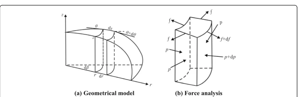
In the model, the cylindrical coordinate (r, z) is used in which thezaxis is directed against the gravity force andris an independent variable. The local slope of the membrane at the same point is defined by the inclination θ between the tangent line and the radial line as illustrated in Fig.1.
The two variablesz(r) andθ(r) are related through [35]:
dz dr¼z
0 r
ð Þ ¼−tanθ ð2:1Þ
Consider the equilibrium of the deformed vesicle model in the r-direction for a micro-block between r
From the equilibrium condition in ther-direction, the formulation is established as follows:
−frdβcosθþðfþdfÞðrþdrÞdβcosðθþdθÞ−2fdssindβ
2
þprzdβ−ðpþdpÞðrþdrÞðzþdzÞdβþ2pzdrsindβ 2 ¼0
ð2:2aÞ
Because dθ and dα are small quantities, the formula-tion is simplified:
sindθ
2 ≈
dθ 2 ; cos
dθ
2 ≈1 ð2:2bÞ
By simplifying and neglecting the small quantities of higher order, the equilibrium differential equation is ob-tained in ther-direction:
f
r−f tanθθ 0
− f s
0
rcosθþf 0
¼ 1
cosθ pz 0
−p0z
ð2:2cÞ
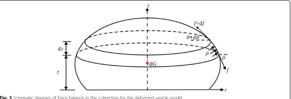
In a similar manner, the equilibrium differential equa-tion in the z-direction for a slice between z and z+ dz
can be obtained as follows. The force analysis diagram is shown in Fig.3.
From the equilibrium condition in thez-direction:
−dGþpπr2−ðpþdpÞ πðrþdrÞ2−f 2πr sinθ
þðf þdfÞ 2πðrþdrÞ sinðθþdθÞ ¼0
ð2:3aÞ
and simply:
Fig. 1Schematic diagram of the deformed vesicle model:Gis gravity applied to the centroid and an internal pressurepacts on the membrane wall.ris the radial distance of any point on the free part of the vesicle model andθis the inclination at the same point. The contact area is circular, andr0is the contact radius.θ0denotes the contact angle, andhis the height of the vesicle model
f θ0þf
r tanθþf 0
tanθ¼ p
cosθ ð2:3bÞ
By solving the system of eqs. (2.1~2.3), the analytic ex-pression of membrane tension is obtained:
f ¼
p tanθ−pz
0
þp0z θ0
sinθ cos
2θþ sinθθ0þs 0
r
ð2:4Þ
In the above equations, the internal pressure p meets the following relationship:
p¼p zð Þ ¼p0−ρgz ð2:5Þ
wherep0is the bottom pressure.
Boundary conditions
It has been shown that the solution may be reduced to solve the differential equations of the equilibrium to-gether with the boundary conditions. To solve the above equations, the global equilibrium condition of the mem-brane is required:
2πr0F0 sinθ0¼πr02p0−G ð2:6Þ
where F0 is the bottom membrane tension determined by using the classic wetting formula of Young’s Eq. (2.7) [34], in which,θ0is the contact angle.
Γ¼ F0ð1−cosθ0Þ ð2:7Þ
Equation solving
In this study, the semi-inverse method is used. In the semi-inverse method, one guesses parts of the solution and then tries to determine the rest rationally so that all of the differential equations and boundary conditions are satisfied. As we know, the guessed solution is an exact solution of this problem. In view of the abovementioned
facts, we assume the shapes of the deformed vesicles model, then obtain the solutions of membrane tension.
Pseudo-ellipsoidal cap
In the model, the un-deformed vesicle is denoted by the sphere, and the deformed vesicle is represented by the ellipsoidal cap obtained by rotating the ellipse about the
zaxis, as shown in Fig.4.
The ellipsoidal cap geometry is a three-parameter model defined in terms of contact radius r0, height h
and contact angle θ0. All three measured quantities are
required to evaluate the volume of the ellipsoidal cap. In the r-z plane, the geometry profile of the vesicle model is an ellipse described by the equation
r2
a2þ
z2
b2¼1 ð3:1Þ
where, aand b are the semi-axis lengths in ther and z directions, respectively. The values of a= 16.67μm and b= 15.90μm are obtained by fitting the experimental data in Table1. The fitting method used in this paper is the polynomial fitting in Origin 8.5. In the fitting process, the contact radius r0 is obtained according to the bottom area in Table 1, and then the values of the semi-axis lengthsaand b is obtained by fitting the con-tact radiusr0and heighthusing Eq. (3.1).
In the ellipse, the relationship between angles θ1and
θ0 satisfies the eq. (3.2),and εr is the ratio of the axes,
εr=b/a[37].
tanθ0¼ε2r tanθ1¼ε2r r0
b−h¼ 2hr0
r02− εhr
2 ð3:2Þ
The volume of the ellipsoidal cap can be obtained [38]
V¼πr0h 3
2r0 tanθ0−h
ð Þ
tanθ0 ð
3:3Þ
The differential surface area of the ellipsoidal cap can be obtained
dA¼ 2πa. b2
ffiffiffiffiffiffiffiffiffiffiffiffiffiffiffiffiffiffiffiffiffiffiffiffiffiffiffiffiffiffiffi
b4þa2−b2z2 q
dz ð3:4Þ
The heights determined by the geometric equation Eq. (3.1) cannot rigorously satisfy the volume formula Eq. (3.3). To overcome this deficiency, a geometry of the pseudo-ellipsoidal cap is offered using the correction parameterm,
as shown below:
tanθ0¼ε2r r0
b−mh¼
2mhr0
r2 0− mhεr
2 ð3:5Þ
By combining Eq. (3.3) and Eq. (3.5), the volume of the pseudo-ellipsoidal cap is obtained
V¼πmh 6ε2
r
3r20ε2rþm2h2
ð3:6Þ
Then, the analytic expression of membrane tension is formulated:
f¼
ffiffiffiffiffiffiffiffiffiffiffiffiffiffiffiffiffiffiffiffiffiffiffiffiffiffiffiffiffiffiffiffiffiffiffiffiffiffiffiffiffiffi a4m2−a2m2r2þb2r2
p
p0a4m2−a2m2r2þb2r2
−ρgabm að 2−r2Þ3=2
h i
b2a4m2−a2m2r2þb2
r2
ð3:7Þ
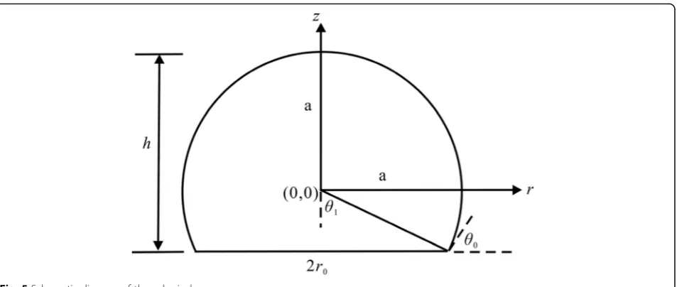
Pseudo-spherical cap
Since the deformed cells are represented by the spherical cap [32], a pseudo-spherical cap is selected to simulate the deformed vesicle model more accurately. In the pseudo-ellipsoidal cap model, when the ratio of the axes
εr is equal to 1, a pseudo-spherical cap is formed; when
the correction parametermis 1, the spherical cap is gen-erated, as shown in Fig.5.
The radius of the spherical cap R=a=b= 16.26μm is obtained by fitting the experimental data in Table 1. Similarly, the heights determined by the geometric equa-tion cannot completely meet the volume formula, and the correction parameter m is needed. In the pseudo-spherical cap, the relationship between the heighthand contact radiusr0satisfies [39]:
mh¼r0 tanθ0
2 ð3:8Þ
It yields the following relationship between the height
hand the contact radiusr0under the condition without
the volume dilatation:
V¼πmh 6 3r0
2þm2h2
ð3:9Þ
The surface area of the pseudo-spherical cap can be obtained:
A¼2πRmh ð3:10Þ
Fig. 4Schematic diagram of an ellipsoidal cap representing the deformed vesicle model on a supporting solid surface
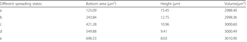
Table 1Dimensions of different spreading states [36]
Different spreading states Bottom area (μm2) Height (μm) Volume(μm3)
a 125.09 15.45 2988.40
b 243.84 12.75 2998.36
c 421.28 10.96 3000.60
d 549.88 9.41 3000.49
In the pseudo-spherical model, the analytic expression of membrane tension is expressed as:
f¼
ffiffiffiffiffiffiffiffiffiffiffiffiffiffiffiffiffiffiffiffiffiffiffiffiffiffiffiffiffiffiffiffiffi
a2m2−m2r2þr2
p
p0ða4m2−a2m2r2þa2r2Þ−ρga2m að 2−r2Þ 3=2
h i
2a4m2−a2m2r2þa2r2
ð Þ
ð3:11Þ
Analytical approximation
The total energyUT of this system is made up of three
terms: the elastic energy of the membraneUE, the
mech-anical energy of gravity UG and the surface energy US
[40].
UT¼UEþUGþUS ð3:12Þ
In this study, the single variable method is adopted, so the fluidity and viscosity of the liquid are ignored. The deformation of the vesicle model is primarily due to gravity. A spherical cap is selected to describe the geom-etry of the deformed vesicle model and the initial bend-ing energy of the un-deformed vesicle model is set to 0. Then, the following equation can be obtained:
US UEþUG¼
−πr02Γ
G R½ 0þR−mh þπκ
mh
RþfΔA
ð3:13Þ
For representative parameter valuesκ= 20kBT≈10−19
J [34], mh/R= 1.8,R0= 8.9μm volume of approximately
3000μm3 [41], f≈4 × 10⁻⁴N/m and G=ρgV= 29.4 × 10⁻¹²N, the value of this ratio is approximately 108. For this reason, the elastic energy of the membrane and the work performed by gravity are ignored throughout the analysis, and the contact radius is determined by the
following equation first solved by Johnson, Kendall, and Roberts (JKR theory) [39,42].
r3¼9π 1−ν
2
ð Þ
2E R
2
0Γ ð3:14Þ
In this article, the Young’s modulusEand the Poisson ratio ν of the membrane are 1000Pa and 0.3, respect-ively [36]. The membrane thickness t is 0.1μm [43]. When the adhesion energy per unit area Г is chosen to be 6 × 10⁻⁴J/m2[33],r0is 8.49μm.
In the pseudo-ellipsoidal cap model, the height h is calculated to be 13.29μm, and the correction parameter
mis equal to 1.027, according to Eq. (3.1) and Eq. (3.6). The contact angleθ0is 73.75° by application of Eq. (3.5).
The tensionF0is 0.83 mN/m by using Eq. (2.7), and p0
is equal to 188.52Paby application of Eq. (2.6).
Furthermore, in the pseudo-spherical situation, the height his 13.02μm and the correction parameter mis equal to 1.071 using Eq. (3.9). The contact angle θ0 is
62.54° by using Eq. (3.8). The tension F0is 0.41 mN/m,
andp0is 86.14Pa.
Results and discussion
Vesicle model deformation from the spherical state to the pseudo-ellipsoidal cap state under the action of gravity is a quasi-static process. The curve of the heighthagainst the radius of the cross sectionris shown in Fig.6. The height
h decreases when the radius of the cross section r in-creases, which agrees well with the experimental data. The results indicate that both the pseudo-ellipsoidal cap and the pseudo-spherical cap can describe the deformed vesicle model by gravity. To evaluate which can better rep-resent the geometry of the deformed vesicle model, the
variance formula is used to estimate the errors. The vari-ance of the pseudo-ellipsoidal cap is equal to 0.83, and that of the pseudo-spherical cap is 1.23, indicating that the pseudo-ellipsoidal cap may be a better representation of the deformed vesicle model.
The relationship between the inclination θ (θ< 90°) and the radius of the cross section r is obtained as shown in Fig. 7. The result shows that the inclinationθ is positively correlated with the radius of the cross sec-tionr. The angle gradually increases as the radius of the cross sectionrincreases. The values of the contact angle
θ0 are 62.43° and 65.86°, respectively. This means that
the different models are chosen to describe the vesicle model deformations; however, the results of the relation-ship between the inclination θ and the radius of the cross sectionrare the same results as shown in Fig.7.
Moreover, the variation of membrane tension f with height his also analysed as shown in Fig.8. The results show that membrane tension increases with decreasing
height h; however, the value of the pseudo-ellipsoidal cap is slightly larger than that of the pseudo-spherical cap. In the pseudo-ellipsoidal cap, the minimum and maximum values of membrane tension are 1.69 mN/m
and 2.95 mN/m, respectively, while the values in the pseudo- spherical cap are 0.75 mN/m and 1.40 mN/m, respectively. This suggests that the membrane tension of the former is approximately twice that of the latter, which may be caused by the bottom pressure. Neverthe-less, in both models, membrane tensions are slightly lar-ger than the reported values, which are approximately 0.2~0.4 mN/m. This means that gravity may have a slight influence on membrane tension when using the single variable method, so considering gravity may contribute to more accurate study of the spreading of vesicles model.
From the above, a macro approximation is used to de-scribe the deformation of the vesicle model under the action of gravity in the present study. It can be used to quantitatively describe the response of membrane ten-sion to gravity. Furthermore, the rationality of using the pseudo-ellipsoidal cap and pseudo-spherical cap to rep-resent the deformed vesicle model is explained from a mathematical point of view.
However, there are some obvious deficiencies in this study. Firstly, due to the very complex structure of eukaryotic cells, the proposed vesicle model may not be suitable for studying the eukaryotic cells. Since some studies have shown that the behavior of bacterial, the ability of bactetia to sense the surrounding environment can change under microgravity, and intestinal microbes can be dysregulated in microgravity. While bacterial be-havior can affect manned spaceflight, and the intestinal microbial disorders can lead to a series of diseases [44]. Given the wide variety of cell types, the relatively simple structure of vesicles and the ability of vesicles to mimic cells, this model can be used to study the changes of
Fig. 6Curve of the heighthagainst the radius of the cross sectionr
Fig. 7Curve of the inclinationθagainst the radius of the cross sectionr
and study whether gravity has a significant influence on the magnitude and distribution of membrane tension compared with other factors.
Conclusions
To summarize, a theoretical model of the deformation of the vesicle under the action of gravity is developed to study the response of membrane tension to gravity. The equilibrium differential equations, mainly consisting of gravity, internal pressure and membrane tension, are established. The analytic expression of membrane ten-sion is obtained. Our findings can be succinctly summa-rized as follows:
a) The deformed geometry of the vesicle model can be represented by both the pseudo-ellipsoidal cap and the pseudo-spherical cap under the action of gravity, and the pseudo-ellipsoidal cap is better from a mathematical point of view.
b) The membrane tension varies with the height: the closer it is to the basement, the greater the membrane tension.
c) The inclinationθbetween the tangent line and radial line is nearly proportional to the radius of the cross sectionrin both models.
d) Considering gravity may be useful to more accurately study the spreading of the vesicle model since gravity can influence the distribution of membrane tension.
The focus of the present work is to quantitatively ana-lyse the response of membrane tension to gravity. These findings may provide certain guidance for cell model spreading and may have some implications for membrane tension-related biological processes, especially under the hypergravity conditions.
Abbreviations
3D:three dimensions;a: major semi axes of ellipse cap (μm);b: minor semi axes of ellipse cap (μm); CSK: cytoskeleton;E: elasticity modulus of membrane (Pa);f: membrane tension (N/m);F0: bottom membrane tension (N/m);f0: initial membrane tension (N/m);G: gravity (N);h: height of deformed vesicle (μm);kB: boltzmann constant;m: correction parameter; p0: bottom pressure (Pa);r: radial distance of any point (μm);R: radius of
commented on, and edited the manuscript; All authors read and approved the final manuscript.
Funding
This work was supported by National Natural Science Foundation of China (Grants No. 11572213).
Availability of data and materials
All data used and analyzed during the current study available from the corresponding author on reasonable request.
Ethics approval and consent to participate Not applicable
Consent for publication Not applicable
Competing interests
The authors declare that they have no competing interests.
Author details
1Shanxi Key Laboratory of Material Strength & Structural Impact, College of Biomedical Engineering, Taiyuan University of Technology, Taiyuan 030024, China.2National Demonstration Center for Experimental Mechanics Education (Taiyuan university of Technology), Taiyuan 030024, China.3Key Laboratory for Space Biosciences & Biotechnology, Faculty of Life Sciences, Northwestern Polytechnical University, Xi’an 710072, China.
Received: 13 December 2018 Accepted: 29 October 2019
References
1. Maier JA, Cialdai F, Monici M, et al. The impact of microgravity and hypergravity on endothelial cells[J]. Biomed Res Int. 2015;2015:1–13. 2. Sieber M, Hanke W, Kohn FP. Modification of membrane fluidity by
gravity[J]. Open Journal of Biophysics. 2014;4(4):105–11.
3. Hemmersbach R, Volkmann D, Häder DP. Graviorientation in Protists and plants[J]. J Plant Physiol. 1999;154(1):1–15.
4. Häder DP, Hemmersbach R, Lebert M. Gravity and the behavior of unicellular organisms[M]. Cambridge: Cambridge University Press; 2005. 5. Wiedemann M, Kohn FPM, Roesner H, et al. Self-organization and
pattern-formation in neuronal systems under conditions of variable gravity[M]. Beijing: Higher Education Press; 2011.
6. Wiedemann M, Rahmann H, Hanke W. Chapter 24-gravitational impact on ion channels incorporated into planar lipid bilayers[J]. Membr Sci Technol. 2003;7(03):669–97.
7. Sieber M, Kaltenbach S, Hanke W, et al. Conductance and capacity of plain lipid membranes under conditions of variable gravity[J]. J Biomed Sci Eng. 2016;9(8):361–6.
8. Häder D, Braun M, Grimm D, et al. Gravireceptors in eukaryotes-a comparison of case studies on the cellular level[J]. Npj Microgravity. 2017; 3(13):1–8.
10. Ciofani G, Ricotti L, Rigosa J, et al. Hypergravity effects on myoblast proliferation and differentiation[J]. J Biosci Bioeng. 2012;113(2):258–61. 11. Tschopp A, Cogoli A. Hypergravity promotes cell proliferation[J]. Experientia.
1983;39(12):1323–9.
12. Signore A, Del Mandillo S, Rizzo A, et al. Hippocampal gene expression is modulated by hypergravity[J]. Eur J Neurosci. 2015;19(3):667–77. 13. Montufar-Solis D, Duke PJ, D'Aunno D. In vivo and in vitro studies of
cartilage differentiation in altered gravities[J]. Adv Space Res. 1996;17(6– 7):193–9.
14. Kawakami S, Kashiwagi K, Furuno N, et al. Effects of hypergravity environments on amphibian development, gene expression and apoptosis[J]. Comp Biochem Physiol A Mol Integr Physiol. 2006;145(1):65–72. 15. Monici M., Marziliano N., Basile V., et. al. Hypergravity affects morphology
and function in microvascular endothelial cells[J]. Microgravity Sci Technol, 2006, 18(3–4): 234–238.
16. Croute F, Gaubin Y, Pianezzi B, et al. Effects of hypergravity on the cell shape and on the organization of cytoskeleton and extracelluar matrix molecules of in vitro human dermal fibroblasts[J]. Microgravity Sci Technol. 1995;8(2):118–24.
17. Gauthier NC, Masters TA, Sheetz MP. Mechanical feedback between membrane tension and dynamics[J]. Trends Cell Biol. 2012;22(10):527–35. 18. Paluch EK, Nelson CM, Biais N, et al. Mechanotransduction: use the force
(s)[J]. BMC Biol. 2015;13(1):47.
19. Gauthier NC, Marc AF, Pere RC, et al. Temporary increase in plasma membrane tension coordinates the activation of exocytosis and contraction during cell spreading[J]. Proc Natl Acad Sci U S A. 2011;108(35):14467–72. 20. Wong IY, Javaid S, Wong EA, et al. Collective and individual migration
following the epithelial–mesenchymal transition[J]. Nat Mater. 2014; 13(11):1063–71.
21. Houk A, Jilkine A, Mejean C, et al. Membrane tension maintains cell polarity by confining signals to the leading edge during neutrophil migration[J]. Cell. 2012;148(1–2):175–88.
22. Morris CE, Homann U. Cell surface area regulation and membrane tension[J]. J Membr Biol. 2001;179(2):79–102.
23. Raucher D, Sheetz MP. Membrane expansion increases endocytosis rate during mitosis[J]. J Cell Biol. 1999;144(3):497–506.
24. Reinhart-King CA, Micah D, Hammer DA. The dynamics and mechanics of endothelial cell spreading[J]. Biophys J. 2005;89(1):676–89.
25. Liu P, Zhang Y, Cheng Q, et al. Simulations of the spreading of a vesicle on a substrate surface mediated by receptor–ligand binding[J]. J Mech Phys Solids. 2007;55(6):1166–81.
26. Lu L, Doak WJ, Schertzer JW, et al. Membrane mechanical properties of synthetic asymmetric phospholipid vesicles[J]. Soft Matter. 2016;12(36):7521–8. 27. Seifert U, Lipowsky R. Adhesion of vesicles[J]. Phys Rev A. 1990;42(8):4768. 28. Jesorka A, Orwar O. Liposomes: technologies and analytical applications[J].
Annu Rev Anal Chem. 2008;1(1):801–32.
29. Pomorski T, Lombardi R, Riezman H, et al. Drs2p-related P-type ATPases Dnf1p and Dnf2p are required for phospholipid translocation across the yeast plasma membrane and serve a role in endocytosis[J]. Mol Biol Cell. 2003;14(3):1240–54.
30. Daleke DL. Phospholipid flippases[J]. J Biol Chem. 2007;282(2):821–5. 31. Pontes B, Monzo P, Gauthier NC. Membrane tension: a challenging but universal
physical parameter in cell biology[J]. Semin Cell Dev Biol. 2017;71:30–41. 32. Thoumine O, Cardoso O, Meister JJ. Changes in the mechanical properties
of fibroblasts during spreading: a micromanipulation study[J]. Eur Biophys J. 1999;28(3):222–34.
33. Thomas F, Olivier T. Predicting the kinetics of cell spreading[J]. J Biomech. 2002;35(8):1137–41.
34. Lin Y, Freund LB. Forced detachment of a vesicle in adhesive contact with a substrate[J]. Int J Solids Struct. 2007;44(6):1927–38.
35. Yang YH, Jiang HY. Cellular volume regulation and substrate stiffness modulate the detachment dynamics of adherent cells[J]. J Mech Phys Solids. 2018;112(1):594–618.
36. Wang LP, Hsu HY, Li X, et al. Effects of frequency and acceleration amplitude on osteoblast mechanical vibration responses: a finite element study[J]. Biomed Res Int. 2016;2016:1–16.
37. Erbil HY, Mchale G, Rowan SM, et al. Analysis of evaporating droplets using ellipsoidal cap geometry[J]. J Adhes Sci Technol. 1999;13(12):1375–91. 38. Erbil HY. Evaporation of sessile drops on polymer surfaces: ellipsoidal cap
geometry[J]. J Phys Chem B. 1997;101(35):6867–73.
39. Meric RA, Erbil HY. Evaporation of sessile drops on solid surfaces: Pseudospherical cap geometry[J]. Langmuir. 1998;14(7):1915–20. 40. Johnson KL, Kendall K, Roberts A. Surface energy and the contact of elastic
solids[J]. Proc R Soc Lon Math Phys Sci. 1971;324(1558):301–13.
41. Mcgarry JG, Prendergast PJ. A three-dimensional finite element model of an adherent eukaryotic cell[J]. Eur Cells Mater. 2004;7:27–34.
42. Johnson KL. Contact mechanics[M]. Cambridge: Cambridge University Press; 1987.
43. Jiang H, Sun S. Cellular pressure and volume regulation and implications for cell mechanics[J]. Biophys J. 2013;105(3):609–19.
44. Higginson EE, Galen JE, Levine MM, et al. Microgravity as a biological tool to examine host–pathogen interactions and to guide development of therapeutics and preventatives that target pathogenic bacteria[J]. Pathog Dis. 2016;74(8):1-9.
45. Chien S. Red cell deformability and its relevance to blood flow[J]. Annu Rev Physiol. 1987;49(1):177–92.
Publisher’s Note