R E S E A R C H A R T I C L E
Open Access
Growth differentiation factor 9 and bone
morphogenetic protein 15 expression in
previtellogenic oocytes and during early
embryonic development of Yellow-tail Kingfish
Seriola lalandi
Jaime Palomino, Giannina Herrera, Phillip Dettleff and Víctor Martínez
*Abstract
Background:During fish oocyte maturation, specific molecules are expressed and accumulated within oocyte until fertilization and embryo development. Special attention have been paid in members of the transforming growth factor (TGF-β) superfamily; growth differentiation factor 9 (GDF9/gdf9) and bone morphogenetic protein 15 (BMP15/bmp15), which exert regulatory functions during oocyte maturation and follicle development. However, little attention has been paid to the involvement of these molecules during embryogenesis considering its importance for the formation of a good quality egg and subsequent embryo survival. The purpose of this study was to analyze the expression ofgdf9andbmp15in previtellogenic oocytes and during early embryonic development inSeriola lalandi, a pelagic fish with increasing prospect for its aquaculture development, which however, show high mortality at embryo and larval stages.
Results:Through RT-qPCR it was found thatgdf9expression was higher in previtellogenic oocytes decreasing after ovulation. This expression profile agrees with its participation in early stages of the follicular development. The transcripts forbmp15also showed the highest levels in previtellogenic oocytes, however this expression was lower than obtained withgdf9. Conversely, in recently spawned oocytes mRNAbmp15levels were highest than observed togdf9.This, is consequent with the main role proposed for this growth factor at the final fish oocyte maturation: avoid the ovulation of an immature oocyte. During embryo development, low levels of mRNA were detected to gdf9, with an increase in 48 H post-fertilization embryos. Thebmp15expression did not change throughout development and was higher thangdf9at 16 cells, blastula and appearance embryos stages.
Conclusions:Both (gdf9andbmp15) expression profiles in previtellogenic oocytes and newly spawned eggs are consistent with the described functions for these growth factors in vertebrate ovarian physiology in early and late stages of the follicular development. So, these genes could be considered as quality biomarkers at these stages. However, further studies of these proteins throughout folliculogenesis, are necessaries to fully understand their functions during the oocyte formation. In addition, the persistent expression of these growth factors during development, allows us to speculate possible roles in embryonic processes, which must also be addressed.
Keywords:gdf9,bmp15, Embryo development, Oocyte maturation, Fish
* Correspondence:[email protected]
FAVET-INBIOGEN, Faculty of Veterinary Sciences, University of Chile, Avda. Santa Rosa 11735, La Pintana, Santiago, Chile
Background
In recent years, evidence regarding to the active role of the oocyte in its maturation during folliculogenesis and the effect on embryonic development, has been accumula-ted [1,2]. This role is performed through molecules secre-ted by the oocyte which could regulate the folliculogenesis and steroidogenesis in both granulose and theca cells [3]. In mammals, some of the key molecules involved in these processes are the members of the transforming growth factor β super family (TGF-β), GDF9 and BMP15 [4,5]. These growth factors stimulate the proliferation of granu-lose cells, but also suppress follicle stimulating hormone (FSH)-induced granulose cell differentiation [6,7]. The effect of GDF9 on FSH action has been presumed by its ability to inhibit FSH-dependent luteinizing hormone (LH) receptor expression, cAMP production, and ste-roids synthesis [6,8,9]. Further, it is appear that GDF9 promotes follicular survival by suppressing granulose cell apoptosis and follicular atresia [10]. Similar to GDF9, BMP15 is an inhibitor of FSH-induced progesterone syn-thesis and in vitro studies have demonstrated that this effect is mediated through the suppression of FSH re-ceptor gene expression [11]. BMP15 is also involved in the cumulus expansion by stimulating the epidermal growth factor (EGF)-like growth factor expression, which is crucial forcumulus cells to be capable of responding to LH-induced granulose cell signal [12]. It has been pur-posed that BMP-5 participate in the regulation ofcumulus cell apoptosis, but the molecular mechanism it is still unknown [13].
Information about GDF9 and BMP15 in non-mammalian vertebrates is limited. However in order to understand the functional roles of these growth factors in those groups of animals, significant contributions principally related with its mRNA expression profiles have been made. In fishes, both gdf9andbmp15 genes have been studied in zebrafish [14-16], carp [17,18], eels [19,20], European sea bass [21] and rainbow trout [22]. In gen-eral, it has been described a decreasing in gdf9 expres-sion during follicular development among fish species. In zebrafish and gibel carp, the highest gdf9expression levels has been observed in ovaries composing by oogo-nias and cells in transition to primary oocytes [16,17]. By the contrary, in European sea bass, eels, rainbow trout and wuchang bream, the highest levels of gdf9 mRNA have been revealed at the previtellogenic stage [18-22].
As well asgdf9, bmp15shows specie-specific patterns in both expression profiles and function during fish fol-liculogenesis. In zebrafish, important roles throughout follicle maturation by inhibiting premature follicles de-velopment and thus avoiding the ovulation of an imma-ture oocyte have been suggested for BMP15 [15,16,23]. In vitro studies using zebrafish follicles, have revealed that incubation with human recombinant BMP15 or over
expression in oocytes, resulted in an inhibition of oocyte maturation induced by both gonadotropins and matur-ation inducing hormone (MIH) [14,15]. Therefore, these findings suggest that BMP15 modulates the follicular growth and prevents premature oocyte maturation in zebrafish, in part, by suppressing the sensitivity of follicles to MIH [23]. Furthermore, similar expression profile com-pared togdf9was observed during fish follicular develop-ment of European sea bass, suggesting that these factors might act cooperatively in their ovarian functions [21].
The yellow-tail kingfishSeriola lalandiis a marine pe-lagic fish with a circumglobal distribution [30]. This spe-cies has been considered essential to diversify the Chilean aquaculture due to its rapid growth rate, good flesh quality and an increasing worldwide demand. However, the full production cycle of this species has been difficult to achieve due to high mortality during embryonic and larval stages [30,31]. Previous studies about the repro-ductive physiology of wild caught individuals estab-lished that males and females reach 50% maturity at a fork length of 812 and 944 mm, respectively. The spawn occur naturally during the austral spring and summer when the seawater temperature was above 17°C and indi-viduals spawn repeatedly during this time [31]. It has been observed a courtship behavior which consisted of a high-speed pursuit punctuated by stalling, nipping and touching between one male and female [30]. There is no scientific evidence regarding disorders during em-bryogenesis in S. lalandi to explain this low survival, and there are not antecedents regarding to aspects in-volved in oogenesis and early development in this spe-cies. Studies performed in marine fish have proposed that low buoyancy of eggs and early embryos [32], skel-etal malformations [33], arrested eye migration [34] and locking of the jaw cartilage [35] could be the result of inappropriate oocyte maturation. Thereby, to eluci-date molecular aspects involved in oocyte maturation and early embryogenesis, appear to be essential in order to understand and eventually improve the reproduction in this species. Therefore, the objective of this work was to study the expression of growth factors gdf9and bmp15 in previtellogenic oocytes and during early em-bryonic development inS. lalandi.
Results
RT-qPCR conditions
The RNA quality was evaluated according to denaturing agarose electrophoresis gels and we only worked with those samples where it was possible clearly to distin-guish both bands that constituted the total RNA (18 and 28 sub-unities) without an important smears (data not
shown). For each primer pairs, non-specific amplifica-tion was evaluated using the shape of the melting curves (Tm), where it was possible to observe a single clear, with a Tm similar to what is expected based on the amplicon sequence. Furthermore, for all primers pairs used in this work, the optimum combination between forward and reverse primers concentration was deter-mined and the selection criteria used was the lower Ct value and smaller standard deviation between replicates of the amplification curves in experiments with the same cDNA template concentration. Finally, it was crucial to validate the qPCR results with efficiencies near to 2 (100%). All reactions displaying similar efficiencies are presented in Table 1.
Growth factors expression during embryonic development
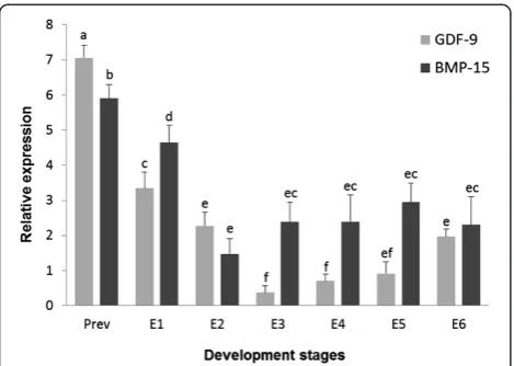
RT-qPCR was performed to assess the relative expression levels of both gdf9and bmp15 in previtellogenic oocytes and throughout the S. lalandi embryonic development. The higher expression levels ofgdf9transcripts were ob-served in previtellogenic oocytes and a significant decrease in the expression of this growth factor was observed after ovulation (Figure 1). Further, as embryonic development progresses, thegdf9mRNA expression declined dramatic-ally at 16 cells stage (E3) maintaining low levels even after gastrulation process (E5) (Figure 1). Interestingly, a signifi-cant increase in the gdf9 expression was observed after 48 hours post fertilization (E6), in spite of this expression level was lower than observed in previtellogenic and recently spawned oocytes (Figure 1). Similar togdf9, tran-scripts to bmp15 showed the highest levels in previtello-genic oocytes, but its expression was lower than gdf9. After spawn and fertilization, was observed a significant decrease in the bmp15 expression, but at this stage (E1), highest mRNA levels thangdf9 was found. In 4 cell em-bryos (E2), a large decrease in the bmp15expression was observed and no changes in these transcripts levels were observed through development (Figure 1). From 16 cells stage (E3) until embryos 24 hour post fertilization (E5), bmp15 expression was higher than gdf9. Finally, in 48
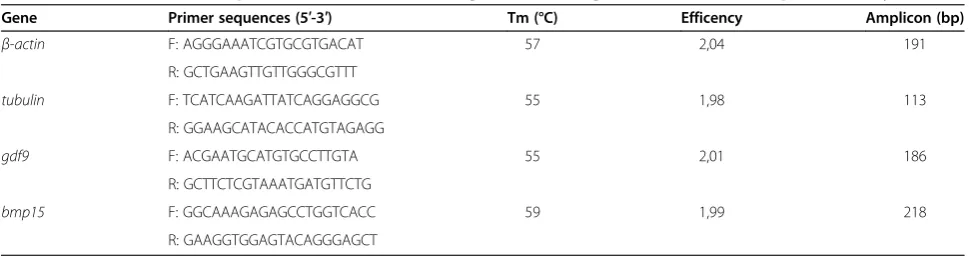
Table 1 PCR characteristic of primers for constitutive and growth factors genes evaluated in the present study
Gene Primer sequences (5′-3′) Tm (°C) Efficency Amplicon (bp)
β-actin F: AGGGAAATCGTGCGTGACAT 57 2,04 191
R: GCTGAAGTTGTTGGGCGTTT
tubulin F: TCATCAAGATTATCAGGAGGCG 55 1,98 113
R: GGAAGCATACACCATGTAGAGG
gdf9 F: ACGAATGCATGTGCCTTGTA 55 2,01 186
R: GCTTCTCGTAAATGATGTTCTG
bmp15 F: GGCAAAGAGAGCCTGGTCACC 59 1,99 218
hour embryos (E6) no differences in the expression of these growth factors were detected (Figure 1).
Discussion
Although mRNA levels to gdf9 and bmp15 during fol-licular development have been analyzed in several teleost species, there is little knowledge regarding the expres-sion of these growth factors after fertilization and their possible biological significance in both embryonic and larval survival. Therefore, given the fundamental role of these TGF-β members in the production of a good-quality oocyte, this study has attempted to give prelimin-ary evidence regarding the expression of these growth factors in previtellogenic oocytes and during early em-bryonic development of yellow tail KingfishS. lalandi.
The expression level ofgdf9was clearly highest in pre-vitellogenic oocytes compared with its expression during embryo development. These results agree with studies in rainbow trout, eels and European sea bass, where the highest level of gdf9 expression at the previtellogenic stage has been revealed, with an important decline along the vitellogenic process [19-22]. Although GDF9 functions inS. lalandiare currently unknown, its high expression in previtellogenic oocytes would propose a role of this growth factor during early ovarian development with potential participation in the follicular recruitment or thereafter during early stages of the vitellogenic process. More accurate conclusions about the functions of this growth factor inS. lalandifollicular growth must be elu-cidated by studying its expression at different stages of ovarian development. However, based on our results and comparing its expression levels, between previtellogenic
occytes and recently spawned eggs (E1), we can infer a de-crease in the expression of this growth factor during vitel-logenesis, which is consistent with the observed in rice field eel [20], zebrafish [16] and European sea bass [21]. Similarly, thebmp15mRNA levels were significantly lower in ovulated oocytes in comparison with previtellogenic oocytes. These results would indicate a decrease in the bmp15 expression during follicular development and more specifically during vitellogenesis and maturation. This interpretation is consistent with studies in gibel carp [36] and European sea bass [21], where a decrease in the bmp15 expression was observed through vitello-genic process. On the contrary,bmp15in zebrafish, was expressed consistently in follicles of different develop-mental stages and no significant differences were ob-served in their expression levels [14].
The expression patterns forgdf9andbmp15in previtel-logenic oocytes confirm the involvement of these proteins in the early stages of follicular development. However, the difference in the expression levels between these growth factors was an interesting result. In previtellogenic oocytes, the levels ofgdf9mRNA were higher than those ofbmp15, which can be explained by its critical role dur-ing early stages of follicular development, specifically during recruitment [16]. Furthermore, this observation supports the hypotheses for European sea bass where it has been suggested that gdf9expression appears not to be regulated by gonadotropins [37]. By contrast, spawned eggs showed higher levels ofbmp15mRNA in comparison withgdf9expression, which agree with the principal role proposed forbmp15in zebrafish, in maintaining egg qual-ity and preventing ovulation of an immature oocyte [23]. Major conclusions regarding the importance of these growth factors and their coordination in processes such as vitellogenesis and oocyte maturation should be obtained by studying their expression during different stages of fol-licular development.
On the other hand, duringS. lalandiembryonic develop-ment time course, bothgdf9andbmp15expression levels significantly decreased after two hours post fertilization (E2). Specifically, gdf9showed the lower expression value in 16 cell embryos (E3), after two and half hours post fer-tilization. This situation, agrees with that observed in wuchang bream [18], but differs which occurs in zebra-fish, where a high gdf9expression is maintained until 6 hours post fertilization, at the blastocyst stage, decreas-ing with the advanced development [16]. We believe that the different lengths of the embryonic processes among fish species could be the explanation for the dif-ferences in thegdf9expression. Therefore, despite there is no evidence to support the biological roles of this growth factor at these development stages, its decreased level of mRNA during the early embryonic development suggest that its gene expression may be affected by the Figure 1Relative mRNA levels of gdf9 and bmp15 throughout
development inS. lalandi.The bars represent the means ± standard deviations of growth factors relative expression in previtellogenic oocytes (Prev) and during different developmental stages inS. lalandi
gradual transition period from oocyte-derivate mRNA and proteins to full embryonic transcriptions [38]. In mammalian embryo development,gdf9has been identi-fied as a part of maternal mRNA to be used during early embryogenesis [25], so a similar situation may also be oc-curring in Seriola, as well as in other fish species. In the other hand, an interesting and significantly increase in the gdf9expression was observed in 48-hour post fertiliza-tion embryos (E6). A different situafertiliza-tion was observed in zebrafish, where gdf9 mRNA virtually disappeared from the gastrula stage, about 10 hours after fertilization [16]. However, our result would agree with those reported in wuchang bream, where despite thegdf9 mRNA is main-tained at a low level, a significant increase was detected in 62 hours embryos compared with those of 50 hours post fertilization [18]. Therefore, the persistent expression of gdf9in earlyS. lalandiembryos would implicate functions of this molecule in early embryogenesis as well as it has been proposed to others growth factors such as GDF1 [39] and GDF5 [40], which have been implicated in the left right patterning and skeletal joint formation, respectively.
Thebmp15level expression decrease significantly after two hours post fertilization (E2) and, unlike gdf9, its mRNA levels did not vary as the embryo development was progressing. Nevertheless, between 16-cell (E3) and appearance embryos (E5) stages, higher expression le-vels for bmp15 in comparison to gdf9, were observed. The involvement of bmp15 in teleost embryogenesis has not been yet described. However, changes in its structure that includes a serine domain, similar to that found in yolk proteins linked to calcium phosphate trans-port during the embryo bone formation, has been men-tioned to speculate its role in this process during fish embryogenesis [21].
Conclusions
The expression of gdf9 and bmp15 in previtellogenic oocytes and during early embryonic development in S. lalandi, has been confirmed. The consistency between their expression profiles in previtellogenic oocytes and newly spawned eggs, with the proposed functions in mammals and other fish species, would allow proposing the possibility of use these genes as potential quality biomarkers at these stages. However, the biological sig-nificance of their expression during embryonic develop-ment should be more depth studied.
Methods
Samples collection
All animals used in the present study as well as the experi-mental procedures were treated according the Chilean Bioethics Committee of the National Commission for Scientific and Technological Research. The samples were provided by Acuinor S. A., a commercial company that
has successfully developed the complete production in captivity in a hatchery production center located in Caldera, Atacama Region, Chile. The broodstock com-prise 40 individuals maintained in two indoor tanks with 2.5 m depth and 20,000 Lts, in a sex proportion of 1:1. Animals were subjected to different photoperiods, temperatures between 18 to 20°C and daily feeding at libitum, in order to assure the availability of eggs around the whole cycle. The spawned eggs were channeled from a skimmer on the surface of each tank into a separate egg collector. Three batches of each tank were observed from spawning and samples of approximately 100 individuals were taken at different development stages as described by Moran et al. [30]. The selected development stages were: newly spawned eggs, 4 cell, 16 cell, blastula, appear-ance embryo and advappear-anced embryo, which were identified as E1, E2, E3, E4, E5 and E6 respectively. In addition, ovary samples obtained by cannulation of the gonophore of three anesthetized adult female individuals were stirred in a petri dish with 3 mL of micro filtered sea water. Then approximately 200 previtellogenic oocytes that ranged between 30 and 50μm were taken with a fine bore pip-ette. All samples were maintained in RNAlater® Solution (Ambion®) and processed to RNA extraction.
RNA extraction and cDNA synthesis
Total RNA was extracted using the column affinity Pu-rification Kit GeneJET™ RNA (Fermentas Life Sciences) following the manufacturer’s instructions. The RNA con-centration was determined using a Qubit® Fluorometer (Invitrogen™) with the quantification kit Qubit® RNA Assay, (Molecular Probes® Invitrogen™). DNA contamin-ation was removed by Dnase I treatment and the total RNA quality was assessed by 1% ethidium bromide agar-ose gels electrophoresis under denaturing conditions. The RNA samples were stored at -80°C until use. The samples were processed for reverse transcription (RT) using the enzyme conjugate SuperScript™ First-Strand Synthesis System (Invitrogen™). In order to corroborate the absence of contaminating DNA, RT negative con-trol was used for each sample. The complementary DNA (cDNA) concentration was determined using the Quantifi-cation Kit ssDNA Qubit® Assay (Invitrogen™ Molecular Probes®). cDNA samples were stored at -20°C until use in qPCR protocols as described below.
Primer design
genes were generated from Seriola quinqueradiata EST sequences (Accession numbers (AN): AB179839.1 and AB461862.1 forβ-actinandtubulin, respectively). Togdf9 primers were designed by sequence alignment from four fish species (Dicentrarchus labrax, AN: AM933667.1; Oncorhynchus mykiss, AN: EU723245.1;Carassius gibelio, AN: HQ454282.1 and Danio rerio, AN: AY833104.1). Primers to bmp15 were designed based on three fish species sequences (Carassius gibelio, AN: HQ454283.1; Cyprinus carpio, AN: JN002405.1 and Dicentrarchus labrax, AN: AM933668.1). The alignment was per-formed using the ClustalX program and the primers were searched in about 200 bp highly conserved re-gions. All primers were designed with Primer 3Plus considering a theoretical optimal annealing temperatures about 60°C, with differences between the forward and re-verse primers not greater than 5°C, guanine and cytosine percentages between 30 and 60%, primers lengths 18 to 20 bp and free of dimmers and hairpins. In addition, all primers were tested by NET primer (http://www.Premier Biosoft.com/netprimer). All the primers are showed in Table 1.
Quantitative real-time PCR (qPCR)
Gene amplifications by qPCR were performed with an Illumina® Eco Real Time PCR System Model EC-100-1001. Each 12.5 μL reaction contained the following: 6.25 μL of Maxima SYBR Green/Fluorescein qPCR Master Mix (2X), a necessary volume to 0.2-0.6μM of each primer, a volume with 10 ng of cDNA and the volume difference was complete with DEPC treated water. Plates were sealed with adhesive optical film and the following PCR conditions were performed: after an initial denaturation step during 15 min at 95°C, 40 amplification cycles were performed according to the following thermo cycling profile: denaturation for 15 s at 94°C, annealing for 30 s at a temperature ac-cording to specific Tm of each primers pair and exten-sion for 30 s at 72°C. A dissociation protocol with a gradient from 60 to 95°C was used to investigate the specificity of the qPCR reaction and the presence of primer dimers. Control samples without reverse tran-scriptase and without the template, were included in each plate. Prior to expression analysis, we standardized and optimized the qPCR conditions for all primers using cDNA obtained from ovarian samples, assessing the op-timal concentration for each primer pair and their ef-ficiency in the PCR. The efef-ficiency (E) was evaluated with six-point standard curves of a six-fold dilution series (1:1-1250). The slope of the standard curve rep-resents the amplification efficiency (E) [41], through the following formula E =10(‒1/slope). We used only primers pairs with efficiencies approaching 2 (which indicate 100%) and over 0.989 of R2value [42].
Data analysis
After standardization of PCR conditions, we evaluated the relative expression ofgdf9andbmp15genes in previ-tellogenic oocytes and during different embryo develop-ment stages inS. lalandi. Three biological replicates that represent three different spawning events were analyzed, which were evaluated in three technical replicates. In each experiment, gene expression levels were recorded as Ct values that corresponded to the number of cycles where the fluorescence signal can be detected above a threshold value. The Cts averages for each biological replicate were calculated and transformed into relative values denominatedQuantity(Q) throughΔΔCt method [43]. Then, the relative quantification in the expression of gdf9 and bmp15 for each developmental stages was estimated as the quotient between Q value of the target gene and a normalization factor (NF), which was calcu-lated based on the geometric mean of reference genes Q values [43]. Relative expression was expressed as the mean ± standard deviation and the data were analyzed by ANOVA using InfoStat Professional Program, Version 2004; National University of Córdoba, Argentina. Signifi-cant differences among means were evaluated using Tukey test and values were considered significantly dif-ferent when P <0.05.
Competing interests
The authors declare that they have no competing interests.
Authors’contributions
JP designed and conceived the study, participating in all experiments and writing the manuscript. GH participated in the primers design and PCR analysis. PD participated in the statistic data analysis and helped to draft the manuscript. VM participated in the design of the experiments and helped to draft the manuscript. All authors read and approved the final manuscript.
Acknowledgements
We gratefully acknowledge financial support of CONICYT by Post-doc FONDECYT grant 3120211 (JP) and doctoral scholarship (PD). We also gratefully to Chilean Aquaculture Diversification Grant 09PDAC-7020 (VM). We thank the Acuinor S.A. Company for their generous collaboration providing us the samples for this study. In a same way, we appreciate to Drs. Mónica De los Reyes, Mario Duchens and Oscar Peralta, by their opinions about our manuscript.
Received: 14 August 2014 Accepted: 28 October 2014 Published: 20 November 2014
References
1. Su YQ, Sugiura K, Wigglesworth K, O’Brien MJ, Affourtit JP, Pangas SA, Matzuk MM, Eppig JJ:Oocyte regulation of metabolic cooperativity between mouse cumulus cells and oocytes: BMP15 and GDF9 control cholesterol biosynthesis in cumulus cells.Development2008,135:111–121. 2. Kidder GM, Vanderhyden BC:Bidirectional communication between
oocytes and follicle cells: ensuring oocyte developmental competence. Can J Physiol Pharmacol2010,88(4):399–413.
3. Matzuk M, Burns KH, Viveiros MM, Eppig JJ:Intercellular communication in the mammalian ovary: oocytes carry the conversation.Science2002,
296:2178–2180.
4. Knight PG, Glister C:TGF-beta superfamily members and ovarian follicle development.Reproduction2006,132:191–206.
6. Vitt UA, Hayashi M, Klein C, Hsueh AJ:Growth differentiation factor-9 stimulates proliferation but suppresses the follicle-stimulating hormone-induced differentiation of cultured granulosa cells from small antral and preovulatory rat follicles.Biol Reprod2000,62:370–377.
7. Otsuka F, Shimasaki S:A novel function of bone morphogenetic protein-15 in the pituitary: selective synthesis and secretion of FSH by gonadotropes.Endocrinology2002,143(12):4938–4941.
8. Spicer LJ, Aad PY, Allen D, Mazerbourg S, Hsueh AJ:Growth differentiation factor-9 has divergent effects on proliferation and steroidogenesis of bovine granulosa cells.J Endocrinol2006,189(2):329–339.
9. Sugiura K, Su YQ, Li Q, Wigglesworth K, Matzuk MM, Eppig JJ:Estrogen promotes the development of mouse cumulus cells in coordination with oocyte-derived GDF9 and BMP15.Mol Endocrinol2010,
24(12):2303–2314.
10. Orisaka M, Orisaka S, Jiang JY, Craig J, Wang Y, Kotsuji F, Tsang BK:
Growth differentiation factor 9 is antiapoptotic during follicular development from preantral to early antral stage.Mol Endocrinol2006,
20(10):2456–2468.
11. Otsuka F, Yamamoto S, Erickson GF, Shimasaki S:Bone morphogenetic protein-15 inhibits follicle-stimulating hormone (FSH) action by suppressing FSH receptor expression.J Biol Chem2001,276(14):11387–11392.
12. Yoshino O, McMahon HE, Sharma S, Shimasaki S:A unique preovulatory expression pattern plays a key role in the physiological functions of BMP-15 in the mouse.Proc Natl Acad Sci U S A2006,103(28):10678–10683. 13. Hussein TS, Froiland DA, Amato F, Thompson JG, Gilchrist RB:Oocytes
prevent cumulus cell apoptosis by maintaining a morphogenic paracrine gradient of bone morphogenetic proteins.J Cell Sci2005,
118(Pt 22):5257–5268.
14. Clelland E, Kohli G, Campbell RK, Sharma S, Shimasaki S, Peng C:Bone morphogenetic protein-15 in the zebrafish ovary: complementary deoxyribonucleic acid cloning, genomic organization, tissue distribution, and role in oocyte maturation.Endocrinology2006,147:201–209. 15. Clelland ES, Tan Q, Balofsky A, Lacivita R, Peng C:Inhibition of premature
oocyte maturation: a role for bone morphogenetic protein 15 in zebrafish ovarian follicles.Endocrinology2007,148:5451–5458. 16. Liu L, Ge W:Growth differentiation factor 9 and its spatiotemporal
expression and regulation in the zebrafish ovary.Biol Reprod2007,
76:294–302.
17. Liu Z, Chen A, Yang Z, Wei H, Leng X:Molecular characterization of growth differentiation factor 9 and its spatio-temporal expression pattern in gibel carp (Carassius auratus gibelio).Mol Biol Rep2012,
39(4):3863–3870.
18. Huang CX, Wei XL, Chen N, Zhang J, Chen LP, Wang WM, Li JY, Wang HL:
Growth differentiation factor 9 ofMegalobrama amblycephala: molecular characterization and expression analysis during the development of early embryos and growing ovaries.Fish Physiol Biochem2014,40(1):193–203.
19. Lokman PM, Kazeto Y, Ozaki Y, Ijiri S, Tosaka R, Kohara M, Divers SL, Matsubara H, Moore LG, Adachi S:Effects of reproductive stage, GH, and 11-ketotestosterone on expression of growth differentiationfactor-9 in the ovary of the eel, Anguilla australis.Reproduction2010,139(1):71–83. 20. He Z, Wu Y, Xie J, Wang T, Zhang L, Zhang W:Growth differentiation
factor 9 (Gdf9) was localized in the female as well as male germ cells in a protogynous hermaphroditic teleost fish, ricefield eelMonopterus albus.Gen Comp Endocrinol2012,178(2):355–362.
21. Halm S, Ibañez AJ, Tyler CR, Prat F:Molecular characterization of growth differentiation factor 9 (gdf9) and bone morphogenetic protein 15 (bmp15) and their patterns of gene expression during the ovarian reproductive cycle in the European sea bass.Mol Cell Endocrinol2008,
291:95–103.
22. Lankford SE, Weber GM:Temporal mRNA expression of transforming growth factor-beta superfamily members and inhibitors in the developing rainbow trout ovary.Gen Comp Endocr2010,166:250–258. 23. Peng C, Clelland E, Tan Q:Potential role of bone morphogenetic
protein-15 in zebrafish follicle development and oocyte maturation. Comp Biochem Phys A2009,153:83–87.
24. Sendai Y, Itoh T, Yamashita S, Hoshi H:Molecular cloning of a cDNA encoding a bovine growth differentiation factor-9 (GDF-9) and expression of GDF-9 in bovine ovarian oocytes and in vitro-produced embryos.Cloning2001,3(1):3–10.
25. Alizadeh Z, Kageyama S, Aoki F:Degradation of maternal mRNA in mouse embryos: selective degradation of specific mRNAs after fertilization. Mol Reprod Dev2005,72(3):281–290.
26. Yeo CX, Gilchrist RB, Thompson JG, Lane M:Exogenous growth differentiation factor 9 in oocyte maturation media enhances
subsequent embryo development and fetal viability in mice.Hum Reprod
2008,23:67–73.
27. Wu YT, Tang L, Cai J, Lu XE, Xu J, Zhu XM, Luo Q, Huang HF:High bone morphogenetic protein-15 level in follicular fluid is associated with high quality oocyte and subsequent embryonic development.Hum Reprod
2007,22:1526–1531.
28. McGrath SA, Esquela AF, Lee SJ:Oocyte-specific expression of growth/ differentiation factor-9.Mol Endocrinol1995,9:131–136.
29. Di Pasquale E, Brivanlou AH:Bone Morphogenetic Protein 15 (BMP15) acts as a BMP and Wnt inhibitor during early embryogenesis.J Biol Chem
2009,284:26127–26136.
30. Moran D, Smith C, Gara B, Poortenaar C:Reproductive behavior and early development in yellowtail kingfish (Seriola lalandiValenciennes 1833). Aquaculture2007,262:95–104.
31. Poortenaar CW, Hooker SH, Sharp N:Assessment of yellowtail kingfish (Seriola lalandilalandi) reproductive physiology, as a basis for aquaculture development.Aquaculture2001,201:271–286.
32. Carnevali O, Mosconi G, Cambi A, Ridolfi S, Zanuy S, Polzonetti-Magni A:
Changes of lysosomal enzyme activities in sea bass (Dicentrarchus labrax) eggs and developing embryos.Aquaculture2001,202:249–256.
33. Lewis LM, Lall SP:Development of the axial skeleton and skeletal abnormalities of Atlantic halibut (Hippoglossus hippoglossus) from first feeding through metamorphosis.Aquaculture2006,257:124–135. 34. Sæle O, Solbakken JS, Watanabe K, Hamre K, Power D, Pittman K:Staging of
Atlantic halibut (Hippoglossus hippoglossus) from first feeding through metamorphosis, including cranial ossification independent of eye migration.Aquaculture2004,239:445–465.
35. Lein I, Tveite S, Gjerde B, Holmefjord I:Effects of salinity on yolk sac larvae of Atlantic halibut (Hippoglossus hippoglossus).Aquaculture1997,
156:291–303.
36. Chen AQ, Liu ZW, Yang ZG, Leng XJ:Characterization of bmp15 and its regulation by human chorionic gonadotropin in the follicle of gibelcarp (Carassius auratus gibelio).Comp Biochem Physiol B2012,163(1):121–128. 37. García-López Á, Sánchez-Amaya MI, Halm S, Astola A, Prat F:Bone
morphogenetic protein 15 and growth differentiation factor 9 expression in the ovary of European sea bass (Dicentrarchus labrax): cellular localization, developmental profiles, and response to unilateral ovariectomy.Gen Comp Endocrinol2011,174(3):326–334.
38. Brevini TA, Cillo F, Antonini S, Tosetti V, Gandolfi F:Temporal and spatial control of gene expression in early embryos of farm animals.Reprod Fertil Dev2007,19(1):35–42.
39. Rankin CT, Bunton T, Lawler AM, Lee SJ:Regulation of left-right patterning in mice by growth/differentiation factor-1.Nat Genet2000,24(3):262–265. 40. Francis-West PH, Abdelfattah A, Chen P, Allen C, Parish J, Ladher R, Allen S,
MacPherson S, Luyten FP, Archer CW:Mechanisms of GDF-5 action during skeletal development.Development1999,126(6):1305–1315.
41. Radonic A, Thulke S, Mackay IM, Landt O, Siegert W, Nitsche A:Guideline to reference gene selection for quantitative real-time PCR.Biochem Biophys Res Commun2004,313:856–862.
42. Bustin S:Quantification of mRNA using real-time reverse transcription (RT-qPCR): trends and problems.J Mol Endocrinol2002,29:23–29. 43. Vandesompele J, De Preter K, Pattyn F, Poppe B, Van Roy N, De Paepe A,
Speleman F:Accurate normalization of real-time quantitative RT-PCR data by geometric averaging of multiple internal control genes.Genome Biol
2002,3(7):research0034.1–research0034.11.
doi:10.1186/0717-6287-47-60
Cite this article as:Palominoet al.:Growth differentiation factor 9 and bone morphogenetic protein 15 expression in previtellogenic oocytes
and during early embryonic development of Yellow-tail KingfishSeriola