University of Pennsylvania
ScholarlyCommons
Publicly Accessible Penn Dissertations
1-1-2014
Membrane Curvature Sorting And Generation By
The Bar Domain Proteins Endophilin And
Syndapin
Chen Zhu
University of Pennsylvania, chenzhu110@gmail.com
Follow this and additional works at:http://repository.upenn.edu/edissertations Part of theChemistry Commons
This paper is posted at ScholarlyCommons.http://repository.upenn.edu/edissertations/1527
For more information, please contactlibraryrepository@pobox.upenn.edu.
Recommended Citation
Zhu, Chen, "Membrane Curvature Sorting And Generation By The Bar Domain Proteins Endophilin And Syndapin" (2014).Publicly Accessible Penn Dissertations. 1527.
Membrane Curvature Sorting And Generation By The Bar Domain
Proteins Endophilin And Syndapin
Abstract
Membrane curvature provides a means to control spatial organization and activity of cells. It is regulated by plenty of peripherally binding membrane proteins, including BAR domain proteins. Two important sub-families of BAR domain containing proteins are NBAR and FBAR domain proteins. However, the current understanding of BAR domain protein membrane curvature (MC) sensing and generation is insufficient. My thesis intends to contribute to alleviating this situation.
We first performed curvature sorting and generation experiments of an NBAR domain protein: endophilin. We found that the endophilin NBAR domain (ENBAR) behaved as a curvature sensor or generator at different concentrations. We studied lateral diffusion of ENBAR and found its diffusion coefficients depending on its membrane density. We developed an analytical model to explain our experimental results. Our theory predicts that the membrane curvature serves as a switch that is modulated by a thermodynamic phase transition.
Then we studied the influence of membrane insertion helices on NBAR domain protein MC sensing and membrane dissociation kinetics by comparing ENBAR WT protein with helices deletion mutants. We found that the two helices had essential contributions for the NBAR domain curvature sorting. The WT and mutant membrane dissociation time were positively correlated with their densities on the membrane. Irreversible binding fractions for both variants were observed. These phenomena suggest higher order structure formation of these variants on the membrane.
Furthermore, we investigated the autoinhibition mechanism of full length endophilin via its H0 variants and the SH3 domain, by fluorescence experiments. We obtained evidence of the interaction between H0 helix and SH3 domain in solution and determined their binding affinities. These results experimentally support an H0/ SH3 domain mediated autoinhibition mechanism.
Finally we explored the regulation of a possible autoinhibition of the tubulation ability of syndapin, an FBAR domain protein. We compared the curvature sensing, generation and initiation abilities of syndapin FBAR, full length and full length with proline rich ligand. We found that the syndapin FBAR curvature initiation ability was larger compared to the full length protein, which was likely due to the differences in their curvature sensing abilities.
Degree Type
Dissertation
Degree Name
Doctor of Philosophy (PhD)
Graduate Group
Chemistry
First Advisor
Tobias Baumgart
Keywords
BAR domain, biophysics, curvature, endocytosis, membrane, protein
Subject Categories
Chemistry
MEMBRANE CURVATURE SORTING AND GENERATION BY THE BAR DOMAIN
PROTEINS ENDOPHILIN AND SYNDAPIN
Chen Zhu
A DISSERTATION
in
Chemistry
Presented to the Faculties of the University of Pennsylvania
in
Partial Fulfillment of the Requirements for the
Degree of Doctor of Philosophy
2014
Supervisor of Dissertation
________________________
Dr. Tobias Baumgart
Associate Professor of Chemistry
Graduate Group Chairperson
________________________
Dr. Gary A. Molander
Hirschmann-Makineni Professor of Chemistry
Dissertation committee
Dr. Jeffery G. Saven, Associate Professor of Chemistry
Dr. Paul A. Janmey, Professor of Physiology
DEDICATION
To Haiying Zhu and Sui Xie
ACKNOWLEDGEMENT
The completion of this thesis is not possible without the help of many others. First of all,
I would like to take the opportunity to thank my thesis advisor Dr. Tobias Baugmart, who
has always been very supportive since I joined the lab and served as an incredible advisor.
His patience and understanding helped me adapt the research at the initial phases of my
research. His enthusiasm about science always inspired me. His depth and breadth of
knowledge always impressed me. His guidance helped me throughout the experimental
part of my PhD studies and writing of this thesis.
I am very grateful to have Dr. Jeffery G. Saven, Dr. Paul A. Janmey, Dr. Zahra Fakhraai
as my committee members. I would like to thank Dr. Saven for serving as my committee
chair. He has always been very kind, patient and gave me useful suggestions. Dr. Janmey
shared his expertise and advice on lipid membranes with me. I am thankful that Dr. Zahra
Fakhraai was willing to join my committee at the late stage of my research.
I thank the past and current lab members in Baumgart’s group. I learned a lot from
several senior colleagues: Dr. Aiwei Tian, Dr. Benjamin R. Capraro and Dr. Michael C.
Heinrich helped me with tether pulling experiments, protein expression and purification,
and the usage of optical tweezers. I also benefited from working with fellow graduate
students: Tingting Wu, Zheng Shi, Zhiming Chen and other group members. It was a
great pleasure and fortunate to work with so many excellent scientists.
Last but not the least, I would never have been able to finish this thesis without
continuous supports and love from my friends and family, especially my parents, who are
always there to cheer me up and stand by me.
ABSTRACT
MEMBRANE CURVATURE SORTING AND GENERATION BY THE BAR DOMAIN
PROTEINS ENDOPHILIN AND SYNDAPIN
Chen Zhu
Dr. Tobias Baumgart
Membrane curvature provides a means to control spatial organization and activity of cells.
It is regulated by plenty of peripherally binding membrane proteins, including BAR
domain proteins. Two important sub-families of BAR domain containing proteins are
NBAR and FBAR domain proteins. However, the current understanding of BAR domain
protein membrane curvature (MC) sensing and generation is insufficient. My thesis
intends to contribute to alleviating this situation.
We first performed curvature sorting and generation experiments of an NBAR domain
protein: endophilin. We found that the endophilin NBAR domain (ENBAR) behaved as a
curvature sensor or generator at different concentrations. We studied lateral diffusion of
ENBAR and found its diffusion coefficients depending on its membrane density. We
developed an analytical model to explain our experimental results. Our theory predicts
that the membrane curvature serves as a switch that is modulated by a thermodynamic
phase transition.
Then we studied the influence of membrane insertion helices on NBAR domain protein
MC sensing and membrane dissociation kinetics by comparing ENBAR WT protein with
helices deletion mutants. We found that the two helices had essential contributions for the
NBAR domain curvature sorting. The WT and mutant membrane dissociation time were
positively correlated with their densities on the membrane. Irreversible binding fractions
for both variants were observed. These phenomena suggest higher order structure
formation of these variants on the membrane.
Furthermore, we investigated the autoinhibition mechanism of full length endophilin via
its H0 variants and the SH3 domain, by fluorescence experiments. We obtained evidence
of the interaction between H0 helix and SH3 domain in solution and determined their
binding affinities. These results experimentally support an H0/ SH3 domain mediated
autoinhibition mechanism.
Finally we explored the regulation of a possible autoinhibition of the tubulation ability of
syndapin, an FBAR domain protein. We compared the curvature sensing, generation and
initiation abilities of syndapin FBAR, full length and full length with proline rich ligand.
We found that the syndapin FBAR curvature initiation ability was larger compared to the
full length protein, which was likely due to the differences in their curvature sensing
abilities.
TABLE OF CONTENTS
ACKNOWLEDGMENT ... III
ABSTRACT ... IV
TABLE OF CONTENTS ... VI
LIST OF FIGURES AND TABLE ... X
CHAPTER 1 Introdution ... 1
A. Clathrin-mediated endocytosis ... 1
B. BAR domain containing proteins ... 2
1. MC S&G by BAR domain proteins ... 3
2. Endophilin ... 5
3. Syndapin ... 7
C. References ... 12
CHAPTER 2 Nonlinear sorting, curvature generation, and crowding of endophilin NBAR on tubular membranes ... 17
A. Background ... 17
B. Materials and methods ... 18
1. Materials ... 18
2. Methods... 18
C. Results ... 25
1. ENBAR curvature sorting on tubular membrane... 25
2. ENBAR curvature sorting is in a concentration dependent manner ... 26
3. ENBAR mobility on the tubular membrane is curvature dependent ... 28
D. Discussion ... 30
1. Introduction of a nonlinear curvature/composition coupling model ... 30
2. Comparison of analytical model to experimental data ... 33
3. Possibility of curvature-induced phase transitions ... 34
E. References ... 47
CHAPTER 3 Membrane interacting helices influencing curvature sorting and membrane kinetics of endophilin NBAR ... 49
A. Background ... 49
B. Materials and methods ... 50
1. Materials ... 50
2. Methods... 50
C. Results ... 51
1. Membrane insertion amphipathic helices are essential for ENBAR curvature sensing ability ... 51
2. Results from membrane dissociation kinetics of ENBAR WT and ΔH1I implied higher order oligomerization on the membrane ... 53
D. Discussion ... 54
E. References ... 62
CHAPTER 4 Endophilin full length H0/SH3 intradimer / intermolecular autoinhibition ... 64
A. Background ... 64
B. Materials and methods ... 65
1. Materials ... 65
2. Methods... 65
C. Results ... 66
1. H0F10FCN/ SH3 FRET experiments showed both donor and acceptor emission
decreasing ... 66
2. Trp in SH3 quenching induced by H0 F10W or H0 WT ... 68
D. Discussion ... 69
E. References ... 78
CHAPTER 5 Syndapin full length BAR/ SH3 intramolecular autoinihbition ... 80
A. Background ... 80
B. Materials and methods ... 81
1. Materials ... 81
2. Methods... 82
C. Results ... 82
1. Curvature sorting of syndapin FBAR/ full length with or without the PRD domain ... 83
2. Curvature generation of syndapin FBAR/ full length with or without the PRD domain ... 84
3. Curvature initiation of syndapin FBAR/ full length with or without the PRD domain 85 D. Discussion ... 85
E. References ... 92
CHAPTER 6 Conclusions and outlook ... 93
A. Nonlinear sorting, curvature generation, and crowding of Endophilin NBAR on tubular membranes ... 93
B. Membrane interacting helices influencing curvature sorting and membrane kinetics of endophilin NBAR ... 93
C. Endophilin full length H0/ SH3 intradimer / intermolecular autoinhibition ... 94
D. Syndapin full length BAR/ SH3 intramolecular autoinihibition ... 95
E. References ... 96
APPENDIX ... 97
BIBLIOGRAPHY ... 100
LIST OF FIGURES AND TABLE
Table 1 Fit parameters for protein sorting and tether radius measurements 46
Figure 1.1 Curvature and clathrin-coated vesicle formation 9 Figure 1.2 Mechanisms of membrane curvature generation and sensing 10 Figure 1.3 Crystal structures of endophilin and syndapin 11 Figure 2.1 Rat endophilin A1 NBAR domains (ENBAR) partition in curvature
gradients generated by tether membranes pulled from GUVs 37
Figure 2.2 ENBAR localization depends on membrane curvature 39 Figure 2.3 ENBAR diffusion on membrane tethers measured via fluorescence recovery after photobleaching (FRAP) slows with increasing curvature 41 Figure 2.4 Diffusion of ENBAR on membrane tethers is faster in tether elongation experiments compared to FRAP experiments at similar membrane tension 42 Figure 2.5 Comparison of curvature/ composition coupling model to experimental
data 43
Figure 2.6 Van der Waals curvature/ composition coupling model predicts
curvature-driven phase transition 44
Figure 3.1 Curvature preference partitioning of ENBAR WT, ENBAR ΔH1I and
ENBAR ΔH0 57
Figure 3.2 Demonstration of ENBAR WT and mutants curvature sorting
reversibilities 58
Figure 3.3 Comparison of curvature sensing abilities of ENBAR WT and mutants at
the same protein membrane density 59
Figure 3.4 ENBAR ΔH1I dissociation follows single exponential decay 60 Figure 3.5 Monitoring ENBAR WT and ΔH1I membrane dissociation processes at
different membrane densities 61
Figure 4.1 H0 F10FCN/ SH3 FRET 71
Figure 4.2 Determination of the linear regime of fluorescence intensity-concentration
relationship for H0 F10FCN 73
Figure 4.3 Incubation of H0 F10FCN and SH3 leads to both donor and acceptor
emission intensities decrease 74
Figure 4.4 Determine linear regime of fluorescence intensity-concentration
relationship for endophilin SH3 domain 75
Figure 4.5 Titration of quenching of Trp in SH3 by H0 F10W or H0 WT 76 Figure 5.1 Curvature-dependent partition of syndapin FBAR (SFB) and syndapin full
length (SFL) on liposome 87
Figure 5.2 Syndapin full length maintains curvature sorting ability, but weaker than
syndapin FBAR 89
Figure 5.3 Syndapin full length could generate the spontaneous curvature of the
membrane, as well as the syndapin FBAR 90
Figure 5.4 Syndapin full length is short of membrane curvature initiation ability 91
Chapter 1: Introductiona
A. Clathrin-mediated endocytosis (CME)
Clathrin-mediated endocytosis (CME) is a fundamental cellular process by which cells
internalize/ uptake molecules, especially nutrients(1).CME is essential for cellular signal
transduction and neurotransmission. It also serves as a controller to many eukaryotic
cellular membrane activities. At the early stage of CME, adaptor and receptor proteins
gather and nucleate on the plasma membrane, followed by clathrin coat assembly and
formation of a nascent clathrin coated pit. Then the curvature sensing/ generating proteins
such as BAR domain proteins, ENTH and dynamin, gradually accumulate around the
neck of the clathrin coated pit, stabilize it, squeeze the neck region, and finally finish the
scission/ release of the clathrin coated vesicle(2-3). Fig.1.1 is a canonical model for the
CME process.
The temporal and spatial organization of dozens of adaptor and accessory proteins
involved in CME is a field of active interest (4-6). Although the temporal and spatial
organization of proteins involved in CME is important, the present work focuses on other
aspects in clathrin-mediated endocytosis. It addresses issues such as: how does the
membrane deform to different curvatures at different stages in CME? How do the above
mentioned adaptor and accessory proteins participate and contribute to finish this
aThis chapter is largely reproduced from previously published work: (1) Zhu, C., Das, S.L., and Baumgart,
T. (2012). Nonlinear sorting, curvature generation, and crowding of endophilin N-BAR on tubular
membranes. Biophys. J.102, 1837-1845. (2) Baumgart, T., Capraro, B.R., Zhu, C. and Das, S.L. (2011).
Thermodynamics and mechanics of membrane curvature generation and sensing by proteins and lipids.
Annu. Rev. Phys. Chem.62, 483-506.
essential cellular process? Here, we focus on the BAR domain proteins (described in
more detail in the following part) and investigate that how they sense and generate
membrane curvature in CME in order to shed light on our understanding of the
mechanism of CME.
B. BAR domain containing proteins.
We are interested in studying the plasma membrane deformation by peripherally binding
membrane proteins involved in CME. We consider three major classes of proteins
involved in membrane curvature sensing and generation (MCS&G), roughly defined by
structural features. The first of these classes is represented by BAR domain family
proteins (Bin/Amphiphysin/Rvs). BAR domains are crescent-shaped dimeric α-helical
bundles that in many cases bind to membranes through both electrostatic and
hydrophobic interactions. Several different types of BAR domains can be distinguished
based on structural characteristics, including classical BAR, N-BAR, F-BAR, I-BAR, and
PX-BAR (7-9).
The second class is represented by dynamin family proteins; these proteins do not contain
BAR domains(10).
Proteins of those two families show structural features believed to generate MC via
scaffolding (see below for a discussion of this mechanism).
The third class considered here includes proteins and protein domains not expected to
exhibit scaffolding on the basis of structure. Rather, proteins belonging to the third class
bear structural units that can lead to MCS&G by inserting (wedging) into the membrane.
Commonly, the inserting regions are intrinsically unfolded appendices that undergo
folding transitions to form amphipathic α-helices (AHs) upon membrane binding.
Members of this class include epsin N-terminal homology (ENTH) domain-containing
proteins such as epsin, which is believed to be involved in clathrin-mediated
endocytosis(11).
In this work we are mostly interested in the first class—BAR domain containing proteins.
This class of proteins is known for its membrane deformation ability, while the
mechanisms for this capability are proposed to be a synergistic contribution from several
following mechanisms:
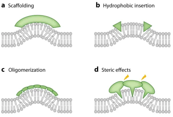
1. MC S&G by BAR domain proteins.
1) Scaffolding (fig. 1.2A):
BAR domain–containing proteins and dynamin are believed to provide scaffolds for
cylindrical curvature(12). Scaffolding is usually defined as an imprinting of protein
monomer or oligomer intrinsic shape of the membrane-binding surface onto the
underlying membrane. The proposal that scaffolding is responsible for MCS&G by BAR
domains was prompted by the determination of the Drosophila amphiphysin N-BAR
domain crystal structure(13). CryoEM reconstructions have supported this mechanism for
the dynamin polymer(14-15).
2) Hydrophobic insertion (fig. 1.2B):
Hydrophobic insertion is another important mechanism proposed to contribute to
membrane deformation induced by proteins. This mechanism is proposing that an
amphipathic helix, which is usually disordered in solution, forms an alpha-helix and
inserts into the bilayer during the membrane binding process, thus causing bilayer
asymmetry and therefore generating membrane curvature. A representative example is
the ENTH domain(11). Membrane insertion of N-terminal AHs is also believed to be
responsible for MC-S&G by Sar1(16), Arf1(17), and Arf6(18), as mutations in their AHs
abolish tubulation. For BAR domain containing proteins, one subfamily are the NBAR
domain proteins, which consist of an N-terminal amphipathic helix adjacent to the BAR
domain. This N-terminal amphipathic helix folds upon membrane binding(13, 19-23)and
is termed H0s. Mutational deletion of H0 from the amphiphysin 2 N-BAR domain
reduced tubulation of vesicle membranes(13, 21).
3) Oligomerization (fig. 1.2C):
For lipids, quantitative experimental evidence(24-25) has shown, in combination with
analytical thermodynamic/ mechanical models, that cooperativity can amplify curvature
sorting(25). The association of F-BAR domain dimers into filaments leads to striated,
lattice-like protein coats on lipid tubes (26-27). The efficacy of such amplification of MC
sensing is further underscored by the dependence of dynamin polymerization on MC(28).
EM imaging(13, 29-31) and molecular dynamics simulations(32) suggest that endophilin
and amphiphysin proteins and N-BAR domains also oligomerize on tubulated membranes.
H0s of these proteins may be involved in this oligomerization, as an amphiphysin H0 has
been shown to form an anti-parallel spontaneous curvature: curvature of a membrane
binding interface, or of a membrane itself, in the absence of any external stresses dimer in
the membrane(20). H0-mediated oligomerization has also been suggested to contribute to
MCS&G for the non scaffolding ENTH domain(33).
4) Steric effect (fig. 1.2D):
It has previously been hypothesized(34-35), and more recently suggested by
experiments(36), that local crowding of peripheral proteins can cause membrane bending.
In these experiments, only when local protein concentration was increased as a result of
lipid-based phase separation was tubulation observed. In this study, proteins were used
that are known to lack intrinsic curvature (i.e., scaffolding) effects, membrane-inserting
AHs, or a tendency for oligomerization(36). Bending by crowding therefore has to be
considered as a synergistic contributor to MCS&G.
2. Endophilin
Endophilin is an N-terminal BAR domain-containing protein(37-40) (crystal structure in
fig. 1.3A&B), which is enriched at neural synapses. Endophilin assembles with dynamin
and synaptojanin around the neck of clathrin-coated membrane invaginations(39, 41).
Endophilin also has been found to be involved in a clathrin-independent endocytic
pathway that is faster than clathrin-dependent endocytosis(42).
The Endophilin N-BAR domain (ENBAR) contains a BAR domain, an N-terminal helix
adjacent to the BAR domain (helix H0) and an additional amphipathic helix (H1 insert
helix, residues ~ 62-86)(19, 43-44). These amphipathic helices are disordered in aqueous
solution and form an α-helix upon membrane insertion(19, 45).
In vitro research has shown that endophilin senses MC and induces the deformation and
tubulation of liposomes (19, 30, 44). The mechanism of MC generation and sensing by
endophilin is not fully understood. Liposome binding and tubulation assays, as well as
results based on electron paramagnetic resonance spectroscopy have suggested that the
concave surface of its BAR domain acts as a rigid, positively charged scaffold(19, 44),
which electrostatically interacts with negatively charged liposomes (12-13, 34). Electron
paramagnetic resonance spectroscopy measurements reported that the concave surface of
the endophilin BAR domain does not penetrate into the acyl chain level of the curved
bilayer, implying that the BAR domain only peripherally interacts with the
membrane(45). The rigidity and spontaneous curvature of the crescent shape are assumed
to bend the membrane(46).
Interestingly, a recently developed single liposome membrane binding assay reported that
the crescent shaped BAR domain dimer is not able to sense MC; instead, MC sensing was
suggested to solely depend on the insertion of amphipathic helices into lipid packing
defects(22). Indeed, H0 and the H1 insert helices are believed to drive MC (19, 44, 47),
via their hydrophobic insertion into the membrane(12, 34, 45, 48). Furthermore,
molecular dynamics simulations have shown that the H1 insert helix orients
perpendicularly to the long axis of the N-BAR domains during membrane binding, and
that the degree of membrane deformation is connected with H1 helix orientation(49).
Besides scaffolding and hydrophobic insertion, higher order oligomerization of BAR
domain dimers may contribute to MC generation(34). Consistent with this hypothesis,
striations have been observed on tubules generated via ENBAR domains(30). Theoretical
characterization of the process of liposome tubulation(50) and vesiculation by N-BAR
domains via mesoscopic simulations and electron microscopy imaging indicate an
intricate coupling between protein density, degree of N-BAR oligomerization, and
membrane deformation(31).
This dissertation sheds light on the understanding of the mechanism of cellular function
of endophilin in CME process via in vitro approaches. For the NBAR domain of
endophilin (ENBAR), we first experimentally characterized the effect of MC on both
ENBAR localization at different protein solution concentrations and translational
diffusion of ENBAR. We then derived an analytical curvature-sorting model that we
compared to our data. Implications of this model for physiologically important membrane
shape transitions are also discussed (Chapter 2).
We then aim to clarify the controversial contributions (see more information about this in
Chapter 3) of amphipathic helices in the MC S&G of endophilin in CME. We express
and purify the endophilin NBAR H0-deletion mutant (ENBAR ΔH0) and H1I-deletion
mutant (ENBAR ΔH1I), respectively. We compare the curvature sensing abilities of
ENBAR WT and their mutants. We find that at the similar protein membrane density
level, both mutants’ curvature sorting efficiencies are lower than WT. This indicates an
essential contribution of either the H0 helix or the H1I helix to the curvature sensing
ability of ENBAR. Likely, this amplifies the scaffolding effect induced by BAR domains.
We also monitor the influence of helices on the potential of ENBAR oligomerization, via
single GUV transfer experiments (Chapter 3).
Besides focusing on the NBAR domain of endophilin, we study how its SH3 domain
(ESH3) plays a role in CME. We observe the systematic quenching of fluorescence
intensity of Trp when the H0 helix is titrated by the SH3 domain. We not only use H0
WT, but also H0-F10FCN and H0-F10W, as the quenchers, to monitor the intensity
decrease of Trp in the ESH3 domain. These findings suggest an interaction of endophilin
H0 and SH3, which a plausible mechanism for the autoinhibition of endophilin (Chapter
4).
3. Syndapin (pacsin)
Syndapin is an FBAR domain containing protein (crystal structure in fig. 1.3C&D).
Among three types of syndapin, syndapin I is enriched in neurons(51). Syndapin I is not
only involved in clathrin-mediated endocytosis, but also interacts with dynamin to
contribute to activity dependent bulk endocytosis (52). Besides, syndapin-N-WASP
interaction could be an important coordinator for local actin polymerization(53).
Syndapin I consists of an FBAR domain, an NPF domain which mostly interacts with EH
domain proteins, and an SH3 domain (7, 51, 53). The FBAR domain has been shown to
be able to induce different types of membrane deformations: tubes, pearling structures,
and small vesicles(54-56). This variety of shapes is likely induced by syndapin’s unique
“S” shaped structure (fig. 1.3D). There is a short loop within helix 2 which could function
as an amphipathic wedge inserted into the bilayer (57-58). This special hinge in pacsin 3
is also proposed to be the key coordinator for the radius of tubes it generated, and it may
regulate the higher order structure arrangement of pacsin 3(59).
The membrane tubulation ability of syndapin I full length is proposed to be inhibited in
vivo(55). In vitro, full length syndapin I also tubulates membranes less efficiently
compared to syndapin FBAR, as quantified by EM experiments (55-56). The current
mechanism suggests an interaction between SH3 domain and FBAR domain in the full
length protein, which inhibits the FBAR membrane bending ability. This mechanistic
hypothesis calls for more for thorough analysis, as described in Chapter 5.
Figures:
Figure 1.1 Curvature and clathrin-coated vesicle formation. Diagram adapted from(1)
with permission from Nature Publishing Group.
Figure 1.2 Mechanisms of membrane curvature generation and sensing. Figure is
adapted from(1) with permission from Annual Reviews Publishing Group.
Figure 1.3 Crystal structures of endophilin and syndapin.
A&B. Ribbon diagram of rat endophilin-A1 BAR domain (Protein Data Bank (PDB)
accession number 2c08) from side view and top view.
C&D. Ribbon diagram of mouse syndapin I FBAR domain (Protein Data Bank (PDB)
accession number 2x3v) from side view and top view.
References:
1. McMahon, H. T., and E. Boucrot. 2011. Molecular mechanism and physiological functions of clathrin-mediated endocytosis. Nature Reviews Molecular Cell Biology 12:517-533
2. Higgins, M. K., and H. T. McMahon. 2002. Snap-shots of clathrin-mediated endocytosis. Trends in Biochemical Sciences 27:257-263.
3. Doherty, G. J., and H. T. McMahon. 2009. Mechanisms of endocytosis. Annual Review of Biochemistry 78:857–902.
4. Taylor, M. J., D. Perrais, and C. J. Merrifield. 2011. A High Precision Survey of the Molecular Dynamics of Mammalian Clathrin-Mediated Endocytosis. Plos Biology 9:e1000604.
5. Cocucci, E., F. o. Aguet, S. Boulant, and T. Kirchhausen. 2012. The first five seconds in the life of a clathrin-coated pit. Cell 150:495–507.
6. Watanabe, S., B. R. Rost, M. Camacho-Pe´rez, M. W. Davis, B. So¨hl-Kielczynski, C. Rosenmund, and E. M. Jorgensen. 2013. Ultrafast endocytosis at mouse hippocampal synapses. Nature 504:242–247.
7. Frost, A., V. M. Unger, and P. De Camilli. 2009. The BAR domain superfamily: membrane-molding macromolecules. Cell 137:191-196.
8. Masuda, M., and N. Mochizuki. 2010. Structural characteristics of BAR domain superfamily to sculpt the membrane. Seminars in Cell & Developmental Biology 21: 391–398.
9. Prinz, W. A., and J. E. Hinshaw. 2009. Membrane-bending proteins. Critical Reviews in Biochemistry & Molecular Biology 44:278-291.
10. Praefcke, G. J. K., and H. T. McMahon. 2004. The dynamin superfamily: universal membrane tabulation and fission molecules? Nature Reviews Molecular Cell Biology 5:133-147.
11. Ford, M. G. J., I. G. Mills, B. J. Peter, Y. Vallis, G. J. K. Praefcke, P. R. Evans, and H. T. McMahon. 2002. Curvature of clathrin-coated pits driven by epsin. Nature 419:361-366.
12. Zimmerberg, J., and M. M. Kozlov. 2006. How proteins produce cellular membrane curvature. Nature Reviews Molecular Cell Biology 7:9-19.
13. Peter, B. J., H. M. Kent, I. G. Mills, Y. Vallis, P. J. G. Butler, P. R. Evans, and H. T. McMahon. 2004. BAR domains as sensors of membrane curvature: the amphiphysin BAR structure. Science 303:495-499.
14. Zhang, P., and J. E. Hinshaw. 2001. Three-dimensional reconstruction of dynamin in the constricted state. Nature Cell Biology 3:922 - 926.
15. Chen, Y.-J., P. Zhang, E. H. Egelman, and J. E. Hinshaw. 2004. The stalk region of dynamin drives the constriction of dynamin tubes. Nature Structural & Molecular Biology:574 - 575.
16. Lee, M. C. S., L. Orci, S. Hamamoto, E. Futai, M. Ravazzola, and R. Schekman. 2005. Sar1p N-terminal helix initiates membrane curvature and completes the fission of a COPII vesicle. Cell 122:605-617.
17. Krauss, M., J.-Y. Jia, A. Roux, R. Beck, F. T. Wieland, D. C. Pietro, and V. Haucke. 2008. Arf1-gtp-induced tubule formation suggests a function of arf
family proteins in curvature acquisition at sites of vesicle budding. The Journal of Biological Chemistry 283:27717-27723.
18. Lundmark, R., G. J. Doherty, Y. Vallis, B. J. Peter, and H. T. McMahon. 2008. Arf family GTP loading is activated by, and generates, positive membrane curvature. Biochemical Journal 414:189-194.
19. Gallop, J. L., C. C. Jao, H. M. Kent, P. J. G. Butler, P. R. Evans, R. Langen, and H. T. McMahon. 2006. Mechanism of endophilin N-BAR domain mediated membrane curvature. The EMBO Journal 25:2898-2910.
20. Fernandes, F., L. M. S. Loura, F. J. Chichon, J. L. Carrascosa, A. Fedorov, and M. Prieto. 2008. Role of helix 0 of the N-BAR domain in membrane curvature generation. Biophysical Journal 94:3065-3073.
21. Löw, C., U. Weininger, H. Lee, K. Schweimer, I. Neundorf, A. G. Beck-Sickinger, R. W. Pastor, and J. Balbach. 2008. Structure and dynamics of helix-0 of the N-BAR domain in lipid micelles and bilayers. Biophysical Journal 95:4315-4323. 22. Bhatia, V. K., K. L. Madsen, P. Bolinger, A. Kunding, P. Hedegard, U. Gether,
and D. Stamou. 2009. Amphipathic motifs in BAR domains are essential for membrane curvature sensing. The EMBO Journal 28:3303-3314.
23. Hatzakis, N. S., V. K. Bhatia, J. Larsen, K. L. Madsen, P. Bolinger, A. H. Kunding, J. Castillo, U. H. Gether, P., and D. Stamou. 2009. How curved membranes recruit amphipathic helices and protein anchoring motifs. Nature Cell Biology 5:835-841.
24. Sorre, B., A. Callan-Jones, J. Manneville, P. Nassoy, J. Joanny, J. Prost, B. Goud, and P. Bassereau. 2009. Curvature-driven lipid sorting needs proximity to a demixing point and is aided by proteins. Proceedings of the National Academy of Science of the United States of America 106:5622-5626.
25. Tian, A., B. R. Capraro, C. Esposito, and T. Baumgart. 2009. Bending stiffness depends on curvature of ternary lipid mixture tubular membranes. Biophysical Journal 97:1636-1646.
26. Shimada, A., H. Niwa, K. Tsujita, S. Suetsugu, K. Nitta, K. Hanawa-Suetsugu, R. Akasaka, Y. Nishino, M. Toyama, L. Chen, Z. Liu, B. Wang, M. Yamamoto, T. Terada, A. Miyazawa, A. Tanaka, S. Sugano, M. Shirouzu, K. Nagayama, T. Takenawa, and S. Yokoyama. 2007. Curved EFC/F-BAR-domain dimers are joined end to end into a filament for membrane invagination in endocytosis. Cell 129:761-772.
27. Frost, A., R. Perera, A. Roux, K. Spasov, O. Destaing, E. H. Egelman, P. De Camilli, and V. M. Unger. 2008. Structural Basis of Membrane Invagination by F-BAR Domains. Cell 132:807-817.
28. Roux, A., G. Koster, M. Lenz, B. Sorre, J.-B. Manneville, P. Nassoy, and P. Bassereaua. 2010. Membrane curvature controls dynamin polymerization. Proceedings of the National Academy of Science of the United States of America 107:4141-4146.
29. Takei, K., V. I. Slepnev, V. Haucke, and P. D. Camilli. 1999. Functional partnership between amphiphysin and dynamin in clathrinmediated endocytosis. Nature Cell Biology 1:33-39.
30. Farsad, K., N. Ringstad, K. Takei, S. R. Floyd, K. Rose, and P. D. Camilli. 2001. Generation of high curvature membranes mediated by direct endophilin bilayer interactions. The Journal of Cell Biology 155:193-200.
31. Mizuno, N., C. C. Jao, R. R. Langen, and A. C. Steven. 2010. Multiple modes of endophilin-mediated conversion of lipid vesicles into coated tubes. The Journal of Biological Chemistry 285:23351-23358.
32. Ayton, G. S., and G. A. Voth. 2010. Multiscale simulation of protein mediated membrane remodeling. Seminars in Cell & Developmental Biology 21:357-362. 33. Yoon, Y., J. Tong, P. J. Lee, A. Albanese, N. Bhardwaj, M. Källberg, M. A.
Digman, H. Lu, E. Gratton, Y.-K. Shin, and W. Cho. 2010. Molecular basis of the potent membrane-remodeling activity of the epsin 1 N-terminal homology domain. The Journal of Biological Chemistry 285:531-540.
34. McMahon, H. T., and J. L. Gallop. 2005. Membrane curvature and mechanisms of dynamic cell membrane remodelling. Nature 438:590-596.
35. Sens, P., and M. S. Turner. 2004. Theoretical Model for the Formation of Caveolae and Similar Membrane Invaginations. Biophysical Journal 86:2049-2057.
36. Stachowiak, J. C., C. C. Hayden, and D. Y. Sasaki. 2010. Steric confinement of proteins on lipid membranes can drive curvature and tubulation. Proceedings of the National Academy of Science of the United States of America 107:7781–7786. 37. Ringstad, N., H. Gad, P. Löw, G. Di Paolo, L. Brodin, O. Shupliakov, and P. De
Camilli. 1999. Endophilin/SH3p4 is required for the transition from early to late stages in clathrin-mediated synaptic vesicle endocytosis. Neuron 24:143-154. 38. Verstreken, P., O. Kjaerulff, T. E. Lloyd, R. Atkinson, Y. Zhou, I. A.
Meinertzhagen, and H. J. Bellen. 2002. Endophilin mutations block clathrin-mediated endocytosis but not neurotransmitter release. Cell 109:101-112.
39. Schuske, K. R., J. E. Richmond, D. S. Matthies, W. S. Davis, S. Runz, D. A. Rube, A. M. van der Bliek, and E. M. Jorgensen. 2003. Endophilin is required for synaptic vesicle endocytosis by localizing synaptojanin. Neuron 40:749-762. 40. Dickman, D. K., J. A. Horne, I. A. Meinertzhagen, and T. L. Schwarz. 2005. A
slowed classical pathway rather than kiss-and-run mediates endocytosis at synapses lacking synaptojanin and endophilin. Cell 123:521-533.
41. Sundborger, A., C. Soderblom, O. Vorontsova, E. Evergren, J. E. Hinshaw, and O. Shupliakov. 2011. An endophilin-dynamin complex promotes budding of clathrin-coated vesicles during synaptic vesicle recycling. Journal of Cell Science 124:133-143.
42. Llobet, A., J. L. Gallop, J. J. E. Burden, G. Camdere, P. Chandra, Y. Vallis, C. R. Hopkins, L. Lagnado, and H. T. McMahon. 2011. Endophilin drives the fast mode of vesicle retrieval in a ribbon synapse. The Journal of Neuroscience 31:8512-8519.
43. Weissenhorn, W. 2005. Crystal structure of the Endophilin-A1 BAR domain. Journal of Molecular Biology 35:653-661.
44. Masuda, M., S. Takeda, M. Sone, T. Ohki, H. Mori, Y. Kamioka, and N. Mochizuki. 2006. Endophilin BAR domain drives membrane curvature by two newly identified structure-based mechanisms. The EMBO Journal 25:2889-2897.
45. Jao, C. C., B. G. Hegde, J. L. Gallop, P. B. Hegde, H. T. McMahon, I. S. Haworth, and R. Langen. 2010. Roles of Amphipathic Helices and the Bin/Amphiphysin/Rvs (BAR) Domain of Endophilin in Membrane Curvature Generation. The Journal of Biological Chemistry 285:20164-20170.
46. Zimmerberg, J. 2004. Membrane curvature: How BAR domains bend bilayers Curr. Biol. 14:R250-R252.
47. Campelo, F., G. Fabrikant, H. T. McMahon, and M. M. Kozlov. 2010. Modeling membrane shaping by proteins: focus on EHD2 and N-BAR domains. FEBS Letters 584:1830-1839.
48. Campelo, F., H. T. McMahon, and M. M. Kozlov. 2008. The hydrophobic insertion mechanism of membrane curvature generation by proteins. Biophysical journal 95:2325-2339.
49. Cui, H., G. S. Ayton, and G. A. Voth. 2009. Membrane binding by the endophilin N-BAR domain. Biophysical Journal 97:2746-2753.
50. Ayton, G. S., E. Lyman, V. Krishna, R. D. Swenson, C. Mim, V. M. Unger, and G. A. Voth. 2009. New insights into BAR domain-induced membrane remodeling. Biophysical journal 97:1616-1625.
51. Modregger, J., B. Ritter, B. Witter, M. Paulsson, and M. Plomann. 2000. All three PACSIN isoforms bind to endocytic proteins and inhibit endocytosis. Journal of Cell Science 113:4511-4521.
52. Clayton, E. L., V. Anggono, K. J. Smillie, N. Chau, P. J. Robinson, and M. A. Cousin. 2009. The phospho-dependent dynamin-syndapin interaction triggers activity-dependent bulk endocytosis of synaptic vesicles. The Journal of Neuroscience 29:7706–7717.
53. Kessels, M. M., and B. Qualmann. 2002. Syndapins integrate N-WASP in receptor-mediated endocytosis. The EMBO Journal 21:6083-6094.
54. Wang, Q., M. V. A. S. Navarro, G. Peng, E. Molinelli, S. Lin Goh, B. L. Judson, K. R. Rajashankar, and H. Sondermann. 2009. Molecular mechanism of membrane constriction and tubulation mediated by the F-BAR protein Pacsin/Syndapin. Proceedings of the National Academy of Science of the United States of America 106:12700-12705.
55. Rao, Y., Q. Ma, A. Vahedi-Faridi, A. Sundborger, A. Pechstein, D. Puchkov, L. Luo, O. Shupliakov, W. Saenger, and V. Haucke. 2010. Molecular basis for SH3 domain regulation of F-BAR-mediated membrane deformation. Proceedings of the National Academy of Science of the United States of America 107:8213-8218. 56. Goh, S. L., Q. Wang, L. J. Byrnes, and H. Sondermann. 2012. Versatile
Membrane Deformation Potential of Activated Pacsin. Plos ONE 7:e51628. 57. Edeling, M. A., S. Sanker, T. Shima, P. K. Umasankar, S. Höning, H. Y. Kim, L.
A. Davidson, S. C. Watkins, M. Tsang, D. J. Owen, and L. M. Traub. 2009. Structural requirements for PACSIN/Syndapin operation during zebrafish embryonic notochord development. Plos ONE 4:e8150.
58. Plomann, M., J. G. Wittmann, and M. G. Rudolph. 2010. A hinge in the distal end of the PACSIN 2 F-BAR domain may contribute to membrane-curvature sensing. Journal of Molecular Biology 400:129–136.
59. Bai, X., G. Meng, M. Luo, and X. Zheng. 2012. Rigidity of wedge loop in PACSIN 3 protein Is a key factor in dictating diameters of tubules. The Journal of Biological Chemistry 287:22387-22396.
Chapter 2: Nonlinear sorting, curvature generation, and crowding of Endophilin
NBAR on tubular membranesa
A. Background
The curvature of biological membranes is controlled by membrane-bound proteins. For
example, during endocytosis, the sorting of membrane components, vesicle budding, and
fission from the plasma membrane are mediated by adaptor and accessory proteins.
Endophilin is a peripherally binding membrane protein that functions as an endocytic
accessory protein.
Endophilin’s membrane tubulation capacity is well-known. However, in order to
understand thermodynamic and mechanical aspects of endophilin function, experimental
measurements need to be compared to quantitative theoretical models.
We present measurements of curvature sorting and curvature generation of the endophilin
A1 NBAR domain on tubular membranes pulled from giant unilamellar vesicles (GUV).
At low concentration, endophilin functions primarily as a membrane curvature sensor; at
high concentrations, it additionally generates curvature. We determine the spontaneous
curvature induced by endophilin, and observe sigmoidal curvature/composition coupling
isotherms that saturate at high membrane tensions and protein solution concentrations.
The observation of saturation is supported by a strong dependence of lateral diffusion
coefficients on protein density on the tether membrane.
a This chapter is largely reproduced from previously published work: Zhu, C., Das, S.L., and Baumgart, T.
(2012). Nonlinear sorting, curvature generation, and crowding of endophilin NBAR on tubular membranes.
Biophys. J.102, 1837-1845.
We develop a non-linear curvature/composition coupling model that captures our
experimental observations. Our model predicts a curvature-induced phase transition
among two states with varying protein density and membrane curvature. This transition
could act as a switch during endocytosis.
B. Materials and methods
1. Materials 1,2-dioleoyl-sn-glycero-3-phosphocholine (DOPC), 1,2-dioleoyl-sn
-glycero-3-phospho-(1’-rac-glycerol) (DOPG) and
1,2-distearoyl-sn-glycero-3-phosphoethanolamine-N-[biotinyl(polyethylene glycol)-2000] (ammonium salt)
(DSPE-Bio-PEG2000) were obtained from Avanti Polar Lipids (Alabaster, AL). Fatty-acid free
bovine serum albumin (BSA) was from Sigma Chemical (St. Louis, MO). Rat endophilin
A1 NBAR-Alexa Fluor 488 (ENBAR-A488, amino acids 1-247, labeled at C108) was
obtained from Pr. Langen, U. Southern California, USA, and stored in buffer (20 mM
HEPES, pH 7.4, 150 mM NaCl). Using vesicle spin down assays(1), we confirmed that
fluorescence labeling at position C108 does not alter membrane binding (data not shown).
Texas Red-1,2-dihexadecanoyl-sn-glycero-3-phosphoethanolamine triethylammonium
salt (TR-DHPE) was from Invitrogen (Carlsbad, CA). Streptavidin conjugated
microspheres with diameter of 6 μm were from Polysciences, Inc. (Warrington, PA).
2. Methods:
Preparation of giant unilamellar vesicles (GUVs): GUVs were prepared by
electroformation in solutions of 300 mM sucrose as described(2). Lipids were mixed in
chloroform at a total concentration of 1 mM. DOPG was used at 25 mol%, DOPC at 74
mol%, TR-DHPE at 0.3 mol%, and DSPE-Bio-PEG2000 content was 0.7 mol%.
ENBAR-A488 was added after electroswelling (so proteins bind to the exterior leaflet of
GUV membrane only) but immediately before micropipette aspiration experiments, to
yield final solution concentrations indicated below.
Preparation of micropipettes:Micropipettes (World Precision Instruments, Sarasota, FL)
were manufactured with a pipette puller; pipette tips were clipped using a microforge.
The inner opening diameters were 3-6 μm. Irreversible membrane/pipette adhesion was
avoided by incubating micropipette tips with 0.5 mg/mL fatty-acid-free BSA dissolved in
1X PBS with a MicroFil needle (WPI), followed by rinsing, and pipettes were finally
filled with 300 mM sucrose solution.
Preparation of chamber and tether pulling: GUV dispersions obtained through
electroswelling were diluted 1:10 in 300 mM sucrose solution. 50 μL diluted GUV
dispersions which contained ~0.5 μL 10X PBS, 0.5-1 μL streptavidin coated polystyrene
bead solution and various concentrations of ENBAR-A488 (rat endophilin A1 NBAR,
amino acids 1-247, labeled at C108 with AlexaFluor 488) solution were injected into a
measurement chamber. The chamber was constructed from microscope slides and
coverslips that allowed access by two perpendicularly oriented micropipettes.
Micropipettes were inserted into the chamber by a three-dimensional motorized
manipulator system (Luigs and Neumann, Ratingen, Germany). Such a vesicle was
pipette-aspirated with an initial suction pressure amounting to 5~10 Pa. The aspiration
pressure was controlled by adjusting the water level of a reservoir connected to the
micropipettes, and monitored by a pressure transducer with a DP-28 diaphragm
(Validyne Engineering, Los Angeles, CA). In order to pull tethers, a second pipette was
used to aspirate a bead. The bead was gently moved toward the aspirated vesicle and
contacted for ~1 min, and then moved away from the vesicle to pull a membrane tether of
5-15 μm in length, depending on vesicle size and excess area. All experiments were
carried out at room temperature (20 ± 2 °C).
Imaging: Vesicles and tethers were imaged with a fluorescence confocal microscopy
(FV3000 scanning system integrated with a motorized inverted microscope IX81;
Olympus, Center Valley, PA), using a 60X, 1.2 NA water immersion lens (Olympus).
Image analysis was carried out via IMAGEJ (National Institutes of Health, Bethesda,
MD). Vesicle fluorescence intensity values were measured after background subtraction
from the average of four randomly chosen equal area regions of interest on the vesicle
equator.
Tether cross section fluorescence intensity measurements: To investigate protein
partitioning driven by MC, we monitored the local fluorescence intensities on the tubular
membrane under varying membrane tensions. We changed membrane tension by
adjusting the height of a water reservoir. Fluorescence intensities of tethers were
measured by obtaining Kalman-averaged images of the tether cross section (xz plane),
which is orthogonal to the axis of the tether (contained in the xy plane; Fig. 2.1A), at a
stepwidth of 0.15 μm to yield a total imaging depth of around 6 μm. Cross-sectional
fluorescence intensity profiles (Fig. 2.1B) were background-corrected, and intensity was
evaluated in an elliptical region of interest.
In order for us to be able to correlate membrane tether fluorescence intensity changes
with changes in protein coverage fraction, we determined the linear range of fluorescence.
For this purpose, tethers were pulled from pipette-aspirated GUVs incubated with
ENBAR-A488 at a concentration of 150 nM. Intensities of tethers at fixed membrane
tension were measured for varying laser powers(3). Consequently, all measurements in
this report were obtained using laser powers within the indicated linear range.
Determination of dissociation constant for ENBAR-A488 binding on GUVs: Vesicles
were prepared as described above. GUV membranes contained 74% DOPC, 25% DOPG,
0.3% Texas Red-DHPE and 0.7% DSPE-Bio-PEG2000, and were diluted 1:10 in 300
mM sucrose. Protein solutions were mixed with vesicles and incubated for 30 min at
room temperature before imaging. Vesicle fluorescence intensities were determined as
described above. Vesicle fluorescence intensity values as a function of protein solution
concentration were fitted by a classical Langmuir adsorption isotherm.
Diffusion measurements on tethers via fluorescence recovery after photobleaching: For
fluorescence recovery after photobleaching (FRAP) measurements on the membrane
tether, tethers with lengths of 12 ± 1 μm were pulled and kept at fixed membrane tension.
Except for a short stretch amounting to a length of ~0.5 μm measured from the
tether/vesicle junctions, the entire tether was photobleached by repeated scanning at
maximal laser intensity (488 nm illumination). Prebleach and postbleach intensities were
measured using excitation with small laser power (0.1%~0.3% of full power) at 488 nm.
The relative photobleaching recovery ratio R(t) at a given time t was defined as
) 0 ( ) ( ) 0 ( ) ( ) ( I I I t I t R − − −
= (1)
where I(t), I(0)and I(−) are the fluorescence intensities of the tether integrated along
length increments dx at time t, at a time immediately after bleaching (t = 0), and before
bleaching, respectively. A one-dimensional diffusion model(4) was fit to the
photobleaching recovery ratio R(t)
)]} 2 ( ) 2 ( [ 2 1 1 { 1 ) ( 0 2 1 Dt L x h erf Dt L x h erf dx L t R L L − + + + − −
=
∫
ρ (2)where D is the diffusion coefficient, ρ0 is the maximal local recovery ratio, which is
positive and no larger than 1. L1 and L2 are the beginning and ending position of the
analysis range on the tether. L is total length of the region of interest, and h is the position
of an image source accounting for the presence of an impermeable boundary at the bead
position(4). Fitting was done via the software Mathematica (Wolfram Research,
Champaign, IL).
Measurement of tube radius: The radii of membrane tubes displayed in Figs. 2.2E and
2.2F in the main text is below the resolution of the optical microscope. It is possible to
estimate it after calibrating the fluorescence intensity measurements of tubes and vesicles
via fluorescently labeled lipid(5). The tube radius (Rt) is proportional to the fluorescence
intensity from the lipid dye on the tube (Itlipid) normalized by the same intensity on the
vesicle (Ivlipid).
lipid V lipid t t I I A
R = ⋅
(3)
We experimentally determined the calibration factor A from a linear fit to a plot of the
theoretically expected tube radius (varied through membrane tension, in the absence of
protein) against this ratio, yielding a slope of A = 229 ± 40 nm (uncertainty is the
standard deviation of three independent experiments). We then used this conversion
factor to extract the tube radius from the curvature sorting experiments.
Error estimation of uncertainty in diffusion coefficient: We use the χ–square test (6) to
calculate uncertainties in diffusion coefficients. To calculate χ2, we varied diffusion
coefficients while fixing the fitting parameters ρ0, as well as L1 and L2 (see main text for
definitions of these parameters) and summed residuals according to
∑
= − = Σ n j fit D j data j i i t R t R D 1 2 2 2 [ ( ) ( ) ])) ( (
σ
χ (4)
where σ is the relative uncertainty of the fluorescence intensity of an unbleached tether
determined by image analysis, and Di are values for the diffusion coefficient close to the
optimal fit value. By calculating the following 2nd order differential, we obtained
uncertainties sfor the diffusion coefficients (see Fig. 2.3 D):
) ) ( /( 2 2 2 2 i i D D s ∂ ∂
= χ (5)
Error estimation of uncertainty in fitting parameters for VdW model: The uncertainties
for fit parameters were determined by χ–square test. To obtain the uncertainty of the fit
parameter ai (i = 1-4 for a, b, θves and Cp, respectively), we held ak(k�i) constant, and
varied aii around aifit,
∑
= Σ − Σ = n j fit a j data j ii ii R R a 1 2 2 2 [ ( ) ( ) ]) (
σ
χ (6)
where σ is the relative uncertainty of the fluorescence intensity as defined above.
Calculating the 2nd order differential of χ2 with respect to the parameter aii gives the
uncertainty si for the fit parameter ai:
) ) ( /( 2 2 2 2 ii ii i a a s ∂ ∂
= χ (7)
Note that this approach neglects covariant terms in the error matrix.
Error propagation for membrane tension: To estimate the uncertainty of membrane
tension Σ (see Figs. 2.2 C-F), we used multivariate error analysis(6-7):
2 2 2
2 2
2 ( ( )) ( ) ( ) ( ) ( ) ) ( v v p v p p p p v v R R R R R R R R R R P
P δ δ
δ δ − + − + ∆ ∆ = Σ Σ (8)
The uncertainty of aspiration pressure (δ(ΔP)) was 0.5 Pa. Since the uncertainty in the
pipette radius contributes a constant error to membrane tension(8), we ignored this
component in our error analysis. The error for the vesicle radius (δRv ) was approximated
as the image resolution, which is around 0.25 µm.
C. Results.
1. ENBAR curvature sorting on tubular membrane
An important goal of this study was to investigate to what extent and under which
conditions the NBAR domain of the peripheral protein endophilin A1 (ENBAR, from rat)
binds to tubular membranes with variable curvature. Electron microscopy observations
(9-11) combined with computational studies (12-14) have already demonstrated that
ENBAR can deform membranes into high curvature assemblies (with varying curvature
radii typically significantly below 50 nm at multi-micromolar protein concentrations). To
be able to understand thermodynamic and mechanical aspects of ENBAR domain
function, however, requires a comparison of measurements to quantitative models. In
order to quantitatively characterize the curvature sensing of ENBAR, we incubated
ENBAR-A488 with negatively-charged GUVs containing the lipid fluorophore
TR-DHPE, and pulled cylindrical tethers from pipette-aspirated vesicles, using
streptavidin-conjugated microspheres. Fig. 2.1A demonstrates qualitatively that green (ENBAR
protein) fluorescence is enriched on highly curved tubular membranes, rather than on the
quasi-flat vesicular membrane (partially shown on the right-hand side in the fluorescence
micrographs of Fig. 2.1A).
The curvature-induced partitioning of ENBAR was determined by confocal microscopy
fluorescence imaging of membrane tether cross sections (Fig. 2.1B) and analyzed as
described in the Materials and Methods section. Fig. 2.1C shows that green fluorescence
intensity (ENBAR) increases on the tether as membrane tension is increased, whereas the
lipid membrane tether fluorescence decreases as a consequence of the shrinking tubular
radius(15).
Thermodynamic interpretation of our data (see below) requires assessment of
reversibility and equilibration times of fluorescence intensities of the protein and the lipid
probe under varying membrane tension. Both green and red fluorescence signals respond
to large rapid (roughly 0.5mN/m/min) increases of membrane tension within about 1 min
and reach equilibrium (see Fig. 2.1C and fluorescence intensity ratio shown in Fig. 2.1D).
Subsequently lowering tension causes corresponding fluorescence intensity changes,
which demonstrates reversibility of the measurements.
2. ENBAR curvature sorting is in a concentration dependent manner
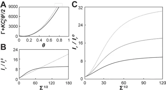
In Fig. 2.2A we display the analysis of a typical ENBAR curvature sorting experiment
using a protein solution concentration of 40 nM. With increasing lateral tension,
fluorescence intensity in the green (protein) fluorescence channel monotonically
increases, whereas the opposite is observed in the red (lipid) channel. In Fig. 2.2,
fluorescence intensity measurements are plotted against the square root of lateral tension
for the following reason. For the case of linear curvature sorting, the square root of lateral
tension can be shown to be proportional to membrane curvature (16). The plots in Fig.
2.2 therefore allow assessment of the linearity of curvature sorting. Importantly, the
results shown here demonstrate non-linear curvature/composition coupling; hence they
deviate from those found for the epsin N-terminal homology (ENTH) domain, where
sorting was observed to be proportional to the square root of membrane tension(17).
The fluorescence intensity of lipid probes in high curvature tether membranes used in this
work is linearly proportional to the MC (see supplementary materials of (3), consistent
with our previous findings(8, 15)); fluorescent lipids therefore are not significantly sorted
by membrane curvature and here serve as a reference for ratiometric fluorescence
intensity measurements. Fig. 2.2B shows the ratio Ir of protein and lipid probe
fluorescence intensities (Ir= Igreen/ Ired) for the same data as shown in Fig. 2.2A.
To facilitate the comparison of our data to a thermodynamic model (see below), the
relative fluorescence intensities Ir were normalized to values Ir0 (
0
r
I = Ives-green / Ives-red)
measured on the vesicle (described in Materials and Methods). A series of individual
sorting experiments were carried out at two different protein solution concentrations, 1
µM and 40 nM, respectively. The results were normalized, binned, and averaged for
multiple tethers; see Figs. 2.2C and D. Again, in contrast to the linear curvature sorting
observed for epsin ENTH, the measurements in Figs. 2.2C and 2.2D show significant
deviations from linearity. Fig. 2.2C shows that for low values of the square root of
tension, the ratiometric parameter Ir / Ir0 increases almost linearly for the case of 1 µM
protein solution concentration. As curvature is further increased, the sorting ratio
becomes nearly constant (Fig. 2.2C). At low protein solution concentration and low
membrane tension (Fig. 2.2D), curvature sorting is significantly weaker compared to
higher tensions (at the same solution concentration). For this concentration, while the
membrane tension increases, the curvature/composition coupling also increases as
indicated by the larger slope of the fluorescence intensity ratios. We note that Figs. 2.2C
and D display relatively large standard deviations comparing different vesicles. The
sources for this variability may include differences in individual vesicle lipid
compositions. However, the main features of our measurements, i.e., non-linear sorting
and saturation of sorting at high membrane curvature and protein solution concentration,
were reproducible for individual vesicles. From fluorescence intensity values of the lipid
dye, measured on vesicle and tether, it is possible to estimate the radius of the tether ((18),
also see Materials and Methods). The results for our two solution conditions are shown in
Figs. 2.2E and F, for the same vesicles shown in Figs. 2.2C and D. The comparison of
the experimental radii to those calculated assuming a bending stiffness of 0.8 ⋅ 10-19J (15)
and absence of spontaneous curvature (dashed lines in Figs. 2.2E and F), reveals
curvature generation at the higher, but not at the smaller protein solution concentration.
These curvature generation measurements, along with the curvature sorting results, were
fitted with a theory (solid lines in Figs. 2.2C to F) detailed below.
3. ENBAR mobility on the tubular membrane is curvature dependent
In addition to the equilibrium curvature sorting measurements described above, we
assessed curvature-dependent diffusion of ENBAR on tubular membranes via FRAP
measurements. Figs. 2.3A and B show examples of fluorescence recovery after
photobleaching of ENBAR on the tether membrane, demonstrating the mobility of
ENBAR on membranes. Individual measurements at a protein solution concentration of 1
µM were recorded for varying membrane tensions and analyzed as described in the
materials and methods section. A one-dimensional diffusion model was then fit to the
time-dependent recovery ratios. Figs. 2.3A and B show experimental results compared to
fitted curves for smallest and largest membrane tensions, respectively. The diffusion
coefficient for the measurement displayed in Fig. 2.3A is 1.47 μm2/s, and the result for
the data in Fig. 2.3B is 0.13 μm2/s. Quantitative photobleaching recovery measurements
were obtained from image analysis of time lapse recordings of tether membrane
fluorescence; see Fig. 2.3C. The results of FRAP measurements for a series of different
membrane tensions are summarized in Fig. 2.3D. As membrane tension increases, the
diffusion coefficient of ENBAR on membrane tethers decreases. As further discussed
below, we hypothesize that the decrease in diffusion coefficients results from an increase
in molecular crowding as the density of protein increases with rising curvature. We
confirmed the hypothesis of protein density affecting diffusion by the following
experiment.
The lateral mobility of ENBAR on tubular membranes was monitored by an alternative
method that consisted of step-wise tether elongations. Membrane tethers previously
equilibrated in the presence of ENBAR (1 μM) were rapidly (10 µm/s) extended by 10
μm, which resulted in a tether region with low protein coverage being pulled from the
aspirated vesicle. ENBAR was observed to diffuse from the vesicle onto the tether (Fig.
2.4A) consistent with the photobleaching results discussed above; see Fig. 2.3C. This
phenomenon could be reproduced several times by repeating the elongation process
described above.
Furthermore, it is observed that for comparable membrane tensions, the diffusion of
ENBAR onto the tubular membrane after tether elongation (Fig. 2.4B) is significantly
faster compared to diffusion observed after photobleaching (Fig. 2.4C). This observation
supports our hypothesis that the lateral mobility of membrane-bound ENBAR depends on
the free area available for diffusion. In this view, diffusion kinetics at high lateral
tensions are slowed (Fig. 2.3 D) due to molecular crowding.
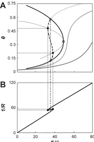
D. Discussion.
In the following sections we outline the derivation of a curvature sorting model that
captures several of our experimental observations. We compare this model to our data,
and discuss the possibility of curvature-induced phase transitions predicted by this model.
1. Introduction of a non-linear curvature/composition coupling model Classical
analytical curvature/composition coupling models assume a linear coupling between local
composition and local MC(19). Similar models have recently been used to interpret the
partitioning of peripheral proteins in curvature gradients(15, 17-18). Our findings for the
curvature partitioning of ENBAR shown in Figs. 2.2 C and D clearly deviate from linear
coupling (note that in linear curvature/composition coupling models the curvature is
proportional to Σ (17)). Thermodynamic terms in linear curvature/composition
coupling models can be interpreted as terms resulting from second order Taylor
expansion in composition and curvature of the free energy(15, 17). In such models, the
coefficients of these expansions are evaluated for the thermodynamic reservoir (i.e., the
GUV) that pulled tethers are in contact with. In the following we replace the expansion
term squared in composition change by Γ, which is a function of fractional protein
coverage θ (ranging from zero to one), to define the tube free energy Ft:
( )
fL CR RL
Ft p −
where R and L are tether radius and length, respectively, κ is membrane bending stiffness,
Cp is a spontaneous curvature of the membrane induced by protein binding, Σ is the
lateral tension, and f is the pulling force acting on the tether. We note that this highly
simplifying model neglects aspects such as the area difference elasticity(20), osmotic
effects(21), membrane undulations, and the possibility of more than one protein binding
mode(10). We also assume that the phenomenological spontaneous curvature Cp does not
depend on membrane curvature.
In Eq. 9, the function Γ results from Legendre transform of a van der Waals free energy
density f0 that describes the thermodynamics of the protein on the membrane:
( )
( )
vesves
b
f − +Π
=
Γθ 0 θ µ θ (10)
where f0 is the mixing free energy density of a two-dimensional van der Waals gas:
2 2 0 1 ln b a b T k b T k
f B B θ θ
θ θ θ − − − − =
. (11)
Here, kB is Boltzmann’s constant, T is temperature, b is the excluded area for protein
coverage and a is a van der Waals interaction term (that here characterizes protein/protein
interactions).
The function Γ shows a non-parabolic dependence on composition (as opposed to the
usual Taylor expansion term(17-19)). This expansion term, which replaces Γ in Eq. 9, is
written for the van der Waals model as follows:
2
2 1χ∆θ
(12a), with
(
)
2 2 22 1 p B C b a b T k κ θ θ χ − + −
= , (12b)