Original Research Article
Evaluation of initial and medium-term follow-up results of balloon
aortic valvuloplasty in severe aortic valve stenosis in newborns,
infants and children in Alexandria, Egypt
Hani Mahmoud Adel*
INTRODUCTION
Aortic valve stenosis (AVS) represents 3%–7% of all congenital heart diseases.1 It may lead to significant morbidity if left untreated. By the mid-80’s, closed or open surgical valvotomy was the standard treatment with average mortality rate of <20% in many centers.2-5 The technique of balloon aortic valvuloplasty (BAoV) was first performed by Labadidi et al.6 Although the early results were encouraging, the concerns of aortic regurgitation and femoral artery injury limited rapid adoption of this technique early after the first report.7,8
With improved catheter technology, BAoV has been established as the procedure of choice for treatment of AVS in infants and children. It has nearly no mortality and minimal morbidity, and has proven to be an effective method for decreasing gradient between the left ventricle and the aorta.9-11 The results of B AoV are comparable to the results of surgical valvuloplasty with the advantage of less risks of morbidity compared to surgery.12,13
The predictors of success as well as the predictors of
acute complications, mortality and long-term
complications, e.g., aortic insufficiency are multifactorial ABSTRACT
Background: Aortic valve stenosis (AVS) represents 3%–7% of all congenital heart diseases. Balloon aortic valvuloplasty (BAoV) has been established as an alternative to surgery for therapy of AVS in infants and children; and has proven to be an effective method for decreasing the gradient between the left ventricle and the aorta. The objective of the study was to evaluate the initial and medium-term results of BAoV in newborns, infants and children with severe AS, treated at the Alexandria University Children’s Hospital.
Methods: Thirty-seven newborns, infants and children with severe AS treated by B AoV between 2009 and 2017 were studied. They were followed-up for at least 1year post- ballooning by clinical and echocardiographic evaluation. Results: The mean AoV annulus diameter by echo was 13.1±4.4 mm and by angiographic measurement was 12.8±4.3 mm. The mean Doppler gradient across AoV was 91.8±14.7 mmHg, compared to mean catheter gradient of 66.1±13.4 mmHg. The mean inflated balloon diameter was 12.1±4.1 mm. The mean balloon/AoV annulus ratio by angiogram was 0.94±0.03 (0.88–1). The mean pressure gradient across the AoV post-ballooning was 21.5±6.9 mmHg by Doppler and was 10.3±4.7 mmHg by catheter, both were significantly less than pre-ballooning values (p<0.001). The procedure was successful in all the cases. Only one case died. Post -ballooning aortic incompetence was moderate in 2 cases (5.4%) and severe in only one case (2.7%).
Conclusions: Balloon aortic valvuloplasty is an effective and safe technique for relieving severe aortic valvular stenosis with acceptable morbidity and minimal mortality, particularly with the new catheter and balloon technology.
Keywords: Aortic stenosis, Aortic valvuloplasty, Balloon dilatation
Department ofPediatrics, University of Alexandria Children’s Hospital, Alexandria, Egypt
Received: 31 March 2019 Accepted: 16 April 2019
*Correspondence: Dr. Hani Mahmoud Adel, E-mail: haniadel70@hotmail.com
Copyright: © the author(s), publisher and licensee Medip Academy. This is an open-access article distributed under the terms of the Creative Commons Attribution Non-Commercial License, which permits unrestricted non-commercial use, distribution, and reproduction in any medium, provided the original work is properly cited.
including valve morphology (bicuspid or trileaflet), thickness of the leaflets, age and clinical condition of the patient and the myocardial function at the time of intervention.11,14,15 In this study we are presenting the initial and medium-term results of BAoV in newborns, infants and children performed in the Alexandria University Children’s Hospital (AUCH).
METHODS
All patients with valvular AS presenting to the AUCH between 2009 and 2017 were subjected to balloon dilatation if they were fulfilling the inclusion criteria. They were 37 patients: 4 neonates,16 infant and 17child (>1-15 years). The male: female ratio was 3.6: 1. Any baby <4 kg with maximum Doppler gradient of 80-100 mmHg or with left ventricular (LV) systolic dysfunction or signs of cardiogenic shock and left ventricular hypertrophy (LVH) irrespective of the Doppler gradient, was included in the study. Any infant >4 kg or child with maximum Doppler gradient >70 mmHg and catheter peak to peak gradient >50 mmHg and with echocardiographic features of LVH (by 2 D- modality and M-mode), was included in the study. Cases associated with other congenital heart defects needing surgery, or associated with more than mild aortic regurgitation, or supravalvular aortic stenosis was excluded from the study. Written informed parental consent was obtained for every patient.
All patients were examined by
Doppler-echocardiography in the long-axis parasternal view, supra-sternal view, apical four chamber view and short axis parasternal view. They were assessed for the AoV morphology regarding the number of cusps and presence of raphae, aortic valve annulus at long axis parasternal view, LVH and its degree, and whether there was initial aortic incompetence (Ai) and its grading (0-4).11
All procedures were done under general anesthesia through retrograde arterial approach as described by Labadidi et al,(except for 2 cases antegradely) as shown in the Figure.6 All patients received IV antibiotics and IV bolus of heparin (100 u/kg). The femoral artery access was used in all the cases. In neonates and infants, femoral vein (FV) access was inserted, while in older children FV access was needed for pacing. For infants less than 1 year or less than 10 kg a 4 French (Fr) (or 5 Fr in a few cases) arterial sheath was inserted. For children between 10-20 kg we used 5 or 6 Fr femoral arterial sheath, and for those above 20-25 kg, we used 7 Fr sheath, for those above 25 kg we used 8 Fr arterial sheaths. We did aortic root angiogram by 4 Fr pigtail to document any Ai and to see the sinuses and help guiding us to direct the multi-purpose (MP) catheter and wires to cross the stenotic valve.
We used Terumo wire to cross the valve (coronary wire in some cases and describe). Then as soon as we crossed the valve and positioned the wire properly, we advance the MP catheter and exchange the wire with ordinary
wire and then exchange the MP catheter with a pigtail catheter for LV injection (left ventriculography in LAO or lateral and PA projections) and measure the AoV annulus then put the ordinary wire and exchange the pigtail catheter with the MP catheter and advance the appropriate wire (whether coronary, or 0.025 inch or 0.035 inch wires) and make sure it is not facing posteriorly in order to minimize the possibility of injuring anterior mitral leaflet (AML).
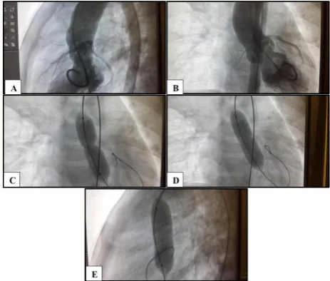
Figure 1: (A and B) Left ventriculography in lateral and frontal projections showing bicuspid stenotic
Aortic valve; (C-E) retrograde balloon aortic valvuloplasty performed in a 2-year-old girl with
severe aortic stenosis (85 mm Hg) and annulus diameter of 11 mm, showing a 10 mm diameter (3 cm long) low pressure balloon dilation catheter across the
valve.
Then we choose a balloon diameter with ratio to the aortic valve annulus by angiography of 0.88-1, (one patient used balloon annulus ratio of 1.1:1), As regards the length of the balloon we choose for newborn and young infants a 2 cm length and for infants (6-12 m, to children up to 15 kg, a 3 cm long balloon and for older or bigger children we used 4 cm long balloons. The balloons used were Tyshak mini and Tyshak II (NuMed Canada, Inc.). For stabilizing the balloon during inflation, we gave adenosine to produce severe bradycardia in neonates, infants and young children up to 15 kg; as described by De Giovanni et al.16 For older children we did RV pacing as described by Daehnert et al.17
Post-ballooning assessment included LV injection to assess AoV mobility and LV systolic function improvement if there was initial impairment, aortic root angiography to assess the aortic regurgitation if increased than before ballooning or the same or did not develop and if developed its grading as mentioned previously by
Moore et al, whether by Doppler or angiography (0-4).14
Successful result if the Doppler gradient is less than 25 mmHg or reduction of gradient by more than 60% if LV systolic function was preserved before ballooning with mild to mild+ only aortic regurgitation, as regards babies with impaired LV systolic function initially the improvement of systolic function is a good indicator of success regardless the gradient as pointed out by Borghi et al.18 If the gradient was still high, re-ballooning was done with a bigger balloon up to 110% of AoV annulus, measured angiographically. If relief of the gradient was inadequate (<50% reduction), in the presence of good systolic function and in the absence of more than mild Ai; serial dilatation with larger size balloons was performed to a maximum of 110%.
Patients were given 3 subcutaneous doses of enoxaparine immediately post-procedure by 2, 12 and 24 hours and were discharged 24 hours post-procedure on baby aspirin (5 mg/kg/day) for 1 month. Follow-up was done after 2 and 6 weeks, and then every 3 months for the first year, and subsequently every 6 months if the follow-up data were satisfactory.
The patients were evaluated every visit for the presence of dyspnea, chest pain and need for drugs and, by echo-Doppler for the pressure gradient, the development of AoV restenosis, the development or progression of Ai and the regression of LVH as well as the myocardial systolic function.
Statistical analysis of the data19
Data were fed to the computer and analyzed using IBM
SPSS software package version 20.0. (Armonk, NY: IBM
Corp) Qualitative data were described using number and
percent.20 Quantitative data were described using range (minimum and maximum), mean, standard deviation and median. Paired t-test for normally distributed quantitative variables, to compare between pre and postoperative. Significance of the obtained results was judged at the 5% level.
RESULTS
The data of the patients and the results of the intervention are shown in the Table. Twenty-seven cases (73%) had bicuspid AoV, 8 cases (21.6%) had trileaflet AoV, and 2 cases (5.4%) had unicuspid AoV.
Three cases (8.1%) were critical AVS, two of these were neonates on prostaglandin E1 (PG E1) infusion and the third was in cardiogenic shock at 5 weeks of age (after the PDA was closed). Fortunately, all the 3 critical cases were saved. Thirty-five cases (95%) were done through retrograde approach and 2 cases through antegrade approach.
The mean AoV annulus diameter by echo was 13.1±4.4 (5.5–22) mm whereas the mean diameter by angiographic measurement was 12.8±4.3 mm (5.9–21). The mean Doppler gradient across AoV was 91.8±14.7 (75–155) mmHg compared to mean catheter gradient of 66.1±13.4 (48–120) mmHg. The mean inflated balloon diameter was 12.1±4.1 (6–20) mm. The mean balloon/AoV annulus ratio by angiogram was 0.94±0.03 (0.88–1). The mean pressure gradient across the AoV by both Doppler and catheter, decreased significantly (<0.001) post-ballooning to 21.5±6.9 (11–35) mmHg and 10.3±4.7 (3– 21) mmHg respectively.
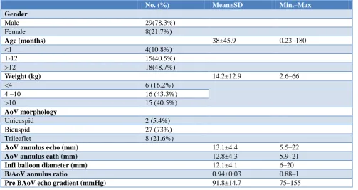
Table 1: Data of the 37 patients with severe aortic valve stenosis.
No. (%) Mean±SD Min.–Max
Gender
Male 29(78.3%)
Female 8(21.7%)
Age (months) 38±45.9 0.23–180
<1 4(10.8%)
1-12 15(40.5%)
>12 18(48.7%)
Weight (kg) 14.2±12.9 2.6–66
<4 6 (16.2%)
4 –10 16 (43.3%)
>10 15 (40.5%)
AoV morphology
Unicuspid 2 (5.4%)
Bicuspid 27 (73%)
Trileaflet 8 (21.6%)
AoV annulus echo (mm) 13.1±4.4 5.5–22
AoV annulus cath (mm) 12.8±4.3 5.9–21
Infl balloon diameter (mm) 12.1±4.1 6–20
B/AoV annulus ratio 0.94±0.03 0.88–1
Pre BAoV echo gradient (mmHg) 91.8±14.7 75–155
No. (%) Mean±SD Min Max
Post BAoV echo gradient (mmHg) 21.5±6.9 11–35
Pre BAoV cath gradient (mmHg) 66.1±13.4 48–120
Post BAoV cath gradient (mmHg) 10.3±4.7 3–21
Procedure time (min) 82±10.5 68–105
Fluroscopy time (min) 11.1±2.8 7–18
Ai post ballooning
No 3 (8.1%)
Trivial 5 (13.5%)
Mild 26 (70.2%)
Moderate 2 (5.4%)
Severe 1 (2.7%)
Complications
Transient loss foot pulse 5 (13.5%)
Transient bradycardia 1 (2.7%)
Mortality after 28 days 1(2.7%)
Latest Doppler gradient (mmHg) 17.95±6.11 10.0–34.0
F/U (months) 36.24±28.99 5.0–84.0
Re-intervention 0 (0%)
The degree of Ai estimated by echo-Doppler and by angiography post-ballooning was trivial in 5 cases (13.5%), mild (Grade 1) in 26 cases (70.2%), moderate (grade III) in 2 cases (5.4%) and severe (Grade IV) in one case (2.7%), and it was absent in 3 cases (8.1%). There was no correlation between the degree of Ai and the balloon/annulus ratio.
The mean fluoroscopy time was 11.1±2.8 (7-18) min and the mean procedure time was 82±10.5 (68–105) min. Two cases (5.4%) were associated with moderate-size PDA and were closed by transcatheter Amplatzer duct occluder (ADO I) in the same setting after BAoV.
Transient loss of foot pulses occurred in five cases (13.5%), one was newborn, three were infants and the fifth was a 2-year-old girl. All responded well to
subcutaneous enoxaparine. Bradycardia needing
resuscitation in one case resolved after 5 mins of resuscitation and adrenaline. One case (2.7%), a neonate, died 2 months post-ballooning of critical stenosis of unicuspid AoV. All the other cases survived.
The follow-up period was 36±29.3 (5.0–84.0) months, 5 (13.5%) patients lost follow-up. The mean of latest Doppler gradient was 17.95±6.11 mmHg. None of the patients required re-intervention or surgery during the follow-up period.
DISCUSSION
Nowadays BAoV is the standard of treatment for AVS in all centers of pediatric cardiology. The procedure is considered safe and effective.21-26 Risk factors for poor outcome include poor condition when starting the procedure, young age and small weight at presentation, endocardial fibroelastosis and borderline LV.22,23
We had 100% success in reduction of the Doppler and catheter peak-to-peak gradient across AoV, and this is comparable with the results of many investigators.21-27 Our results are comparable with those of Al Marsafawy et al, in Al-Mansoura, Egypt who reported significant reduction of the gradient in all their cases.26 However, the mortality was higher in their series (10%) compared to ours 2.7%. Also, similarly, their cases did not need re-intervention by ballooning or surgical AoV replacement. Our results are also similar regarding the success compared with those of Pedra et al, who had a mortality of 1.1% (one case in 87 cases) but they had re-intervention in 36.7% of cases.23
There was no correlation between the balloon diameter to aortic annulus ratio and the occurrence of or increase in aortic regurgitation. This finding is concordant with those of Moore et al, and O’Connor et al.11,27
In contrast, the data reported by the Valvoplasty and Angioplasty of Congenital Anomalies Registry showed, some influence of the average balloon to aortic annulus ratio on aortic regurgitation.14 In that series of patients the maximum balloon to annulus ratio was 1.5, while in our cohort it was 1. The incidence of early severe aortic regurgitation in our series was extremely low (2.7%). In contrast, Witsenburg et al, using a 0.9 to 1 annulus to balloon ratio, reported a 14% occurrence of early severe aortic regurgitation.28 Thus, the possible role played by different valve morphologies has to be considered as suggested by Moore et al, and Sholler et al.11,29
and bilateral groin hematoma in the other patient because of failed trial in another center before.
The venous access approach was first reported by Housdorf et al, in order to avoid the risks of femoral artery compromise, possible easier passage of the wire across the valve, better stability of the wire and balloon and also to decrease the risk of inadvertent perforation the valve leaflets.30 Their results were successful with effective reduction of the gradient with no complications in all their 9 patients. Subsequently, Magee et al,(31) reported their experience and showed similar good results using the venous approach. We did not use the carotid approach as was used initially by Fisher et al, and followed by several cardiologist groups including Borghi et al.18,32-34
No case needed re-dilatation (re-intervention) and no case
needed surgical AoV replacement. In contrast,
McElhinney et al, reported that 35% of their cases developed moderate to severe aortic incompetence and 13% of their cohort needed aortic valve replacement, this is attributed to the fact that they used bigger balloon/annulus ratio.35 The mortality was 14%, but this could be attributed to the fact that all their cases were neonates and infants less than 60 days old.
In conclusion, balloon aortic valvuloplasty is effective and safe technique in relieving aortic valvular stenosis with acceptable morbidity and minimal mortality particularly with the new catheter and balloon technology. The most important complication is the development of significant aortic incompetence.
Funding: No funding sources Conflict of interest: None declared
Ethical approval: The study was approved by the Institutional Ethics Committee
REFERENCES
1. Wolf D, Daniëls O. Management of valvar aortic stenosis in children. Pediatr Cardiol. 2002;23:375–7.
2. Trinkle JK, Grover FL, Arom KV. Closed aortic
valvotomy in infants: Late results. J Thorac Cardiovasc Surg 1978;76:198–201.
3. Messina LM, Turley K, Stanger P, Hoffman JE,
Ebert P. Successful aortic valvotomy for severe congenital valvular aortic stenosis in the newborn infant. J Thorac Cardiovasc Surg. 1984;88:92–6. 4. Sink JD, Smallhorn JF, Macartney FJ, Taylor JN,
Stark J, DeLeval MR. Management of critical aortic stenosis in infancy. J Thorac Cardiovasc Surg. 1984;87:82–6.
5. Zeevi B, Keane JF, Castaneda AR, Perry SB, Lock
JE. Neonatal critical valvar aortic stenosis. A comparison of surgical and balloon dilation therapy. Circulation. 1989;80:831–9.
6. Lababidi Z, Wu RI, Walls TJ. Percutaneous balloon
aortic valvulotomy: results in 23 patients. Am J Cardiol. 1984;53:194-7.
7. Kasten-Sportes CH, Piechaud JF, Sidi D, Kachaner
J. Percutaneous balloon valvuloplasty in neonates with critical aortic stenosis. J Am Coll Cardiol. 1989;13:1101–5.
8. Wren C, Sullivan I, Bull C, Deanfield J.
Percutaneous balloon dilatation of aortic valve stenosis in neonates and infants. Br Heart J. 1987;58:608–12.
9. Roc chini AP, Beekman RH, Ben Shachar G,
Benson L, Schartz D, Kan JS. Balloon aortic
valvuloplasty: results of valvuloplasty and
angioplasty of congenital anomalies registry. Am J Cardiol. 1990;65:784–9.
10. Beekman RH, Rocchini AP, Andes A. Balloon
valvuloplasty for critical aortic stenosis in the newborn: Influence of new catheter technology. J Am Coll Cardiol. 1991;17:1172–6.
11. Moore P, Egito E, Mowrey H, Perry SB, Lock JE, Keane JF. Midterm results of balloon dilation of congenital aortic stenosis: predictors of success. J Am Coll Cardiol. 1996;l27:1257–63.
12. Justo RN, McCrindle BW, Benson LN, Williams
WG, Freedom RM, Smallhorn JF. Aortic valve insufficiency after surgical versus percutaneous balloon valvotomy for congenital aortic valve stenosis. Am J Cardiol. 1996;77:1332–8.
13. McCrindle BW, Blackstone EH, Williams WG,
Sittiwangkul R, Spray TL, Azakie A, et al. Are outcomes of surgical versus transcatheter balloon valvotomy equivalent in neonatal critical aortic stenosis? Circulation. 2001;104:I152–8.
14. McCrindle B. For the Valvoplasty, Angioplasty of
Congenital Anomalies (VACA) Registry
Investigators. Independent predictors of immediate results of percutaneous balloon aortic valvotomy in childhood. Am J Cardiol. 1996;77:286–93.
15. Reich O, Tax P, Marek J, Rázek V, Gilík J, Tomek
V, et al. Long-term results of percutaneous balloon
valvoplasty of congenital aortic stenosis:
independent predictors of outcome. Heart.
2004;90:70–6.
16. De Giovanni JV, Edgar RA, Cranston A. Adenosine
induced transient cardiac standstill in catheter interventional procedures for congenital heart disease. Heart. 1998;80:330–3.
17. Daehnert I, Rotzsch C, Wiener M, Schneider P. Rapid right ventricular pacing is an alternative to adenosine in catheter interventional procedures for congenital heart disease. Heart. 2004;90:1047–50. 18. Borghi A, Agnoletti G, Valsecchi O, Carminati M.
Aortic balloon dilatation for congenital aortic stenosis: report of 90 cases (1986–98). Heart. 1999;82:e10.
20. Kirkpatrick LA, Feeney BC. A simple guide to IBM SPSS statistics for version 20.0. Student ed. Belmont, Calif.: Wadsworth, Cengage Learning; 2013.
21. Mc Lean KM, Lorts A, Pearl JM. Current treatments
for congenital aortic stenosis. Curr Opin Cardiol. 2006;21:200–4.
22. Lofland GK, McCrindle BW, Williams WG,
Blackstone EH, Tchervenkov CI, Sittiwangkul R, et al. Critical aortic stenosis in the neonate: a multi-institutional study of management, outcomes, and
risk factors. J Thorac Cardiovasc Surg.
2001;121:10–27.
23. Pedra CA, Sidhu R, McCrindle BW, Nykanen DG,
Justo RN, Freedom RM, et al. Outcomes after balloon dilation of congenital aortic stenosis in
children and adolescents. Cardiol Young.
2004;14:315–21.
24. Alva C, Sanchez A, David F, Jiménez S, Jiménez D,
Ortegén J, et al. Percutaneous aortic valvoplasty in congenital aortic valvar stenosis. Cardiol Young. 2002;12:328–32.
25. Weber HS. Catheter management of aortic valve
stenosis in neonates and children. Catheter Cardiovasc Interv. 2006;67:947–55.
26. Al Marshafawy H, Al Sawah GA, Hafez M, Matter
M, El Gamal A, Sheishaa AG, et al. Balloon valvuloplasty of aortic valve stenosis in childhood: Midterm results in a Children’s Hospital, Mansoura University, Egypt. Clin Med Insights Cardiol. 2012;6:57–64.
27. O’Connor BK, Beekman RH, Rocchini AP,
Rosenthal A. Intermediate-term effectiveness of balloon valvulotomy for congenital aortic stenosis.
A prospective follow-up study. Circulation.
1991;84:732–8.
28. Witsenburg M, Cromme-Dijkhuis AH,
Frohn-Mulder ME, Hess J. Short- and midterm results of balloon valvulotomy for valvular aortic stenosis in children. Am J Cardiol. 1992;69:945–50.
29. Sholler GF, Keane JF, Perry SB, Sanders SP, Lock
JE. Balloon dilation of congenital aortic valve stenosis. Results and influence of technical and morphological features on outcome. Circulation. 1988;78:351–60.
30. Hausdorf G, Schneider M, Schirmer KR, Schulze-Neick I, Lange PE. Antegrade balloon valvuloplasty of aortic stenosis in children. Am J Cardiol. 1993;71:460–3.
31. Magee AG, Nykanen D, McCrindle BW, Wax D,
Freedom RM, Benson LN. Balloon dilation of severe aortic stenosis in the neonate: Comparison of antegrade and retrograde catheter approaches. J Am Coll Cardiol. 1997;30:1061–6.
32. Fischer DR, Ettedgui JA, Park SC, Siewers RD, del
Nido PJ. Carotid artery approach for balloon dilation of aortic valve stenosis in the neonate: a
preliminary report. J Am Coll Cardiol.
1990;15:1633–6.
33. Maeno Y, Akagi T, Hashino K, Ishii M, Sugimura T, Takagi J, et al. Carotid artery approach to balloon aortic valvuloplasty in infants with critical aortic valve stenosis. Pediatr Cardiol. 1997;18:288–91.
34. Weber HS, Mart CR, Kupferschmid J, Myers JL,
Cyran SE. Transcarotid balloon valvuloplasty with
continuous transesophageal echocardiographic
guidance for neonatal critical aortic valve stenosis: An alternative to surgical palliation. Pediatr Cardiol. 1998;19:212–7.
35. McElhinney DB, Lock JE, Keane JF, Moran AM,
Colan SD. Left heart growth, function, and reintervention after balloon aortic valvuloplasty for neonatal aortic stenosis. Circulation. 2005;111:451-8.