Asian Journal of Pharmaceutical Research and Development
(An International Peer-Reviewed Journal of Pharmaceutical Research and Development)
www.ajprd.com
ISSN 2320-4850
Review Article
EBOLA VIRUS: AN OVERVIEW
Shubham Gautam
*, Ranu Sharma, M.P. Khinchi, Mohd. Shahid Khan,
Ashiya Ansari
Department of Pharmaceutical Chemistry, Kota College of Pharmacy, Kota, Rajasthan, India
ABSTRACT
Ebola virus disease (EVD), also known as Ebola hemorrhagic fever (EHF) or simply Ebola, is a viral hemorrhagic fever of humans and other primates caused by ebola viruses. Signs and symptoms typically start between two days and three weeks after contracting the virus with a fever, sore throat, muscular pain, and headaches. Then, vomiting, diarrhea and rash usually follow, along with decreased function of the liver and kidneys. At this time some people begin to bleed both internally and externally. The disease has a high risk of death, killing between 25 and 90 percent of those infected, with an average of about 50 percent. This is often due to low blood pressure from fluid loss, and typically follows six to sixteen days after symptoms appear.
The virus spreads by direct contact with body fluids, such as blood, of an infected human or other animals. This may also occur through contact with an item recently contaminated with bodily fluids. Spread of the disease through the air between primates, including humans, has not been documented in either laboratory or natural conditions. Semen or breast milk of a person after recovery from EVD may carry the virus for several weeks to months. Fruit bats are believed to be the normal carrier in nature, able to spread the virus without being affected by it. Other diseases such as malaria, cholera, typhoid fever, meningitis and other viral hemorrhagic fevers may resemble EVD.
Keywords: EVD, Disease, Viral hemorrhagic fever, bleed.
INTRODUCTION
ontrol of outbreaks requires
coordinated medical services,
alongside a certain level of
community engagement. The medical
services include rapid detection of cases of disease, contact tracing of those who have come into contact with infected individuals, quick access to laboratory services, proper healthcare for those who are infected, and proper disposal of the dead through cremation or burial. Samples of body fluids and tissues from people with the disease should be handled with special caution. Prevention includes limiting the spread of disease from infected animals to humans. This may be done by handling potentially infected bush meat only while wearing protective clothing and by thoroughly cooking it before eating it.
*Corresponding author:
Shubham Gautam
Kota College of Pharmacy, Kota Sp-1 RIICO Industrial Area, Ranpur, Kota Mob. 8955980636
E mail- invincibleshubh18@gmail.com
It also includes wearing proper protective clothing and washing hands when around a
person with the disease.[1] No specific
treatment or vaccine for the virus is available, although a number of potential treatments are being studied. Supportive efforts, however, improve outcomes. This includes either oral
rehydration therapy (drinking slightly
sweetened and salty water) or giving intravenous fluids as well as treating symptoms.
deaths. It was declared no longer an emergency on 29 March 2016.
SIGNS AND SYMPTOMS Onset
The length of time between exposure to the virus and the development of symptoms (incubation period) is between 2 to 21 days, and usually between 4 to 10 days. However, recent estimates based on mathematical models predict that around 5% of cases may take greater than 21 days to develop.
Bleeding
In some cases, internal and external bleeding may occur. This typically begins five to seven days after the first symptoms. All infected people show some decreased blood clotting. Bleeding from mucous membranes
or from sites of needle punctures has been reported in 40–50 percent of cases. This may cause vomiting blood, coughing up of blood, or blood in stool. Bleeding into the skin may create petechiae, purpura, ecchymoses or
hematomas (especially around needle
injection sites).Bleeding into the whites of the eyes may also occur.
Recovery and death
Recovery may begin between 7 and 14 days after first symptoms. Death, if it occurs, follows typically 6 to 16 days from first symptoms and is often due to low blood pressure from fluid loss. In general, bleeding often indicates a worse outcome, and blood loss may result in death. People are often in a coma near the end of life.
Figure 1: Two nurses standing near Mayinga N’Seka a nurse with Ebola virus diseases in the 1976 outbreak in Zaire. N’Seka died a few days later.
CAUSES
EVD in humans is caused by four of five viruses of the genus Ebola virus. The four are Bundibugyo virus (BDBV), Sudan virus (SUDV), Taï Forest virus (TAFV) and one simply called Ebola virus (EBOV, formerly Zaire Ebola virus).EBOV, species Zaire ebola virus, is the most dangerous of the
known EVD-causing viruses, and is
responsible for the largest number of outbreaks. The fifth virus, Reston virus (RESTV), is not thought to cause disease in humans, but has caused disease in other primates. All five viruses are closely related to marburgviruses.
VIROLOGY
Ebola viruses contain single-stranded, non-infectious RNA genomes. Ebola virus genomes contain seven genes including 3'- UTR-NP-VP35-VP40-GP-VP30-VP24-L-5'-UTR.The genomes of the five different ebola viruses (BDBV, EBOV, RESTV, SUDV and TAFV) differ in sequence and the number and location of gene overlaps. As with all filo viruses, ebola virus virions are filamentous particles that may appear in the shape of a shepherd's crook, of a "U" or of a "6," and they may be coiled, toroid or branched. In general, ebola virions are 80 nanometers (nm) in width and may be as long as 14,000 nm.
Figure 3: Electron micrograph of an Ebola virus virion
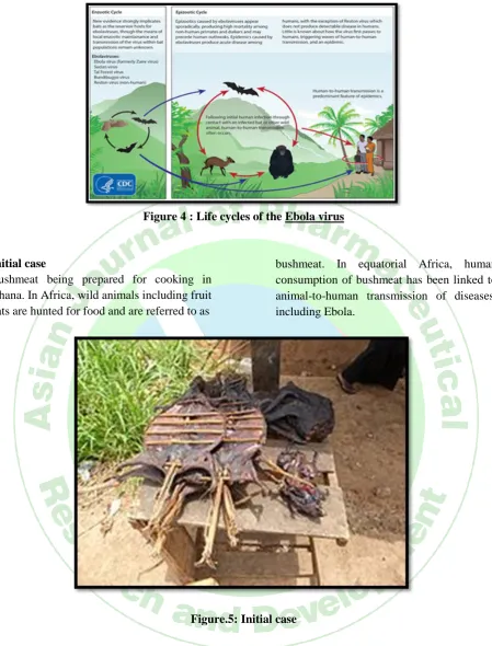
Transmission
It is believed that between people, Ebola disease spreads only by direct contact with the blood or other body fluids of a person who has developed symptoms of the disease. Body fluids that may contain Ebola viruses include saliva, mucus, vomit, feces, sweat, tears, breast milk, urine and semen. The WHO states that only people who are very sick are able to spread Ebola disease in saliva, and whole virus has not been reported to be transmitted through sweat. Most people spread the virus through blood, feces and
Figure 4 : Life cycles of the Ebola virus
Initial case
Bushmeat being prepared for cooking in Ghana. In Africa, wild animals including fruit bats are hunted for food and are referred to as
bushmeat. In equatorial Africa, human consumption of bushmeat has been linked to animal-to-human transmission of diseases, including Ebola.
Figure.5: Initial case
Reservoir
The natural reservoir for Ebola has yet to be confirmed; however, bats are considered to be the most likely candidate species. Three types of fruit bats (Hypsignathus monstrosus,
Epomops franqueti and Myonycteris
torquata) were found to possibly carry the virus without getting sick. As of 2013, whether other animals are involved in its spread is not known. Plants, arthropods and
birds have also been considered possible viral reservoirs.
PATHOPHYSIOLOGY
chemical signals and leads to a septic state. EBOV is thought to infect humans through contact with mucous membranes or through skin breaks. Once infected, endothelial cells (cells lining the inside of blood vessels), liver cells, and several types of immune cells such as macrophages, monocytes, and dendritic cells are the main targets of infection. Following infection with the virus, the immune cells carry the virus to nearby lymph nodes where further reproduction of the virus
takes place. From there, the virus can enter the bloodstream and lymphatic system and spread throughout the body. Macrophages are the first cells infected with the virus, and this infection results in programmed cell death. Other types of white blood cells, such as lymphocytes, also undergo programmed cell death leading to an abnormally low concentration of lymphocytes in the blood. This contributes to the weakened immune response seen in those infected with EBOV.
Figure.6: Pathogenesis schematic
DIAGNOSIS
When EVD is suspected in a person, his or her travel and work history, along with an exposure to wildlife, are important factors to consider with respect to further diagnostic efforts.
Laboratory testing
Possible non-specific laboratory indicators of EVD include a low platelet count; an initially decreased white blood cell count followed by an increased white blood cell count; elevated
levels of the liver enzymes alanine
aminotransferase (ALT) and aspartate
aminotransferase (AST); and abnormalities in
blood clotting often consistent with
disseminated intravascular coagulation (DIC)
such as a prolonged prothrombin time, partial thromboplastin time, and bleeding time. Filovirions, such as EBOV, may be identified by their unique filamentous shapes in cell cultures examined with electron microscopy, but this method cannot distinguish the various filo viruses.
Differential diagnosis
Early symptoms of EVD may be similar to those of other diseases common in Africa, including malaria and dengue fever. The symptoms are also similar to those of other viral hemorrhagic fevers such as Marburg virus disease.
fever, shigellosis, rickettsial diseases, cholera, sepsis, borreliosis, EHEC enteritis, leptospirosis, scrub typhus, plague, Q fever,
candidiasis, histoplasmosis, trypanosomiasis, visceral leishmaniasis, measles, and viral hepatitis among others.
PREVENTION
Infection control
Figure 7: British woman wearing protective gear
People who care for those infected with Ebola should wear protective clothing including masks, gloves, gowns and goggles. The US Centers for Disease Control (CDC) recommend that the protective gear leaves no skin exposed. These measures are also recommended for those who may handle objects contaminated by an infected person's body fluids. In 2014, the CDC began
recommending that medical personnel
receive training on the proper suit-up and removal of personal protective equipment (PPE); in addition, a designated person, appropriately trained in biosafety, should be watching each step of these procedures to ensure they are done correctly. In Sierra Leone, the typical training period for the use of such safety equipment lasts approximately 12 days.
Isolation
Isolation refers to separating those who are sick from those who are not. Quarantine refers to separating those who may have been
exposed to a disease until they either show signs of the disease or are no longer at risk. Quarantine, also known as enforced isolation, is usually effective in decreasing spread. Governments often quarantine areas where the disease is occurring or individuals who may transmit the disease outside of an initial area. In the United States, the law allows quarantine of those infected with ebola viruses.
Prognosis
EVD has a high risk of death in those infected which varies between 25 percent and 90 percent of those infected. As of September 2014, the average risk of death among those infected is 50 percent. The highest risk of death was 90 percent in the 2002–2003 Republic of the Congo outbreak.
Death, if it occurs, follows typically six to sixteen days after symptoms appear and is often due to low blood pressure from fluid loss. Early supportive care to prevent dehydration may reduce the risk of death.
REFERENCES
1. "Ebola virus disease Fact sheet No. 103". World Health Organization. September 2014.
2. Ruzek, edited by Sunit K. Singh, Daniel (2014). Viral hemorrhagic fevers. Boca Raton: CRC Press, Taylor & Francis Group. p. 444. ISBN 9781439884294.
3. "2014 Ebola Virus Disease (EVD) outbreak in West Africa". WHO. 21 April 2014. Retrieved 3 August 2014.
4. "Preliminary study finds that Ebola virus fragments can persist in the semen of some survivors for at least nine months". CDC. 14 October 2015.
5. "Recommendations for Breastfeeding/Infant Feeding in the Context of Ebola". cdc.gov. 19 September 2014. Retrieved 26 October 2014.
6. "Guidance for Safe Handling of Human Remains of Ebola Patients in U. S. Hospitals and Mortuaries". Retrieved 10 October 2014.
8. "Ebola Viral Disease Outbreak — West Africa, 2014". CDC. 27 June 2014. Retrieved 26 June 2014.
9. Cite error: The named reference 30who2016 was invoked but never defined (see the help page).
10. "CDC urges all US residents to avoid nonessential travel to Liberia, Guinea and Sierra Leone because of an
unprecedented outbreak of Ebola.". CDC. 31 July 2014. Retrieved 2 August 2014.