R E S E A R C H
Open Access
Array CGH improves detection of mutations in
the
GALC
gene associated with Krabbe disease
Alice K Tanner
1, Ephrem L H Chin
1, Patricia K Duffner
2and Madhuri Hegde
1*Abstract
Background:Krabbe disease is an autosomal recessive lysosomal storage disorder caused by mutations in the
GALCgene. The most common mutation in the Caucasian population is a 30-kb deletion of exons 11 through 17. There are few other reports of intragenicGALCdeletions or duplications, due in part to difficulties detecting them.
Methods and results:We used gene-targeted array comparative genomic hybridization (CGH) to analyze theGALC
gene in individuals with Krabbe disease in whom sequence analysis with 30-kb deletion analysis identified only one mutation. In our sample of 33 cases, traditional approaches failed to identify two pathogenic mutations in five (15.2%) individuals with confirmed Krabbe disease. The addition of array CGH deletion/duplication analysis to the genetic testing strategy led to the identification of a second pathogenic mutation in three (9.1%) of these five individuals. In all three cases, the deletion or duplication identified through array CGH was a novelGALCmutation, including the only reported duplication in theGALCgene, which would have been missed by traditional testing methodologies. We report these three cases in detail. The second mutation remains unknown in the remaining two individuals (6.1%), despite our full battery of testing.
Conclusions:Analysis of theGALCgene using array CGH deletion/duplication testing increased the two-mutation detection rate from 84.8% to 93.9% in affected individuals. Better mutation detection rates are important for improving molecular diagnosis of Krabbe disease, as well as for providing prenatal and carrier testing in family members.
Keywords:Krabbe disease, GALC, Deletion, Duplication, Array CGH
Background
Krabbe disease, also called globoid cell leukodystrophy, is an autosomal recessive lysosomal storage disorder in-volving progressive damage to the white matter of the central and peripheral nervous systems (reviewed in [1]). The disease is caused by deficiency of the enzyme galac-tocerebrosidase (GALC), which leads to an inability to degrade galactolipids found mainly in the myelin sheath [2]. Symptoms include spasticity, irritability, and devel-opmental delay and regression, which progress to a severe decerebrate condition with no voluntary move-ments [3]. While the age of onset and progression of the disease is variable, 85-90% of affected individuals de-velop symptoms in the first six months of life, with a median survival of 17 months [1,4]. Death is often due
to respiratory infections or cerebral hyperpyrexia. Late infantile- and juvenile- onset forms with longer survival periods are also seen, and adults may present with weak-ness, loss of manual dexterity, and paresthesia in their extremities [1]. The prevalence of Krabbe disease is ap-proximately one in 100,000 in the US and Europe [5] with higher frequencies in the Druze and Muslim Arabs in Israel [6].
Mutations in the GALC gene (14q31) cause Krabbe disease [7], and numerous nonsense, missense, small in-sertion, and small deletion mutations spanning the en-tire length of the GALC gene have been described [3]. The most common mutation, consisting of approxi-mately 40% of alleles from affected individuals with European ancestry and 35% of alleles from those with Mexican ancestry, is a 30-kb deletion beginning in in-tron 10 and extending nine kb beyond the polyadenyla-tion signal [3,8,9]. The 30-kb delepolyadenyla-tion mutapolyadenyla-tion results in the classic infantile form of the disease in the * Correspondence:[email protected]
1
Emory Genetics Laboratory, Department of Human Genetics, Emory University, Atlanta, GA, USA
Full list of author information is available at the end of the article
homozygous state or when in trans (on opposite chro-mosomes) with another mutation associated with severe disease [3].
There are few reports of any other large deletions or large duplications encompassing the GALC gene. The techniques currently available for detecting single- and multi-exon deletions and duplications include multiplex PCR, quantitative PCR, Southern blotting, multiplex ligation-dependent probe amplification (MLPA), detec-tion of virtually all mutadetec-tions-SSCP (DOVAM-S), and single condition amplification/internal primer sequen-cing (SCAIP). Due to the difficulty and complexity of test development to routinely and reliably detect dosage differences, clinical laboratories have not offered this testing, and therefore data on the presence of these types of mutations are lacking.
To further explore the deletion/duplication mutation spectrum of the GALCgene, we have developed a com-prehensive mutation detection strategy which begins with sequence analysis of theGALCgene in combination with mutation-specific testing for the 30-kb deletion. If two mutations are not found using this approach, des-pite an established biochemical diagnosis for the patient, this testing is followed by targeted array comparative genomic hybridization (CGH) to look for copy number changes within the GALC gene. Here we present muta-tion identificamuta-tion statistics forGALC analysis at Emory Genetics Laboratory (EGL); we also identify two novel
GALCdeletions and describe the only largeGALC dupli-cation reported in an individual with Krabbe disease.
Results
Emory Genetics Laboratory (EGL) started offering array CGH-based deletion/duplication testing for the GALC gene in January of 2008. From January of 2008 through August of 2011, approximately 100 samples were sub-mitted to EGL for full GALC gene analysis. There were 33 cases in which we performed complete GALC gene analysis (sequence analysis with 30-kb deletion analysis followed by array CGH deletion/duplication analysis, if necessary). These cases included samples from indivi-duals reported to be enzymatically or clinically affected by Krabbe disease, and also paired parental samples from cases in which the affected individual was deceased. The remainder of the samples were not in-formative for our study and included samples for which no clinical information was submitted, samples sent for carrier testing in individuals with a family history of Krabbe disease in which the familial mutation was not known, and samples sent for comprehensive carrier test-ing from individuals with partners known to be carriers for Krabbe disease. Analysis began with sequencing of the 17 exons and flanking intronic regions of the GALC gene, along with allele-specific PCR analysis for the
common 30-kb deletion. If a mutation(s) was not found, the ordering physician had the option of reflex testing with array CGH to detect single and multiple exon dele-tions and duplicadele-tions.
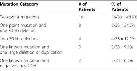
Results ofGALCanalysis for the 33 cases of confirmed Krabbe disease are given in table 1. In 28 of the 33 cases (84.8%), both mutations were identified via sequence analysis in combination with analysis for the 30-kb dele-tion. Sixteen of these cases had two mutations identified through sequence analysis, eight had one mutation iden-tified through sequence analysis and one copy of the 30-kb deletion, and four had a homozygous 30-30-kb deletion. In an additional three of the 33 cases (9.1%), sequence analysis with analysis of the 30-kb deletion identified one mutation, while reflex deletion/duplication testing identified a second mutation; these cases are discussed in more detail below as cases 1-3. The overall detection rate for two mutations using a combination of sequence analysis and array-CGH deletion/duplication testing was therefore 31/33 (93.9%).
In two of the 33 cases (6.1%), comprehensive GALC mutation analysis, including sequence analysis, 30-kb de-letion analysis, and dede-letion/duplication analysis, failed to identify two known mutations. When only one muta-tion is identified in individuals with a biochemical diag-nosis of a recessive disease, it is EGL’s customary practice to design a second set of PCR primers for the entire gene involved. Resequencing of the GALC gene with an alternative primer set also failed to identify a second mutation in these individuals, reducing the prob-ability of allele drop out. In one of the two cases, the proband was deceased but reportedly had clinical fea-tures consistent with Krabbe disease and an enzymatic diagnosis of Krabbe disease from another laboratory (la-boratory reports of the enzyme analysis were not pro-vided to EGL). Sequence analysis with 30-kb deletion analysis of the parents identified one copy of the 30-kb deletion in the mother, but was negative in the father. Reflex deletion/duplication testing in the father was also
Table 1GALCMutations Identified in 33 Biochemically Confirmed Krabbe Disease Patients at Emory Genetics Laboratory
Mutation Category # of
Patients
% of Patients
Two point mutations 16 16/33 = 48.5%
One point mutation and one 30-kb deletion
8 8/33 = 24.2%
Two 30-kb deletions 4 4/33 = 12.1%
One known mutation and one large deletion or duplication
3 3/33 = 9.1%
One known mutation and negative array CGH
negative. In the second case, the proband had a bio-chemical diagnosis of Krabbe disease from another la-boratory; no other information on the proband was provided. Sequence analysis with 30-kb deletion analysis identified one missense mutation and one intronic vari-ant of unknown clinical significance (c.909-10A>G; IVS8-10A>G) in the affected individual. Reflex dele-tion/duplication testing was negative. Testing the par-ents of the second individual was recommended to aid in interpretation of the unknown variant, but has not been ordered at EGL to date.
Case 1
Case 1 was an eight-month-old Caucasian male reported to have deficient GALC enzyme activity. Sequence ana-lysis with 30-kb deletion anaana-lysis identified one copy of a five-bp deletion in exon 16 of theGALCgene. A second mutation was not identified. After the affected individual passed away, samples from the parents were sent to EGL for testing. The individual’s mother was found to carry the five-bp deletion. Array-CGH deletion/duplication testing of the individual’s father detected a 6.9-kb dele-tion of exon 8 (see figure 1).
Case 2
Case 2 was a one-year-old Caucasian female reported to have deficient GALC enzyme activity by blood assay and by fibroblast assay, and clinical features consistent with Krabbe disease. Sequence analysis with 30-kb deletion analysis identified one copy of the 30-kb deletion in this individual. A second mutation was not identified. Test-ing of the parents indicated that the child’s mother was the carrier of the 30-kb deletion. Array-CGH deletion/ duplication analysis of the individual’s father revealed a novel 11-kb duplication encompassing exons 11 through 14. Subsequent deletion/duplication testing of the affected individual revealed a complex copy number pat-tern in the GALC gene resulting from the overlapping combination of the 30-kb deletion, which extends from exons 11 through the end of the gene, and a duplication of exons 11 through 14 (see figure 2). In this individual, the combination of a deletion and a duplication resulted in an array result with normal copy number for exons 11 through 14, while exons 15 through the end of the gene were deleted. In the absence of parental testing, this combined set of copy number changes would have been difficult to detect and interpret. This individual was confirmed to be the same individual reported in [10] and is the only report of a large duplication in the
GALCgene to date.
Case 3
Case 3 was a 17-month-old East Indian female. Testing in another laboratory indicated deficient GALC activity
and a homozygous single base pair deletion in exon 1 of theGALCgene. Testing of this individual’s parents indi-cated that the mother was a carrier of the single base pair deletion, but testing for the mutation in the father was negative. Samples were submitted to EGL for array-CGH deletion/duplication testing for the affected indi-vidual and her father. This testing revealed a deletion of exons 1 through 6 in both individuals (see figure 3). The deletion that the affected individual inherited from her father caused the mutation inherited from her mother to appear homozygous by sequence analysis.
Discussion
Array CGH is currently being used successfully in many molecular cytogenetic laboratories to detect gross altera-tions in the human genome. Most cytogenetic arrays, however, are not designed to detect small (intragenic) deletions and duplications. The development of a clinical test for detecting intragenic copy number changes has the potential to raise the mutation detection rate for tested diseases, thereby improving molecular diagnosis of affected individuals. Gene-targeted array CGH offers a powerful alternative to the current methods used for detecting these mutations.
We have adapted array CGH technology and success-fully shown that it can detect intragenic deletions and duplications in a large set of genes, including GALC, using a single array [11]. A study by Aradhya et al. demonstrated that gene-targeted array CGH was able to identify partial or whole gene deletions and duplications in approximately 5% of a clinical cohort sent to a diag-nostic laboratory for testing [12]. When broken down by mode of inheritance, the positive rate approximately 5% for autosomal dominant genes, approximately 10% for autosomal recessive genes, and approximately 3.5% for X-linked genes. They conclude that intragenic copy number mutations are more prevalent that previously suspected in Mendelian disorders and should be part of their routine diagnostic workup. In addition, a study by Wang et al. reported a 3% positive rate when using gene targeted array CGH to identify deletions and duplication in a mitochondrial and metabolic patient cohort, and conclude that gene targeted array CGH is useful as a complementary diagnostic test for gene sequence ana-lysis [13].
out the presence of one copy of the mutation on one allele and a deletion on the other allele. Of the 11 individuals in this study with apparently homozygous mutations identi-fied by sequence analysis with 30-kb deletion analysis (seven point mutations and four 30-kb deletions), parental samples were submitted on only three; in all three cases, the parents were shown to each carry one copy of the now verified homozygous mutation identified in the child. Without parental testing on the other eight cases, it is not possible to rule out the presence of a deletion, possibly leading to an underrepresentation of the frequency of novel deletions in theGALCgene.
In addition, the novel kb duplication of exons 11-14 in case 2 would not have been detected by the use of traditional methodologies. This duplication normalizes
the copy number in the proband from exons 11-14, in spite of the fact that the proband carries the common 30-kb deletion on the other allele. The detection of a de-letion and duplication in trans in the same gene with partially overlapping exons clearly proved the power of aCGH to detect gene-targeted deletions and duplica-tions. The better able we are to detect mutations, the more options there will be for molecular prenatal testing and carrier testing in family members.
Conclusions
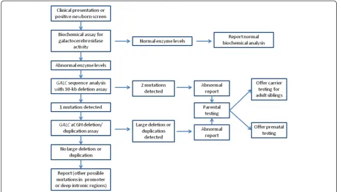
An algorithm for molecular testing for Krabbe disease is given in figure 4. First, genetic counselling and molecu-lar testing for Krabbe disease can be offered to indivi-duals with clinical symptoms, low levels of GALC
enzyme, or positive newborn screen results. Initial testing should begin with GALC gene sequence analysis, along with allele-specific PCR for the 30-kb deletion, as these methods have the highest detection rate and are cost effective. If these methods do not identify two known mutations,GALCarray CGH deletion/duplication analysis should be offered. If two mutations are identified, prenatal testing can be offered after parental testing to confirm that the two mutations arein trans. Since sequence ana-lysis alone cannot distinguish a homozygous mutation from a sequence mutation that lies intranswith a dele-tion in the same gene, mutadele-tion-specific testing of both parents of individuals with an apparently homozygous mutation is especially important. Parental testing should
be followed by deletion/duplication testing in those cases in which one parent tests negative for the muta-tion. Once two mutations have been identified and confirmed in parental samples, testing by known mu-tation analysis would also be possible for other affected family members or as carrier testing in adult family members.
This study of 33 Krabbe cases reveals that array CGH deletion/duplication analysis of theGALCgene increased the rate of detection of two mutations from 84.8%, for sequence analysis with 30-kb deletion analysis, up to 93.9%, and enabled us to uncover three previously un-reported copy number mutations. In addition to the 33 cases presented here, there were also three other
cases for which sequence analysis with 30-kb deletion analysis failed to identify two known mutations, but for which reflex deletion/duplication testing was not ordered. In one case, only one mutation was detected though sequence analysis, whereas in the other two cases, one known mutation and one variant of unknown clinical significance were detected through sequence analysis. In all three cases, deletion/duplication analysis and parental testing were recommended in the hope that a second mutation could be identified or the signifi-cance of the variants might be clarified; none of this test-ing, however, has been ordered at EGL to date. Ultimately, further GALC array CGH deletion/duplication testing in
clinical laboratories will likely refine these statistics and identify other novel mutations.
Methods Sequencing
Oligonucleotide primers were designed to amplify the 17 coding exons of theGALCgene in 16 fragments. (Oligo-nucleotide sequences are available from the authors upon request.) PCR products were analyzed on a 2% agarose gel, after which the remainder of the PCR prod-uct was purified using a Millipore Ultrafiltration PCR purification kit (Millipore, Billerica, MA). Sequencing reactions were prepared in a 10-ul reaction volume using
the BDv3.1W sequencing kit (Applied Biosystems, Foster City, CA). Each PCR product was sequenced in both directions according to ABI recommendations using uni-versal M13 sequencing primers. Sequenced PCR pro-ducts were purified using SephadexW cleanup plates by Edge Biosystems (Gaithersburg, MD) according to the manufacturer’s recommendation. Samples were heat-denatured for 5 minutes and loaded onto an ABI 3730xl sequencer. Sequence data were analyzed using Mutation SurveyorW v3.1 (Softgenetics, PA) and SeqScapeW (Ap-plied Biosystems, Foster City, CA).
Allele-specific PCR
The allele-specific PCR assay for the 30-kb GALC dele-tion was designed by Rafi et al., 1995. Three primers, one sense and two antisense, were used to amplify the wild-type and mutant alleles. The PCR products were analyzed on a 2% agarose gel. A wild-type allele yields a 615 bp PCR product whereas a mutant allele yields a 320 bp PCR product.
Array CGH
Array design
The targeted gene high-resolution oligonucleotide CGH array was custom designed on Oxford Gene Technolo-gies (OGT) 180 K platform to detect deletions and duplications in 175 genes associated with various genetic
disorders. Long oligonucleotides (~45–60 mer) were used to design the array, with repeat sequence masking implemented to ensure greater sensitivity and specificity. The GALC gene was covered by 431 probes with 116 probes covering the 17 exons at an average spacing of 15 bp between probes. The intronic region was covered by 315 probes at an average spacing of 25 bp between probes. Use of intronic oligonucleotide probes allows us to detect dosage changes within the entire genomic re-gion of the gene and determine the approximate breakpoints.
Experimental set up
DNA extraction was performed on patient DNA using a Gentra Puregene DNA extraction kit according to the manufacturer’s instructions. Male and female wild-type control DNA was obtained from Promega, Inc. Each pa-tient and reference DNA sample was sonicated, such that the DNA fragment size was between 200-5,000 bases and verified on a 1% agarose gel. Patient and refer-ence DNA samples were labeled using Klenow enzyme (NEB) and Cy3 or Cy5 9mer wobble primers (TriLink Technologies), respectively. After labeling, each sample was purified by isopropanol precipitation and reconsti-tuted in ultra-pure water. We combined 4 ug each of la-beled patient and reference DNA, and the products were desiccated in a vacufuge (Savant DNA 120), then
resuspended in appropriate hybridization buffer along with Cy3 and Cy5 control CPK6 50mer oligonucleotides. This mixture was hybridized to a NimbleGen targeted gene CGH array for 16-20 hours at 42 °C in a Maui Hybridization system (BioMicro Systems). The array has 389,587 unique sequence probes with an average spacing of 10-bp within coding regions and 25-bp within in-tronic regions, allowing for detection of copy number changes and breakpoints as small as 100-bp within the entire coding region. Arrays were then washed according to the manufacturer’s recommendation and immediately scanned on a GenePix 4000 scanner (Molecular Devices).
After scanning, data were extracted from images, and within-array normalization was accomplished using manufacturer-provided software (NimbleScan). Normal-ized log(2) ratio data were analyzed using two different analysis programs: SegMNT and DNA copy NimbleScan (NimbleGen Systems, Inc.). Both software programs re-port breakpoints for predicted deletions or duplications in the patient or test sample relative to the reference and also display results in a bar graph where the y-axis indicates gain or loss of material (1 = gain, 0 = normal, -1 = loss), while the x-axis indicates the position of each feature on the chromosome.
Data files (.gff ) generated from different averaging windows using NimbleScan software were parsed using a custom program (Nimkit) that was developed in-house. Nimkit enables the laboratory to select and analyze only the gene of interest. Nimkit generates a gene-specific report summarizing breakpoints detected in the gene of interest, the respective log(2) ratios, and the exons present at each region. All other regions are masked and not analyzed by Nimkit, preventing genetic analysis of genes for which clinical testing was not requested, in compliance with HIPAA requirements.
Array quality was assessed by control resequencing oli-gonucleotides on each array that correspond to synthetic sequences designed to have no cross-hybridization po-tential to any known sequence. This sequence was designed to have three distinct sequencing domains with different characteristics: A, B, and C domains. Resequen-cing was performed on both the forward and reverse strands, so that the resequencing report has six different scores for the Cy3 channel and six distinct scores for the Cy5 channel: A-forward and A-reverse, B-forward and B-reverse, C-forward and C-reverse.
The“A” domain contained long runs of G nucleotides that can be difficult to synthesize. The “B”domain con-tained a large perfect hairpin sequence. The“C”domain contained a straightforward domain that should always resequence. Failure of domain “C” indicated a cata-strophic failure. Control DNA was spiked into each ex-periment for both CGH and resequencing arrays. A score from 0-100% was obtained that indicated the
sequence fidelity and correlated well with the overall performance of a microarray experiment [14].
Abbreviations
CGH: Comparative genomic hybridization; GALC: Galactocerebrosidase; MLPA: Multiplex ligation-dependent probe amplification; DOVAM-S: Detection of virtually all mutations-SSCP; SCAIP: Single condition amplification/internal primer sequencing; EGL: Emory Genetics Laboratory.
Competing interests
The authors declare no competing interests.
Acknowledgements
The authors would like to thank Dr. Kathryn Garber for editorial assistance.
Author details
1Emory Genetics Laboratory, Department of Human Genetics, Emory
University, Atlanta, GA, USA.2Hunter James Kelly Research Institute, Department of Neurology, School of Medicine, State University of New York at Buffalo, Buffalo, NY, USA.
Authors’contributions
AKT participated in data analysis and drafted the manuscript. ELHC participated in performing the sequence analysis and array CGH analysis. PKD collaborated on the case of the largeGALCgene duplication. MH conceived of the study, and participated in its design and coordination and helped to draft the manuscript. All authors read and approved the final manuscript.
Received: 17 January 2012 Accepted: 15 June 2012 Published: 15 June 2012
References
1. Wenger DA, Rafi MA, Luzi P, Datto J, Costantino-Ceccarini E:Krabbe disease: genetic aspects and progress toward therapy.Mol Genet Metab
2000,70:1–9.
2. Suzuki K, Suzuki Y:Globoid Cell Leukodystrophy (Krabbe's Disease): Deficiency of Galactocerebroside beta-Galactosidase.Proc Natl Acad Sci
1970,66:302–309.
3. Wenger DA, Rafi MA, Luzi P:Molecular genetics of Krabbe disease (globoid cell leukodystrophy): diagnostic and clinical implications.Hum Mutat1997,10:268–279.
4. Kemper AR, Knapp AA, Green NS, Comeau AM, Metterville DR, Perrin JM: Weighing the evidence for newborn screening for early-infantile Krabbe disease.Genet Med2010,12:539–543.
5. Wenger DA:Krabbe disease online NIH gene review. 2011.Available at http:// wwwncbinlmnihgov/books/NBK1238/Accessed February 25.
6. Korn-Lubetzki I, Nevo Y:Infantile Krabbe disease.Arch Neurol2003, 60:1643–1644.
7. Chen YQ, Rafi MA, de Gala G, Wenger DA:Cloning and expression of cDNA encoding human galactocerebrosidase, the enzyme deficient in globoid cell leukodystrophy.Hum Mol Genet1993,2:1841–1845. 8. Luzi P, Rafi MA, Wenger DA:Characterization of the large deletion in the
GALC gene found in patients with Krabbe disease.Hum Mol Genet1995, 4:2335–2338.
9. Rafi MA, Luzi P, Chen YQ, Wenger DA:A large deletion together with a point mutation in the GALC gene is a common mutant allele in patients with infantile Krabbe disease.Hum Mol Genet1995,4:1285–1289. 10. Duffner PK, Barczykowski A, Jalal K, Yan L, Kay DM, Carter RL:Early infantile
Krabbe disease: results of the World-Wide Krabbe Registry.Pediatr Neurol
2011,45:141–148.
11. Tayeh MK, Chin EL, Miller VR, Bean LJ, Coffee B, Hegde M:Targeted comparative genomic hybridization array for the detection of single-and multiexon gene deletions single-and duplications.Genet Med2009, 11:232–240.
12. Aradhya S, Lewis R, Bonaga T, Nwokekeh N, Stafford A, Boggs B, Hruska K, Smaoui N, Compton JG, Richard G, Suchy S:Exon-level array CGH in a large clinical cohort demonstrates increased sensitivity of diagnostic testing for Mendelian disorders.Genet Med2012,14:594–603. 13. Wang J, Zhan H, Li FY, Pursley AN, Schmitt ES, Wong LJ:Targeted array
diagnosis of mitochondrial and metabolic disorders.Mol Genet Metab
2012,106:221–230.
14. Hegde MR, Chin EL, Mulle JG, Okou DT, Warren ST, Zwick ME: Microarray-based mutation detection in the dystrophin gene.Hum Mutat2008, 29:1091–1099.
doi:10.1186/1750-1172-7-38
Cite this article as:Tanneret al.:Array CGH improves detection of
mutations in theGALCgene associated with Krabbe disease.Orphanet
Journal of Rare Diseases20127:38.
Submit your next manuscript to BioMed Central and take full advantage of:
• Convenient online submission
• Thorough peer review
• No space constraints or color figure charges
• Immediate publication on acceptance
• Inclusion in PubMed, CAS, Scopus and Google Scholar
• Research which is freely available for redistribution