Ex vivo Expansion of
Hematopoietic Stem Cells
ISBN: 978-94-6203-334-4
© Elnaz Farahbakhshian
The research presented in this thesis was performed at the Department of Hematology within the Erasmus University Medical Center, Rotterdam, The Netherlands.
The research has been supported by the European Commission's 5th, 6th, and 7th Framework Programs (contracts QLK3-CT-2001-00 427-INHERINET, LSHB-CT-2004-005242-CONSERT and grant agreement no. 222878-PERSIST), and by The Netherlands Organization for Health Research ZonMW (program grant 434-00-010).
Cover Design: Elnaz Farahbakhshian Layout: Sima Kheradmand Kia
Ex vivo
Expansion of
Hematopoietic Stem Cells
Proefschrift
ter verkrijging van de graad van doctor aan de Erasmus Universiteit Rotterdam op gezag van de rector magnificus
Prof. dr. H. G. Schmidt
en volgens besluit van het College voor Promoties
De openbare verdediging zal plaatsvinden op Dinsdag 9 april 2013 om 15.30
door
Elnaz Farahbakhshian
Promotie commissie
Promotor: Prof.dr. F.G. Grosveld
Overige leden: Prof.dr. J.N.J. Philipsen Prof.dr. H.R. Delwel Dr. M. von Lindern
To my parents and
my grandmother
Contents
Chapter1 Introduction 9
Outline of the thesis 25
Chapter2 Angiopoietin-like protein 3 promotes the proliferation and expansion of hematopoietic stem cells 35 Chapter3 The effect of over-expressing of angiopoietin-like protein 3 (Angptl3) on the proliferation and expansion of hematopoietic stem cells 51 Chapter4 The effect of Angiopoietin-like protein 3 (Angptl3) on the proliferation and expansion of umbilical cord blood cells 65 Chapter5 Effects of hypoxia on the ex vivo expansion of murine and human repopulating hematopoietic stem and progenitor cells 79 Chapter6 Discussion 99 Summary 113 Samenvatting 117 Abbreviations 119 Acknowledgements 121 Curriculum Vitae 128 PhD portfolio 129
Chapter 1
Introduction
11
Hematopoiesis
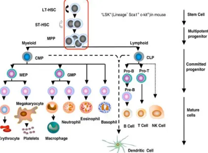
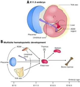
Hematopoiesis is a complex cellular differentiation process resulting in the formation of all blood cell types. In this process, hematopoietic stem cells (HSCs) reside at the top of the hematopoiesis hierarchy and have the capacity to differentiate into all blood cell lineages (multipotency) as well as maintaining themselves (self-renewal) during the lifespan of an individual [1, 2] (Fig.1). Mouse primitive HSCs are first found in the blood islands of the extra-embryonic yolk sac at day 7.5 of gestation. At day 10.5 of gestation the earliest HSCs can be detected in the dorsal aorta region of aorta gonad mesonephros (AGM) [3]. The first HSC precursors cluster in the aortic endothelium and it has been shown that these definitive HSCs originate from specialized heamogenic endothelial cells (ECs) of the mouse embryonic aorta [4] (Fig.2). Recently, it has been reported that similar to the AGM at day 10.5-11.5, ECs of mouse embryonic head also have hemogenic capacity and give rise to HSCs [5]. HSCs then migrate from these initial sources into the fetal liver around day 11, where they undergo dramatic expansion during fetal development [6]. At next stage, around day 12 to16, HSCs are mobilized from the fetal liver into the bone marrow and spleen; although the fetal liver remains an important organ of definitive hematopoiesis during the embryonic period. From birth and throughout adult life, the bone marrow becomes the major hematopoetic tissue [7].
Figure 1: The hierarchy of hematopoietic cells. LT-HSC, long-term repopulating HSC; ST-HSC, short-term repopulating HSC; MPP, multipotent progenitor; CMP, common myeloid progenitor; CLP, common lymphoid progenitor; MEP, megakaryocyte/erythroid progenitor; GMP, granulocyte−macrophage progenitor. The encircled pluripotent population, LT-HSC, ST-HSC and MPP are Lin-, Sca-1+, c-kit+ as shown (Taken from: Larsen et al. Oncogene 2005) [8]
Medvinsky A et al. Development 2011;138:1017-1031
A
B
Figure 2: Continuity in embryonic hematopoietic development. (A) A schematic representation of an E11.5 mouse embryo (anterior uppermost), showing development of embryonic hematopoietic stem cell (HSC) niches: placenta, umbilical cord and aorta-gonad-mesonephros (AGM) region. The yolk sac expands and encompasses the embryo. (B) A model of multisite hematopoietic development, showing hematopoietic progenitors and definitive. HSCs from the sites shown in A colonizing the liver rudiment and each other (as shown by the circular arrow). Disagreement exists about which of these sites is the genuine source of dHSCs and the adult hematopoietic system. After expansion in the fetal liver, dHSCs colonize the bone marrow, spleen and thymus. In adulthood, the thymus and spleen are colonized by bone marrow progenitors (not shown) (Taken from: Medvinsky A et al. Development 2011) [7].
Murine hematopoietic stem cells
Murine HSCs were initially identified based on their ability to form colonies in the spleens of lethally irradiated mice following bone-marrow transplantation [2, 9, 10]. Subsequently, the long-term repopulating (LTR) assay has been introduced to examine the HSC potential of lifelong reconstitution of all blood cell lineages after transplantation into lethally irradiated recipients. In the most stringent version of this assay, known as serial transplantation, HSC-containing donor bone marrow can be re-transplanted into secondary, and tertiary, recipients while maintaining both self-renewal and multi lineage differentiation capacity. These functional assays provided opportunities to identify the cell-surface phenotype of mouse HSCs, allowing their purification by fluorescence-activated cell sorting (FACS) [2, 11].
Introduction
13
The cell surface profile of murine hematopoietic stem cells
Lineage Markers. Primitive hematopoietic cells, including HSCs, do not express the surface markers that are associated with the terminal maturation of specific blood cell types.
Lack of expression of these lineage (lin) markers (i.e., CD3, B220, Gr1, Mac1, and Ter119), which includes all of the mature hematopoietic cell types (T-cells, B-cells, granulocytes, monocytes, and RBCs) can be used to isolate immature cells from the more differentiated cells. Further, murine bone marrow (BM) HSCs were distinguished as cells that do not express cell-surface markers of lineage-committed hematopoietic cells (Lin-) but do express high levels of stem-cell antigen 1 (Sca-1) and c-Kit, which is known as the LSK (Lin– Sca-1+c-Kit+) subset. Because only some phenotypic LSK HSCs have LTR activity, they can be further subdivided into long-term (LT)-HSCs, which are CD34–, fms-related tyrosine kinase 3 (FLT3)– CD150+, and short-term (ST)-HSCs, which are CD34+FLT3– and have only limited self-renewal capacity [12].
Human hematopoietic stem cells
Although, it has been shown that HSCs can proliferate and differentiate in vivo, there is as yet no in vitro assay that specifically detects HSCs. They divide poorly in semisolid media. They are phenotypically separable from most of the cells able to form mature myeloid colonies, erythroid, and/or megakaryocyte progeny in vitro. These colony-forming cells (CFCs) are considered to comprise a large, intermediate progenitor compartment that spans the entire stepwise process of lineage restriction. Long-term culture initiating cell (LTC-IC) assays can detect some [13, 14]but not all HSCs, which are more primitive than CFCs. For detecting human HSCs, the use of immunodeficient mice can be also a helpful assay [15]. The NOD/SCID mouse assay has provided a useful tool in the in vivo analysis of human hematopoietic stem cells, although only the short-term repopulating ability of stem cells can be analyzed due to the development of lymphomas and leakiness of murine B and T cells in aging NOD/SCID mice.
The cell surface profile of human hematopoietic stem cells
HSCs and primitive progenitors in human, like those in mice, are also distinguished from the majority of the cells in hematopoietic tissues by their lack of expression of various markers specific to maturing blood cells (i.e., CD3, CD4, CD8, CD14, CD15, CD16, CD19, CD20, CD56, and CD66b).
CD34 was the first differentiation marker to be recognized on primitive human hematopoietic cells and is still the most commonly used marker to obtain enriched populations of human HSCs and progenitors for research or clinical use. CD34 is expressed on ~1-4% of the nucleated cells in normal human bone marrow (BM) aspirate samples and <0.1% of nucleated cells in steady-state human peripheral blood [16]. Human HSCs can also be purified based on functional markers, such as the detoxifying enzyme aldehyde dehydrogenase
(ALDH) [17]. Hess et al. showed that Lin- ALDH high from umbilical cord blood cells (UCB) can repopulate and result in human cell engraftment in NOD-SCID mice whereas UCB-Lin- ALDH low hardly repopulate [17].
Hematopoietic stem cell nice
The stem cell niche is a term occasionally used to describe the stem cells location, however, the niche is actually composed of cellular components surrounding stem cells and signaling molecules provided by supporting cells [2, 18].
Within the human body, stem cell niches maintain adult stem cells in a quiescent state, but at situations like tissue injury, the surrounding microenvironment actively signals to stem cells to either increase self renewal or differentiation to form new tissue.
When stem cells are cultured in vitro, they differentiate, change their morphology and lose their self-renewal ability. Hence there is a lot of activity to study the various components of the niche to try to replicate the in vivo niche conditions in vitro. Several factors have been shown to be important for the regulation of stem cell characteristics within the niche but it is as yet not possible to effectively maintain/expand HSCs in vitro [2, 19, 20].
Interactions between stem cells and neighbor differentiated cells
The bone marrow consists of hematopoietic and non-hematopoietic (stromal) cells. It has been demonstrated that stromal cells have an active role in the regulation of HSCs differentiation into all blood cell lineages[21]. In 1978 Schofield predicted that there is a specific hematopoietic stem cell niche in BM, which “fix” the stem cells in place and prevent their maturation, allowing the stem cell to proliferate and retain its stemness [22]. In 2003, two groups reported that the size of the HSC pool in vivo is influenced by the bone-forming osteoblast cells, identifying that osteoblast cells are the critical component of the HSC niche [23, 24]. In these studies it was shown that the high level expression of bone morphologenetic protein (BMP) receptor type IA and notch ligand jagged 1 on oesteoblast cells increase the number of HSCs through stimulating the BMP pathway and Notch signaling, respectively [23, 24].
Products of osteoblasts that have been shown to be positive regulators of HSCs include angiopoietin-1 (Ang1), thrombopoietin (TPO) and Jagged-1 (under conditions of parathyroid hormone/parathyroid hormone-related peptide receptor (PPR) activation) whereas osteoblast-associated negative regulators of the HSC niche include osteopontin (OPN) and dikkopf1 [25-30].
Osteoblasts also abundantly express CXCL12, a chemokine that is involved in chemotaxis, homing, survival and maintenance of HSCs in the bone marrow. However, it should be noted that other non-osteoblast “reticular” cells in the marrow also express high levels of CXCL12 and may participate in the niche [31, 32]. Osteoblastic cells are a heterogeneous population thought to derive from multipotent mesenchymal progenitors. The specific role of each subset of osteoblastic cells in the niche has yet to be determined.
Introduction
15
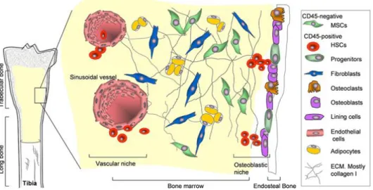
In this context, other cell types derived from mesenchymal progenitors, such as adipocytes and osteoclasts are also involved in the regulation of HSCs activity (Fig.3). In one study it was demonstrated that adipocytes secret adiponectin molecule that increases proliferation of HSCs, while maintaining their immature state [33]. In contrast, another study showed that the content of adipocytes in the marrow negatively correlates with the hematopoietic activity [34]. Osteoclasts also play a role in the mobilization of hematopoietic progenitors in the circulation through the cathepsin K-mediated cleavage of CXCL12. Furthermore, the osteoclastic bone absorption actively changes the Ca+2 concentrations in the BM microenvironment, which is important in HSC engraftment. HSCs deficient in the calcium sensing receptor were unable to engraft bone marrow [35]. Besides osteoclasts, other cell types participate in HSC regulation in the marrow, such as endothelial cells[36]. All blood vessels are covered by endothelial cells and in the BM they build a barrier between the developing hematopoietic cells and the blood [37,38]. BM endothelial cells constitutively express cytokines, such as CXCL12, and adhesion molecules, such as endothelial-cell (E)-selectin and vascular cell-adhesion molecule 1 (VCAM1), which are important for HSC mobilization, homing, and engraftment.
Figure 3. The stem cell niches in bone marrow. In the bone marrow, HSCs and their progeny populate the vascular niche which is surrounded by stromal cells derived from MSCs. Naïve MSCs with true stem cell attributes are part of the stroma while MSCs which are committed osteoprogenitor cells reside in the osteoblastic niche (Taken from Grassel et al. FBS 2007).
The sympathetic nervous system (SNS) also plays an important role in regulation of HSC mobilization between BM and bloodstream [39, 40]. It was shown that the SNS neurotransmittor, norepinephrine (NE), controls G-CSF-induced osteoblast suppression, bone CXCL12 downregulation, and HSC mobilization [39]. Additional studies in the mouse revealed
that the cyclical release of HSCs and expression of CXCL12 in the bone marrow microenvironment was regulated through circadian NE secretion by the SNS [40, 41]. Neurotransmittors also have roles in human HSC mobilization, proliferation and differentiation [41, 42].
Based on all these studies, it is clear that the HSC niche is much more complex than consisting of one or two cell types and further studies will be required to fully clarify how each of these cell types contributes to the HSC niche.
Extracellular matrix components
The concept of extracellular matrix (ECM) regulating primitive cells is a longstanding debate. As part of the bone marrow niche, it has been shown that ECM proteins influence human haematopoiesis.
One in vitro study explored the capacity of human HSCs adhesion to the ECM secreted by human marrow fibroblasts, including fibronectin, and discovered that adherence to the matrix varied both with the cell lineage and maturation stage of the progenitor cell [43]. A study on the spatial location of ECM proteins including fibronectin, collagen types I, III and IV, and laminin in murine femoral bone marrow (BM) by immuno-fluorescence labeling has revealed distinct locations of each protein, supporting the notion that they have an important role in the homing and lodgment of transplanted cells [44]. Whether there is a preference for a specific subset of primitive hematopoietic cells to adhere to certain ECM proteins within the adult BM remains to be explored. Many reports have documented the importance of β1 integrins, particularly α4β1 (VLA-4) and α5β1 (VLA- 5), in modulating adhesive interactions between HSCs, cellular and ECM components that comprise the stem cell niche [45-47]. One study demonstrated that VLA-5 is expressed on mouse and human long-term repopulating hematopoietic cells. It binds to fibronectin in the ECM and disruption of this binding can lead to decreased engraftment in the BM [48]. The deletion of α4 integrin induced by Mx1-Cre has shown that α4 integrin-deficient HSCs accumulate in the peripheral blood [49]. Furthermore, in transplantation studies, α4-/- cells displayed impaired homing to the BM, and short-term engraftment was severely delayed [49].
Another study on the cell–matrix interaction focused on laminins, a group of ECM proteins expressed in BM, that bind HSCs through their cognate receptors [50]. Specific expression of p67 laminin receptor was found on erythroid HPCs and blocking p67 binding on donor cells with antifunctional antibody leads to reduced BM homing of BFU-Es [51]. Whether non-erythroid HSCs can interact with laminins through other receptors is still unanswered.
Growth factors
Stem cell factor (SCF)
The gene encoding stem cell factor (SCF) is found in the Sl locus in mice and on chromosome 12q22-12q24 in humans [52]. SCF is produced by fibroblasts and endothelial cells and is active in both soluble and trans-membrane forms [53, 54]. SCF binds to c-kit (SCF
Introduction
17
receptor, CD117) and induces the homodimerization of c-kit receptor, activating the intracellular domain of the receptor, and triggering cellular pathways that regulate HSC activity [55]. Even before identifying the SCF and c-kit as a receptor-ligand pair, mouse studies revealed that mutation in c-kit kinase domain reduces colony-forming units spleen/HSCs activity [55].
The role of c-kit was also examined in adult mice by applying the anti c-kit antibody, ACK2, which resulted in a striking decrease in the number of HSCs and other hematopoietic progenitor cells in the bone marrow [56]. It has been shown that SCF promotes HSCs survival and migration but not proliferation by activating the AKT and ERK1/2 pathways. c-Kit, is a type III receptor protein-tyrosine kinase and activates the phosphoinositide 3-kinase (PI3K) pathway, which indirectly causes AKT phosphorylation followed by phosphorylation and inhibition of death proteins (FOXO3 and BAD). SCF also can effectively promote growth and survival signaling pathways by downstream effectors of the PI3K/AKT signaling pathway like mTOR, which has downstream effectors such as p70S6K and 4E-binding protein 1. Parallel to the AKT pathway, ERK1/2 is phosphorylated by a kinase cascade (RAF to MEK to ERK1/2) that is activated by RAS [57].
Thrombopoietin (TPO)
The human and mouse thrombopoietin (TPO) gene is located on chromosome 3 (q26.3-27) and 16, respectively. TPO is produced primarily in the liver and its receptor, Mpl, is expressed on platelets, megakaryocytes and the HSC compartment, including long-term HSCs (Fig.4). Approximately 50% of the murine fetal liver stem cell-enriched population, 70% of the murine marrow LSK cells, and 70% of the human bone marrow CD34+CD38- express c-mpl [58]. TPO production is constant and the concentration of available circulating TPO is controlled by the Mpl receptors available on the surface of megakaryocytes and platelets to internalize the cytokine [59-62]. Transplantation assays also revealed that LSK cells from wild type mice engraft almost 7-fold better than LSK cells from c-mpl deficient mice [58]. The distinct role of TPO in postnatal HSC maintenance is accompanied by accelerated HSC cell-cycle kinetics in Tpo-/- mice [62] and reduced expression of the cyclin-dependent kinase inhibitors p57Kip2 and p19INK4D as well as multiple Hox transcription factors. Thus, TPO regulates post-transplantation HSC expansion as well as the maintenance of adult quiescent HSCs. TPO is also the primary regulator of platelet production. Taken together, these observations highlight the key role for TPO signaling in control of platelet number and regulation of HSCs [59,62,63].
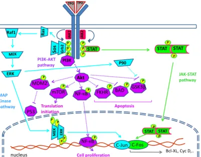
TPO works through inducing receptor dimerization which results in tyrosine phosphorylation, and a series of signaling events including activation of JAK/STAT, Shc/Ras/MAPK and PI3K/Akt. These pathways overlap with those induced by other cytokines , but the differences that lead to the unique biological effects of TPO that can maintain long term reconstitution cells without combination with other growth factor, have not been well characterized [27, 64, 65].
PI3K Akt STAT TPO TPO Grb 2 Sos Ra s Raf1 ERK JAK-STAT pathway MAP Kinase pathway PI3K-AKT pathway P P P P P P STAT STAT P P STAT STAT P P P P ME K ERK P P JAK 2 JAK 2 Bcl-XL, Cyc D,.. P P P P mTOR BAD P FKHR MDM2 GSK3β P NF-КB C-Fos P90 Translation initiation Cell proliferation P53 P NF-КB Apoptosis C-Jun nucleus
Figure 4: signalling pathway of TPO in HSCs. In the absence of ligands, cytokine receptor (Mpl) is preassembled with tyrosine kinase JAK in inactive complexes. Cytokine binding to extracellular domains of receptors induces a conformational change which allows the appended JAKs to cross- phosphorylate and activate each other. In turn, JAKs phosphorylate tyrosine residues (Py) on the cytosolic regions of receptors, which attract SH2- and PTB-containing signaling proteins. These proteins become phosphorylated themselves and either translocate to the nucleus to regulate gene expression (such as STATs, Signal Transducers and Activators of Transcription) or initiate kinase signaling cascades (such as Mitogen Activated Protein-Kinases, MAPK, phosphatydylinositol-3-kinase, PI3K, and Akt) (Taken from Stefan Constantinescu Signal transduction and molecular hematology group [Structure and function of cytokine receptors]).
Fms-related tyrosine kinase 3 ligand (Flt3L)
Fms-related tyrosine kinase 3 ligand (Flt3L) belongs to a small group of growth factors that regulates proliferation of early hematopoietic cells. The chromosomal location of the Flt3L gene is preserved on chromosome 7 in mouse and chromosome 19 in human [66, 67]. Flt3L is expressed in bone marrow fibroblasts and stromal cells of adherent layers of long-term bone marrow culture (BMC). While multiple Flt3L isoforms have been identified the transmembrane isoform is predominant.When proteolytically cleaved, the transmembrane isoform becomes soluble and biologically active [66]. The Flt3L receptor, CD135, is a tyrosine kinase type III receptor and is predominantly expressed on immature hematopoietic stem and progenitor. When this receptor binds Flt3L, it forms a homodimer and activates signaling through second messengers and supports precursors survival in different lineages of blood-forming cells such as CFU-GM, CFU-GEMM, and the very primitive high proliferative potential colony-forming cells, HPP-CFC [68]. Although, Flt3L alone cannot stimulate proliferation, it synergizes well with other CSFs and interleukins (G-CSF, GM-CSF, M-CSF, IL3, IL6, IL7, IL11,
Introduction
19
IL12 and SCF) to induce growth and differentiation [69, 70]. In adult murine BM cells FLt3L is a marker of short term hematopoietic stem cells ST-HSCs. ST-HSCs have more limited self-renewal capacity and are capable of giving rise to blood lineages for 8–12 weeks [71]. However, using Flt3L-deficient mice it was shown that HSC expansion in fetal liver or after transplantation is Flt3L independent. Moreover characterization of HSCs in Flt3(-/-) mice revealed that the FLT3 receptor is not required for HSC steady-state maintenance or expansion after transplantation [72], suggesting that Flt3 receptor and ligand are not critical regulators of mouse HSCs, neither in steady state nor during fetal or posttransplantation expansion [72].
Insulin like growth factor 2
Insulin-like growth factor-2 (IGF-2), also known as somatomedin A, is a member of the insulin family of polypeptide growth factors. The Mouse Igf2 gene on chromosome 7 and human IGF-2 gene on chromosome 11 both extend over 30 kb, containing 8 exons (5 noncoding and 3 coding exons).
In rodents IGF-2 expression is widespread in prenatal tissues and dramatically diminishes after birth, therefore, IGF- 2 functions as a fetal growth factor. In humans however, IGF-2 is expressed in various tissues throughout life and helps to maintain self-renewal and pluripotency of stem cells [73-75].
IGF2 activates both the IGF-1 receptor and a splice variant of the insulin receptor (IR) known as IR-A. IGF2 action through the IR-A, promotes the self-renewal of stem/progenitor cell. IGF2 binds to the IGF-2R, which mediates IGF2 shuttling to lysosomes for degradation. Although, its role in signaling has not been clearly demonstrated [76, 77], Zhang et al. reported that HSC-supportive mouse fetal liver CD3+ cells express IGF2 and the angiopoietin like protein family that can stimulate HSCs expansion [78]. The recent study by Chou et al., clarified that these cytokines are expressed from SCF+DLK+ stromal cells of fetal liver and not from CD3+ cells [79]. IGF-2 in combination with other growth factors, promoted expansion of day-15 fetal liver Lin-Sca-1+c-Kit+ long-term (LT)-HSC numbers about ~ 2-fold [80].
Fibroblast growth factor-1 (FGF-1)
The human Fibroblast growth factor-1 (FGF-1) gene maps to Chromosome 5 at position q31 [81]. Acidic fibroblast growth factor is derived from beta-endothelial cell growth factor by posttranslational processing [82]. FGF-1 belongs to the families of FGFs of which, to date, 22 FGFs and four FGF receptors have been identified in vertebrate genomes. All FGFs have a high affinity for heparin and for cell surface heparin sulfate proteoglycan. This complex is crucial for high-affinity binding of FGF to its receptors [83].
FGF-1 is a potent stimulator of stem cell activity in vitro. All long-term repopulating HSC activity is contained in the lineage-depleted, FGF receptor (FGFR)-positive cell population in mouse bone marrow (BM). Initial studies demonstrated that FGF-1 induces granulopoiesis and megakaryocytopoiesis [84]. Both FGF-1 and FGF-2 can sustain the proliferation of hematopoietic progenitor cells and maintaining their primitive phenotype [85-87]. FGF-1 was
shown to be involved in the expansion of multilineage, serially transplantable long-term repopulating HSCs. Most noticeable is that the culturing of unfractionated mouse bone marrow in serum-free media supplemented with only FGF-1 results in a significant and sustained expansion of cells with both lymphoid and myeloid repopulating capacity [88].
Although the mechanism of FGF-induced signal transduction is not well-defined, FGF causes rapid FGFR dimerization on the cell surface, resulting in intracellular polypeptides phosphorylation, e.g. MAP kinases which correlates with cell proliferation induction, and inducing tyrosine phosphorylation of Src during the entire G1 period, which correlates with migration [89]. FGF stimulation of the Erk1/2 signalling cascade triggers transition of pluripotent embryonic stem cells from self-renewal to lineage commitment [90].
Angiopoietin like protein 3
Angiopoietin like protein 3 (Angptl3) is a 455 amino acid protein, encoded by seven exons on mouse chromosome 4, and human chromosome 1. This polypeptide has the characteristic structure of Angiopoietin: a signal peptide, an extended helical domain to form dimeric or trimeric coiled-coils, a short linker peptide, and a globular fibrinogen homology domain (FHD). However, unlike the other angiopoietins, Angptl3 does not bind to the tyrosine kinase receptor Tie-1 or Tie-2, and was suggested that Angptl3 functions differently from the other family members [91].
Several members of the Angptl family play roles in regulating lipid metabolism and angiogenesis [92]. In particular, Angptl3 inhibits lipoprotein lipase activity in vitro and in vivo, and mice deficient in Angptl3 display defects in lipid metabolism[92, 93]. In addition, it was shown that Angptl3 is capable of binding to the cell-surface integrin αVβ3, and stimulating the adhesion and migration of endothelial cells and inducing blood vessel formation. Angptl3 also activates protein kinase B and increases the permeability of glomerular endothelial cells [93, 94]. Several studies suggested that Angptl3 plays a role in the regulation of HSC activity [78, 95].
HSCs in Angptl3-/- mice were decreased in numbers, quiescence, and repopulating activity. In addition Angptl3, which is secreted by BM endothelial and other stromal cells, binds directly to the HSC cell surface and regulates HSC activity by repressing the expression of the Ikaros transcription factor. Its over-expression diminishes the repopulation activity of HSCs [95].
Recently, it has been shown that the immune-inhibitory receptor human leukocyte like receptor B2 (LILRB2) and its mouse orthologue paired immunoglobulin-like receptor (PIRB) are receptors for several ANGPTLs including Angptl3[96]. Therefore, Angptl3 as an extrinsic factor directly binds to HSCs and supports expansion of repopulating cells in coculture experiments. As a consequence, combination of saturated levels of SCF, TPO, IGF2, FGF1 and Angptl3 were able to promote expansion of LT-HSCs up to ~30-fold [78].
Introduction
21
Physiochemical nature of the niche environment
Ca 2+concentrationThe osteoblast lineage, responsible for bone formation, is one of the key cellular components of the HSC niche [23,97, 98]. The endosteum, where active bone modeling and remodelling takes place, has a high extracellular concentration of calcium-ions, reaching up to 40mM near the resorbing osteoclasts. This is more than 20-fold the calcium-ion concentration in serum[99]. Hence, a unique features of bone that contribute to the microenvironmental niche for stem cells might include the high concentration of calcium ions at the HSC-enriched endosteal surface [99, 100].
Because Ca2+ Receptor (CaR) is also expressed on haematopoietic cells, including monocyte/ macrophage lineage cells, it has been postulated that CaR might affect stem cell responses to the unique endosteal microenvironment [35, 101]. To prove this hypothesis, CaR-/- mice were studied, which revealed that these mice have abundant primitive HSCs in their spleen and in the circulation but few HSCs in BM. However, the number of HSCs in fetal liver was normal. In addition, the numbers of progenitor cells were relatively low compared to other blood lineages in BM of CaR-/- mice. CaR-/- mice do not have any problem in transporting HSCs from fetal liver to BM but that they cannot home properly in the endosteal niche. Taken all together while the calcium concentration proved to be an important factors in HSCs niche biology, but the mechanism still needs to be elucidate [35].
Oxygen gradients
The oxygen concentration within the human bone marrow has a gradient from below 1% in hypoxic niches up to 6% in the sinusoidal cavity [102]
.
Most slow cycling hematopoietic stem cells are found in the hypoxic zones close to the bone surface and distant from capillaries, raising the possibility that these hypoxic niches are important for diminished HSC proliferation [103]. That quiescent HSCs situated in a hypoxic environment has recently been confirmed in mice by injecting Hoechst 33342 dye which increases the in vivo level of oxygen. Bone marrow cells from injected mice with Hoechst 33342 dye were isolated and divided according to dye diffusion gradients. To investigate the stem cell characteristics, cells with low and high Hoechst fluorescence were transplanted into lethally irradiated mice. The results showed that the bone marrow fraction with the lowest Hoechst-dye uptake, referred as hypoxic, had the highest amount of long-term repopulating cells [104].Therefore, it was postulated that hypoxic niches in the bone marrow microenvironment provide optimal conditions for HSC maintenance.The oxygen level in the hypoxic niche at the molecular level regulates the heterodimer transcription factor hypoxia-inducible factor-1 (HIF-1). HIF-1 consists of 2 subunits, α and β, of which α is oxygen sensitive and β is oxygen insensitive. Three regulatory HIF subunits were characterized as HIF-1α, HIF-2α, and HIF-3α. At oxygen levels above 5%, hydroxylation of the proline residues 402 and 564 of HIF-1α enables binding of the ubiquitination ligase von Hippel-Lindau tumor suppressor protein, which leads to HIF-1α proteosome degradation
[105]. In contrast, at oxygen levels below 5%, hydroxylation is inhibited leading to HIF-1α stabilization. Consequently, HIF-1 heterodimer transcription factor targets several genes that are involved in HSC maintenance, including erythropoietin (EPO), erythropoietin receptor (EPO-R) transferrin and its receptor, VEGF and its receptor VEGF-R (Flt1) [106, 107].
It has also been proposed that oxidative stress suppresses N-cadherin-mediated HSC adhesion to osteoblasts and induces HSCs to exit the niche [108].
Hematopoietic stem cell transplantation (HSCT)
Hematopoietic deficiencies are potentially curable with hematopoietic stem cell transplantation (HSCT) replacing defect HSCs with healthy HSCs. Subsequently these healthy HSCs generate different hematopoietic lineages in the patients’ body for lifelong [109-111]. Stem cell transplantation can be performed with cells from afamily member or an unrelated (matched) volunteer (allogeneic transplantation)or with stem cells previously collected from the patient (autologoustransplantation) [111, 112].
In autologous transplantation the cells from the patient are used, and there is no need to look for a human leukocyte antigen (HLA) match and no risk for graft-versus-host disease (GvHD). In case of leukemias, however the disadvantage of an autologous transplant is the possibility that the graft contains malignant cells and the lack of a graft-versus-tumor effect
.
Therefore autologous transplantation is not the best alternative for patients with a malignant disease. For these patients an allogenic transplantation is the most suitable treatment [113, 114]. However, the outcome of allogenic transplantation is strongly affected by the degree of HLA-matches which can result in a higher or lower chance of graft rejection (Host-versus-Graft reaction (HvG)) and (Host-versus-Graft-versus-Host Disease (GvHD)[115, 116]. Furthermore, patients who receive allogenic transplantation require a longer recovery period. This is due to the interaction of the donor-host immune system and immune suppression treatment prior to transplantation, resulting in an increased infections risk in allogenic compared to autologous transplantation [116]. Taken together, allogenic and autologous transplantation each has its own advantages and disadvantages. Hence, a decision on the type of HSCT is made in each case based on various factors [116].Sources and procurement of HSC
The sources of HSC are BM, peripheral blood, or umbilical cord blood (UCB) for autologous or allogeneic transplantation.
Bone marrow
Bone marrow is the internal tissue of large bones and consists of two types of stem cells, hematopoietic stem cells and stromal stem cells. Hematopoietic stem cells give rise to all different blood cells and stromal stem cells generate fat, cartilage and bone. On average there is about 2.6kg of bone marrow in adult humans.
Introduction
23
For transplantation BM is obtained from the posterior iliac crests of the donor under epidural or under general anesthesia. If the amount of bone marrow aspirate is insufficient, the anterior iliac crest or sternum can also be used [117]. BM aspirate can be used immediately for transplantation or alternatively be stored at 4°C for 24 hours with no loss of stem cell viability [117]. Generally, 2 × 10⁸ nucleated BM cells per kg of patients’ body is sufficient to provide a stable long-term engraftment, although this number can be reduced to 1 × 10⁸/kg [117, 118].
Peripheral blood stem cells
Peripheral blood stem cells (PBSCs) are currently the most widely used source of HSCs for allogenic transplantation. PBSCs are isolated from donor peripheral blood by apheresis. In this method peripheral blood is withdrawn and after isolation of a particular constituent, PBSCs in this case, the remainder is returned to the donor. Prior to apheresis, donors are treated with several sub-cutaneous injection of granulocyte-colony stimulation factor to increase the number of HSC and mobilize them from BM to PB [119-121].It is known that PBSCs engraft faster than BM-derived stem cells. For instance neutrophil and platelet engraftment after PBSC transplantation is seen 14 and 18 days, respectively, compared to 19 and 25 days in case of BM transplantation [119, 122-124].
Umbilical cord blood
Umbilical cord blood (UCB) has a higher HSC frequency compared to immobilized adult peripheral blood. Hence, UCB is an important HSC source to support myeloblative or non-myeloblative therapies. UCB can be accessed rapidly and easily and has a lower GVHD incidence regardless of HLA disparity. Laghlin et al. showed that HLA-mismatched UCB transplanted in adult patients or children required a longer period of time to engraft compared to an HLA-matched sibling or unrelated marrow transplant, whereas a UCB transplant resulted in a lower incidence of GvHD [125]. because UCB provide a greater flexibility of HLA disparity than BM or PBSC grafts, finding UCB donors in minority populations that are currently under-represented in donor registries is a high priority [126-128]. These special properties make UCB an attractive option when BM or PBHSC donors are not the best possible alternative [109]. Although the use of UCB as a stem cell source has increased significantly in recent years, especially in children and early adulthood, it is not without downsides. One of the most important drawbacks of UCB for HSC therapy is the limited available HSCs for transplantation [129, 130]. The outcome of HSC transplantation is correlated with the total nucleated cell (TNC) number transplanted per kilogram (kg) of recipients body weight [131]. As a consequence, UCB transplantation (UCBT) is likely more successful in children. To overcome the low HSC problem, two approaches have been introduced. One has utilized more than one UCB unit in order to achieve a higher number of available TNC for infusion [128, 132-134] while the second approach has attempted to expand UCB units ex vivo [129, 135-137].
Combining more than one cord blood unit may overcome the drawback of low cell dose in a single cord blood unit [138]. However, combining UCB units could raise concerns such as
a higher chance of GvHD or a lack of engraftment as a result of immune reactivity among the transplanted units [131, 139]. Moreover, another concern of double-unit UCB transplantation is ‘graft-versus-graft’ effect. In an early double-unit UCB transplantation attempt both UCB units engrafted [140]; however, only one unit contributed to long-term hematopoiesis repopulation [141, 142].
The second approach to resolve the issue of limited available HSCs for transplantation in UCB units is ex vivo expansion of HSCs [129, 135-137]. Many clinical applications of HSCs would become feasible if there would be a culture system that expands HSC numbers while maintaining stem-cell pluripotency [143-145]. In particular, the ex vivo expansion of human cord blood HSCs would make this important source of cells useful for adult applications. During the past two decades several methodshave been used for ex vivo UCB cell expansion, such as liquid suspension culture application [146].
In liquid suspension culture, isolated UCB stem cells are exposed to a cocktail of cytokines, and growth factors for a specific period of time[129]. To establish an optimum growth factor cocktail for in vitro expansion of human HSCs, different combinations of growth factors have been studied. In this regard, the analysis of 16 cytokines on stroma-free cultures of CD34+CD38- bone marrow cells indicated that the combination of FLT3L, SCF and IL3 yield the highest (30-fold) expansion of starting long-term culture-initiating cells (LTC-IC) [147]. In another study addition of Flt-3 maintained the ability of human CD34+ cells to sustain long-term HSCs [148]. TPO has also been reported to increase the multi lineage growth of CD34+CD38- bone marrow cells under stroma-free conditions when added to the cytokine combination of Flt3-L and SCF [149]. Levac et al. reported that the combination of SCF, TPO, and Flt3L (STF) can successfully expand UCB-derived primitive HSCs [150]. IGF2 and FGF1 were also introduced as promising growth factors in ex vivo expansion and proliferation of HSC [87, 151]. Zhang et al. reported that using a new combination of growth factors that included SCF, TPO, IGF-2 and FGF-1(STIF) supplemented with Angiopoietin-like proteins (Angptls) for 10 days resulted in a 24- to 30-fold expansion of murine LT-HSCs [78, 95]. Furthermore, IGF-binding protein 2 (IGFBP2) and Angptl5 (A5) were introduced as additional secreted proteins that support human HSC expansion [152]. Using SCF [153], TPO [154], and FGF-1 supplemented with IGFBP2 and Angptl5 the number of human SCID-repopulating cells (SRCs) increased ~20-fold [155]. However, the ideal mixture of growth factors or cytokines, and the optimum culture duration for the ex vivo expansion of HSCs has not yet been determined.
As a gene therapeutic modality
Gene therapy has been introduced as a new concept for the treatment of certain human diseases, which may replace or assist traditional drug therapies. In contrast to traditional drug therapy life manufactured chemicals, gene therapy aims to make use of the patients own cells after introducing the desired genetic material into the patient. Gene therapy can use several approaches such as inserting a wild type gene into a non-specific location
Introduction
25
within the genome to compensate for nonfunctional gene, or using homologous recombination to exchange a mutated sequence with wild type DNA sequence which is more difficult.
Initial trials were focused on diseases that are caused by a single-gene defect, such as cystic fibrosis [156], hemophilia [157], and muscular dystrophy [158]. In future, the main aim would be toward curing diseases that are caused by multiple genes.
The most common strategy that has been applied in gene therapy is introducing the normal gene by viral vectors into a type of stem cell [159], lymphocyte [160] or fibroblast which is removed from patients' body. Human hematopoietic stem cells have been used in vast majority of cell-based gene therapy for blood disorders. This is due to the self-renewal and multiputent ability of HSCs which allows reduction or elimination of repeated administrations of the gene therapy. Moreover, HSCs migrate to strategic locations, such as BM, spleen, liver and lymph node that are strategic locations for liver and metabolic diseases as well as the hematopoietic disorders.
However, there are still several major concerns before gene therapy could be considered as a regular treatment approach in the clinic. One major issue is the risk of insertional mutagenesis caused by the gene therapy vector. This may cause over-expression of an oncogene(s) or inactivate a tumor suppressor gene(s). This is evaluated by following the
ex vivo expansion of the modified stem cells. The probability of over-expression of oncogenes or inactivation of tumor suppressor genes can be dramatically decreased by such procedure.
Outlines of this thesis
Ex vivo expansion of HSCs is one of the best ways to extend the boundaries of HSCs applications in cell and gene therapy area. The technical interests of the ex vivo expansion of hematopoietic stem cells (HSCs) are numerous. The aims of ex vivo expansion of HSCs are to increase the number of mature cells, to produce specific cells for adoptive therapy, and to increase the number of primitive stem cells for SCT or gene therapy.
The culture conditions and combination of different growth factors can indeed be very detrimental for the final outcome of HSCs ex vivo expansion. In this respect, one of the major questions in the HSC expansion field is whether different combinations of hematopoietic growth factors (HGFs) promote proliferation of HSCs in such a way that their stemness is maintained and/ or whether it directs them toward different differentiation pathways.
It has been an enormous challenge to establish a well-defined ex vivo expansion condition that can proliferate HSCs and sustain their stemness. Based on the observation that HSCs undergo a huge expansion in the fetal liver during embryonic development, it was found that SCF+DLK+ stromal cells of fetal liver supports HSC expansion [79]. Microarray studies on this sub-population showed that insulin like growth factor 2 and the angiopoietin like protein family, notably Angptl2 and 3, are highly expressed in these cells. Furthermore, it was shown that cultures of mouse bone marrow (BM) side population (SP) CD45+Sca-1+ cells in serum-free medium containing SCF,TPO, IGF-2, and FGF-1 supplemented with one of several Angptls (Angptl2, Angptl3, Angptl5, Angptl7, or Mfap4) stimulated the expansion of mouse long-term (LT) HSC [78].
The first aim of this thesis was to find an optimal growth factor combination for ex vivo
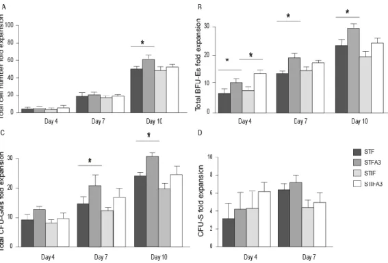
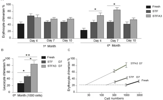
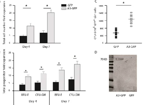
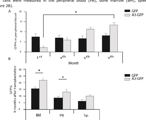
expansion of murine and human hematopoietic stem cells under serum free culture condition, with a specific focus on the effect of angiopoietin like protein 3. We compared the effect of two distinct combinations of growth factors on expansion and proliferation of murine and human progenitors, ST- and LT-HSCs in vitro and in vivo. These are the HSC established growth factor combination,SCF, TPO, Flt3L (STF), and a new combination of SCF, TPO, FGF-1, IGF-2 (STIF), which was introduced previously by Lodish et al. [78] at the time of starting the study presented in this thesis. We elucidated the effect of murine Angptl3 supplemented with STF or STIF on the number of progenitors, ST, and LT-HSCs of fresh, 4-,7-, and 10-day cultured murine BM-Lin-/LSK cells by colony forming units, spleen colony forming and long term repopulation assays, respectively. Furthermore, the effect of over-expression of Angptl3 in murine-HSCs in vitro and in vivo was studied. Initially, we examined the effect of over-expression of Angptl3 on murine BM-HSCs in vitro. Next, we studied the influence of ectopicly expressed Angptl3 on ST- and LT-HSCs.
In human studies, the consequence of including Angptl3 into the most common growth factors mixtures to ex vivo expansion of human UCB-HSCs in serum-free culture condition was investigated. We used UCB-CD34+ as a starting population and compared the effect of STF and STIF on human HSCs. The effect of Angptl3 supplemented with STF or STIF combinations of growth factors was examined on progenitors and ST-HSCs by the colony forming unit assay and by transplantation of the serially diluted cells into NOD-SCID mice.
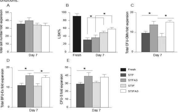
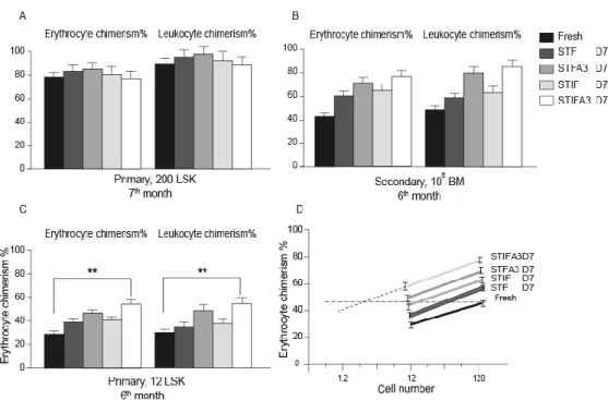
The second aim of this thesis was to ex vivo expand stem cells using well-defined culture conditions, including an up-to-date growth factor cocktail, focusing on hypoxia. We determined the effect of hypoxia (5% O2) on the expansion and proliferation of human UCB-HSCs or murine UCB-HSCs during 7 days in vitro culturing in the presence of the STIFA3 cocktail (SCF) [161, 162], thrombopoietin (TPO) [27, 63, 65], insulin like growth factor 2 (IGF-2) [151, 155], fibroblast growth factor 1 (FGF-1) [85, 163], and angiopoietin like protein 3 (A3) [78, 95]). We evaluated the effects of hypoxia on human cord blood HSCs, by performing in vitro colony assays, and bone marrow reconstitution assays to determine whether the oxygen tension alters the functioning of human cord blood HSCs in recipient immunodeficient mice. We also investigated the effects of hypoxic culture conditions on murine bone marrow HSCs.
In vitro colony assays, colony forming unit spleen (CFU-S) assays, and transplantation of sublethally irradiated mice with graded numbers of fresh or cultured cells under hypoxic or normoxic conditions was used to compare the effect of the different oxygen levels on progenitor cells, ST- and LT-HSC.
Taken together, we wished to develop an optimal condition which makes it possible to obtain the best grafts that might be useful for therapeutic purposes in adults as well as in children in the future.
Introduction
27
References
1. Moore, K.A. and I.R. Lemischka, Stem Cells and Their Niches. Science, 2006. 311(5769): p. 1880-1885.
2. Wilson, A. and A. Trumpp, Bone-marrow haematopoietic-stem-cell niches. Nat Rev Immunol, 2006. 6(2): p. 93-106.
3. de Bruijn, M.F.T.R., et al., Definitive hematopoietic stem cells first develop within the major arterial regions of the mouse embryo. EMBO J, 2000. 19(11): p. 2465-2474.
4. Boisset, J.-C., et al., In vivo imaging of haematopoietic cells emerging from the mouse aortic endothelium. Nature, 2010. 464(7285): p. 116-120.
5. Li, Z., et al., Mouse Embryonic Head as a Site for Hematopoietic Stem Cell Development. Cell Stem Cell, 2012. 11(5): p. 663-675.
6. Ema, H. and H. Nakauchi, Expansion of hematopoietic stem cells in the developing liver of a mouse embryo. Blood, 2000. 95(7): p. 2284-2288.
7. Medvinsky, A., S. Rybtsov, and S. Taoudi, Embryonic origin of the adult hematopoietic system: advances and questions. Development, 2011. 138(6): p. 1017-1031.
8. Larsson, J. and S. Karlsson, The role of Smad signaling in hematopoiesis. 0000. 24(37): p. 5676-5692.
9. Till and McCulloch, A direct measurement of the radiation sensitivity of normal mouse bone marrow cells. Radiat Res., 1961 Feb. 14: p. 213-22.
10. Wu., et al., Evidence for relationship between mouse hematopoietic stem cells and cells forming coloniesin culture. Proc Nat Aca Sciences, 1968. 59(4): p. 1209-15.
11. Domen, J. and I.L. Weissman, Self-renewal, differentiation or death: regulation and manipulation of hematopoietic stem cell fate. Molecular Medicine Today, 1999. 5(5): p. 201-208.
12. Yang, L., et al., Identification of Lin-Sca1+kit+CD34+Flt3- short-term hematopoietic stem cells capable of rapidly reconstituting and rescuing myeloablated transplant recipients. Blood, 2005.
105(7): p. 2717-2723.
13. Ploemacher, R., et al., Use of limiting-dilution type long-term marrow cultures in frequency analysis of marrow-repopulating and spleen colony-forming hematopoietic stem cells in the mouse. Blood, 1991. 78(10): p. 2527-2533.
14. Dexter TM, A.T., Lajtha LG, Conditions controlling the proliferation of haemopoietic stem cells in vitro. J Cell Physiol, 1997 june. 3: p. 335-44.
15. Greiner, D.L., R.A. Hesselton, and L.D. Shultz, SCID Mouse Models of Human Stem Cell Engraftment. Stem Cells, 1998. 16(3): p. 166-177.
16. Eaves, C., et al., Characterization of Human Hematopoietic Cells with Short-Lived in Vivo Repopulating Activity. Annals of the New York Academy of Sciences, 2001. 938(1): p. 63-71. 17. Hess, D.A., et al., Functional characterization of highly purified human hematopoietic
repopulating cells isolated according to aldehyde dehydrogenase activity. Blood, 2004. 104(6): p. 1648-1655.
18. Scadden, D.T., The stem-cell niche as an entity of action. Nature, 2006. 441(7097): p. 1075-1079.
19. Avecilla, S.T., Chemokine-mediated interaction of hematopoietic progenitors with the bone marrow vascular niche is required for thrombopoiesis. Nature Med., 2004. 10: p. 64-71.
20. Xie, T. and A.C. Spradling, A Niche Maintaining Germ Line Stem Cells in the Drosophila Ovary. 2000. p. 328-330.
21. JJ, T., Role of the hematopoietic inductive microenviroments (HIM) in xenogeneic bone marrow transplantation. Transplantation, 1973. 15(6).
22. R, S., The relationship between the spleen colony-forming cell and the haemopoietic stem cell.
Blood Cells, 1978. 4(1-2): p. 7-25.
23. Calvi, L.M., et al., Osteoblastic cells regulate the haematopoietic stem cell niche. Nature, 2003.
425(6960): p. 841-846.
24. Zhang, J., et al., Identification of the haematopoietic stem cell niche and control of the niche size.
Nature, 2003. 425(6960): p. 836-841.
25. Arai, F., et al., Tie2/Angiopoietin-1 Signaling Regulates Hematopoietic Stem Cell Quiescence in the Bone Marrow Niche. Cell, 2004. 118(2): p. 149-161.
26. Wilson, A., et al., c-Myc controls the balance between hematopoietic stem cell self-renewal and differentiation. Genes & Development, 2004. 18(22): p. 2747-2763.
27. Yoshihara, H., et al., Thrombopoietin/MPL Signaling Regulates Hematopoietic Stem Cell Quiescence and Interaction with the Osteoblastic Niche. Cell Stem Cell, 2007. 1(6): p. 685-697.
28. Fleming, H.E., et al., Wnt Signaling in the Niche Enforces Hematopoietic Stem Cell Quiescence and Is Necessary to Preserve Self-Renewal In Vivo. Cell stem cell, 2008. 2(3): p. 274-283. 29. Nilsson, S.K., et al., Osteopontin, a key component of the hematopoietic stem cell niche and
regulator of primitive hematopoietic progenitor cells. Blood, 2005. 106(4): p. 1232-1239. 30. Kiel, M.J. and S.J. Morrison, Uncertainty in the niches that maintain haematopoietic stem cells.
Nat Rev Immunol, 2008. 8(4): p. 290-301.
31. Papayannopoulou, T. and D.T. Scadden, Stem-cell ecology and stem cells in motion. Blood, 2008.
111(8): p. 3923-3930.
32. Sugiyama, T., et al., Maintenance of the Hematopoietic Stem Cell Pool by CXCL12-CXCR4 Chemokine Signaling in Bone Marrow Stromal Cell Niches. Immunity, 2006. 25(6): p. 977-988. 33. DiMascio, L., et al., Identification of Adiponectin as a Novel Hemopoietic Stem Cell Growth Factor.
The Journal of Immunology, 2007. 178(6): p. 3511-3520.
34. Naveiras, O., et al., Bone-marrow adipocytes as negative regulators of the haematopoietic microenvironment. Nature, 2009. 460(7252): p. 259-63.
35. Drüeke, T.B., Haematopoietic stem cells—role of calcium-sensing receptor in bone marrow homing. Nephrology Dialysis Transplantation, 2006. 21(8): p. 2072-2074.
36. Kiel, M., et al., SLAM family receptors distinguish hematopoietic stem and progenitor cells and reveal endothelial niches for stem cells. Cell, 2005. 121(7): p. 1109-21.
37. Sipkins, D.A., et al., In vivo imaging of specialized bone marrow endothelial microdomains for tumour engraftment. Nature, 2005. 435(7044): p. 969-973.
38. Winkler, I.G. and J.-P. Lévesque, Mechanisms of hematopoietic stem cell mobilization: When innate immunity assails the cells that make blood and bone. Experimental hematology, 2006.
34(8): p. 996-1009.
39. Katayama, Y., et al., Signals from the Sympathetic Nervous System Regulate Hematopoietic Stem Cell Egress from Bone Marrow. Cell, 2006. 124(2): p. 407-421.
40. Mendez-Ferrer, S., et al., Haematopoietic stem cell release is regulated by circadian oscillations.
Nature, 2008. 452(7186): p. 442-447. 41. Purton, L.E., The hematopoietic stem cell niche.
42. Spiegel, A., et al., Catecholaminergic neurotransmitters regulate migration and repopulation of immature human CD34+ cells through Wnt signaling. Nat Immunol, 2007. 8(10): p. 1123-1131. 43. Coulombel, L., et al., Lineage- and stage-specific adhesion of human hematopoietic progenitor
cells to extracellular matrices from marrow fibroblasts. Blood, 1988. 71(2): p. 329-334.
44. Nilsson, S.K., et al., Immunofluorescence Characterization of Key Extracellular Matrix Proteins in Murine Bone Marrow In Situ. Journal of Histochemistry & Cytochemistry, 1998. 46(3): p. 371-377.
45. Williams, D.A., et al., Fibronectin and VLA-4 in haematopoietic stem cell-microenvironment interactions. Nature, 1991. 352(6334): p. 438-441.
46. Simmons, P., et al., Vascular cell adhesion molecule-1 expressed by bone marrow stromal cells mediates the binding of hematopoietic progenitor cells. Blood, 1992. 80(2): p. 388-395.
47. Teixidó, J., et al., Role of beta 1 and beta 2 integrins in the adhesion of human CD34hi stem cells to bone marrow stroma. The Journal of Clinical Investigation, 1992. 90(2): p. 358-367.
48. van der Loo, J.C., et al., VLA-5 is expressed by mouse and human long-term repopulating hematopoietic cells and mediates adhesion to extracellular matrix protein fibronectin. The Journal of Clinical Investigation, 1998. 102(5): p. 1051-1061.
49. Scott, L.M., G.V. Priestley, and T. Papayannopoulou, Deletion of {alpha}4 Integrins from Adult Hematopoietic Cells Reveals Roles in Homeostasis, Regeneration, and Homing. Mol. Cell. Biol., 2003. 23(24): p. 9349-9360.
50. Siler, U., et al., Characterization and functional analysis of laminin isoforms in human bone marrow. Blood, 2000. 96(13): p. 4194-4203.
51. Bonig, H., et al., The p67 laminin receptor identifies human erythroid progenitor and precursor cells and is functionally important for their bone marrow lodgment. Blood, 2006. 108(4): p. 1230-1233.
52. Geissler, E.N., et al., Stem cell factor (<i>SCF</i>), a novel hematopoietic growth factor and ligand for c-kit tyrosine kinase receptor, maps on human chromosome 12 between 12q14.3 and 12qter. Somatic Cell and Molecular Genetics, 1991. 17(2): p. 207-214.
53. Broudy, V.C., Stem Cell Factor and Hematopoiesis. Blood, 1997. 90(4): p. 1345-1364.
54. Keller, J., M. Ortiz, and F. Ruscetti, Steel factor (c-kit ligand) promotes the survival of hematopoietic stem/progenitor cells in the absence of cell division. Blood, 1995. 86(5): p. 1757-1764.
Introduction
29
55. Kent, D., et al., Regulation of Hematopoietic Stem Cells by the Steel Factor/KIT Signaling Pathway. Clinical Cancer Research, 2008. 14(7): p. 1926-1930.
56. Hoffman, R., et al., The in vitro and in vivo effects of stem cell factor on human hematopoiesis.
STEM CELLS, 1993. 11(S2): p. 76-82.
57. Matsui, J., et al., Stem Cell Factor/c-kit Signaling Promotes the Survival, Migration, and Capillary Tube Formation of Human Umbilical Vein Endothelial Cells. Journal of Biological Chemistry, 2004.
279(18): p. 18600-18607.
58. Solar, G.P., et al., Role of c-mpl in Early Hematopoiesis. Blood, 1998. 92(1): p. 4-10.
59. Sitnicka, E., et al., The effect of thrombopoietin on the proliferation and differentiation of murine hematopoietic stem cells. Blood, 1996. 87(12): p. 4998-5005.
60. Qian, S., et al., Primary Role of the Liver in Thrombopoietin Production Shown by Tissue-Specific Knockout. Blood, 1998. 92(6): p. 2189-2191.
61. Murone, M., D.A. Carpenter, and F.J. de Sauvage, Hematopoietic Deficiencies in c-mpl and TPO Knockout Mice. Stem Cells, 1998. 16(1): p. 1-6.
62. de Graaf, C.A., et al., Regulation of hematopoietic stem cells by their mature progeny.
Proceedings of the National Academy of Sciences, 2010. 107(50): p. 21689-21694.
63. Yagi, M., et al., Sustained ex vivo expansion of hematopoietic stem cells mediated by thrombopoietin. USA. PNAS., 1999. 96(14): p. 8126-8131.
64. Geddis, A.E., H.M. Linden, and K. Kaushansky, Thrombopoietin: a pan-hematopoietic cytokine.
Cytokine & growth factor reviews, 2002. 13(1): p. 61-73.
65. Matsunaga, T., et al., Thrombopoietin Promotes the Survival of Murine Hematopoietic Long-Term Reconstituting Cells: Comparison With the Effects of FLT3/FLK-2 Ligand and Interleukin-6. Blood, 1998. 92(2): p. 452-461.
66. McClanahan T., C.J., Campbell D., Wagner J., Franz-Bacon K., Mattson J., Tsai S., Luh J., Guimaraes M.J., Mattei M.-G., Rosnet O., Birnbaum D. and Hannum C.H., Biochemical and genetic characterization of multiple splice variants of the Flt3 ligand. Blood, 1996. 88(9): p. 3371-3382.
67. Hannum, C., et al., Ligand for FLT3/FLK2 receptor tyrosine kinase regulates growth of haematopoietic stem cells and is encoded by variant RNAs. Nature, 1994. 368(6472): p. 643-648.
68. Banu, N., et al., MODULATION OF HAEMATOPOIETIC PROGENITOR DEVELOPMENT BY FLT-3 LIGAND. Cytokine, 1999. 11(9): p. 679-688.
69. Sudo, Y., C. Shimazaki, and E. Ashihara, Synergistic effect of FLT-3 ligand on the granulocyte colony-stimulating factor-induced mobilization of hematopoietic stem cells and progenitor cells into blood in mice. Blood, 1997. 89: p. 3186-3191.
70. Robinson, S.N., et al., Delivery of Flt3 ligand (Flt3L) using a poloxamer-based formulation increases biological activity in mice. Bone Marrow Transplant, 0000. 31(5): p. 361-369.
71. Christensen, J.L. and I.L. Weissman, Flk-2 is a marker in hematopoietic stem cell differentiation: A simple method to isolate long-term stem cells. Proceedings of the National Academy of Sciences, 2001. 98(25): p. 14541-14546.
72. Buza-Vidas, N., et al., FLT3 receptor and ligand are dispensable for maintenance and posttransplantation expansion of mouse hematopoietic stem cells. Blood, 2009. 113(15): p. 3453-3460.
73. LI, Y., et al., A potential role for insulin-like growth factor signaling in induction of pluripotent stem cell formation. Vol. 20. 2010, Kidlington, ROYAUME-UNI: Elsevier. 8.
74. Favelyukis, S., et al., Structure and autoregulation of the insulin-like growth factor 1 receptor kinase. Nat Struct Mol Biol, 2001. 8(12): p. 1058-1063.
75. Munshi, S., et al., Crystal Structure of the Apo, Unactivated Insulin-like Growth Factor-1 Receptor Kinase. Journal of Biological Chemistry, 2002. 277(41): p. 38797-38802.
76. Ziegler A., R.A.M., Forbes B., Wood T.L. and Levison S.W. and 2010, IGF2 promotes murine forebrain neural stem/progenitor cell self-renewal via the insulin receptor. Growth Hormone and IGF Research, 2010.
77. Zumkeller, W., The Insulin-like Growth Factor System in Hematopoietic Cells. Leukemia & Lymphoma, 2002. 43(3): p. 487-491.
78. Zhang, C.C., et al., Angiopoietin-like proteins stimulate ex vivo expansion of hematopoietic stem cells. Nat Med, 2006. 12(2): p. 240-245.
79. Chou, S. and H.F. Lodish, Fetal liver hepatic progenitors are supportive stromal cells for hematopoietic stem cells. Proc Nat Aca Sciences, 2010. 107(17): p. 7799-7804.
80. Zhang, C.C. and H.F. Lodish, Insulin-like growth factor 2 expressed in a novel fetal liver cell population is a growth factor for hematopoietic stem cells. 2004. p. 2513-2521.
81. Mergia, A., et al., The genes for basic and acidic fibroblast growth factors are on different human chromosomes. Biochemical and Biophysical Research Communications, 1986. 138(2): p. 644-651.
82. W H Burgess, T.M., D R Marshak, B A Fraser, and T Maciag, Structural evidence that endothelial cell growth factor beta is the precursor of both endothelial cell growth factor alpha and acidic fibroblast growth factor. PNAS, 1986. 83(19): p. 7216-7220.
83. Forough, R., et al., Differential transforming abilities of non-secreted and secreted forms of human fibroblast growth factor-1. Journal of Biological Chemistry, 1993. 268(4): p. 2960-2968. 84. Yang, M., et al., Platelet-Derived Growth Factor Enhances Granulopoiesis Via Bone Marrow
Stromal Cells. International Journal of Hematology, 2001. 73(3): p. 327-334.
85. de Haan, G., et al., In Vitro Generation of Long-Term Repopulating Hematopoietic Stem Cells by Fibroblast Growth Factor-1. Developmental Cell, 2003. 4(2): p. 241-251.
86. Crcareva, A., et al., Hematopoietic stem cells expanded by fibroblast growth factor-1 are excellent targets for retrovirus-mediated gene delivery. Experimental Hematology, 2005. 33(12): p. 1459-1469.
87. Yeoh, J.S.G., et al., Fibroblast Growth Factor-1 and -2 Preserve Long-Term Repopulating Ability of Hematopoietic Stem Cells in Serum-Free Cultures. 2006. p. 1564-1572.
88. Yeoh, J.S.G., et al., Fibroblast Growth Factor-1 and -2 Preserve Long-Term Repopulating Ability of Hematopoietic Stem Cells in Serum-Free Cultures. Stem Cells, 2006. 24(6): p. 1564-1572. 89. LaVallee, T.M., et al., Activation of the MAP Kinase Pathway by FGF-1 Correlates with Cell
Proliferation Induction While Activation of the Src Pathway Correlates with Migration. The Journal of Cell Biology, 1998. 141(7): p. 1647-1658.
90. Kunath, T., FGF stimulation of the Erk1/2 signalling cascade triggers transition of pluripotent embryonic stem cells from self-renewal to lineage commitment. Development, 2007. 134: p. 2895-2902.
91. Arai, F., Tie2/angiopoietin-1 signalling regulates hematopoietic stem cell quiescence in the bone marrow niche. Cell, 2004. 118: p. 149-161.
92. Hato, T., M. Tabata, and Y. Oike, The Role of Angiopoietin-Like Proteins in Angiogenesis and Metabolism. Trends in Cardiovascular Medicine, 2008. 18(1): p. 6-14.
93. Ge, H., et al., Differential regulation and properties of angiopoietin-like proteins 3 and 4. 2005. p. 1484-1490.
94. Yunling Li, a.X.Z., Angiopoietin-like protein 3 modulates barrier properties of human glomerular endothelial cells through a possible signaling pathway involving phosphatidylinositol-3 kinase/protein kinase B and integrin aVb3. ABBS, 2008(40): p. 459-465.
95. Zheng, J., et al., Angiopoietin-like protein 3 supports the activity of hematopoietic stem cells in the bone marrow niche. Blood, 2011. 117(2): p. 470-479.
96. Zheng, J., et al., Inhibitory receptors bind ANGPTLs and support blood stem cells and leukaemia development. Nature, 2012. 485(7400): p. 656-660.
97. Zhang, J., Identification of the haematopoietic stem cell niche and control of the niche size.
Nature, 2003. 425: p. 836-841.
98. Chattopadhyay N., V.P.M.a.B.E.M., Calcium-sensing receptor: Roles in and beyond systemic calcium homeostasis. Biological Chemistry, 1997. 378(8): p. 759-768.
99. Adams, G.B., et al., Stem cell engraftment at the endosteal niche is specified by the calcium-sensing receptor. Nature, 2006. 439(7076): p. 599-603.
100. Ito, K., Calcium influx triggers the sequential proteolysis of extracellular and cytoplasmic domains of E-cadherin, leading to loss of [beta]-catenin from cell–cell contacts. Oncogene, 1999. 18: p. 7080-7090.
101. House, M.G., et al., Expression of an Extracellular Calcium-Sensing Receptor in Human and Mouse Bone Marrow Cells. Journal of Bone and Mineral Research, 1997. 12(12): p. 1959-1970. 102. Chow, D.C., et al., Modeling pO2 Distributions in the Bone Marrow Hematopoietic Compartment.
II. Modified Kroghian Models. Biophysical journal, 2001. 81(2): p. 685-696.
103. Kubota, Y., K. Takubo, and T. Suda, Bone marrow long label-retaining cells reside in the sinusoidal hypoxic niche. Biochemical and Biophysical Research Communications, 2008. 366(2): p. 335-339.
104. Parmar, K., et al., Distribution of hematopoietic stem cells in the bone marrow according to regional hypoxia. Proceedings of the National Academy of Sciences, 2007. 104(13): p. 5431-5436.
105. Maxwell, P.H., et al., The tumour suppressor protein VHL targets hypoxia-inducible factors for oxygen-dependent proteolysis. Nature, 1999. 399(6733): p. 271-275.