0 1989 by The American Society for Biochemistry and Molecular Biology, Inc. Printed in U. S . A.
Complete Amino Acid Sequence
and Structure Characterization
of the
Taste-modifying Protein,
Miraculin*
(Received for publication, July 21,1988)
Sarroch TheerasilpS, Hiromu Hitotsuya, Shigeo NakajoQ, Kazuyasu NakayaQ, Yasuharu NakamuraQ,
and Yoshie Kuriharall
From the Department of Chemistry, Faculty of Education, Yokohama National University, Yokohamu 240, Japan and the §Laboratory of Biological Chemistry, School of Pharmaceutical Sciences, Showa University, Tokyo 152 Japan
The taste-modifying protein, miraculin,
has
the
un-
usual property of modifying sour taste
into sweet taste.
The complete amino acid sequence of miraculin puri-
fied from miracle fruits by a newly developed method
(Theerasilp,
S., and Kurihara,
Y. (1988)
J.
Biol. Chern.
263, 11636-11639) was determined by an automatic
Edman degradation method. Miraculin was a single
polypeptide with 191 amino acid residues. The calcu-
lated molecular weight based on the amino acid
se-
quence and the carbohydrate content
(13.9%) was
24,600. Asn-42 and Asn-186 were linked N-glycosid-
ically to carbohydrate chains. High homology
was
found between the amino acid sequences of miraculin
and soybean trypsin inhibitor.
Richadella dulcifica is
a native shrub
of
tropical West Africa.
It
yields red berries which have an unusual property in mod-
ifying sour taste into sweet taste. For example, lemons elicit
sweet taste after chewing pulps of the berries. Because of this
unusual property, the berry has been called “miracle fruit.”
Kurihara and Beidler
(1)first isolated the active principle
of
miracle fruit and showed that it is a basic glycoprotein.
Brouwer
et
al.
(2),
Giroux and Henkin (3), and Kurihara and
Terasaki
(4)also isolated the active principle, and Brouwer
et
al.
(2)
named it “miraculin.” The miraculin samples isolated
in all these studies were not completely pure, and hence any
attempt to determine miraculin’s primary structure has not
been made. Recently Theerasilp and Kurihara
(5)
established
a new method to obtain miraculin in very pure form. They
showed that it is a basic glycoprotein having a molecular
weight of about 28,000 as estimated
by
SDS-PAGE,’
while
molecular weights reported in previous papers
(1-4)
ranged
from 40,000 to 48,000. Miraculin contained as much as 13.9%
of
carbohydrate
(5).
In the present study, we have determined the complete
amino acid sequence of miraculin.
It
is a single polypeptide
*
This work was supported by grants from the Ministry of Educa- tion of Japan, Mitajiri Chemical Industry Co., and Mishima Science Foundation. The costs of publication of this article were defrayed in part by the payment of page charges. This article must therefore be hereby marked “advertisement” in accordance with 18 U.S.C. Section1734 solely to indicate this fact.
$Present address: Dept. of Chemistry, Faculty of Science and Technology, Sri-Ayudhya United Colleges at Pranakornsri-Ayudhya, Ayudhya 13000, Thailand.
ll To whom all correspondence should be addressed Dept. of Chem- istry, Faculty of Education, Yokohama National University, Toki- wadai, Hodogaya-ku, Yokohama, 240 Japan.
The abbreviations used are: SDS-PAGE, sodium dodecyl sulfate- polyacrylamide gel electrophoresis; HPLC, high performance liquid chromatography; LEP, lysine endopeptidase; Ch, chymotrypsin.
with 191 amino acid residues. The calculated molecular weight
of miraculin based on the amino acid sequence and the car-
bohydrate content is 24,600.
EXPERIMENTAL PROCEDURES
Materials-Miracle fruits (R. dulcifica) were obtained from plants grown in the green house of Yokohama National University. Mira- culin was purified from pulps of the fruits free from seeds and skins as described in the previous paper (5).
The sources of proteases are as follows. Achrornobacter lyticus protease I (lysyl endopeptidase), Wako Pure Chemicals Industries, Ltd.; l-chloro-3-tosylamido-7-amino-2-heptanone-chymotrypsin and carboxypeptidase A, Sigma; Staphylococcus aurew V8 protease, Miles Laboratories, Inc.; ~-l-tosylamido-2-phenylethyl chloromethyl ke- tone-trypsin, Worthington.
All other chemicals used were of analytical grade.
Preparation of S-Carboxyamidomethylted Miraculin-Seven mil- ligrams of the purified miraculin were dissolved in 5 mi of 0.4 M Tris buffer, pH 8.2, containing 6 M guanidine hydrochloride, 2 mM EDTA, and 60 mM dithiothreitol. The solution was incubated at 37 “C under atmosphere of nitrogen gas for 24 h. Iodoacetamide, 0.2 g, was added to the solution, mixed, let stand at room temperature for 10 min, and then placed in an ice bath for 60 min. The obtained S-carboxyami- domethylated miraculin was desalted by using a Sephadex G-25
column (1.6 X 5 cm) equilibrated with 50 mM EDTA.
Enzymatic Cleavage-Lysyl endopeptidase digestion of S-carbox- yamidomethylated miraculin was performed in 50 mM ammonium bicarbonate buffer, pH 8.0, containing 2 M urea and 2 mM EDTA at
37 “C for 20 h. The protein concentration was 1 mg/ml, and the enzyme:substrate ratio was 1:lOO (w/w). The reaction was terminated by addition of HCI to give a final pH 2.0. There was no insoluble material formed after digestion. The solution was injected into a HPLC to isolate the peptides.
Chymotrypsin digestion of the modified miraculin was performed under the same conditions as those for lysyl endopeptidase digestion except that the digestion time was 90 min. The reaction was also terminated in the same manner. There was no insoluble material formed, and the solution was injected into a HPLC to isolate the peptides.
S-Carboxyamidomethylated miraculin was digested by S. aureus
V8 protease in 50 mM ammonium bicarbonate buffer, pH 7.8, con- taining 2 M urea, and 2 mM EDTA at 37 “C for 3 h. The protein concentration was 1 mg/ml, and the enzyme:substrate ratio was 1:30,
w/w. Precipitates formed after digestion. Solid urea was added to the reaction mixture until the solution became clear. The solution was injected in a HPLC to isolate the peptides.
Peptide Isolation-The peptides in hydrolysates of the enzymatic digestion were separated by HPLC (Tosoh PC 8000) with a TSK- ODs-IZOT column (0.46 X 25 cm) (Tosoh). The peptides were eluted from the column by a linear gradient of acetonitrile containing 0.05% trifluoroacetic acid a t a flow rate of 1 ml/min. The eluted peptides were monitored by measuring absorption at 210 nm, and each peak was collected manually.
Amino Acid Analysis-Amino acid compositions of the peptides were determined by a Waters Picotag system (6). The peptide was hydrolyzed by HCl vapor at 110 “C for 22 h. The obtained amino acids were converted to phenylthiocyanate derivatives, and their contents were analyzed by HPLC using a TSK-ODs-80TM column
6656
Amino Acid Sequence
of
Miraculin
(0.46 X 15 cm) (Tosoh). Phenylthiocyanate-amino acid derivatives were eluted from the column by a linear gradient from solution A (3% acetonitrile in 50 mM phosphate buffer of pH 7.0 containing 0.1 M sodium perchlorate) to a mixture of solution A and 40% acetonitrile (1:4, v/v) for 20 min. The elution was carried out at column temper- ature of 40 "C and at a flow rate of 1 ml/min. The eluted phenylthio- cyanate-amino acid derivatives were monitored by measuring absorp- tion at 254 nm.
Sequence Analysis of Protein and Peptides-Amino-terminal amino acid sequence analysis by automatic Edman degradation was per- formed with an Applied Biosystem Protein Sequencer (model 470A). Phenylthiohydantoin amino acid derivatives were analyzed by HPLC using a TSK-ODs-12OT column, 0.46 X 25 cm, as described by Tsunasawa et al. (7) or by an Applied Biosystem on-line HPLC system. I ""
A
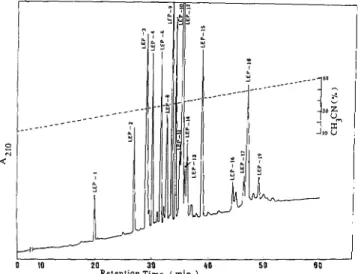
0 IO 20 30 40 5 0 6 0 R e t e n t i o n T i m ( m i " . )FIG. 1. HPLC separation of peptides obtained by lysine en- dopeptidase digestion of S-carboxyamidomethylated miracu- lin. Separation was carried on a reverse phase column, TSK-ODS- 120T (0.46 X 25 cm). The peptides were eluted by a linear gradient
of acetonitrile containing 0.05% trifluoroacetic acid from 10 to 60% at a flow rate of 1 ml/min for 1 h. The eluted peptides were monitored by absorption at 210 nm.
Carboxyl-terminal amino acid sequence was determined by using carboxypeptidase A as described by Ambler (8). Miraculin, 200 pg, was dissolved in 0.9 ml of 0.1 M N-ethylmorpholine acetate buffer, pH 8.0. Carboxypeptidase A, 1 pg, was added, and the reaction mixture was incubated at room temperature. Aliquots were taken at 15, 30, 60, and 120 min, and the proteins were precipitated by addition of trichloroacetic acid to give a final concentration of 10%. The precip- itates were removed by centrifugation, and the supernatant was subjected to analysis of the released amino acids by a Waters Picotag system as described above.
Detection of Carbohydrates-Carbohydrates in peptides were de- tected by orcinol-sulfuric acid reaction (9). In addition, the carbohy-
drate-containingpeptides (LEP-6 and LEP-12) were subjected to acid hydrolysis and the presence of carbohydrates in the hydrolysate was confirmed by HPLC on an ISA-O7/S2504 column (5).
RESULTS
Amino acid sequence from the amino terminus of miraculin
to the 46th amino acid residue has been determined with the
whole molecule of
S-carboxyamidomethylated
miraculin (Fig.
3).
Aspartic acid is the amino-terminal amino acid.
Several peptides were obtained by digestion of the modified
miraculin with lysyl endopeptidase (Fig.
1).The peptides were
well separated on
HPLC
except
LEP-3
which contained two
peptides,
LEP-3A
and
LEP-3B.
These peptides were sepa-
rated from each other upon rechromatography on the same
column but using a narrow gradient of acetonitrile and a
longer running time.
The sequences of minor peaks
(LEP-11, LEP-13,
and
LEP-
14)
from the amino-terminal to at least
10
residues toward
their carboxyl terminus were identical with that of
LEP-12.
Therefore these minor peaks seem to be either a part of
LEP-
12 peptide or
LEP-12
peptide with some extending amino
acid sequences from its carboxyl terminus. Similarly, peptides
from the minor peaks,
LEP-16, LEP-17,
and
LEP-19,
had the
same sequence as
LEP-18
at least 10 residues from their
amino terminus toward the carboxyl terminus. Amino acid
sequences and amino acid compositions of all major peaks
were determined as shown in Fig.
3
and Table
I,
respectively.
The modified miraculin was cleaved by lysyl endopeptidase
TABLE
IAmino acid compositions of miraculin and peptides obtained from digestion of S-carboxyamidomethyluted miraculin
with lysyl endopeptidase
Amino LEP-lb LEP-2 LEP-3A LEP-3B LEP-4 LEP-6 L E P S LEP-9 LEP-10 LEP-12 LEP-15 LEP-18 (170-179) (159-169) (1-14) (145-156) (134-144) (180-191) (170-187) (57-72) (105-120) (15-56) (121-133) (73-104) Asx(D/N) Glx(E/Q) CYS(C) Ser(S) GMG) His(H) Thr(T) Ala(A) Pro(P) Tyr(Y) Val(V) Met(M) Ile(1) Leu(L) Phe(F) Lys(K) T d W ) Total 0.9(1) 1.9(2) 1.0(1) 0.8(1) 0.8(1) 0.9(1) 1.8(2) 0.9(1) 2.7(3) 0.8(1) 1.0(1) 3.4(4) 1.2(1) 2.6(3) 0.9(1) 0.9(1) 1.1(1) 1.1(1) 0.8(1) 2.1(2) 1.0(1) 1.3(1) 0.9(1) 2.1(2) residuesjmolecule' 0.6(1) 1.7(2) 2.7(3) 0.9(1) 1.8(2) 0.7(1) 0.8(1) 2.1(2) 2.1(2) 0.8(1) 0.9(1) 0.9(1) 0.8(1) 1.9(2) 1.4(1) 3.0(3) 1.4(1) 1.6(2) 0.6(1) 1.9(2) 1.1(1) 0.5(1) 0.9(1) 2.9(3) 2.8(3) 0.9(1) 0.7(1) 1.2(1) 1.5(2) 0.9(1) 1.1(1) 1.2(1) 2.2(2) 2.3(2) 1.0(1) 1.5(1) 1.4(1) 2.2(2) 0.9(1) 0.9(1) 2.4(2) 0.9(1) 0.9(1) 2.1(2) 3.6(4) 2.7(3) 1.9(2) 1.2(1) 1.3(1) 1.4(1) 0.9(1) 1.0(1) 1.2(1) 1.1(1) 2.1(2) 1.4(1) 1.1(1) (10) (11) (14) (12) (11) (12) (18) (16) (16) a One-letter symbols are given in parentheses.
*
Name of peptides and the sequence position (in parentheses).e Determined by amino acid analysis or from the sequence (in parentheses). ND, not determined. 2.6(3) 1.0(1) 0.9(1) 0.9(1) 4.3(5) 1.2(1) 6.4(7) 1.3(1) 4.4(4) 4.2(4) 1.7(2) 7.4(6) 0.8(1) 2.6(3) 1.7(1) 1.1(1) (42) 0.9(1) 4.7(5) 0.9(1) 1.2(1) 1.4(1) 1.8(2) 4.5(5) 2.1(2) 0.9(1) 2.8(3) 1.3(1) 2.2(2) 1.2(1) 2.7(3) 3.7(4) 1.2(1) 1.1(1) 1.3(1) 2.0(2) 1.0(1) 2.1(2) 0.9(1) 1.2(1) ND(1)" ND(1) (13) (32) 20.1(21) 11.8(12) 6.5(7) 12.8(13) 15.6(17) 1.8(2) 14.6(15) 6.2(6) 13.9(14) 10.8(11) 7.3(7) 21.3(19) 0.9(1) 8.4(8) 9.5(10) 14.5(14) 12.4(12) ND(2) (191)
r"
IT
1h
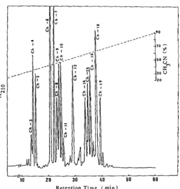
-70 20 30 40 50 60 Retention Time ( m i n )FIG.
2.HPLC
separation of peptides obtained from diges- tion of S-carboxyamidomethylated miraculin by chymotryp- sin.Separation
wascarried
on areverse
phasecolumn, TSK-ODS-
120T (0.46 X 25 cm).The peptides
wereeluted
bya linear gradient
ofacetonitrile containing
0.05%trifluoroacetic acid
from 20 to 90%at
a flow rate of 1ml/min
for 1 h.The
elutedpeptides
weremonitored
by absorptionat
210 nm.at the
carboxyl-terminal site of all lysine residues. Only
peptide bonds at two positions (Lys-Pro at position 179-180
and Lys-Thr at position 187-188) showed partial resistance
to the
digestion as indicated by amino acid sequences
of
LEP-
1, LEP-6, and LEP-8
(Fig.
3
and Table I). Amino acid
compositions
of
all LEP-peptides were in agreement with
their compositions as determined from the sequence. Mira-
culin contained 2 tryptophan residues, one in LEP-18 and the
other in LEP-15. Only LEP-6 had no lysine as the carboxyl-
terminal amino acid. There was no
phenylthiohydantoin
amino acid observed after phenylalanine. This result suggests
that phenylalanine is the carboxyl-terminal amino acid of
miraculin.
LEP-6 and LEP-12 showed orcinol-sulfuric acid reaction,
suggesting that these peptides contain carbohydrates.
In
ad-
dition, the peptides were subjected to acid hydrolysis, and the
presence
of
carbohydrates in the hydrolysates was confirmed
by HPLC. Amino acid residue at position 186 in LEP-6 could
not be identified by the automatic sequencing. It was, how-
ever, identified as aspartic acid by determination of amino
acid composition of LEP-6. Thus the amino acid a t position
186 is asparagine to which a carbohydrate chain is
linked.
LEP-12 was a long peptide composed
of
42 amino acid resi-
dues; and hence, to identify the amino acid residue to which
a carbohydrate chain is linked, LEP-12 isolated was digested
with chymotrypsin, and a carbohydrate-containing
peptide
was collected. The automatic sequencing of this peptide
in-
dicated that its sequence is
Thr-Val-Ser-Ala-Thr-Pro-X-Gly-
Thr-Phe. The results of determination
of
the amino acid
composition was consistent with this sequence if
X
(position
42) is aspartic
acid. Therefore it was concluded that the
amino
acid at position
42
is asparagine to which a carbohydrate
chain is linked.
Attempt to overlap LEP-peptides was performed by diges-
tion of the modified miraculin with chymotrypsin. The results
1 10 20 3 0 D S A P N P V L D I D G E K L R T G T N Y Y I V P V L R D H I I LEP-31 LEP-12 1 31 40 50 60 G G G L T V S A T T P N G T F V C P P R V V Q T R K E V D H Ch-8
-
LCh-15"- N -Ch-15- -Ch-14 61 70 8 0 D R P L A F F P E N P K E D V V R V S T D L N I N F S A F M 90 -LEP-9- -LEP-182
a
Ch-16- L C h - 9 - 91 P C R W T S S T V S R L D K Y D E S T G Q Y F V T I C G V K 100 110 1 2 0 121 G N P G P E T I S S W F K I E E F C G S G F Y K L V F C P T -LEP-15- L L E P - 4 - LLEP-3B- 130 110 150 L"Ch-6- I v-10 """""* 1 V-6 151 V C G S C K V K C G D V G I Y I D Q K G R R R L A L S D K P 160 110 180 1 L L E P - Z - L LEP-I- LEP-8-
Ch-7- I C h - 1 2 """""""""""""""_
F A F E F N K T V Y F 181 191-
LEP-6 I "-J _ _ - - I 1 L A LFIG.
3. The proof amino acid sequence of miraculin.The
sequence ofthe peptides is
represented by a one-letter code. LEPand
Ch
denote peptides derived from lysyl endopeptidase
andchymotryp-
sin digestions
of S-carboxyamidomethylatedmiraculin, respectively.
Their numbers also correspond
tothose
inFigs.
1and
2. V denotespeptides derived
fromVS-protease digestion, and their numbers cor-
respond tothose
inFig.
4. Solid linesindicate the
aminoacid
sequenceobtained
fromthe automatic sequencing, and
dashed lines indicatethe
sequencewhich was not determined
bythe sequencing. The
urrow(L)
indicates
the sequencefrom the carboxyl terminus determined
bycarboxypeptidase
Adigestion.
obtained are shown in Fig. 2. The peptides, Ch-4 to Ch-9, Ch-
12, Ch-14, Ch-16, and Ch-18 were subjected to determination
of amino acid sequences and amino acid compositions (Fig. 3
and Table
11).
Amino acid compositions of the Ch-peptides
were in agreement with their compositions as determined
from the sequences. Ch-14 linked LEP-12 to LEP-9, which
extended the sequence from 1st to 72nd positions. Ch-16
overlapped LEP-9 and LEP-18. Ch-18 overlapped three pep-
tides,
i.e.
LEP-18, LEP-10, and LEP-15. Ch-7 clearly linked
LEP-3B to LEP-2, indicating that there
is a Val-Lys peptide
(position 157-158) between the two peptides. This linkage
and amino acid sequences of Ch-6 and Ch-7 suggested that
LEP-15 links to LEP-4 which further links t o LEP-3B. Ch-
12 overlapped LEP-2, LEP-1,
and
LEP-6. Thus the Ch-
peptides overlapped LEP-peptides to give the complete amino
acid sequence from the amino-terminal amino acid (Asp-1) to
the carboxyl-terminal amino acid (Phe-191).
Connection between LEP-15 with LEP-4 with LEP-3B was
established by amino acid sequences of V-6 and V-10 peptides
which were obtained from digestion
ofthe modified miraculin
6658
Amino Acid Sequence
of
Miraculin
TABLE I1
A m i n o acids compositions of peptides obtained from digestion of S-carboxyamidomethylated miraculin with
chymotrypsin Amino acids" Ch-4b Ch-5 Ch-6 Ch-7 Ch-8 Ch-9 Ch-12 Ch-14 Ch-15 Ch-16 Ch-18 (95-100) (166-181) (133-143) (144-165) (1-22) (87-94) (166-191) (46-64) (23-45) (65-86) (101-132) Asx(D/N) Glx(E/Q) CYS(C) Ser(S) GMG) His(H) Thr(T) Ala(A) Pro(P) Arg(R) Tyr(Y) Val(V) Met(M) Ile(1) Leu(L) Phe(F) Lys(K) T d W ) Total 1.9(2) 1.3(1) 2.7(3) 1.1(1) 1.2(1) 2.1(2) 0.9(1) 0.9(1) 2.9(3) 1.2(1) 0.8(1) 1.7(2) 1.2(1) 1.9(2) (6) (16)
c
Q 0.7(1) 1.7(2) 0.9(1) 3234) 1.1(1) 0.7(1) 2.4(2) 3.6(3) 1.2(1) 1.3(1) 1.0(1) 1.0(1) 4.4(4) 1.3(1) 0.7(1) 0.7(1) 1.5(2) 1.4(1) 1.0(1) 2.5(3) (11) (22) residueslmolecule' 3.8(5) 2.2(3) 1.0(1) 2.1(2) 1.2(1) 0.9(1) 1.2(1) 0.8(1) 2.4(2) 1.1(1) 2.2(2) 1.3(1) 2.4(2) 1.2(1) 2.0(2) 1.4(1) 0.5(1) 1.6(2) 0.6(1) (22) 1.1(1) 1.2(1) 1.8(2) 1.4(1) 1.1(1) 1.3(1) 2.6(3) 1.4(1) 1.0(1) 0.9(1)o m )
1.6(2) 1.5(1) 3.6(4) 3.3(3) ND(l)d (8) (26)a One-letter symbols are given in parentheses.
*
Name of peptides and the sequence position (in parentheses).e Determined by amino acid analysis or from the sequence (in parentheses). ND, not determined.
^""
N
20 30 40 !
R e t e n t i o n Time ( m i n )
FIG.
4. HPLCseparation of peptides obtained from
S. au-reus Vi3
protease digestion of
S-carboxyamidomethylatedmiraculin.
Separation was carried on a TSK-ODS-120T column. The peptides were eluted by a linear gradient of acetonitrile contain- ing 0.05% trifluoroacetic acid from 20 to 70% at a flow rate of 1 ml/ min for 1 h. The eluted peptides were monitored by absorption at 210 nm.with S.
aureus
VS protease (Figs.
3
and
4).Determination of the amino acid sequence from the car-
boxyl-terminal was performed by digestion of native miraculin
with carboxypeptidase
A.
The results are shown in Fig. 5. The
rate of release of amino acids is decreased in the order:
phenylalanine
>
tyrosine
>
valine. There was no other amino
acid released during 2 h digestion. These results indicate that
the amino acid sequence from the carboxyl terminus of mir-
aculin is Phe-Tyr-Val which is in accordance with the se-
2.3(3) 2.2(2) 0.8(1) 0.8(1) 0.9(1) 2.5(3) 2.8(3) 4.3(3) 0.9(1) 1.2(1) (19) 1.7(2) 1.2(1) 3.3(4) 0.8(1) 3.2(4) 1.1(1) 2.1(2) 1.2(1) 3.5(3) 0.9(1) 2.1(2) 1.5(1) (23) 4.8(5) 1.6(2) 1.3(1) 1.2(1) 1.1(1) 2.2(2) 1.1(1) 2.8(3) 0.8(1) 1.3(1) 2.8(3) 1.4(1) (22) 3.5(3) 2.9(3) 2.2(2) 3.8(5) 2.5(3) 2.4(2) 1.3(1) 1.9(2) 2.1(2) 1.9(2) 1.5(1) 2.9(2) 2.2(2) NDU) (32)
TY
r
"""""""-=-
Val
-*
""_"""_
*-
" I 0.0 0 30 6 0 9 0 1 2 0 Time (min)FIG.
5.Amino acids
releasedfrom the carboxyl terminus
ofmiraculin after digestion with carboxypeptidase
A. Native miraculin was digested by carboxypeptidase A in 0.1 M N-ethylmor- pholine acetate buffer of pH 8.0 at room temperature with an en- zyme:substrate ratio of 1:200 (w/w). 8 nmol of miraculin was used for the digestion. The ordinate represents moles of amino acids released/ mol of miraculin.quence determined by automatic sequencing of Ch-12 and
LEP-6 peptides.
The complete sequence of 191 amino acid residues (Fig.
3)
is approximately in accordance with the amino acid compo-
sition of the whole protein (Table
I).
The sum of the molecular
weights of amino acid residues is
21,257.
In Fig. 6, the hydro-
pathic profile (10) is plotted, which suggests that there are a
number of hydrophobic domains in the miraculin molecule.
DISCUSSION
In the present study, we have determined the complete
sequence of
191amino acid residues of miraculin. It is a single
Residue
I
4 0 80 120 160 200No.
+ 4
r
I
E
n
FIG. 6.
Hydropathic profile calculated
bythe method
ofKyte and Doolittle
(10). The global average hydropathic index is indicated by the horizontal line. In the upper part, the location of positive and negative charges along the polypeptide chain of miraculin is represented.polypeptide chain with molecular weight of 21,257. Carbohy-
drate content was as much as 13.9% (5), and hence the total
calculated molecular weight is 24,600 which is 8 8 % of the
molecular weight (28,000) estimated by SDS-PAGE
( 5 ) .
Ap-
parent molecular weight of a glycoprotein on SDS-PAGE is
often larger than the actual one
(ll),and hence SDS-PAGE
of miraculin seems to have given a larger molecular weight
than the actual one. Resistance of Lys-Thr at position 187-
188to lysyl endopeptidase may be due to the presence of the
carbohydrate chain at Asn-186. Similar effect of the carbo-
hydrate chain on the proteolytic action was observed in car-
boxypeptidase
A
digestion.
One characteristic of the miraculin structure is that the
amino-terminal half of the molecule is enriched in proline
residues; it contains 10
of
14 proline residues. Another char-
acteristic of miraculin structure is that 5 half-cystines of a
total 7 half-cystines are located between positions 138 and
159. That is, 2 half-cystine residues are located at positions
47
and 92, and
5
residues are at positions 138, 148, 152, 155,
and 159. The fact that the region containing 5 half-cystine
residues is highly resistant to the protease digestion unless
dithlothreitol is applied to miraculin suggests that these half-
cystine residues form disulfide bonds.
The amino acid sequences of thaumatin and
monellin,
which are sweet-tasting proteins,
were already determined
(12, 13). We looked for homology of amino acid sequence
between miraculin and these sweet-tasting proteins
using
homology search program (IDEAS) developed by Kanehisa
(14), but there
was no particular homology between miraculin
and the sweet proteins. This is consistent with the fact that
the antibody to miraculin did not exhibit cross-reactivity with
thaumatin (data not
shown). However, high homology was
found between the sequences of miraculin and soybean trypsin
inhibitors
A
and
C
(Kunitz) was 36.3%
(the sequence of
miraculin from the amino terminus
to 60th residue) and
51.1%
(the sequence of miraculin from 143rd to the carboxyl termi-
nus).
It
is interesting to note that both proteins are produced
in plants and have similar molecular weights of about 20,000.
Acknowledgments-We are indebted to Professor Lloyd M. Beidler of The Florida State University for supply of seeds of miracle fruit and for constant encouragement. We also thank Professor Takeo Aso
of Yokohama National University who cultured miracle fruit. We thank Professor Noriko Takahashi of Nagoya City University for valuable suggestions on carbohydrate chains of miraculin.
REFERENCES
1. Kurihara, K., and Beidler, L. M. (1968) Science 1 6 1 , 1241-1242 2. Brouwer, J. N., Van der Wel,
H.,
Francke, A., and Henning, G.3. Giroux, E. L., and Henkin, R. I. (1974)
J.
Agric. Food Chem. 22,4. Kurihara, Y., and Terasaki, S. (1982) Biochim. Biophys. Acta
5. Theerasilp, S., and Kurihara, Y. (1988) J. Biol. Chem. 2 6 3 , 6. Bidlingmeyer, B. A., Cohen, S. A., and Tarvin, T. L. (1984)
J.
7. Tsunasawa, S., Kondo, J., and Sakiyama, F. (1985)J.
Biochern.8. Ambler, R. P. (1967) Methods Enzymol. 1 1 , 155-166
9. Francois, C., Marshall, R. D., and Neuberger, A. (1962) Biochem.
J . (1968) Nature 220,373-374 594-601 719,444-449 11536-11539 Chromatogr. 336,93-104 (Tokyo) 9 7 , 701-704
J.
83,335-34110. Kyte, J., and Doolittle, R. F. (1982) J. Mol. Biol. 1 5 7 , 105-132 11. Segrest, J. P., and Jackson, R. L. (1972) Methods Enzymol. 28,
12. Frank, G., and Zuber, H. (1976) Hoppe-Seyler’s
2.
Physiol. Chem.13. Iyengar, R. B., Smits, P., Van der Ouderaa, F., Van der Wel, H., van Brouwershaven, J., Ravestein,
P.,
Richters, G., and Van Wassenaar, P.D.
(1976) Eur. J. Biochem. 9 6 , 193-204 54-63357,585-592