Multimodality approach to mediastinal staging in
non-small cell lung cancer. Faults and benefits of
PET-CT: a randomised trial
Barbara M Fischer,
1,2Jann Mortensen,
1Hanne Hansen,
3Peter Vilmann,
4Søren S Larsen,
5Annika Loft,
1Anne K Bertelsen,
1Jesper Ravn,
6Paul Clementsen,
7Asbjørn Høegholm,
8Klaus R Larsen,
9Asger Dirksen,
7Birgit G Skov,
10Mark Krasnik,
11Liselotte Højgaard,
1Ulrik Lassen
12 ABSTRACTBackgroundCorrect mediastinal staging is a cornerstone in the treatment of patients with non-small cell lung cancer. A large range of methods is available for this purpose, making the process of adequate staging complex. The objective of this study was to describe faults and benefits of positron emission tomography (PET)-CT in multimodality mediastinal staging.
MethodsA randomised clinical trial was conducted including patients with a verified diagnosis of non-small cell lung cancer, who were considered operable. Patients were assigned to staging with PET-CT (PET-CT group) followed by invasive staging (mediastinoscopy and/or endoscopic ultrasound with fine needle aspiration (EUS-FNA)) or invasive staging without prior PET-CT (conventional work up (CWU) group). Mediastinal involvement (dichotomising N stage into N0e1 versus N2e3) was described according to CT, PET-CT, mediastinoscopy, EUS-FNA and consensus (based on all available information), and compared with the final N stage as verified by thoracotomy or a conclusive invasive diagnostic procedure.
ResultsA total of 189 patients were recruited, 98 in the PET-CT group and 91 in the CWU group. In an intention-to-treat analysis the overall accuracy of the consensus N stage was not significantly higher in the PET-CT group than in the CWU group (90% (95% confidence interval 82% to 95%) vs 85% (95% CI 77% to 91%)). Excluding the patients in whom PET-CT was not performed (n¼14) the difference was significant (95% (95% CI 88% to 98%) vs 85% (95% CI 77% to 91%), p¼0.034). This was mainly based on a higher sensitivity of the staging approach including PET-CT.
ConclusionAn approach to lung cancer staging with PET-CT improves discrimination between N0e1 and N2e3. In those without enlarged lymph nodes and a PET-negative mediastinum the patient may proceed directly to surgery. However, enlarged lymph nodes on CT needs confirmation independent of PET findings and a positive finding on PET-CT needs confirmation before a decision on surgery is made.
Clinical trial numberNCT00867412.
INTRODUCTION
Staging is a complex and critical event in the care of patients with lung cancer; precise description of the extent of the disease is important for the selection
of the proper treatment modality as well as in predicting prognosis.1
In patients without distant metastases, the most significant factor for deciding treatment is the status of mediastinal lymph nodes, as medi-astinal spread (N2e3 disease) excludes the patient from primary surgery. The European Society of Thoracic Surgery (ESTS) as well as the American College of Chest Physicians (ACCP) has published guidelines for proper preoperative mediastinal staging,2e4and there seems to be a cross-continent
consensus on the approach to preoperative staging. However, scratching the surface of the consensus reveals several unresolved problems and contro-versies:
1. Positron emission tomography (PET)-CT is
recommended if available.2 3 However, two
randomised clinical trials have demonstrated that staging by means of PET-CT significantly reduces the number of thoracotomies as well as the frequency of futile thoracotomies.5 6Is this only due to detection of unknown distant metastases or does PET-CT also increase the diagnostic accuracy of mediastinal staging? 2. It has been suggested that mediastinoscopy or
other invasive staging can be omitted in certain cases where mediastinum is PET negative.2 4 However, recent data suggest that by doing this, 16% of these patients have occult N2 disease.7 3. Does the combination of knowledge of lymph
node size on CT and [18F]fluorodeoxyglucose (FDG) accumulation on PET increase the diag-nostic accuracy of PET?8
4. Does mediastinoscopy increase diagnostic accu-racy in the case of positive imaging and negative endoscopic ultrasound (EUS)?2
The purpose of the current study is to suggest answers to and discuss these problems, based on results from a randomised clinical trial.5
METHODS
Setting and participants
Patients were recruited from three departments of pulmonology in the area of Copenhagen, Denmark. Patients were between 18 and 80 years of age with a newly diagnosed non-small cell lung cancer (NSCLC) and considered operable after
conven-tional staging procedures.5 9 The study was
approved by the Ethics Committee and Institu-tional Review Board of Copenhagen hospitals 1
Department of Clinical Physiology, Nuclear Medicine and PET, Rigshospitalet, Copenhagen University Hospital, Copenhagen, Denmark 2Department of Clinical Physiology and Nuclear Medicine, Hvidovre Hospital, Hvidovre, Denmark 3
Department of Radiology, Bispebjerg Hospital, Copenhagen, Denmark 4Department of Surgery, Herlev Hospital, Endoscopic Unit at Gentofte Hospital, Hellerup, Denmark
5Department of Surgery, Hvidovre Hospital, Hvidovre, Denmark
6Department of Thoracic Surgery, Rigshospitalet, Copenhagen University Hospital, Copenhagen, Denmark 7Department of Pulmonology, Gentofte Hospital, Hellerup, Denmark
8
Department of Pulmonology, Naestved Hospital, Næstved, Denmark 9 Department of Pulmonology, Bispebjerg Hospital, Copenhagen, Denmark 10Department of Pathology, Bispebjerg Hospital, Copenhagen, Denmark 11 Department of Development and Quality, Copenhagen University Hospital, Copenhagen, Denmark 12Department of Oncology, Rigshospitalet, Copenhagen University Hospital, Copenhagen, Denmark Correspondence to
Barbara M Fischer, Department of Clinical Physiology and Nuclear Medicine, Hvidovre Hospital, section 239, Kettegaard Alle´ 30, 2650 Hvidovre, Denmark; bjerregaard.fischer@gmail.com Received 4 November 2010 Accepted 11 November 2010
Lung cancer
(reference 11-118/01 and 01-252/00) and conducted according to the Declarations of Helsinki and Tokyo (http://ClinicalTrials.gov number, NCT00867412). Written informed consent was obtained from all patients.
Randomisation and intervention
After initial CT, eligible patients were 1:1 randomly assigned to (1) PET-CT followed by invasive diagnostic procedures (PET-CT group) or (2) invasive diagnostic procedures alone (conventional work-up (CWU) group). Randomisation was done centrally by a permuted block design, stratified by sex and recruiting centre. Standard staging procedures were governed by local routine based on current guidelines; however, mediastinoscopy was considered mandatory. TNM stage was assigned according to the revised Mountain classification.10
PET-CT imaging
All PET-CT scans were performed in the Department of Clinical Physiology, Nuclear Medicine and PET, Rigshospitalet, Copen-hagen University Hospital. After a fasting period of 6 h, 400 MBq of FDG was given intravenously and the patient rested for 1 h. The patient was scanned from the head to the upper thigh on an integrated PET-CT system (GE Discovery LS, General Electric Medical Systems, Milwaukee, Wisconsin, USA). A diagnostic CT protocol with intravenous contrast was applied.5 PET-CT images were evaluated side by side by an experienced radiologist and nuclear medicine specialist. Invasive staging
Mediastinoscopy was performed in all patients regardless of CT and PET-CTfindings, unless this procedure was contraindicated for other reasons. Specialists in cardiothoracic surgery or laryngologists performed the mediastinoscopy under general anaesthesia. Material from regions 2/4R, 2/4L and 7 were sampled if visible or palpable.10 At one trial site a randomised trial evaluating the value of EUS with fine needle aspiration
(EUS-FNA) was ongoing,11 thus approximately half of
the patients had an EUS-FNA procedure performed, either during the same general anaesthesia as used for mediastinoscopy (EUS-FNAfirst) or as an outpatient procedure with the patient under conscious sedation. The EUS examination was performed with an electronic linear transducer with an adjustable ultra-sonic frequency of 5 or 7.5 MHz and with a penetration depth of 7e8 cm (Olympus ultrasonic endoscope (GF-UC160P-OL5) or Pentax EG 3870). All lymph nodes with at least one criterion suggestive of malignancy were sampled (22 gauge needle, MEDI-Globe, type Sono-tip 2). The cytological specimens were stained by the MayeGrünwaldeGiemsa method and examined by an experienced pathologist. Only results from EUS-FNA proce-dures were used for staging and reported in the following. At the same site, prototype equipment for endobronchial ultrasound-guided transbronchial needle aspiration (EBUS-TBNA) was under development, and some patients also underwent this procedure.
At thoracotomy, samples were taken from all available mediastinal and hilar lymph node stations. N stage defined by the pathologist after thoracotomy served as the reference
(N-final). For patients in whom thoracotomy was not performed, N stage assigned by mediastinoscopy, EUS-FNA or EBUS-TBNA served as the reference. If imaging modalities suggested N2e3 disease and this could not be confirmed by mediastinoscopy or any other modality and the patient did not have a thoracotomy, an N-final stage was not assigned (Nx).
Outcomes and follow-up
Before the decision was made to operate, a consensus TNM stage was assigned by a pulmonologist and a thoracic surgeon on the basis of all available information (clinical data, initial CT, PET-CT, bronchoscopy, mediastinoscopy and, if available, EUS-FNA or EBUS-TBNA). Mediastinoscopy, EUS-FNA or EBUS-TBNA served as the gold standard when assessing mediastinal lymph node involvement (consensus N stage). Positivefindings on PET-CTwere evaluated by histology or other imaging modality at the discretion of the referring clinician. Follow-up data were retrieved from patientfiles and the local patient registry. Statistical analysis
The primary analysis on diagnostic accuracy of a staging approach with and without PET-CT was done as an intention-to-treat analysis, including all randomised patients with an
N-final stage. For the purpose of comparing the performance of individual modalities, only data on patients in whom the rele-vant examination was performed were included. Diagnostic accuracy, sensitivity, specificity, positive predictive value (PPV) and negative predictive value (NPV) with corresponding 95% CIs (Wilson score method without continuity correction) were calculated. The variables were compared by hypothesis testing for proportions in two independent groups, assuming normal distribution of the differences and without continuity correc-tion.12Clinical characteristics were compared with the use of an independent t test for continuous variables (age) and a
c
2test or Fisher exact test for mutually exclusive categorical variables (sex, histology, N-final and confirmation of N-final). Proportions of patients in the two groups on whom different staging procedures were performed were compared by hypothesis testing as describedabove. Agreement between a modality and N-final within
a patient group (correlated data) was measured by Cohens kappa (
k
) and McNemars test. Kappa values were interpreted as suggested by Altman: <0.20 poor agreement; 0.21e0.40 fair agreement; 0.41e0.60 moderate agreement; 0.61e0.80 good agreement; and 0.81e1.00 excellent agreement.13All reported p values are two-sided and a significance level of 0.05 was applied. Statistical analysis was performed with the use of SPSS software, version 16.RESULTS Patients
A total of 189 patients were recruited to the trial (98 in the
PET-CT group and 91 in the CWU group, figure 1). CT was
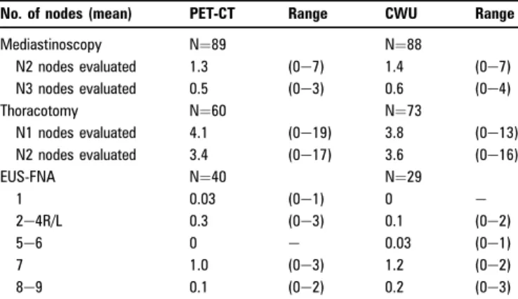
performed in all patients. A PET-CT scan was not performed in 14 patients in the PET-CT group due to an unacceptably long waiting time or technical problems with the PET-CTequipment. Patient characteristics were well balanced (table 1). Media-stinoscopy was performed in 89 patients (91%) in the PET-CT group and 88 (97%) in the CWU group. On average 1.3 and 1.4 biopsies from N2 lymph nodes and 0.5 and 0.6 biopsies from N3 lymph nodes were performed in each patient in the PET-CT and CWU group, respectively. EUS-FNA was performed in 40 (41%) and 29 (32%) patients, respectively, in each group. In the PET-CT group, an FNA from lymph node station 7 was performed in 70% of the procedures and an FNA from at least one of the lymph nodes stations 1, 2e4R/L, 5e6 or 8e9 was performed in 39% of the procedures. The values in the CWU group were 90% and 21%, respectively (table 2).
Diagnostic accuracy
Diagnostic accuracy of PET-CT, CT, mediastinoscopy and EUS-FNA in assigning correct N stage (N0e1 vs N2e3) is described in
Appendix 1. Consensus N stage is the N stage assigned based on all available information prior to decision on surgery. N-final served as the reference, excluding six and two patients, respec-tively, in each group due to lack of confirmation of thefinal N stage (Nx). By intention-to-treat analysis (table 3) the accuracy of the staging strategy with PET-CT appears only slightly superior to the CWU staging strategy (90% (95% CI 82% to 95%) vs 85% (95% CI 77% to 91%), p¼0.322), mainly based on
an improved sensitivity (75% (95% CI 59% to 86%) vs 59% (95% CI 41% to 74%), p¼0.162). Excluding the 14 patients in the PET-CT group on whom a PET-PET-CT scan was not performed, the diagnostic accuracy of the consensus N stage was significantly higher in the PET-CT group compared with the CWU group (difference of 10% (95% CI 0.2% to 20%), p¼0.034, table 3), again primarily based on the improved sensitivity as both groups had equally high specificity, based on the results of the invasive staging methods.
In 82 patients in the PET-CT group an exact description of the localisation (central, intermediate and peripheral) of the primary tumour on initial CT scan was available (table 4). In patients with a central tumour (n¼24) the agreement between N stage on PET-CT and N-final was fair (
k
¼0.39), whereas it was moderate in patients with a peripheral or intermediate tumour (kappa¼0.54). There was no systematic disagreement between N at PET-CT and N-final (eg, consequent overstaging or under-staging) in either subgroup (McNemar, p¼1.0).PET-negative lymph nodes
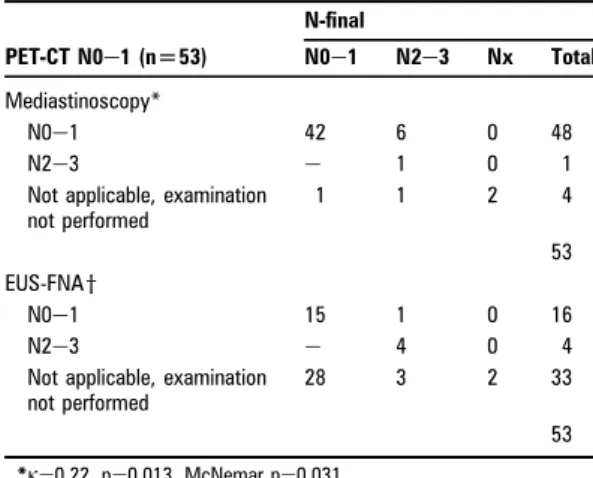
In the PET-CT group 53 (54%) patients were staged N0e1 on PET-CT (table 5). Mediastinoscopy was performed in 49 of these patients, one with a positive finding (N2 disease). Of the remaining 48 patients who were PET negative and negative on mediastinoscopy, six patients had mediastinal metastases (three confirmed by EUS-FNA, two by thoracotomy and one by EBUS-TBNA). Twenty patients with N0e1 on PET-CT had EUS-FNA
Figure 1 Study flow diagram. N = 189
Patients with NSCLC RANDOMIZATION PET/CT 98 Mediastinoscopy and/or EUS-FNA Mediastinoscopy and/or EUS-FNA N0-1 68 N2-3 30 56 False negative 9 True positive 27 False positive 0 Conventional work-up 91 Nx 3 Nx 3 N0-1 73 N2-3 18 True negative 59 False negative 12 True positive 17 False positive 1 Nx 2 Nx 0 RANDOMIZATION True negative N0-1 73
Nx: A final N-stage could not be assigned
Table 1 Patient characteristics
Patient PET-CT (n[98) CWU (n[91) p Value
Mean age in years (range) 62 (42e80) 64 (38e80) 0.222
Sex (male (%)) 53 (54) 49 (54) 0.974 Staging procedures, n (%) CT 98 (100) 91 (100) e PET-CT 84 (86) 0 (0) <0.001 Mediastinoscopy 89 (91) 88 (97) 0.097 EUS 47 (48) 35 (38) 0.187 EUS-FNA 40 (41) 29 (32) 0.201 EBUS 8 (8) 11 (12) 0.368 Histology at operation (n¼60/n¼73) Adenocarcinoma 29 29 0.330
Squamous cell carcinoma 20 22
Large cell carcinoma 4 12
Bronchioalveolar carcinoma 0 1 NSCLC, other 7 6 Benign 0 3 Final N stage N0e1 56 60 0.268 N2e3 36 29 Nx 6* 2y
Final N stage confirmed by (n)
Thoracotomy 60 72 0.014
Mediastinoscopy 9 10
EUS-FNA 19 7
EBUS-TBNA 4 0
Not applicable 6* 2y
*In 4 patients M1 was found on PET-CT and further confirmation of N stage was not sought. One patient was N0e1, but considered inoperable due to co-existing disease. Explorative thoracotomy was performed in one patient due to inoperable T4 disease; N stage not confirmed.
yExplorative thoracotomy was performed in two patients due to inoperable T4 disease; N stage not confirmed.
CWU, conventional work-up; EBUS, endobronchial ultrasound; EUS, endoscopic ultrasound; FNA, fine needle aspiration; NSCLC, non-small cell lung cancer; PET, positron emission tomography; TBNA, transbronchial needle aspiration.
Table 2 Details on invasive procedures
No. of nodes (mean) PET-CT Range CWU Range
Mediastinoscopy N¼89 N¼88 N2 nodes evaluated 1.3 (0e7) 1.4 (0e7) N3 nodes evaluated 0.5 (0e3) 0.6 (0e4) Thoracotomy N¼60 N¼73 N1 nodes evaluated 4.1 (0e19) 3.8 (0e13) N2 nodes evaluated 3.4 (0e17) 3.6 (0e16) EUS-FNA N¼40 N¼29 1 0.03 (0e1) 0 e 2e4R/L 0.3 (0e3) 0.1 (0e2) 5e6 0 e 0.03 (0e1) 7 1.0 (0e3) 1.2 (0e2) 8e9 0.1 (0e2) 0.2 (0e3)
CWU, conventional work-up; EUS, endoscopic ultrasound; FNA, fine needle aspiration; PET, positron emission tomography.
performed. The cytology showed N2e3 disease in four of these patients and was false negative in one patient. In patients with N0e1 on PET-CT, agreement between mediastinoscopy and
N-final was fair (
k
¼0.22) with a tendency to underdiagnose (McNemar, p¼0.031). Agreement between EUS-FNA andN-final was excellent (
k
¼0.857). Overall PET-CT gave a false-negative result in eight patients (false-false-negative rate, 8/53 15% (95% CI 8% to 27%)). Two of the latter patients proceeded to thoracotomy, and inspection of the surgical specimen (macro-scopic as well as micro(macro-scopic) revealed carcinoma in lymph node station 7, and in 7 and 2L, respectively.Lymph node size
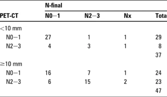
In table 6 the results of PET-CT are categorised according to the size of mediastinal lymph nodes on CT (<10 mm or$10 mm). The diagnostic accuracy of PET-CT was 86% (30/35, 95% CI 71% to 94%) in patients with normal-sized lymph nodes on CT, and 70% (31/45, 95% CI 56% to 82%) in patients with enlarged lymph nodes. The prevalence of N2e3 disease was 11% (95% CI 4% to 25%) and 47% (95% CI 33% to 61%) in the two groups. Twenty-nine patients had normal-sized lymph nodes on CT as well as negative mediastinum on PET-CT (table 6); among these patients one had N2e3 as thefinal stage (false-negative rate 4% (95% CI 0.01% to 0.18)).
Positive imaging and negative EUS-FNA
Sixty-six patients had both CT and EUS-FNA performed, and 36 patients had both PET-CTand EUS-FNA performed. The yield of EUS-FNA and mediastinoscopy in the case of positive CT or PET-CT is illustrated in table 7. The NPV of EUS-FNA in the entire study population as well as in the subgroups with positive CT or PET-CT exceeds the NPV of mediastinoscopy. All patients with a positive PET-CT or CT and negative EUS-FNA but N2e3
as N-final had a negative mediastinoscopy. Among the 24
patients who had N2e3 disease on CT, negative mediastino-scopy and N2e3 disease as N-final, N2e3 was confirmed by EUS-FNA in 12 patients and by EBUS-TBNA in one. Similarly,
EUS-FNA confirmed N2e3 disease in 7 of 10 patients with
N2e3 disease on PET-CT, despite negative mediastinoscopy. Twenty-six patients had N2e3 disease on both CT and PET-CT; seven of these patients had final N0e1 (false-positive rate of 27% (95% CI 14% to 46%)).
DISCUSSION
The results presented in this manuscript are based on an analysis of data on mediastinal staging from a prospective randomised clinical trial. The primary end point was futile thoracotomy and was previously reported.5One of the strengths of our data is the
Table 3 232 tables and diagnostic accuracy for consensus N stage
Table 4 Tumour localisation at CT and N stage at
PET-CT PET-CT N-final N0e1 N2e3 Nx Total Central tumour* N0e1 10 3 1 14 N2e3 3 5 2 10 Peripheraleintermediatey N0e1 33 5 1 39 N2e3 6 12 1 19
Not applicable, no information on exact tumour lacalisation
N0e1 e e e
N2e3 1 1 e 2
Total 84
*k¼0.39, p¼0.07, McNemar p¼1.0. yk¼0.54, p<0.001, McNemar p¼1.0. PET, positron emission tomography.
Table 5 Yield of invasive staging procedures in
PET-CT-negative patients PET-CT N0e1 (n[53) N-final N0e1 N2e3 Nx Total Mediastinoscopy* N0e1 42 6 0 48 N2e3 e 1 0 1
Not applicable, examination not performed 1 1 2 4 53 EUS-FNAy N0e1 15 1 0 16 N2e3 e 4 0 4
Not applicable, examination not performed
28 3 2 33
53
*k¼0.22, p¼0.013, McNemar p¼0.031. yk¼0.857, p<0.001, McNemar p¼1.0.
EUS, endoscopic ultrasound; FNA, fine needle aspiration; PET, positron emission tomography.
consecutive and prospective recruitment of patients as well as the systematic application of mediastinoscopy, and to a certain extent, EUS-FNA, regardless of the results of the imaging test. However, the invasive diagnostic procedures have evolved significantly since the initiation of this study, especially with regard to a broader use of endoscopic techniques. The major limitation of this material is the relatively low number of patients, which particularly in subgroup analysis results in broad CIs and hence makes clear-cut conclusions difficult to achieve. Based on the presented results we will discuss some of the remaining problems in mediastinal staging of patients with lung cancer as outlined in the Introduction section.
Diagnostic accuracy
The overall diagnostic accuracy of a multimodality staging approach was improved by adding PET-CT. This is mainly based on the improved sensitivity of the PET-CT approach. Based on
these findings as well as previously published studies demon-strating the ability of PET-CT to decrease the number of futile thoracotomies,5 6we suggest that PET-CT should be performed in all patients with NSCLC under consideration for surgery. However, in patients with centrally located primary tumours, PET-CT is only moderately better than chance in predicting the N stage (
k
¼0.39).PET-negative lymph nodes
Can mediastinoscopy be omitted in cases where there is no sign of mediastinal metastases on PET-CT?We found a frequency of false-negative mediastinum on PET-CT of 15% (8e27%), roughly equal to what has been reported previously.7In the case of a PET-CT-negative mediastinum, our data suggest EUS-FNA (false-negative rate of 6%) as the most rational choice.
Adding a mediastinoscopy to a negative PET-CT hardly improves accuracy, as the false-negative rate in this setting was 13%. It should however be noted that the sensitivity of media-stinoscopy in our material (30%) is significantly below what has been previously reported in the literature.3 14Two conditions in the present study can explain some of the divergence between the performance of mediastinoscopy in our study and what is generally published in the literature. First, the present study included mediastinoscopy, but did not focus on this examina-tion. Thus the mediastinoscopies was done in a setting reflecting everyday routine and were not always done by dedicated experts. Further, the largest participating centre had an on-going trial during the study period, evaluating the diagnostic value of EUS-FNA, often performing EUS-FNA and mediastinoscopy during the same general anaesthesia, which could have resulted in a less thorough mediastinoscopy, for example after a positive EUS-FNA.
The impact of incidental N2 disease on patient prognosis can be disputed. In this study, two of eight patients with false-negative mediastinal PET-CT had incidental N2 disease (found intraoperatively; surgery was completed with lymph node resection); both patients were alive at follow-up after 3 and 4.3 years.
Lymph node size
Two things demand attention when discussing the relationship of size of mediastinal lymph nodes to PET-CTfindings:first the problem of partial volume effect, which can cause false-negative PET-CT results in smaller foci (<10 mm),15 potentially leading to understaging. Secondly the higher prevalence of malignancy in larger lymph nodes, which can affect the estimated PPV and NPV. In the present study we found a higher diagnostic accuracy of PET-CT assessing small lymph nodes compared with enlarged lymph nodes (>10 mm) (86% vs 70%) despite a lower preva-lence of malignancy among the smaller nodules. A closer look reveals that sensitivity and specificity are similar in the two groups, but in concordance with the low prevalence NPV is significantly higher in the group with small nodules (96% vs 70%) and PPV is lower (43% vs 71%). Thus, in patients with normal-sized mediastinal lymph nodes a negative PET-CT is highly valid, whereas the risk of a false-negative diagnosis is substantial in enlarged lymph nodes without FDG uptake (30%). It also demonstrates that the performance of PET-CT or any other test is never independent of the disease prevalence. Positive imaging and negative EUS-FNA
Current recommendations suggest that in the case of positive
findings on PET-CT or CT followed by a negative EUS-FNA or EBUS-TBNA,final confirmation by mediastinoscopy should be
Table 6 Lymph node size and N stage at PET-CT
PET-CT N-final N0e1 N2e3 Nx Total <10 mm N0e1 27 1 1 29 N2e3 4 3 1 8 37 $10 mm N0e1 16 7 1 24 N2e3 6 15 2 23 47
Table 7 Results of invasive tests in CT or PET-CT
negative patients
N-final
N0e1 N2e3 Nx Total
(a) Yield of mediastinoscopy
CT N2e3 Mediastinoscopy* N0e1y 51 24 3 78 N2e3 e 15 0 15 93 PET-CT N2e3 Mediastinoscopy N0e1z 9 10 2 21 N2e3 e 6 0 6 27 (b) Yield of EUS-FNA CT N2e3 EUS-FNAx N0e1{ 20 5 0 25 N2e3 e 21 0 21 46 PET-CT N2e3 EUS-FNA N0e1** 6 0 0 6 N2e3 e 10 0 10 16
*Overall NPV of mediastinoscopy was 76% (107/141, 95% CI 68% to 82%).
yNPV of mediastinoscopy when CT is positive: 68% (57% to 77%). zNPV of mediastinoscopy when PET-CT is positive: 47% (27% to 68%). xOverall NPV of EUS-FNA is 85% (35/41) (72% to 93%).
{NPV of EUS-FNA when CT is positive: 80% (61% to 91%). **NPV of EUS-FNA when PET-CT is positive: 100% (61% to 100%). EUS, endoscopic ultrasound; FNA, fine needle aspiration; NPV, negative predictive value; PET, positron emission tomography.
sought.2 4 16In the present study we did notfind any additional value of mediastinoscopy in the case of positive imaging and negative EUS-FNA. However, as discussed above, the low sensitivity of mediastinoscopy in our study may not be repre-sentative for other settings. Further, the high accuracy of EUS-FNA reported here might be difficult to achieve once the modality becomes part of daily clinical routine and is no longer performed solely by dedicated experts in the setting of an accuracy study. It should also be noted that the good perfor-mance of EUS-FNA in this study is based mainly on evaluation of lymph node station 7 (table 2) and not station 2e4R or 10e11 which is better reached by EBUS.17
A meta-analysis has reported a relatively high false-negative rate of EUS-FNA of 19%, compared with mediastinoscopy of 11%.4 However, the prevalence of N2e3 disease was higher in the EUS-FNA studies (61%) compared with the mediastino-scopy studies (39%). Two studies have looked at the diagnostic value of combined PET and EUS-FNA,finding an overall accu-racy of>90% and a false-negative rate of EUS-FNA in the case of a positive PETof 0e7%.18This is confirmed by the present study where the false-negative rate of EUS-FNA after positive PET was zero, but 20% after a positive CT. Due to the relatively small sample size, this should be confirmed in a larger study. Conclusion
In accordance with current recommendations we strongly recommend preoperative staging by PET-CT of patients with lung cancer. In patients without enlarged lymph nodes and a PET-negative mediastinum our data suggest that the patient may proceed directly to surgery. However, enlarged lymph nodes on CT needs confirmation independently of PET findings and a positivefinding on PET-CT needs confirmation before a deci-sion on surgery is made.
FundingDanish Cancer Society and the Danish Center for Health Technology Assessment.
Competing interestsNone.
Ethics approvalThis study was conducted with the approval of the Ethics Committee and Institutional Review Board of Copenhagen Hospitals.
Provenance and peer reviewNot commissioned; externally peer reviewed.
REFERENCES
1. Tanoue L.Staging of non-small cell lung cancer.Semin Respir Crit Care Med
2008;29:248e60.
2. De Leyn P,Lardinois D, Van Schil P,et al. ESTS guidelines for preoperative lymph node staging for non-small cell lung cancer.Eur J Cardiothoracic Surg
2007;32:1e8.
3. Silvestri GA,Gould MK, Margolis ML,et al. Noninvasive staging of non-small cell lung cancer. ACCP evidenced-based clinical practice guidelines (2nd edition).Chest
2007;132(3 Suppl):178Se201S.
4. Detterbeck FC,Jantz MA, Wallace M,et al. Invasive mediastinal staging of lung cancer. ACCP evidence-based clinical practice guidelines (2nd edition).Chest
2007;132(3 Suppl):202se20s.
5. Fischer B,Lassen U, Mortensen J,et al. Preoperative staging of lung cancer with combined PET-CT.N Engl J Med2009;361:32e9.
6. Maziak DE,Darling GE, Inculet RI,et al. Positron emission tomography in staging early lung cancer. A randomized trial.Ann Intern Med2009;151:221e8. 7. Al-Sarraf N,Aziz R, Gately K,et al. Pattern and predictors of occult mediastinal
lymph node involvement in non-small cell lung cancer patients with negative mediastinal uptake on positron emission tomography.Eur J Cardiothoracic Surg
2008;33:104e9.
8. de Langen AJ,Raijmakers P, Riphagen I,et al. The size of mediastinal lymph nodes and its relation with metastatic involvement: a meta-analysis.Eur J Cardiothorac Surg2006;29:26e9.
9. Danish Lung Cancer Group.Lung CancerdDiagnosis and Therapy. A˚rhus, Denmark: Dansk Lungecancer Gruppe, 2001. (ISBN: 87-988368-0-3)
10. Mountain CF.Revisions in the international system for staging lung cancer.Chest
1997;111:1710e17.
11. Larsen SS,Vilmann P, Krasnik M,et al. Endoscopic ultrasound guided biopsy performed routinely in lung cancer staging spares futile thoracotomies: preliminary results from a randomised clinical trial.Lung Cancer2005;49:377e85. 12. Altman DG.Comparing groupsdcategorical data. In: Altman DG, ed.Practical
Statistics for Medical Research. London, UK: Chapman & Hall, 1991:229e76. 13. Altman DG.Some common problems in medical research. In: Altman DG, ed.
Practical Statistics for Medical Research. London, UK: Chapman & Hall, 1991:396e439.
14. Toloza EM,Harpole L, Detterbeck F,et al. Invasive staging of non-small cell lung cancer.Chest2003;123(1 Suppl):157Se66S.
15. Fischer BM,Mortensen J. The future in diagnosis and staging of lung cancer: positron emission tomography.Respiration2006;73:267e76.
16. Tournoy KG,De Ryck F, Vanwalleghem LR,et al. Endoscopic ultrasound reduces surgical mediastinal staging in lung cancer. A randomized trial.Am J Respir Crit Care Med2008;177:531e5.
17. Vilmann P,Annema J, Clementsen P. Endosonography in bronchopulmonary disease.Best Pract Res Clin Gastroenterol2009;23:711e28.
18. Annema JT,Hoekstra OS, Smit EF,et al. Towards a minimally invasive staging strategy in NSCLC: analysis of PET positive mediastinal lesions by EUS-FNA.Lung Cancer2004;44:53e60.
APPENDIX 1: DIAGNOSTIC ACCURACY IN ASSIGNING N0-1 VS N2-3 PET-CT CWU Sensitivity 95% CI Sensitivity 95% CI Consensus (n[92*) 0.75 0.59 to 0.86 Consensus (n[89y) 0.59 0.41 to 0.74 PET-CT (n¼79z) 0.69 0.50to0.84 CT (n¼92) 0.78 0.62to0.88 CT (n¼89) 0.72 0.54to0.85 Mediastinoscopy (n¼86) 0.28 0.16to0.45 Mediastinoscopy (n¼86) 0.38 0.23to0.56 EUS-FNA (n¼39) 0.94 0.73to0.99 EUS-FNA (n¼29) 0.67 0.42to0.84 Specificity Specificity Consensus 1 0.94 to 1 Consensus 0.98 0.91 to 1 PET-CT 0.81 0.69to0.89 CT 0.52 0.39to0.64 CT 0.53 0.41to0.65 Mediastinoscopy 1 0.93to1 Mediastinoscopy 0.98 0.91to1 EUS-FNA 1 0.85to1 EUS-FNA 1 0.77to1
Positive predictive value Positive predictive value
Consensus 1 0.88 to 1 Consensus 0.94 0.74 to 0.99
PET-CT 0.64 0.46to0.79
CT 0.51 0.38to0.64 CT 0.43 0.30to0.57
Mediastinoscopy 1 0.70to1 Mediastinoscopy 0.92 0.65to0.99
EUS-FNA 1 0.80to1 EUS-FNA 1 0.72to1
Negative predictive value Negative predictive value
Consensus 0.86 0.76 to 0.93 Consensus 0.83 0.73 to 0.90 PET-CT 0.84 0.72to0.92 CT 0.78 0.63to0.89 CT 0.80 0.65to0.90 Mediastinoscopy 0.70 0.59to0.79 Mediastinoscopy 0.76 0.65to0.84 EUS-FNA 0.96 0.79to0.99 EUS-FNA 0.72 0.49to0.88 Accuracy Accuracy Consensus 0.90 0.82 to 0.95 Consensus 0.85 0.77 to 0.91 PET-CT 0.77 0.67to0.85 CT 0.62 0.52to0.71 CT 0.60 0.49to0.69 Mediastinoscopy 0.73 0.63to0.81 Mediastinoscopy 0.78 0.68to0.85 EUS-FNA 0.97 0.87to1 EUS-FNA 0.82 0.64to0.92
*Only patients with a final N-stage. yOnly patients with a final N-stage.