Multi-Atlas based Segmentation of
Multi-Modal Brain Images
by
Keyvan Kasiri
A thesis
presented to the University of Waterloo in fulfillment of the
thesis requirement for the degree of Doctor of Philosophy
in
Systems Design Engineering
Waterloo, Ontario, Canada, 2016
c
I hereby declare that I am the sole author of this thesis. This is a true copy of the thesis, including any required final revisions, as accepted by my examiners.
Acknowledgements
I would like to express my sincere gratitude to my supervisors Professor David Clausi and Professor Paul Fieguth for their guidance, advice, and moral support throughout my Ph.D. studies under their supervision. They have contributed enormously to my growth as a researcher.
I wish to thank my doctoral committee members, Prof. Daniel Stashuk, Prof. Jeff Orchard, Prof. Ed Vrscay, and Prof. Anant Madabhushi for their valuable comments and suggestions.
I wish to acknowledge the University of Waterloo Faculty of Engineering, and the Nat-ural Sciences and Engineering Research Council (NSERC) of Canada for financial support of my research.
I would also like to thank my friends and my colleagues at University of Waterloo for their support during these years.
Finally, I would like to express my deepest gratitude and love to my family for their unconditional love and support. My special gratitude and love goes to my parents, my beloved wife Nazanin, and my dear brother Iman for all their continuous support and encouragement.
Dedication
Abstract
Brain image analysis is playing a fundamental role in clinical and population-based epi-demiological studies. Several brain disorder studies involve quantitative interpretation of brain scans and particularly require accurate measurement and delineation of tissue volumes in the scans. Automatic segmentation methods have been proposed to provide reliability and accuracy of the labelling as well as performing an automated procedure.
Taking advantage of prior information about the brain’s anatomy provided by an atlas as a reference model can help simplify the labelling process. The segmentation in the atlas-based approach will be problematic if the atlas and the target image are not accurately aligned, or if the atlas does not appropriately represent the anatomical structure/region. The accuracy of the segmentation can be improved by utilising a group of atlases. Em-ploying multiple atlases brings about considerable issues in segmenting a new subject’s brain image. Registering multiple atlases to the target scan and fusing labels from reg-istered atlases, for a population obtained from different modalities, are challenging tasks: image-intensity comparisons may no longer be valid, since image brightness can have highly differing meanings in different modalities.
The focus is on the problem of multi-modality and methods are designed and devel-oped to deal with this issue specifically in image registration and label fusion. To deal with multi-modal image registration, two independent approaches are followed. First, a similarity measure is proposed based upon comparing the self-similarity of each of the im-ages to be aligned. Second, two methods are proposed to reduce the multi-modal problem to a mono-modal one by constructing representations not relying on the image intensi-ties. Structural representations work on the basis of using un-decimated complex wavelet representation in one method, and modified approach using entropy in the other one. To handle the cross-modality label fusion, a method is proposed to weight atlases based on atlas-target similarity. The atlas-target similarity is measured by scale-based comparison taking advantage of structural features captured from un-decimated complex wavelet co-efficients. The proposed methods are assessed using the simulated and real brain data from computed tomography images and different modes of magnetic resonance images. Experimental results reflect the superiority of the proposed methods over the classical and state-of-the art methods.
Table of Contents
Abstract v
Table of Contents vi
List of Tables x
List of Figures xi
List of Abbreviations xiii
List of Symbols xv
1 Introduction 1
1.1 Multi-modal Multi-Atlas Segmentation Problem . . . 3
1.2 Challenges . . . 4
1.3 Objectives and Contribution . . . 5
1.4 Thesis Outline . . . 5
2 Background 7 2.1 Brain Tissue Segmentation . . . 7
2.2.1 Types of Atlases . . . 9 2.2.2 Segmentation Strategies . . . 10 2.3 Multi-Atlas-Based Segmentation . . . 13 2.3.1 Image Registration . . . 13 2.3.2 Label Fusion . . . 19 2.4 Problem of Multi-Modality . . . 21
2.4.1 Multi-Modal Image Registration . . . 22
2.4.2 Multi-Modal Label Fusion . . . 23
2.5 Summary . . . 24
3 Problem Formulation 26 3.1 Overview of the Problem . . . 26
3.2 Existing Limitations . . . 28
3.3 Objectives . . . 29
3.3.1 Defining a Similarity Measure for Multi-Modal Image Registration . 29 3.3.2 Reducing the Multi-Modal Image Registration . . . 30
3.3.3 Extending the Problem to Cross Modality Multi-Atlas Segmentation 30 4 Similarity Measure 32 4.1 Introduction . . . 32
4.2 Related Research . . . 33
4.2.1 Mutual Information . . . 33
4.2.2 Local Mutual Information . . . 34
4.2.3 Conditioned Mutual Information . . . 34
4.2.4 Self-Similarity Measures . . . 35
4.3.1 Motivation . . . 37
4.3.2 Patch Similarity . . . 37
4.3.3 Patch Selection . . . 39
4.3.4 Multi-Modal Similarity Measure . . . 40
4.4 Summary . . . 40
5 Structural Representation 43 5.1 Modality Independent Image Representation . . . 44
5.2 Complex Wavelet Representation . . . 45
5.2.1 Complex Amplitude and Phase . . . 46
5.2.2 Phase Congruency . . . 48
5.2.3 Representation Based on Complex Wavelets . . . 50
5.3 Entropy-based Representation . . . 53
5.3.1 Entropy Image . . . 56
5.3.2 Problem of Distinctiveness . . . 57
5.3.3 Modified Entropy Representation . . . 60
5.4 Summary . . . 62
6 Multi-Modal Image Registration 64 6.1 Introduction . . . 64
6.2 Experimental Data . . . 65
6.3 Self-similarity measure . . . 66
6.3.1 Rigid Registration . . . 68
6.3.2 Non-Rigid Registration . . . 70
6.4 Structural Representation for Image Registration . . . 70
6.4.2 Modified Entropy Image . . . 74
6.5 Discussion . . . 78
6.6 Summary . . . 80
7 Label Fusion 81 7.1 Introduction . . . 81
7.2 Weighted Label Voting . . . 83
7.3 Cross-Modality Label Fusion . . . 85
7.4 Results and Discussion . . . 88
7.4.1 Data . . . 88 7.4.2 Experimental setup . . . 89 7.4.3 Results . . . 90 7.4.4 Discussion . . . 92 7.5 Summary . . . 93 8 Conclusions 94 8.1 Thesis Contributions . . . 94 8.2 Future Research . . . 96
8.2.1 Performance Investigation Under Different Circumstances . . . 96
8.2.2 Unified Framework for Multi-Atlas-Based Segmentation . . . 96
8.2.3 Joint Multi-modal Registration . . . 97
List of Tables
6.1 Multi-modal rigid registration (translation and rotation) using the self-similarity measure for BrainWeb dataset . . . 69 6.2 Multi-modal rigid registration (translation and rotation) using the self-similarity
measure for RIRE dataset . . . 69 6.3 Multi-modal deformable registration using the self-similarity measure for
RIRE dataset . . . 70 6.4 Quantitative comparison of registration errors (in mm) obtained by MI and
the proposed complex wavelet representation method . . . 74 6.5 Multi-modal rigid registration (translation and rotation) using modified
en-tropy for BrainWeb dataset . . . 75 6.6 Multi-modal rigid registration (translation and rotation) using modified
en-tropy for RIRE dataset . . . 77 6.7 Multi-modal deformable registration using modified entropy for RIRE dataset 77 6.8 Comparison of computation time for different registration approaches. . . . 79 7.1 Segmentation results when the atlas database consists of T1 and T2 scans
and the target scan is in PD mode . . . 91 7.2 Segmentation results when the atlas database consists of T1 scans and the
List of Figures
1.1 Block diagram illustrating the atlas-based segmentation procedure used for
brain tissue segmentation . . . 2
1.2 Multi-atlas segmentation approach . . . 3
2.1 Deterministic atlas . . . 10
2.2 Probabilistic atlas . . . 11
2.3 Multi-atlas-based segmentation process . . . 14
2.4 Different parts of the images can have different intensity relations in multi-modal images . . . 22
3.1 Block-diagram of the multi-atlas-based segmentation framework. . . 27
4.1 Self-similarity in different modes of MR images . . . 42
5.1 2D Gabor complex wavelets in spatial domain with different orientations . 47 5.2 Fourier components of a step in a square wave . . . 49
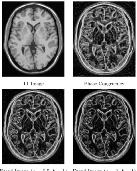
5.3 Complex wavelet representation for images with different structural contrast 51 5.4 Effect of applying gradient magnitude on PC for a slice of T1 brain MR image 54 5.5 Structural representation for different MR modes based on a combination of phase congruency and gradient information . . . 55
5.6 Overview of the modified entropy approach for constructing the structural representation . . . 56
5.7 Entropy as a representation for image structures . . . 58 5.8 Problem of distinctiveness for entropy-based image representation . . . 59 5.9 Applying a location dependent weighting to differentiate patches with
dif-ferent structures and the same entropy . . . 59 5.10 Applying function f on the patch histogram . . . 61 5.11 Structural representation for different MR modes using modified entropy . 62 6.1 Comparing the usage of MI and sorted patch intensity comparison in
mea-suring self-similarity . . . 67 6.2 Similarity plots of complex wavelet representations for BrainWeb dataset . 72 6.3 Cross-modal registration using the proposed method based on complex wavelet
representation . . . 73 6.4 Similarity plots of entropy-based representations for BrainWeb dataset . . 76 7.1 Block-diagram of the multi-atlas-based segmentation for multi-modal atlases 84 7.2 Similarity measure for multi-modal images based on structural features . . 86 7.3 Structural features from different MR modes . . . 87 7.4 Multi-modal versus single-mode segmentation . . . 91 7.5 Single-mode multi-atlas segmentation results . . . 92
List of Abbreviations
ANN artificial neural networksCC cross correlation
cMI conditional mutual information
CoCoMI contextual conditioned mutual information CR correlation ratio
CSF cerebrospinal fluid CT computed tomography DoF degree of freedom
DT-CWT dual tree-complex wavelet transform eSSD entropy sum of squared differences FFD free-form deformation
fMRI functional magnetic resonance imaging Gm gradient magnitude
GM gray matter
IR infra-red
LMI local mutual information LWV local weighted voting MI mutual information
MIND modality independent neighbourhood descriptor MR magnetic resonance
MRF Markov random field
MRI magnetic resonance imaging MV majority voting
NCC normalised cross correlation NLM non-local means
NMI normalised mutual information PC phase congruency
PD proton density
RIRE retrospective image registration evaluation SAD sum of absolute differences
SeSaMI self-similarity α-mutual information SSD sum of squared differences
SPECT single photon emission computed tomography TPS thin plate spline
UDWT undecimated wavelet transform WM white matter
List of Symbols
A atlas
B B-spline function
D Dice coefficient D pixel descriptor
Dsort sorted pixel descriptor
Dp patch distance ˜
Dp sorted patch distance
E Energy Function
f pairwise pixel self-similarity function
fs complex sinusoid function
fg 2D Gaussian function
fR representation function
F spatial transformation F label fusion function
G Gaussian kernel
Gx gradient along x direction
Gy gradient along y direction
Gm gradient magnitude
h weighted pixel information
H entropy of a random variable ˜
H modified entropy of a random variable
I image
If fixed image
Im moving image
IT target image
L label
m order of polynomial function
M number of the most similar pixels
MI mutual information of two random variables
N spatial neighbourhood
NA number of atlases
Nb number of neighbourhoods
N L denoised image using non-local means
NMI normalised mutual information of two random variables
Px patch centring x
˜
Px sorted patch centring x
p probability density function
P C phase congruency
Rc complex wavelet representation
Re entropy representation
RM e modified entropy representation
s Scale
S pixel self-similarity
SM pixel similarity
SMF similarity in label fusion
Tr threshold
w weight
W weight set
WP C phase congruency weight
Zn normalisation factor
µ mean of a random variable
σ standard deviation
α amplitude of complex wavelet coefficient
φ phase of complex wavelet coefficient
ω frequency
γ Gabor filter
γe even-symmetric Gabor filter
γo odd-symmetric Gabor filter
ζ phase order
ρ similarity measure
ψ polynomial function
χ self-similarity map construction function Ω image grid
Γ log-Gabor filter
Υ complex wavelet transform response Ξ scale-based label fusion function
Chapter 1
Introduction
Brain image analysis is playing a fundamental role in clinical and population-based epi-demiological studies. Several brain disorder studies involve quantitative interpretation of brain scans and particularly require accurate measurement and delineation of tissue vol-umes in the scans [1, 2, 3, 4, 5]. Manual labelling of brain images by human experts is inconsistent and time-consuming, specifically for large datasets [6]. Automatic segmenta-tion methods have been proposed to provide reliability and accuracy of the labelling as well as performing an automated procedure.
Automatic segmentation of brain images is a challenging task due to undesirable arte-facts such as noise, partial volume effect or non-uniformity in the intensity of the image. Therefore, using a priori information about the anatomy of the brain, which is provided by a reference image/volume, called an atlas, can help simplify this procedure [7]. In the literature, the term ’atlas’ is referred to both an intensity image, which is a brain template, or the segmented image, which is the labelled one [7, 8].
In traditional atlas-based segmentation, a target scan is labelled by referring to an atlas where the target is aligned to the atlas using deformable registration and atlas labels are then propagated to the target image space [9]. However, if either the mapping between images is not accurate or the atlas is not anatomically an appropriate representative for a specific structure/region, the segmentation will be problematic. Fig. 1.1 illustrates the process of atlas-based segmentation used for delineation of brain tissues. The atlas-based
Figure 1.1: Block diagram illustrating the atlas-based segmentation procedure used for brain tissue segmentation. Segmentation is based on registering the atlas to the target patient image and using the resulting spatial transformation F to propagate atlas labels to target space to attain a segmentation.
segmentation is shown as a registration-based segmentation approach where F stands for the spatial transformation between the atlas and the target scan.
The error caused by any single atlas registration will be effectively reduced by using a group of segmented images. There are two different categories of approaches in using multiple atlases for segmentation. In the first class of methods, information from several atlases are combined to create an average or a probabilistic atlas [8, 10, 11]. Then, the constructed atlas is warped to the target image to provide prior information. The second category of methods tries to combine labels from some number of registered atlases [11, 12, 13]; this work has led to an active literature on multi-atlas approaches [13, 14].
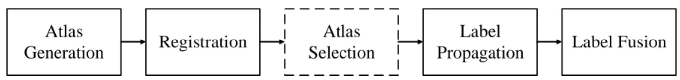
Label Fusion Label Propagation Registration Atlas Selection Atlas Generation
Figure 1.2: Multi-atlas segmentation approach: The overall block-diagram of multi-atlas segmentation procedure and its major components. Atlas selection is shown in a dashed box as an optional step in the multi-atlas segmentation framework.
1.1
Multi-modal Multi-Atlas Segmentation Problem
The multi-atlas approach takes advantage of more information from multiple atlases and is more robust to anatomical variations than single atlas-based approach [12, 15]. The multi-atlas segmentation approach can be subdivided into several steps. In general, key steps of any multi-atlas segmentation framework are atlas generation, registration, atlas selection, label propagation, and label fusion. These components are generally implemented sequentially in independent steps, however, there are many exceptions to this sequential organization. The overall block-diagram of multi-atlas segmentation procedure and its major components are presented in Fig. 1.2. Here, several already segmented images from different subjects, i.e., atlases, are registered to the patient input image resulting in a set of transformations. A subset of registered atlases may be selected to either reduce the complexity or exclude irrelevant atlases. Atlas labels are required to be propagated to the target space using the obtained transformation. Then, propagated labels are fused for each pixel to form a final segmentation result. Atlas selection is not a necessary step in every multi-atlas segmentation framework, and therefore it is shown in dashed line in this figure. The multi-atlas approaches are promising, however, these methods remain problematic in those cases where the atlases and the target scan are obtained from different sensors or from different acquisition modalities: image-intensity comparisons may no longer be valid, since image brightness can have highly differing meanings and circumstances in different modes [16, 17]. The goal of this thesis is to focus on the multi-modality issue and design and develop methods to deal with this issue specifically in major steps of the multi-atlas segmentation framework: image registration and label fusion.1.2
Challenges
As described in Section 1.1, the general form of multi-atlas segmentation framework consists of major steps of atlas generation, registration, label propagation, and label fusion. Since in most cases atlases, i.e., segmented scans, are already available, we skip atlas generation for the rest of thesis. To deal with cross-modality in the multi-atlas segmentation problem, the major components to cope with the issue of intensity variation are registration and label fusion. Thus, the major challenges to address in this problem are
• Multi-modal registration: To segment the target image, the atlases, which might exploit multiple imaging modalities, are required to be registered to the target space. The intensity variations across modalities has been an issue in the multi-modal reg-istration. Statistical metrics, such as those based on mutual information (MI), have been proposed in the literature as the solution to address this issue [18, 19, 20]. However, MI-based measures are intrinsically global and therefore may suffer from many false local optima. Moreover, the optimisation of these statistical measures for registration is computationally more complex compared to simple intensity dif-ference metrics [20]. This can be more of a concern when the number of atlases to be registered are increasing in the database [14].
• Cross-modality label fusion: A key challenge associated with the multi-atlas approach is label fusion. Most label fusion approaches are limited by the assumption that they depend on the consistency of voxel intensities across different scans. Many label fusion methods, such as majority voting (MV) [13] and weighted voting [21, 22, 23] do not consider image intensities after being warped to the target image. The multi-atlas approaches are promising, however, these methods remain problematic in those cases where the atlases and the target scan are obtained from different acquisition modalities: image-intensity comparisons may no longer be valid, since image brightness can have highly differing meanings and circumstances in different modes [16].
1.3
Objectives and Contribution
The objectives of this thesis target the multi-modal registration and cross-modality label fusion in a multi-atlas segmentation framework. The thesis makes the following contribu-tions:
• Defining a novel similarity measure based on measuring the image self-similarity for registration of multi-modal images, which is described in Chapter 4 and evaluated in Chapter 6,
• Reducing the multi-modal registration problem to a mono-modal one and hence, lowering the complexity of the registration problem by proposing structural repre-sentations not relying on the intensity mapping, which is described in Chapter 5 and evaluated in Chapter 6,
• Extending the existing label fusion approach to cross modality multi-atlas segmen-tation by making cross-modality image comparison based on extracted structural features, which is described and assessed in Chapter 7.
1.4
Thesis Outline
The structure of this thesis closely follows the sequence of mentioned contributions. In Chapter 2, we present an overview of the atlas-based segmentation and multi-atlas-based approach. A review of methods in image registration and label fusion as two major components of multi-atlas framework is also presented in this chapter.
Chapter 3 states and formulates the problem we are targeting in multi-atlas-based segmentation. Challenges and limitations related to the existing approaches followed by the objectives and contributions in this problem are presented.
Chapter 4 presents a new similarity measure for registering multi-modal images. The concept of self-similarity and measures for multi-modal image registration is presented. Following that, we present the proposed self-similarity measure based on taking the most significant image self-similarities into account.
In Chapter 5, two independent image representations are presented to map multi-modal images into common intensity space. First, complex wavelets is used to present the proposed image representation. Second, independent of the first representation, a modification to the formulation of entropy is applied to build an alternative structural representation.
Experiments to measure the accuracy of multi-modal image registration based on struc-tural representations are presented in Chapter 6. Strucstruc-tural representations in Chapter 5 based on complex wavelets and modified entropy are assessed in the same framework but independent of each other. In the following, employing the self-similarity presented in Chapter 4 is evaluated in the multi-modal image registration framework.
In Chapter 7, the problem of cross-modality label fusion is of focus. The weighted voting label fusion followed by the proposed method for combining labels from multi-modal images are presented. Experiments to evaluate the proposed method comparing with the conventional approach are given later in this chapter.
Chapter 2
Background
This chapter is devoted to reviewing the materials and methods required for the purpose of segmentation of MR images based on using multiple atlases. First, in Section 2.1, a general overview of brain tissue segmentation and different approaches are explained. Second, in Section 2.2, a generic form of atlas-based approach and its components are presented. Third, the multi-atlas-based approach, as a specific case of atlas-based segmentation, its components, and related challenges are presented in Section 2.3. Lastly, the problem of dealing with multiple modalities in this approach is given in Section 2.4.
2.1
Brain Tissue Segmentation
Segmentation is the process of partitioning an image into constituent regions whose el-ements (pixels in each region) have the same characteristics, such as color, intensity, or texture [6, 7, 24]. Since most studies on medical data highly rely on large datasets, a manual image segmentation approach by a human expert is a time-consuming procedure. Moreover, a manual approach highly depends on intra- and inter-observer variability which results in the degradation of credibility in the segmentation analysis. Therefore, attempts have been made towards an automatic segmentation of medical images to provide a repro-ducible, accurate, and robust segmentation framework.
liter-ature [6, 7, 24, 25, 26]. Pham et al. categorised segmentation methods into eight main categories of thresholding, region growing, pattern recognition methods, clustering, Markov random field (MRF) model, artificial neural network (ANN) methods, deformable models, atlas-based and other methods [6].
Among them, atlas-guided approaches aim to reduce human interaction and have a fully automatic and accurate segmentation approach. This category of methods, which is described in more detail in Section 2.2, incorporates additional higher level knowledge that can be prior information about the image under consideration or any predefined model [15, 25]. The atlas, which is generally a segmented image, is used as a reference model for the image to be segmented. The simplest atlas-based paradigm finds a one-to-one mapping between the atlas and the image to be segmented. Using the one-one-to-one mapping, all information available in the atlas is transferred to the target image to help label the image [8]. The typical atlas-based method along with different types of atlases and segmentation strategies are explained in the following.
2.2
Atlas-Based Segmentation
The automatic segmentation of brain images has been always a challenging problem [8, 27, 28, 29, 30]. Therefore, usinga priori information about the anatomy of the brain can help simplify this procedure. Prior information can be provided by a reference model, called an atlas, which either is a manually segmented version of brain scans where contains label information at specific locations.
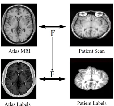
In atlas-based segmentation, the segmentation problem turns into a registration one. The atlas, A, is registered to the target patient image, IT, resulting in a transformation
F. Using the transformation F, labels of the atlas, denoted as L, are then propagated to the target image space. However, if either the atlas is not anatomically an appropriate representative for a specific structure/region or there exist labelling errors in the atlas seg-mentation, the error will be propagated during the registration procedure. In the following, two types of atlases as well as approaches under the atlas-based category are explained in more detail.
2.2.1
Types of Atlases
The construction and application of brain atlases are of great importance in neuroimaging and human brain research [8, 29, 31, 32]. This is due to the need for a standardized template which is the key concept in the field of human brain mapping. Creation of a realistic brain atlas, considering anatomical details and variability, is a time-consuming step. Therefore, many efforts have been recently made to provide this field of research with manually segmented data.
Topological Atlases: The first version of the atlas constructed for human brain research is the topological atlas which, in the literature, is also called the brain template, single-subject, or deterministic atlas. The topological atlas is referred to a volume image chosen from a population of brain scans to represent the whole population in terms of size, shape or intensity. The construction of a template to describe how different parts and structures are organized in the brain is the first step in creation of any probabilistic, region or disease-specific atlases.
The first attempt in creating atlas of the human brain led to the Talairach atlas [31] by which deep brain structures were identified in a space independent from individual dif-ferences in the size and overall shape of the brain. Fig. 2.1 shows an example of the deter-ministic atlas which is a brain template from the BrainWeb simulated brain database [33]. This image indicates the 143th axial slice of one of the twenty anatomical models of 20 normal brains. In each model, a set of “fuzzy” tissue membership volumes is presented. This set consists of different classes of background, cerebrospinal fluid (CSF), gray mat-ter (GM), white matmat-ter (WM), fat, muscle, muscle/skin, skull, blood vessels, connective (region around fat), dura matter and bone marrow.
Probabilistic Atlas: The major factor which is not considered in deterministic atlases is the diversity of human brain anatomy. In order to address the anatomical variability in the human brain, a population of brain scans is used to form the brain atlas. This type of atlas is often referred to as population-based, probabilistic, or statistical atlas [8]. In the construction of probabilistic atlases, the population can be subdivided into different groups based on different factors such as age, sex, or handedness. Such a population-based atlas is
Figure 2.1: An example of deterministic atlas: a slice of a 3D anatomical model of a normal brain from the BrainWeb [33] database. A set of different tissue classes are distinguishable by using different gray-scale values. The gray scale values from dark to bright indicate twelve classes of background, CSF, GM, WM, fat, muscle, muscle/skin, skull, vessels, around fat, dura matter, and bone marrow.
constructed using a set of segmented MRI data sets. For this purpose, all segmented images in the database are registered into a standard space and then the tissue probability of each voxel for a specific structure or region is computed. In Fig. 2.2, a sample probabilistic atlas for brain tissues is shown. This figure shows the 74th axial slice of the ICBM452 [34] atlas from the LONI database [35] which includes T1 mean, WM, GM and CSF probability maps.
2.2.2
Segmentation Strategies
The atlas-based segmentation approach tries to deform a brain atlas into a patient’s brain scan to create a labelled version of patient’s scan. The so-called atlas is a labelled scan which is previously segmented.
To use a priori information available in the atlas A, a transformation is required to map the atlas space into target imageIT space which forms a registration problem. Having found the transformation F from atlas space into target space, it is possible to map the reference (atlas) labelled image L to the patient’s image (target) space and obtain the labelled version of patient’s scanLT. The labelled volume is defined byLunique segments:
T1 average CSF GM WM
Figure 2.2: An example of probabilistic atlas: ICBM452 [34] probabilistic atlas showing the average topology of the brain and probabilistic map of CSF, GM, and WM.
wherex is the location in the label map Lcorresponding to the same location in atlas A.
Label Propagation
Having done the registration step, the easiest and fastest way to do the final labelling process is to propagate atlas labels to the input image space. In typical label propagation, the estimated transformation ˆF resulting from the registration step is used to deform the atlas labels, then the labels mapped to the coordinate system of the input image are simply assigned to input image voxels:
LT(x) =L Fˆ(x)
. (2.2)
In this way, the labelling error relies on the error that happened at the registration step and the whole segmentation procedure will basically be transformed into a registration problem. Since large anatomical differences will lead to a large registration error, this method is feasible for the cases in which the atlas is sufficiently similar to the input image. When dealing with intra-subject registration in medical applications, such as registra-tion of multi-modal images for radiotherapy or progression in a specific disease, global rigid registration and affine transformation will perform sufficiently well. Inter-subject registration which highly involves anatomical variations requires high degree of freedom
and therefore more complicated methods, non-rigid registration techniques, are employed. However, the risk of getting stuck in local extrema during the optimization procedure will be increased [8].
Probabilistic Atlas-based Segmentation
Typically, probabilistic atlases are used in a Bayesian framework to maximise the condi-tional probability of intensities in each class. The classical Bayesian approach for classifi-cation is defined by ˆ L(x) = argmax l∈{1,···,L} p L(x) = l|A(x) = argmax l∈{1,···,L} p I(x)|L(x) = l ·p(L(x) =l), (2.3)
wherep I(x)|L(x) = lstands for conditional probability of the voxel intensities given the class label and p(L(x) = l) represents the label prior. In this approach, class priors are provided by the probabilistic atlas and either parametric or non-parametric methods can be used to estimate the conditional probability.
Multi-Atlas Label Propagation
In a typical label propagation, when the atlas anatomy is far different from the input patient image, the accuracy of the segmentation will decrease. To overcome the registration error and therefore improve the segmentation accuracy, one possible solution is to employ multiple atlases. As was first shown by Heckemann et al. [13], as new atlases are taken into consideration, the accuracy of segmentation procedure will increase. Not only is the number of atlases used in the segmentation important to have an acceptable segmentation accuracy, but also the quality of atlases.
The first important issue associated with multi-atlas-based segmentation is the number of atlases and also how to choose them. Atlases should be selected in such a way that maximum anatomical variety in a population of atlases can be achieved. If a large database of atlases is available, the more efficient way will be selecting a subset of atlases which are very close to the input image to be segmented in terms of similarity. Further improvements are achieved by clustering atlases into different classes based on different structures and organs. Atlas ranking is another possibility to deal with using multiple atlases.
Another important issue in multi-atlas-based segmentation is the number of registra-tions required for segmentation. Typically, all atlases are warped into a common space to reduce the number of registrations and hence reduce the computations. However, the result will always be biased towards the initial selected space. For this reason, groupwise registration techniques are employed to suggest a better way for this problem. These meth-ods try to build an average reference template and register all of available atlases into this common space.
Having aligned all atlases, all deformed labels should be combined in some way. This step can be considered as a specific case of classifier fusion. Weighted voting is the typical way to apply on warped labels which are used both globally and locally.
2.3
Multi-Atlas-Based Segmentation
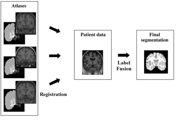
As described in Section 2.2.2, in multi-atlas-based segmentation approach, each atlas is available and potentially utilised for segmenting the target image. The overall framework of the approach for segmentation of medical images is illustrated in Fig. 2.3. The conventional approach involves registering each atlasAi, i= 1,· · · , NA, from a database of NA atlases, to the target (patient’s) image IT, propagating the atlas labels Li, i = 1,· · · , NA, to the target image coordinates, resulting in atlases and labels in the target image coordinates,
A0i and L0i, and then fusing the propagated labels. This section focuses on registration and label fusion as the main components of multi-atlas-based segmentation procedure.
2.3.1
Image Registration
Image registration, which is also named image matching or alignment, is the process of aligning two or more different images by finding one-to-one spatial correspondence between images [36]. Image alignment, as an image processing step, plays an important role in processing 2D/3D data in a variety of applications including robot vision, remote sensing, and medical imaging [37, 38, 39]. In particular, image registration is considered one of the fundamental problems in processing of medical images. Tracking temporal evolution and change detection, fusing image data, and 3D image construction are some examples medical applications [37, 39].
Figure 2.3: Multi-atlas-based segmentation procedure.
The process of registering images in the particular case of medical applications be-comes more challenging due to the variety of the imaging modalities and the fact that each modality can deliver the particular type of information [40]. For example, in medi-cal imaging, some modalities provide anatomimedi-cal information (i.e., computed tomography (CT) and MRI) and some other provide functional information (i.e., positron emission to-mography (PET), single photon emission computed toto-mography (SPECT), and functional MRI (fMRI)) about a specific tissue, structure or organ [41]. The anatomical informa-tion provides clinicians with spatial informainforma-tion such as shape, size and spatial relainforma-tion- relation-ship between structures and pathology, while the functional information leads clinicians
to studying the relationship between the underlying structure and physiology. Moreover, establishing a model for the relationship between images of human organs or structures is quite difficult, due to the highly complex transformations required.
To overcome the problems and challenges related to registering medical images, different approaches have been proposed in the literature [20, 36, 37, 40, 42]. In this subsection, an overview of the framework for medical image registration and its fundamental components are introduced.
In general, a registration framework involves finding a deformation transform F from a moving image Im to a fixed image If in order to maximise (minimise) an objective (cost) function ρ. The cost function combines a measurement of spatial alignment with a regulariser that quantifies the plausibility of the deformation:
ˆ F = argmax F ρ If, F(Im) (2.4) Thus, the three main component of registration framework are the deformation model, the objective function, and the optimizer.
Transformation Model
Transformation models are geometric models that establish a one-to-one mapping between the movingIm and fixedIf domains. The transformation model used during the registra-tion process relies on the accuracy to be satisfied, the deformaregistra-tion and the images to be registered. These models can be classified into three fundamental categories; rigid, affine, and non-rigid transformations.
Rigid transformation in three dimensions involves three degrees of freedom (DoFs) for rotation and three for translation. Transformation function can be expressed in matrix form as Frigid(x, y, z) = x0 y0 z0 1 = r11 r12 r13 tx r21 r22 r23 ty r31 r32 r33 tz 0 0 0 1 x y z 1 , (2.5)
where rij determine rotations about each coordinate axis and tx, ty, and tz stand for the translation along x, y, and z axes, respectively.
In addition to translation and rotation expressed in rigid transformation, scaling and shearing may be also necessary for aligning images. The matrix form of scaling transfor-mation in a 3D space and a shearing matrix in the (x, y) plane can be expressed in the following way: Fscale= sx 0 0 0 0 sy 0 0 0 0 sz 0 0 0 0 1 (2.6) Fshearxy = 1 0 hx 0 0 1 hy 0 0 0 1 0 0 0 0 1 , (2.7)
wheresx,sy andszstand for the scaling in each of the coordinate axes, andhx,hy represent the shearing in each of those axes. The overall linear mapping to cover the rigid, shearing, and scaling transformations is affine transformation that can be obtained by multiplying the rigid transformation, scaling and shearing matrices:
Faffine(x, y, z) =Fshear·Fscale·Frigid·
h
x y z 1
iT
. (2.8)
The resulting transformation provides twelve DoFs specifying translation, rotation, scaling and shearing.
In medical image registration, it is common to use rigid transformations to relate images when registering images of rigid parts of the body such as bones. Rigid models are global in nature and are not able to model local differences between images. Since rigid and affine models are of low complexity, they are often limited to registration of rigid structures and organs or only used as a pre-registration process prior to more complex registration procedures [36]. Since human body organs and structures are mostly deformable structures, non-rigid registration approaches are used in medical applications to build flexible elastic models [36, 40].
Basically, two types of deformations are considered in medical image registration: free-form and guided defree-formations. In free-free-form defree-formation models, any kind of defree-formation is allowed, whereas guided deformations are controlled by a physical model caused by the material properties of the organ or structure [43, 44, 45].
In free-from deformation (FFD) approaches, the registration is mainly performed by defining a grid of control points to determine the deformation between images. For the point located between the grid points, the deformation vector is obtained using any of interpolation methods. The use of B-spline tensor products as the deformation function was first proposed by Rueckertet al [45]. If the domain of the image volume is defined as Ω ={x= (x, y, z)|0≤x < X,0≤y < Y,0≤z < Z}, (2.9)
the transformation field by FFD with mesh of control points di,j,k with uniform control point spacingδ can be expressed as the 3D tensor product of the 1D cubic B-splines:
F(x) = 3 X l=0 3 X m=0 3 X n=0 Bl(u)Bm(v)Bn(w)di+l,j+m,k+n (2.10)
where Bl represents the l-th basis function of the B-spline, i = bδxc −1, j = byδc −1,
k=bz δc −1,u= x δ − b x δc, v = y δ− b y δc, andw= z δ− b z
δc. This deformation model requires a few degrees of freedom to describe local deformations and can efficiently provide smooth deformations.
Guided deformation models such as elastic models consider objects in the image as elastic solids [46, 47]. Therefore, the model is defined based on internal and external forces applied to the deformation fields. The internal static forces are applied to oppose the deformation, while the external forces caused by similarity metric helps the deformation to fit the configuration. Both forces are applied to deform the image until they reach an equilibrium. Guided deformations are non-parametric models that characterise the deformation at every voxel of the image volume.
Objective Function
The objective function is typically based on either metrics that measure the degree of similarity or the spatial distance between corresponding landmarks to quantify the accuracy of alignment in image registration. In the latter case, the landmarks are manually placed or detected automatically before performing the alignment. Similarity measures can be classified into intensity- and feature-based categories.
Measures based on image intensity in image registration [48] are usually based on intensity differences, intensity cross correlation, and information theory [48, 49]. The simplest intensity-based measure is based on sum-of-squared-differences (SSD) between the intensities in I1 and I2:
ρSSD =
X
(I1−I2)2. (2.11)
Metrics based on intensity difference are basically assuming the same characteristics for the images to be aligned and restricted to uni-modal image registration. A more general assumption than of having identical modalities is to have a linear relationship between im-age intensities. In this case, similarity can be measured using normalised cross correlation (NCC) as ρN CC = P (I1 −µ1)(I2−µ2) pP (I1−µ1)2 P (I2−µ2)2 (2.12) whereµ1 andµ2 are the average pixel intensities in the imagesI1 andI2, respectively. Nev-ertheless, the NCC is largely restricted to applications in registering mono-modal images. Information theoretical metrics such as mutual information [20], which are based on Shannon’s entropy [50], can be applied to both uni- and multi-modal registration frame-works and measure how well one image is able to explain the other image. Mutual infor-mation for two images I1 and I2 is defined based on the Shannon entropy as
MI(I1, I2) = H(I1) +H(I2)−H(I1, I2) (2.13) whereH(I1) andH(I2) represent the entropy of random variables I1 and I2, andH(I1, I2) stands for the joint entropy of these two random variables. MI can be equivalently expressed as MI(I1, I2) = X i X j p(i, j) log p(i, j) p(i)p(j), (2.14) where p(i, j) is the joint probability distribution function of I1 and I2, and p(i) and p(j) are the marginal probability distribution functions ofI1 and I2 respectively.
Feature-based metrics are usually based on landmarks, salient points, edges, contours, corners and/or surfaces [48, 49]. Distances between the corresponding features are con-sidered as a criterion to measure the alignment. It is required to extract features and estimation of correspondences prior to computing the distance. As an advantage of using feature-based registration is that it can be also used for multi-modal registration. However,
feature-based registration may need a prior segmentation to extract landmarks or features in the images. Furthermore, errors produced during the feature extraction procedure will be propagated into the registration and affect the accuracy of the procedure [36, 40, 42].
Numerical Optimization
The problem of image registration can be expressed as an optimization problem in which the goal is to minimise the cost or maximise the similarity between two images. The method tries to search for the optimum of an objective/cost function in the mapping model. Choosing a global or local optimization technique depends on the form of the objective/cost function, computational complexity, robustness, speed of the algorithm, and the accuracy required for the underlying application [36, 40, 49].
In the case of rigid and affine transformations, there is no constraint as the cost function and the optimisation problem aims to maximise the similarity between images. In non-rigid transformations, the role of the cost function plays the role of regularization or penalty term to constrain the transformation relating both images [36].
A common family of optimisation approaches is based on gradient descent that opti-mise the objective function by following the negative energy gradient, the direction that decreases the energy. Gradient descent has been utilised to solve various registration problems including the FFD registration algorithm. Conjugate gradient, Gauss-Newton method, stochastic gradient descent, and graph-based methods are the examples of ap-proaches that have been used widely in the application of image processing.
2.3.2
Label Fusion
As described in Section 2.2.2, the key challenge associated with the multi-atlas approach is “label fusion” — the strategy by which atlas labels are combined into a single segmen-tation [12]. To formulate the problem of label fusion, we consider a set ofNAatlases{An} with labels {Ln}, wheren = 1,· · · , NA, and IT as the target image to be segmented. The label alphabet contains L unique segments:
where x denotes the location in the label map Li corresponding to the i-th atlas. The atlases and the target image are assumed to be aligned using the transformations {Fn} corresponding to the {An} atlases. Given these transformations, each input, whether image or label field, can be transformed to the common space that is the target image space. Thus, {A0n}and {L0n}are the atlases and labels in the target image frame such that
A0n(x) =An Fn(x) , (2.16) L0n(x) =Ln Fn(x) . (2.17)
A final segmentation resultLT associated withIT is generated by combining all propagated labels {L0n} using a label fusion method.
Majority Voting
The simplest and most widely used label fusion method is majority voting (MV) [13], which asserts an equal contribution for each atlas. Considering each atlas as a classifier providing class labels, no prior information about each classifier’s accuracy is taken into account. In this approach, each voxel is assigned with the label that most classifiers select. Thus, the combination result can be expressed as
ˆ LT(x) = argmax l∈{1,···,L} NA X i=1 Lli(x), (2.18) where Ll
i(x) represents the vote for label l produced by the ith atlas as
Lli(x) = 1 if Li(x) = l, 0 otherwise. (2.19) Weighted Voting
As the image intensity is not taken into account during label fusion, a higher accuracy can be achieved by some form of weighting, based on the similarities between the atlases and the target image.This optimization problem can be solved by simply comparing numbers at each voxel: the fused label of each voxel is computed via a local weighted voting strategy.
The local image likelihood terms serve as weights and the label prior values serve as votes. Therefore, at each voxel, training images that are more similar to the test image at the voxel after registration are weighted more:
ˆ LT(x) = argmax l∈{1,···,L} NA X i=1 wi(x)Lli(x), (2.20)
wherewi(x) is a local weight assigned to the ith atlas and
NA X
i=1
wi(x) = 1. (2.21)
Fixing the weights across all atlases to a constant, wi(x) =C ignores the atlas similar-ities and leads to majority voting. Fixing the weights within a single atlas to a constant,
wi(x) = Ci globally expresses the similarity between the target and atlas, which models the atlas selection strategy [51, 52].
Global label fusion approaches perform generally better than single atlas-based seg-mentation. However, as weights are assigned globally, it is impossible for the atlases to have higher contribution in the areas where the registration performs successfully, even if the registration was inaccurate in the rest of the image.
2.4
Problem of Multi-Modality
In medical image analysis, multiple modalities of the same subject or organ provide com-plementary information that is very important for medical diagnosis and computer-aided surgery [53]. In a multi-atlas-based segmentation problem, of particular interest is dealing with atlases acquired from different sensors, imaging protocols, or modalities [17]. An-other scenario could be the cross-modality segmentation of a patient’s image with the single-mode atlas database. In either cases, both the registration and label fusion steps would be challenging since image-intensity comparisons may no longer be valid across dif-ferent modalities [16]. This section reviews the multi-modality challenge and approaches dealing with cases in multi-modal image registration and label fusion.
(a) T1 mode (b) T2 mode (c) labelled anatomy (d) joint histogram Figure 2.4: Different parts of the images can have different intensity relations in multi-modal images. Perfectly aligned slices in T1 (a) and T2 (b) from simulated BrainWeb [33] database are shown. The brain anatomy in different colors is described in (c). Image (d) is the joint histogram of (a) and (b). Images (c) and (d) show how the brain anatomy relates to the joint histogram by mapping pixel intensities from T1 to T2.
2.4.1
Multi-Modal Image Registration
A key component in every image registration tool is defining a way of measuring the similarity of images to be aligned. As described in Section 2.3.1, for images captured from the same modality, classical similarity measures, such as SSD and cross-correlation coefficient (CC), assume a linear relationship between intensities of the corresponding pixels across the whole image domain. This assumption will not be valid for images obtained from different modalities or imaging sensor types [53]. Since different physical phenomena are measured in different imaging systems, no functional relation between the image intensities can be defined to map the corresponding elements from one image to another. As shown in Fig. 2.4 illustrates how the intensities in two modes of MR brain images are related. Perfectly aligned slices of T1 and T2 modes are shown along with the segmented anatomical parts corresponding to the joint histogram of those images. The joint histogram shows the simultaneous occurrences of intensities between the two images. In Fig. 2.4(c) and (d), the intensity of different tissues are related differently in the two modes.
Traditionally, multi-modal image registration employs mutual information, which uses the statistical dependency of the intensity values between images for evaluating the reg-istration results [20]. Mutual information has been first introduced for rigid alignment of
multi-modal images [18] and later used for deformable registration [45].
In calculating MI, in Eq. 2.13, for measuring image similarity, changing the overlap between two images during the registration process affects the MI value, therefore, nor-malised mutual information (NMI) has been introduced to cope with this issue [54]. A di-rect approach to normalisation is presented to evaluate the ratio of the joint and marginal entropies
NMI(I1, I2) =
H(I1) +H(I2)
H(I1, I2)
. (2.22)
A major drawback of mutual information and its variants for image registration is that they do not take spatial information into account. For those cases in which the intensity relations are not spatially invariant or there is a complex intensity relationship, MI-based approaches may suffer from local maxima and an incorrect global maximum problem [55]. Further works have been proposed to overcome this problem by integrating spatial and contextual information in the MI formulation in expense of higher computational time and complexity [56, 57, 58, 59].
Structural information has been also used in the literature of multi-modality problem for improving the robustness of similarity measures to image intensity variations [60, 61, 62, 63, 64]. Thus, the multi-modal registration problem will be transformed to registering two image representations using a simple intensity-based similarity/dissimilarity measure. The registration problem formulated in Eq. 4.1 will be changed into
ˆ F = argmax F ρ Rf, F(Rm) , (2.23)
where Rf and Rm are the image representation of the fixed image If and moving Im, respectively. The challenge is still how to find a mapping function that transforms image intensities from different modalities into a new intensity space, so that all images can share similar features in the new space.
2.4.2
Multi-Modal Label Fusion
The multi-atlas approaches are promising compared to single atlas-based segmentation [14]; however, these methods remain problematic in those cases where the atlases and the target
scan are obtained from different sensors or from different acquisition modalities: measuring intensity-based proximity may no longer be valid, since image brightness can have highly differing meanings and circumstances in different modes [16].
Many label fusion methods have been introduced in the medical atlas literature [22]. As described in Section 2.3.2, the simplest and most widely used one is MV [13], which asserts an equal contribution for each atlas. As the image intensity is not taken into account during label fusion, a higher accuracy can be achieved by some form of weighting, based on the similarities between the atlases and the target image. Weighting strategies can be applied in both global and local forms [65, 66], where local weighted voting (LWV) outperforms global strategies when dealing with high contrast anatomical structures [21, 22, 23].
Most label fusion approaches are limited by the assumption that they depend on the consistency of voxel intensities across different scans. In these cases, approaches based on MI do help [67] by assigning weights to atlas labels based on the similarity between the target and the atlases. Thus, the weights in Eq. 7.3 will be defined by
wi(x) = MI(A0i, IT). (2.24) However the inherent non-locality in MI make it problematic for local weighted label fusion. This issue will be highlighted when atlases and target image are acquired with different modalities [16, 21].
2.5
Summary
This chapter provided a review of the background required for brain image segmentation in a multi-based framework. The brain image segmentation in the context of atlas-based segmentation as a registration-atlas-based method, the advantage of using prior knowledge available in atlases, and the issue regarding the atlas-target registration were discussed. The multi-based segmentation framework, which aims to cope with the basic atlas-target registration problem, was reviewed. As described in this chapter, the key steps in performing the multi-atlas segmentation are the image registration and label fusion. Due to the growth of atlas databases and availability of scans from different modalities, multi-atlas approaches are required to deal deal with multi-modality issue. Multi-modal
registration of brain scans and cross-modal combination of labels from registered atlases are the remaining challenges in multi-atlas problem.
Chapter 3
Problem Formulation
This chapter formulates the problem of multi-atlas-based segmentation and states the motivation, limitations, and the objectives to contribute to the conventional framework. An overview of the problem, the general framework, and its components are given in Section 3.1. Section 3.2 overviews the existing limitations and challenges of the multi-atlas segmentation framework. To address these limitations, the objectives, which are pursued in the following chapters, are introduced in Section 3.3.
3.1
Overview of the Problem
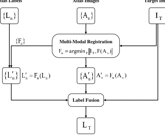
As described in Section 2.3, a general multi-atlas segmentation framework consists of two major components, image registration and label fusion. Fig. 3.1 shows the block diagram of the general multi-atlas-based segmentation framework, in which {An}, {Ln}, and IT respectively represent the set ofNA atlases, the labels corresponding to these atlases, and the target image. In the first stage, the atlases are all warped to the target image resulting in the inferred transformations {Fn}. Given these transformations, each input, whether image or label field, can be transformed to the common reference of the target space. Thus {A0n} and {L0n} are the atlases and labels in the common reference frame. All warped labels are then combined together to form the final segmentationLT based on information obtained from warped atlases and the target image.
Multi-Modal Registration
Label Fusion
Atlas Images Target Image
Atlas Labels
Target Label
Figure 3.1: Block-diagram of the multi-atlas-based segmentation framework.
In this general framework, the problem is how to perform each of the blocks ‘Multi-Modal Registration’ and ‘Label Fusion’ to attain accurate segmentation of the target image. Performing an accurate registration of atlases to the target image and propagating the atlas labels to the target space is crucial for the next step which is the label fusion. The regis-tration is generally defined as an optimisation problem to find the optimal transformation
F which maximises the similarity ρ between the moving image Im and a fixed image If: ˆ F = argmax F ρ If, F(Im) . (3.1)
In the context of multi-atlas segmentation problem, Im and If are An and IT. Given the atlases aligned with the target image, accurate segmentation of the target image requires
a method of combining labels from multiple atlases in the database:
LT =F(L0n, A
0
n), (3.2)
wheren is the atlas index, F represents the fusion method, and
A0n =An Fn(x)
, L0n =Ln Fn(x)
. (3.3)
In the following, the limitations related to the problem of multi-atlas segmentation are reviewed.
3.2
Existing Limitations
As described in Section 2.4, the general multi-atlas segmentation approach is limited to mono-modal cases. From the discussion in Chapter 1, Section 2.3, and Section 2.4, the cross-modality atlas segmentation has brought major challenges regarding the multi-modality problem that can be summarised in the multi-modal image registration and cross-modality label fusion.
The first major challenge in cross-modality multi-atlas segmentation is to register mul-tiple atlases from different modalities. Conventional multi-modal registration methods use the statistical dependency of the intensity values between images for evaluating the align-ment accuracy. When the image intensity relations are not spatially invariant or there is a complex intensity relationship, these measures may suffer from local maxima and an incor-rect global maximum problem. Performing the registration framework based on employing similarity measures robust to complex intensity relationships requires more complicated procedures, specifically in the optimization step. The amount of computation will increase at least linearly with the number of atlases in the database [11].
Cross-modality label fusion is the second major challenge in the multi-atlas segmenta-tion problem. Existing label combinasegmenta-tion strategies either use only atlas labels independent of image intensities or rely on the intensity similarity of each atlas to the target volume. While existing label fusion methods can achieve very good segmentation accuracy for im-ages captured from the same modality, extending them for those cases in which the atlases and the target image are in different intensity mappings is challenging: image brightness can have highly differing meanings and circumstances in different modes.
3.3
Objectives
The objectives introduced in Section 1.3 are listed below for reference and the details are presented in Sections 3.3.1, 3.3.2, and 3.3.3.
• Defining a new similarity measure ρ for multi-modal image registration in Eq. 3.1
• Reducing the multi-modal registration problem in Eq. 3.1 to a mono-modal problem
– Create a structural representation R not relying on the intensity of the images to be aligned (Im and If)
– Reduce the complexity of the registration problem
• Extending the label fusion problem in Eq. 3.2 to cross modality multi-atlas segmen-tation
– Extract structural features not depending on the intensity of atlases {An}
– Define a measureρF to make a cross-modality comparison
3.3.1
Defining a Similarity Measure for Multi-Modal Image
Reg-istration
Section 2.3.1 presents a general framework and components for registering two images, in either the same or different intensity mappings. To deal with complex intensity rela-tionship in multi-modal images, one should define an appropriate similarity measure in 3.1 which is robust to those intensity variations. The objective is to define a similarity measure independent of image intensity based on assessing the image self-similarity S — the similarity of a pixel to other pixels in an image:
S(I,x) = f I(x), I(x+ ∆x), x+ ∆x∈ N(x), (3.4)
where f reflects the pairwise similarity between the pixels x and x+ ∆x in an image I, whileN(x) specifies a neighbourhood aroundx. The similarity measure in Eq. 3.4 can be calculated by comparing the self-similarities in each of the images to be aligned:
ρ(I1, I2) = Ψ S(I1,x),S(I2,x)
where ρ(I1, I2) measures the proximity between two images I1 and I2 and Ψ denotes a function to compare two self-similarities. Chapter 4 provides the proposed approach for measuring the similarity based on image self-similarity. The proposed approach will be evaluated in a registration framework in Chapter 6.
3.3.2
Reducing the Multi-Modal Image Registration
For the cases where images are from different modalities, defining the objective function in Eq. 3.1 to measure the image similarity is a challenging part of the problem. Here, the goal is to count on structural features, which are invariant to image intensity in different modal-ities, instead of intensity relationship. We aim to find a new structural representation, R, of different modalities, which will be a common intensity space for images of different modalities and can reduce the problem of multi-modal registration to a mono-modal one, so that a simple measure can effectively be employed to assess the degree of alignment. Reducing the multi-modal problem will result in using simple L1 or L2 distance metrics that are computationally less expensive than statistical or structural similarity measures. For the representation R, the registration problem stated in Eq. 3.1 will be reformulated as ˆ F = argmax F ρ Rf, F(Rm) , (3.6)
where Rf and Rm stand for the representation of images If and Im, respectively. This objective and details about presenting two structural representations are pursued in Chap-ter 5, Sections 5.2 and 5.3. Structural representation will be employed in a registration framework and the accuracy of alignment is assessed in Chapter 6. The structural repre-sentations proposed in Sections 5.2 and 5.3 are presented respectively by Kasiri et al. [68] and Kasiriet al. [69].
3.3.3
Extending the Problem to Cross Modality Multi-Atlas
Seg-mentation
The problem of label fusion and its conventional solutions are discussed in Section 2.3.2 and is formulated in Eq. 3.2. The goal is to design a label combination methodF to form
a final segmentation result LT, with the assigned labels on the basis of the similarity of the transformed atlases {A0n} and the target IT. In the weighted voting equation
ˆ LT(x) = argmax l∈{1,···,L} X i wi(x)Lli(x), (3.7)
the labels from each atlas are weighted relying on how the similarity of each atlas’ structures to the ones from the target image. The weighting approach can be either global, which makes it an atlas ranking approach, or local. The set of weightsW(x) ={wi(x)}ii==1NA for a locationxin the target image can locally be assigned as
W(x) = nwi(x);wi(x) =ρF A0i(x), IT(x)
o
, (3.8)
whereρF(I1, I2) measures the similarity of two images I1 and I2 in the label fusion frame-work. Details about the label fusion paradigm, how to extract structural features, and measuring the similarity of structures in images are given in Chapter 7 and has been also presented by Kasiriet al. [70].
Chapter 4
Similarity Measure
This chapter describes the overall design of the proposed similarity measure for multi-modal image registration. An introduction to the problem of assessing cross-modal similarity in medical images is presented. An overview of the multi-modal similarity measures, specif-ically related works based on mutual information, is presented to illustrate the challenges and issues that need to be addressed in designing a similarity measure. Following the described methods and issues, a new similarity measure is proposed based on the concept of self-similarity, the proximity of patches within an image, motivated by the assumption that similar structures are more probable to undergo similar intensity transformations1.
4.1
Introduction
In multi-modal image registration, a challenge is to deal with the large spectrum of inten-sity variations originating from illumination changes, inhomogeneities, or simply imaging modalities. Since different physical phenomena are measured in different imaging systems, no functional relation between the image intensities can be defined to map the correspond-ing elements from one image to another. To deal with this issue, one should define an appropriate similarity/dissimilarity measure which is robust to those intensity variations.
Conventional multi-modal approaches tend to assess the accuracy of the alignment by measuring a similarity based on statistical dependency of the intensity values between
images. Traditionally, mutual information and its variants such as normalized mutual information (NMI) [18, 19, 20] are used to measure the statistical dependency by assum-ing a functional or statistical relationship between image intensities [53]. However, these measures do not consider local structures and would be problematic in those cases with complex and spatially dependent intensity relations [55, 73]. Conditioning MI calculation on the spatial information [57, 56, 74], measuring patch similarities [58, 59], estimating local entropies and aligning the structural representations [75] are some examples of taking local contextual information into account for registering multi-modal images.
In this chapter, we propose a self-similarity measure based on estimating the similarity of a point in an image to other points in the same image. A similarity map for the image is made from the pixel similarities measured based on the patch-based estimation of mutual information. The similarities corresponding to each pixel are ranked and the higher ones are considered to describe the pixel of interest. Having a pixel descriptor, independent of pixel values, will allow us to measure the similarity of two images with different intensity mappings.
4.2
Related Research
As described in Chapter 2, the registration of a moving image Im to a fixed image If is formulated as ˆ F = argmax F ρ If, F(Im) , (4.1)
whereIm, If : Ω−→I,ρstands for the similarity measure to assess the degree of alignment, andF represents the spatial transformation. Dissimilarity measures such as sum of squared differences (SSD) take their minimum when the images are aligned, therefore, the negative of dissimilarity measure is used as the similarity in the Eq. 4.1. In the following, an overview of measuring cross-modal similarity is described.
4.2.1
Mutual Information
As described in Section 2.4.1, mutual information is the traditional measure to evaluate the similarity of images obtained from different imaging sensors by measuring the statistical
dependency of images to be aligned. Mutual information for two imagesI1 andI2is defined based on the Shannon entropy as
MI(I1, I2) = H(I1) +H(I2)−H(I1, I2) (4.2) whereH(I1) andH(I2) represent the entropy of random variables I1 and I2, andH(I1, I2) stands for the joint entropy of these two random variables.
A major drawback of mutual information and its variants for image registration is that they do not take spatial information into account. This drawback can degrade the quality of registration when there is an intensity distortion such as a non-stationary bias field in an MR image [76].
4.2.2
Local Mutual Information
To overcome the problem related to non-locality of MI, one approach is to take spatial information into account and integrate it in the joint and marginal histogram compu-tation. One approach is to use spatial kernels as box filters to implement the localised mutual information (LMI) [56]. In LMI, the average of MI computed over multiple local neighbourhoods is returned as the similarity measure:
LMI(Im, If; Ω) = 1 Nb Nb X i=1 MI(Im, If;N(xi)). (4.3) whereN(xi)⊂Ω is the spatial neighbourhood for pixeli andNb stands for the number of neighbourhoods.
4.2.3
Conditioned Mutual Information
To deal with the sensitivity of MI to intensity non-uniformities, Studholmeet al. [73] intro-duced a third channel to the joint histogram containing the regional label. Conditioning MI
![Figure 2.2: An example of probabilistic atlas: ICBM452 [34] probabilistic atlas showing the average topology of the brain and probabilistic map of CSF, GM, and WM.](https://thumb-us.123doks.com/thumbv2/123dok_us/417663.2547878/28.918.137.791.184.406/figure-example-probabilistic-probabilistic-showing-average-topology-probabilistic.webp)

![Figure 5.1: 2D Gabor complex wavelets in spatial domain with different orientations: the even symmetric component of the Gabor filters are shown when θ ∈ [0, π].](https://thumb-us.123doks.com/thumbv2/123dok_us/417663.2547878/64.918.163.773.184.522/figure-complex-wavelets-spatial-different-orientations-symmetric-component.webp)
![Figure 5.3: Complex wavelet representation for images with different structural con- con-trast: The top row shows the original MR images in T1, T2, and PD modes from RIRE database [100] and the second row shows the PC computed for the three modes](https://thumb-us.123doks.com/thumbv2/123dok_us/417663.2547878/68.918.141.797.184.635/figure-complex-representation-different-structural-original-database-computed.webp)