PDF hosted at the Radboud Repository of the Radboud University
Nijmegen
The following full text is a publisher's version.
For additional information about this publication click this link.
http://hdl.handle.net/2066/169260
Please be advised that this information was generated on 2017-12-07 and may be subject to
change.
Sialic Acid Mimetics for Cancer Immunotherapy
Christian Büll
Sialic Acid Mimetics for Cancer Immunotherapy
Proefschrift
ter verkrijging van de graad van doctor aan de Radboud Universiteit Nijmegen
op gezag van de rector magnificus prof. dr. J.H.J.M. van Krieken, volgens besluit van het college van decanen
in het openbaar te verdedigen op donderdag 11 mei 2017 om 14.30 uur precies
door
Christian Büll geboren op 5 maart 1987
Promotor Prof. dr. G.J. Adema Copromotoren Dr. M.H.M.G.M den Brok
Dr. T.J. Boltje
Manuscriptcommissie Prof. dr. L. B. Hilbrands (voorzitter) Dr. H. Dolstra
Prof. dr. L. Nitschke (Universität Erlangen, Duitsland)
The research performed in this thesis was performed at the Department of Tumor Immunology, Radboud Institute for Molecular Life Sciences, Radboud university medical center, Nijmegen, The Netherlands. The studies described in this thesis were supported by the Radboud university medical center, the Radboud Institute for Oncology, the Dutch Cancer Society (KWF), the STOPhersentumoren Foundation, the Prinses Beatrix Spierfonds, the Netherlands Research School for Chemical Biology (NRSCB), the European Research Council (ERC), and the Netherlands Organization for Scientific Research (NWO).
Cover design by C. Büll & Hidde Elferink
Royality-free stock images obtained from Adobe Stock Printed by Gildeprint, Enschede
He knew now, he said to Kunth, what he wanted to concern himself with: Life. He couldn't give his approval, said Kunth. One had more tasks on earth than mere existence. Life in and of itself did not supply the content for existence.
That wasn´t what he´d meant, he replied. He wanted to investigate Life, to understand its strange grip on the world. He wanted to uncover its tricks!
6
Contents
Scope of the thesis 7
Outline of the thesis 8-9
Introduction
Chapter 1 Sialic acids sweeten a tumor’s life 10-25
Chapter 2 Sialic acid mimetics to target the sialic acid-siglec axis 26-53 Part I Sialic acid mimetics to remodel the sialome
Chapter 3 Targeting aberrant sialylation in cancer cells using a fluorinated sialic acid analog impairs adhesion, migration, and in vivo tumor growth
54-77
Chapter 4 Sialic acid glycoengineering using an unnatural sialic acid for the detection of sialoglycan biosynthesis defects and on-cell synthesis of Siglec ligands
78-107
Chapter 5 Steering sialic acid-Siglec interactions of living cells using bioorthogonal chemistry
108-135 Part II Sialic acids and Siglecs as targets for cancer immunotherapy
Chapter 6 Targeted delivery of a sialic acid-blocking glycomimetic to cancer cells inhibits metastatic spread
136-167 Chapter 7 Expression profiling of the sialic acid-Siglec axis in
patients with glioma 168-187
Chapter 8 Metabolic sialic acid blockade lowers the activation threshold of moDCs for TLR stimulation
188-203 Chapter 9 Sialic acid blockade suppresses tumor growth by
enhancing T cell-mediated tumor immunity
204-239 Summarizing discussion and future perspective
Chapter 10 Sialic acids and Siglecs: Key targets to modulate the immunosuppressive tumor microenvironment
240-269
Dutch summary 270-281
German summary 282-285
Acknowledgements 286-289
Curriculum Vitae 290-291
7
Scope of the thesisAll living cells, from bacteria to the cells of a multicellular organism, are covered with glycans, chains of sugar molecules that are linked to one another. Glycans show a tremendous diversity, because they can be composed of many different types of sugars and they vary enormously in length and structure. Many glycans have in common that they are capped with a particular type of sugar − sialic acid. Located at the outer end of cell surface glycans, sialic acids effectively participate in numerous physiological processes at the molecular and cellular level for instance in cell-cell or cell-extracellular matrix interactions. In the immune system, sialic acids can mediate leukocyte trafficking, prevent complement activation on sialylated host cells, and regulate immune cell activation by interacting with the immunoinhibitory Siglec receptor family. Sialic acids are also involved in different pathologies including infection, autoimmunity and cancer. Especially in cancer, sialic acids contribute to multiple aspects of the disease. Cancer cells express aberrantly high levels of sialic acids on their cell surface that support cancer cell development, migration and metastasis. Moreover, cancer cell-derived sialic acids appear to contribute to the formation of an immunosuppressive tumor microenvironment which limits the immune system's natural capacity to detect and eradicate cancer cells. Overcoming the immunosuppressive tumor microenvironment is therefore a crucial step for the development of effective cancer immunotherapies. In the tumor microenvironment, sialic acids support immune inhibitory cell types (e.g. regulatory T cells an myeloid-derived suppressor cells) while dampening effector immune cells (NK cells, cytotoxic T cells). This process is not yet understood in great detail, but involves the interaction of cancer cell sialic acids with immunoinhibitory Siglecs on immune cells. Detailed studies on sialic acids and sialic acid-Siglec interactions in cancer and cancer immunology have been hampered by the limited number of glycotools available.
In this thesis, the role of sialic acids and Siglecs is investigated in cancer and immunotherapy using chemically modified sialic acids. These so called sialic acid mimetics either block sialic acid expression on cells or alter sialic acid behavior and recognition by sialic acid binding receptors like the Siglecs that are abundantly present on immune cells in the immunosuppressive tumor microenvironment
8
Outline of the thesisIntroduction
This part introduces sialic acids and Siglecs and their role in the immune system and cancer. Furthermore, an introduction to sialic acid mimetics their development and therapeutic use is provided. Chapter 1 summarizes the molecular mechanisms and functional consequences of aberrant sialic acid expression in tumor cells. Approaches to target aberrant sialic acid expression in tumor cells are discussed, including the blockade of sialic acid expression using sialic acid mimetics. Chapter 2 introduces the Siglec family and the role of sialic acid-Siglec interactions in the immune system as well as various diseases including cancer. The chemical design and potential therapeutic applications of sialic acid mimetics to target sialic acid-Siglec interactions are explained.
Part I Sialic acid mimetics to remodel the sialome
Sialic acid mimetics are great tools to manipulate cell surface sialic acid expression, generally referred to as the ‘sialome’. In this part, two types of sialic acid mimetics are characterized; i) a sialic acid mimetic that blocks sialic acid expression and ii) sialic acid mimetics that can be employed to study binding to Siglecs. Chapter 3 describes a fluorinated sialic acid mimetic that blocks sialic acid expression in tumor cells and the consequences hereof on tumor cell adhesion and migration in vitro as well as tumor outgrowth in vivo. Chapter 4 reports on a group of reactive sialic acid mimetics that can be metabolically incorporated into cell surface sialoglycans. At the cell surface, the reactive sialic acids can be conjugated to fluorophores for sialic acid detection or small molecules that alter the binding to Siglecs. Chapter 5 further explores the use of this system to generate a library of cells with different binding properties to members of the Siglec family. The thereby modified cells can be used to mimic the interaction of surface sialic acids with a particular member of the Siglec family to study its function in immune cells.
Part II Sialic acids and Siglecs as targets for cancer immunotherapy
This part focuses on the role of sialic acids and Siglecs in tumor immunology and the application of the sialic acid-blocking mimetic in cancer immunotherapy. Chapter 6 describes the use of this sialic acid mimetic to prevent cancer cell metastasis. Targeted delivery of the sialic acid mimetic is achieved through incorporation into tumor-targeting nanoparticles that specifically block sialic acid expression in tumor cells. Chapter 7 ascertains sialic acid-Siglec interactions in the tumor microenvironment of glioma patients. Specifically, the expression of Siglecs on myeloid-derived suppressor cells and their sialic acid ligands on glioma cells
9
was investigated. Chapter 8 assesses the effect of the sialic acid-blocking mimetic on human monocyte-derived dendritic cell maturation, cytokine production and T cell activation capacity. Furthermore the effect of this mimetic on sialic acid-Siglec interactions on the dendritic cell surface is investigated. Chapter 9 reports on the use of the sialic acid-blocking mimetic in immunocombination therapy for cancer. The consequences of intratumoral injections on tumor growth and the tumor microenvironment are examined in mouse tumor models. In particular, the effects on tumor cell killing by CD8+ T cells after treatment with the sialic acid mimetic alone or in combination with CpG immune adjuvant are scrutinized.
Summarizing discussion and prospective
Chapter 10 summarizes the findings obtained in this thesis and places them in the context of the immunosuppressive tumor microenvironment and tumor immune evasion. Emerging concepts and future implications of the work described in this thesis are discussed with emphasis on the use of sialic acid mimetics for cancer immunotherapy.
Chapter 1
Sialic Acids Sweeten a Tumor's Life
Christian Büll
Marieke A. Stoel
Martijn H. den Brok
Gosse J. Adema
Introduction
12
AbstractOver four decades ago, specific tumor characteristics were ascribed to the increased expression of sialic acid sugars on the surface of cancer cells, and this led to the definition of sialic acids as potential therapeutic targets. Recent advances in glycobiology and cancer research have defined the key processes underlying aberrant expression of sialic acids in cancer, and its consequences, more precisely. These consequences include effects on tumor growth, escape from apoptosis, metastases formation and resistance to therapy. Collectively, these novel insights provide further rationale for the design and development of therapeutic approaches that interfere with excessively high expression of sialic acids in cancer cells. Strategies to target aberrant sialylation in cancer, however, have evolved comparatively slowly. Here, we review recent findings that emphasize the detrimental effects of hypersialylation on multiple aspects of tumor growth and behavior. We also discuss novel therapeutic strategies.
Introduction
Tumor cells of various origins feature increased expression of sialic acid sugars on membrane glycoproteins and glycolipids and their secretion into the tumor microenvironment. Sialic acids are synthesized in and expressed by essentially every vertebrate cell, and are involved in multiple different physiologic processes. However, hypersialylation of tumor cells relative to their untransformed normal counter parts specifically benefits tumor cell growth and correlates with a poor prognosis for patients with cancer.1,2 Sialic acids comprise a family of more than 50 carbohydrates that share a nine-carbon backbone (C1-9) to which specific chemical modifications are enzymatically attached inside the cell. The most common sialic acid derivate found in mammals is N-Acetylneuraminic acid (Neu5Ac) which bears an acetyl group on the fifth carbon atom (C5). In general, sialic acids terminate the outer end of glycans (sialoglycans), where they are enzymatically linked to other carbohydrates, such as the monosaccharide galactose, by glycosidic bonds. This enzymatic process is carried out by more than 20 distinct Golgi-resident sialyltransferases (STs) that link sialic acids via their second carbon (C2), to the carbon atom at position C3 (ST3Gal I-VI), C6 (ST6Gal I,II and ST6GalNAc I-VI) or to C8 (ST8Sia I-VI) of carbohydrates, yielding α2,3-,
α2,6- or α2,8-linked sialic acids, respectively.3 The latter enzymes are also involved
in creating α2,8-linked polysialic acids. Together, the multifarious chemical substitutions, the different linkages to underlying sugars and their spatial and temporal organization result in a tremendous diversity of sialoglycans, termed the sialome byAjit Varki.4
Sialic Acids Sweeten a Tumor’s Life
13
Though we have just begun to understand the biology of the sialome and its implications in pathology, sialoglycans are known to regulate glycoprotein and glycolipid structure, stability, trafficking and function. Moreover, their prominent position on the cell membrane allows sialoglycans to effectively participate in cell-cell and cell-cell-extracell-cellular matrix interactions, including adhesion, migration and immune recognition.3 There are receptor families that specifically recognize sialoglycans such as selectins or sialic acid-binding immunoglobulin-like lectins (Siglecs). Selectins are expressed on endothelial cells and leukocytes and enable extravasation of immune cells to sites of inflammation and, also allow hematogenous spread of cancer cells. Siglecs are expressed on most cells of the immune system and can transmit immunosuppressive signals upon binding to sialic acid ligands. Increased expression of Siglec ligands by tumor cells could thus contribute to tumor immune evasion.5-7
In line with their vital role in many physiologic processes, several lines of evidence imply that aberrant expression of sialic acids confers major advantages to tumor cells, ranging from inhibition of apoptosis to resistance to cancer therapy. For these reasons, strategies to block aberrant sialylation on tumors may be highly beneficial, not only to directly limit tumor growth, but also to enhance the effect of cancer therapeutics.
Mechanisms of Aberrant Sialylation in Cancer
To date, three key mechanisms have been reported to cause aberrant sialylation in cancer cells. First, overexpression and/or altered activity of sialyltransferases results in increased sialylation of glycans and expression of specific tumor-associated carbohydrate antigens (e.g. sLex/a, STn, GD2, GD3 or PSA).8,9 The proto-oncogenes Ras and c-Myc have been shown to control transcription of the sialyltransferases ST6Gal I and ST3Gal I, II, IV, respectively. This has been
reported to result in increased α2,6-sialylation of β1 integrin (Ras) and high expression of sLex/a antigens (c-Myc), both facilitating tumor cell motility.10,11 In addition, low oxygen levels and high hormone levels have been found to upregulate expression of STs. Hypoxia eventually selects highly aggressive tumor cells and is associated with a poor prognosis in patients. Low oxygen levels were found to induce ST3Gal I expression and subsequently sLex/a antigen synthesis in colon cancer cells, eventually favoring binding to selectins and entry into the blood stream.12 Hatano and colleagues demonstrated that in hormone-sensitive prostate cancer cells androgens control transcription of ST3Gal II sialyltransferase by inducing promoter demethylation resulting in high GD1a expression, a sialoganglioside involved in tumor progression.13 Multiple other studies have
Introduction
14
reported overexpression of STs in tumor tissue indicating that sialyltransferase upregulation is a dominant mechanism underlying hypersialylation in cancer. The second mechanism was proposed by Almaraz and colleagues, who provided evidence that the metabolic flux through the sialic acid synthesis pathway is enhanced in cancer cells due to increased substrate availability or overexpression of genes involved in sialic acid biosynthesis. They demonstrated that sialylation of glycoproteins increased dramatically when enhancing the flux rate through the sialic acid pathway by the addition of sialic acid precursors to cancer cells in vitro.14 Interestingly, mainly glycoproteins affiliated with extracellular matrix (ECM) interactions and cell migration were found to be hypersialylated, instead of gross upregulation of protein sialylation. Whether this is truly a tumor-specific phenomenon remains to be answered, but these data imply that metabolic changes in sialic acid biosynthesis in the tumor microenvironment can lead to hypersialylation of cancer cells, and therefore alter the expression of molecules involved in migration and metastasis.
Increased tumor cell sialylation caused by differential expression of endogenous sialidases has been put forward as a third mechanism. Sialidases can enzymatically cleave sialic acids from glycans and thereby regulate shedding, plasticity and degradation of sialoglycans. At present, four human sialidases have been identified, located in the lysosome (NEU1, 4), cytosol (NEU2) or plasma membrane (NEU3). Expression of NEU1, 2 and 4 has been reported to be decreased in malignancies leading to accumulation of sialoglycans in cancer cells. Interestingly, NEU3 is found to be upregulated in some cancer cell types, but the functional consequences on sialylation are not yet understood.15
Although the molecular mechanisms responsible for hypersialylation are starting to be unraveled, many questions remain, for instance how sialic acid and sialoglycan synthesis or hydrolysis rates relate to the function of individual glycoproteins and glycolipids in the cell membrane. Another unresolved issue is whether hypersialylation can have a causative role in tumorigenesis or is a bystander effect of malignant transformation. Overexpression of ST3Gal I in cancer cells has been reported to be sufficient to drive tumorigenesis in a mouse model for breast cancer, and Swindall and colleagues found indications that ST6Gal I upregulation is associated with cancer stem cell maintenance.16,17 These findings at least suggest that sialyltransferase overexpression may have a significant role in promoting tumorigenesis.
Sialic Acids Sweeten a Tumor’s Life
15
Sialic Acids and Apoptosis Evasion in Cancer
Resisting cell death is one hallmark of cancer cells.18 Mutations or downregulation of molecules involved in the Fas receptor-Fas ligand (FasR-FasL) apoptotic pathway are well-known mechanisms exploited by cancer cells to escape apoptosis. Swindall and Bellis discovered a novel strategy whereby tumor cells escape from Fas mediated apoptosis: Hypersialylation of the Fas receptor was shown to disable apoptosis induction in cancer cells.19 They identified FasR, which serves as a substrate for ST6Gal I. Silencing expression of ST6Gal I in cancer cells enhanced Fas ligand-induced apoptosis, whereas overexpression of ST6Gal I hindered Fas-mediated apoptosis. Detailed analysis revealed that α2,6-sialylation of the FasR prevents the initiation of the death-inducing signaling complex (DISC) by hindering the binding of the Fas-associated adaptor molecule FADD to the FasR
death domain. In addition, it was shown that α2,6-sialylation impaired internalization of the Fas receptor. Normally, internalization of FasR leads to further DISC complex formation and acts as a positive feedback loop for Fas-mediated apoptosis.20 Sialylation of FasR prevented this signal amplification loop and disrupted the downstream apoptotic signaling cascade allowing tumor cells to disable a major mechanism of apoptosis.
Next to Fas-mediated apoptosis, hypersialylation has been reported to mediate resistance to anoikis, a cell death process triggered when cells detach from adjacent cells or the ECM.21 Amano and colleagues and Sanchez-Ruderisch and colleagues suggested an on/off switch model in which sialylation of the fibronectin
receptor α5β1 integrin controls galectin-1-mediated anoikis.22,23 In this model, galectin-1 binds α5β1 integrin following detachment from fibronectin and triggers
pro-apoptotic signals leading to caspase 8 activation and subsequent cell death.
α2,6-hypersialylation of α5β1 integrin prevented its binding to galectin-1 and the subsequent induction of anoikis. These findings support the concept that α2,6 -linked sialic acids generally prevent binding of galectins to cell surface glycans and inhibit galectin signaling.24 Interestingly, in these studies a correlation was observed between the anoikis-inducing tumor suppressor p16INK4a and expression of genes involved in sialic acid synthesis. p16INK4a selectively upregulates
expression of both α5β1 integrin and galectin-1 and reduces α2,6 sialylation by
downregulation of two rate-limiting enzymes in sialic acid biosynthesis, UDP-GlcNAc-2-epimerase/ManAc kinase (GNE) and sialic acid synthase (NANS).22,25 In line with these findings, GNE overexpression has been shown to counteract p16INK4a-induced anoikis, most likely via increasing the metabolic flux through the sialic acid pathway.22,26
Introduction
16
Figure 1 Aberrant sialylation favors tumor growth and progression.
Several factors (e.g. oncogenes, hormones or chemotherapy) increase the expression of sialyltransferases (STs) and downregulate the expression of sialidases (Neu) in cancer cells (red). As a result, sialoglycan synthesis in the Golgi system by sialyltransferases is enhanced and hydrolysis of sialoglycans by sialidases in the lysosome is reduced leading to accumulation of hypersialylated structures on the cell membrane. The aberrant high expression of sialoglycans impairs apoptotic signaling by the Fas receptor or integrins following detachment from the ECM (green); facilitates binding to the ECM or selectins allowing migration/tissue invasion and metastasis formation, respectively (orange); mediates resistance to chemo- and radiotherapy (blue).
Sialic Acids Sweeten a Tumor’s Life
17
Sialic Acids and Cancer Progression and Metastasis
Elevated expression of sialoglycans correlates with tumor aggressiveness and their capacity to metastasize and invade surrounding tissue, and therefore correlates with a poor prognosis for patients with cancer 27. Cell biologic aspects of high sialoglycan expression on cell adhesion and motility are well documented and have been extensively reviewed elsewhere.28 Much less is known about the molecular changes during tumor progression that lead to high expression of sialoglycans and a metastatic phenotype. Recent insights reveal that the changes that occur during epithelial-mesenchymal transition (EMT) are associated with altered expression of sialoglycans. EMT is essential for tumor progression and is a prerequisite for cancer cells to invade surrounding tissues and to metastasize. EGF-induced EMT in colon cancer cells resulted in high expression of the sialoglycans sialyl Lewis x (sLex) and sialyl Lewis a (sLea) due to increased expression of ST3Gal I, III, IV 11. SLex/a are both ligands for selectins expressed on endothelial cells that allow adherence to blood vessels, facilitate extravasation into surrounding tissue and trigger angiogenesis. Indeed, high sLex/a expression correlates with tumor aggressiveness and patient survival.29 In another comprehensive study, Maupin and colleagues analyzed gene expression in a model of TGF-β-induced EMT. Here EMT induction caused upregulation of the enzymes ST3Gal II, ST6GalNAc IV and ST8Sia IV, which are involved in the synthesis of the adhesion molecules GD1a and PSA.30 These studies indicate that the upregulation of sialyltransferases and subsequent expression of sialoglycans during EMT represent an important step underlying the migratory phenotype of metastasizing cancer cells.
Further support for the importance of sialic acids in cancer metastasis comes from the scant information that is available about the contribution of sialidases to tumor migration and metastasis. Generally it is believed that downregulation of sialidases in cancer increases their metastatic ability. For example downregulation of the lysosomal sialidases NEU1 and NEU4 favors tumor metastasis through hypersialylation and enhanced signaling of the laminin receptor β4 integrin or
reduced hydrolysis of sLex antigens, respectively.31,32 Additional work is necessary to understand the individual and combined contribution of STs and sialidases in cancer cell motility, tissue invasion and metastasis formation.
Sialic Acids and Resistance to Cancer Therapy
Tumor cell resistance to chemotherapeutics or radiotherapy forms a major barrier toward effective cancer therapy. Recently, several groups provided evidence that aberrant sialylation and especially overexpression of ST6Gal I contribute to therapy resistance in cancer. Schultz and colleagues reported that overexpression of ST6Gal I confers resistance to cisplatin, a platinum-based chemotherapeutic drug
Introduction
18
frequently used in the clinics. They showed that ST6Gal I knockout sensitizes cancer cells to cisplatin treatment, while overexpression confers resistance. Moreover, they demonstrated that ST6Gal I is highly expressed in cisplatin resistant cells compared to non-resistant cells.33 However the effects on α 2,6-sialylation of surface sialoglycans, for example, FasR, need to be determined. Lately, this group also indicated that expression of ST6Gal I regulates cancer stem cell resistance to chemotherapy with irinocetan.17 Importantly, the sialidase NEU3 has recently been found to mediate resistance to the topoisomerase inhibitor etoposide, potentially by affecting surface expression of b1 integrins and increased FAK/AKT signaling.34
Other than chemotherapy, Lee and colleagues reported that radiotherapy induces high ST6Gal I expression in both cancer cells and healthy tissue. Radiation of
cancer cells increased α2,6-sialylation of b1 integrins and could be linked to increased cell adhesion and migration. As is the case with cisplatin resistance, ST6Gal I expression mediated resistance to radiation-induced cell death, but could be reversed upon knockout of ST6Gal I or expression of the plasma sialidase NEU2. Irradiation induced the expression of other sialyltransferases (ST3Gal I-IV, ST8Sia I) as well.35,36 However, their role in radiation resistance remains to be investigated. Together, the data available so far provide preliminary evidence that altered sialyltransferase and possibly sialidase expression, and hypersialylation of cancer cells can modulate the efficacy of anticancer drugs and confer resistance to chemo and radiation therapy. These intriguing findings imply that it may be highly rewarding to study the role of sialic acids in cancer therapy resistance further. Sialic Acids as Targets for Cancer Therapy
In the late 1960s several groups reported that high expression of sialic acids on tumors favors tumor growth and therefore defined sialic acids as a potential therapeutic target for cancer. Consequently, bacterial sialidases that remove surface sialic acids were utilized for cancer therapy and sialidase-treated tumor cells were used to therapeutically vaccinate patients with cancer in clinical trials, although with limited success.37 For several decades, bacterial sialidases constituted the only robust approach to remove sialic acids from cells and are still widely used in research. However, the fact that tumor cells can rapidly replenish sialic acid expression on their cell surface following enzymatic removal severely limits the usage of sialidases for cancer therapy.38
Today, advances in glycobiology and carbohydrate chemistry have boosted the development of novel strategies to target aberrant sialylation in cancer. Current strategies include specific or global inhibition of sialyltransferases and other enzymes involved in sialic acid biosynthesis (e.g. GNE), overexpression and
1
Sialic Acids Sweeten a Tumor’s Life
19
selective inhibition of sialidases, incorporation of unnatural (antigenic) sialic acid analogues into sialoglycans and delivery of drugs to tumors using sialic acid-recognizing antibodies or newly developed phenylboronic acid-installed polymeric micelles.39-42 Experimentally, some approaches have already produced promising results in vitro or in tumor mouse models. Our group has recently evaluated the therapeutic potential of a novel sialyltransferase inhibitor, P-3Fax-Neu5Ac, that was developed by Rillahan and colleagues.43 P-3Fax-Neu5Ac is a fluorinated sialic acid analogue that globally inhibits sialyltransferases and prevents the de novo synthesis of sialoglycans with high potency. P-3Fax-Neu5Ac treatment is significantly more effective in prolonged reduction of surface expression of sialic acids on tumor cells when compared with bacterial sialidases. Moreover, in agreement with the proposed consequences of aberrant sialylation for tumor growth, we were able to demonstrate that blockage of sialylation with P-3Fax-Neu5Ac strongly hinders tumor cell adhesion to ECM ligands and migration in vitro and tumor engraftment in a mouse model in vivo.38 Accordingly, Chen and colleagues reported that another ST inhibitor Lith-O-Asp attenuates spontaneous metastasis formation in a mouse breast cancer model.44 These examples compellingly suggest that the recent approaches to selectively block aberrant sialylation in cancer have the potential to counteract tumor growth and metastasis formation and should be further explored for anticancer therapy.
Conclusions and Future Directions
Sialic acids promote tumorigenesis and enhance tumor progression at multiple levels by facilitating escape from apoptosis, formation of metastasis and resistance to therapy. Selective approaches interfering with sialic acid expression would therefore affect multiple different key processes in cancer cells simultaneously and hold great promise for cancer treatment. For a long time, only a few compounds interfering with sialic acids were available and data from preclinical trials have been limited. Recently, novel sialyltransferase inhibitors have been developed that show specific and potent blockade of the aberrant sialylation of tumor cells. Moreover, the potential of this new class of inhibitors is emphasized by their ability to antagonize tumor growth and metastasis formation in mouse tumor models. These findings will not only stimulate further basic research into the mechanisms and consequences of aberrant sialylation in cancer, but should also boost further studies on the therapeutic window of opportunities to apply these inhibitors in (pre)clinical models for cancer therapy. It may turn out to be highly rewarding to evaluate the therapeutic potential of these inhibitors for clinical application, either as stand-alone treatment or in combination with other cancer treatment modalities.
Introduction
20
AcknowledgementsThis work was supported by a Radboud University Medical Center PhD grant awarded to C. Büll and a grant from the Dutch Cancer Society (KUN2009-4402). References
1. Varki, A., Kannagi, R. & Toole, B.P. Glycosylation Changes in Cancer. in Essentials of Glycobiology (eds. Varki, A., et al.) (Cold Spring Harbor NY) (2009).
2. Schneider, F., Kemmner, W., Haensch, W., et al. Overexpression of sialyltransferase CMP-sialic acid:Galbeta1,3GalNAc-R alpha6-Sialyltransferase is related to poor patient survival in human colorectal carcinomas. Cancer Research61, 4605-4611 (2001). 3. Varki, A. & Schauer, R. Sialic Acids. in Essentials of Glycobiology (eds. Varki, A., et al.)
(Cold Spring Harbor NY) (2009).
4. Cohen, M. & Varki, A. The sialome - far more than the sum of its parts. OMICS 14, 455-464 (2010).
5. Pillai, S., Netravali, I.A., Cariappa, A., et al. Siglecs and immune regulation. Annual Review of Immunology30, 357-392 (2012).
6. Jandus, C., Boligan, K.F., Chijioke, O., et al. Interactions between Siglec-7/9 receptors and ligands influence NK cell-dependent tumor immunosurveillance. The Journal of Clinical Investigation124, 1810-1820 (2014).
7. Hudak, J.E., Canham, S.M. & Bertozzi, C.R. Glycocalyx engineering reveals a Siglec-based mechanism for NK cell immunoevasion. Nature Chemical Biology 10, 69-75 (2014).
8. Astronomo, R.D. & Burton, D.R. Carbohydrate vaccines: developing sweet solutions to sticky situations? Nature Reviews Drug Discovery9, 308-324 (2010).
9. Hauselmann, I. & Borsig, L. Altered Tumor-Cell Glycosylation Promotes Metastasis.
Frontiers in Oncology4, 28 (2014).
10. Seales, E.C., Jurado, G.A., Singhal, A., et al. Ras oncogene directs expression of a differentially sialylated, functionally altered beta1 integrin. Oncogene 22, 7137-7145 (2003).
11. Sakuma, K., Aoki, M. & Kannagi, R. Transcription factors c-Myc and CDX2 mediate E-selectin ligand expression in colon cancer cells undergoing EGF/bFGF-induced epithelial-mesenchymal transition. Proc. Natl. Acad. Sci. U. S. A. 109, 7776-7781 (2012).
12. Koike, T., Kimura, N., Miyazaki, K., et al. Hypoxia induces adhesion molecules on cancer cells: A missing link between Warburg effect and induction of selectin-ligand carbohydrates. Proc. Natl. Acad. Sci. U. S. A.101, 8132-8137 (2004).
13. Hatano, K., Miyamoto, Y., Mori, M., et al. Androgen-regulated transcriptional control of sialyltransferases in prostate cancer cells. PloS One7, e31234 (2012).
14. Almaraz, R.T., Tian, Y., Bhattarcharya, R., et al. Metabolic flux increases glycoprotein sialylation: implications for cell adhesion and cancer metastasis. Molecular & Cellular Proteomics11, M112 017558 (2012).
Sialic Acids Sweeten a Tumor’s Life
21
15. Miyagi, T., Takahashi, K., Hata, K., et al. Sialidase significance for cancer progression.
Glycoconjugate Journal29, 567-577 (2012).
16. Picco, G., Julien, S., Brockhausen, I., et al. Over-expression of ST3Gal-I promotes mammary tumorigenesis. Glycobiology20, 1241-1250 (2010).
17. Swindall, A.F., Londono-Joshi, A.I., Schultz, M.J., et al. ST6Gal-I protein expression is upregulated in human epithelial tumors and correlates with stem cell markers in normal tissues and colon cancer cell lines. Cancer Research73, 2368-2378 (2013).
18. Hanahan, D. & Weinberg, R.A. Hallmarks of cancer: the next generation. Cell 144, 646-674 (2011).
19. Swindall, A.F. & Bellis, S.L. Sialylation of the Fas death receptor by ST6Gal-I provides protection against Fas-mediated apoptosis in colon carcinoma cells. Journal of Biological Chemistry286, 22982-22990 (2011).
20. Lee, K.H., Feig, C., Tchikov, V., et al. The role of receptor internalization in CD95 signaling. EMBO Journal25, 1009-1023 (2006).
21. Guadamillas, M.C., Cerezo, A. & Del Pozo, M.A. Overcoming anoikis--pathways to anchorage-independent growth in cancer. Journal of Cell Science 124, 3189-3197 (2011).
22. Amano, M., Eriksson, H., Manning, J.C., et al. Tumour suppressor p16(INK4a) - anoikis-favouring decrease in N/O-glycan/cell surface sialylation by down-regulation of enzymes in sialic acid biosynthesis in tandem in a pancreatic carcinoma model. FEBS Journal 279, 4062-4080 (2012).
23. Sanchez-Ruderisch, H., Detjen, K.M., Welzel, M., et al. Galectin-1 sensitizes carcinoma cells to anoikis via the fibronectin receptor alpha5beta1-integrin. Cell Death & Differentiation18, 806-816 (2011).
24. Zhuo, Y. & Bellis, S.L. Emerging role of alpha2,6-sialic acid as a negative regulator of galectin binding and function. Journal of Biological Chemistry286, 5935-5941 (2011). 25. Plath, T., Detjen, K., Welzel, M., et al. A novel function for the tumor suppressor
p16(INK4a): induction of anoikis via upregulation of the alpha(5)beta(1) fibronectin receptor. The Journal of Cell Biology150, 1467-1478 (2000).
26. Kemmner, W., Kessel, P., Sanchez-Ruderisch, H., et al. Loss of UDP-N-acetylglucosamine 2-epimerase/N-acetylmannosamine kinase (GNE) induces apoptotic processes in pancreatic carcinoma cells. FASEB Journal26, 938-946 (2012). 27. Julien, S., Ivetic, A., Grigoriadis, A., et al. Selectin ligand sialyl-Lewis x antigen drives
metastasis of hormone-dependent breast cancers. Cancer Research 71, 7683-7693 (2011).
28. Schultz, M.J., Swindall, A.F. & Bellis, S.L. Regulation of the metastatic cell phenotype by sialylated glycans. Cancer and Metastasis Reviews31, 501-518 (2012).
29. Nakamori, S., Kameyama, M., Imaoka, S., et al. Increased expression of sialyl Lewisx antigen correlates with poor survival in patients with colorectal carcinoma: clinicopathological and immunohistochemical study. Cancer Research53, 3632-3637 (1993).
Introduction
22
30. Maupin, K.A., Sinha, A., Eugster, E., et al. Glycogene expression alterations associated with pancreatic cancer epithelial-mesenchymal transition in complementary model systems. PloS One5, e13002 (2010).
31. Uemura, T., Shiozaki, K., Yamaguchi, K., et al. Contribution of sialidase NEU1 to suppression of metastasis of human colon cancer cells through desialylation of integrin beta4. Oncogene28, 1218-1229 (2009).
32. Shiozaki, K., Yamaguchi, K., Takahashi, K., et al. Regulation of sialyl Lewis antigen expression in colon cancer cells by sialidase NEU4. Journal of Biological Chemistry 286, 21052-21061 (2011).
33. Schultz, M.J., Swindall, A.F., Wright, J.W., et al. ST6Gal-I sialyltransferase confers cisplatin resistance in ovarian tumor cells. Journal of Ovarian Research6, 25 (2013). 34. Tringali, C., Lupo, B., Silvestri, I., et al. The plasma membrane sialidase NEU3
regulates the malignancy of renal carcinoma cells by controlling beta1 integrin internalization and recycling. Journal of Biological Chemistry287, 42835-42845 (2012). 35. Lee, M., Lee, H.J., Bae, S., et al. Protein sialylation by sialyltransferase involves
radiation resistance. Molecular Cancer Research6, 1316-1325 (2008).
36. Lee, M., Lee, H.J., Seo, W.D., et al. Sialylation of integrin beta1 is involved in radiation-induced adhesion and migration in human colon cancer cells. International Journal of Radiation Oncology, Biology, Physics76, 1528-1536 (2010).
37. Sedlacek, H.H. & Seiler, F.R. Immunotherapy of Neoplastic Diseases with Neuraminidase - Contradictions, New Aspects, and Revised Concepts. Cancer Immunology Immunotherapy5, 153-163 (1978).
38. Büll, C., Boltje, T.J., Wassink, M., et al. Targeting Aberrant Sialylation in Cancer Cells Using A Fluorinated Sialic Acid Analogue Impairs Adhesion, Migration and In Vivo Tumor Growth. Molecular Cancer Therapeutics74, 1935-1946 (2013).
39. Deshayes, S., Cabral, H., Ishii, T., et al. Phenylboronic Acid-installed polymeric micelles for targeting sialylated epitopes in solid tumors. Journal of the American Chemical Society135, 15501-15507 (2013).
40. Fuster, M.M. & Esko, J.D. The sweet and sour of cancer: glycans as novel therapeutic targets. Nature Reviews Cancer5, 526-542 (2005).
41. Ochoa-Alvarez, J.A., Krishnan, H., Shen, Y.Q., et al. Plant Lectin Can Target Receptors Containing Sialic Acid, Exemplified by Podoplanin, to Inhibit Transformed Cell Growth and Migration. PloS One7, e41845(2012).
42. O'Shea, L.K., Abdulkhalek, S., Allison, S., et al. Therapeutic targeting of Neu1 sialidase with oseltamivir phosphate (Tamiflu(R)) disables cancer cell survival in human pancreatic cancer with acquired chemoresistance. OncoTargets and Therapy7, 117-134 (2014).
43. Rillahan, C.D., Antonopoulos, A., Lefort, C.T., et al. Global metabolic inhibitors of sialyl- and fucosyltransferases remodel the glycome. Nature Chemical Biology 8, 661-668 (2012).
44. Chen, J.Y., Tang, Y.A., Huang, S.M., et al. A novel sialyltransferase inhibitor suppresses FAK/paxillin signaling and cancer angiogenesis and metastasis pathways.
Cancer Research71, 473-483 (2011).
1
Chapter 2
Sialic Acid Mimetics to Target
the Sialic Acid-Siglec Axis
Christian Büll*
Torben Heise*
Gosse J. Adema
#Thomas J. Boltje
#*
, #Contributed Equally
Introduction
28
AbstractSialic acid sugars are vital regulators of the immune system through binding to immunosuppressive sialic acid-binding immunoglobulin-like lectin (Siglec) receptors on immune cells. Aberrant sialic acid-Siglec interactions are associated with an increasing number of pathologies including infection, autoimmunity and cancer. Therefore, the sialic acid-Siglec axis is an emerging target to prevent or affect the course of several diseases. Chemical modifications of the natural sialic acid ligands have led to sialic acid mimetics (SAMs) with improved binding affinity and selectivity towards Siglecs. Recent progress in glycobiotechnology allows the presentation of these SAMs on nanoparticles, polymers and living cells via bioorthogonal synthesis. These developments now enable the detailed study of the sialic acid-Siglec axis including its therapeutic potential as an immune modulator.
The sialic acid-Siglec axis in health and disease
Following nucleic acids, lipids and proteins, almost half of the ‘building blocks of life’ are glycans, sugar chains comprising various monosaccharides.1 Glycans cover the membrane of every living cell and display a staggering structural diversity (the glycome), greatly exceeding the diversity of the genome and proteome. One family of monosaccharides that stands out from other glycan components is the sialic acid family. Sialic acids literally stand out, because they are usually found at the terminal position of so-called sialoglycans.2,3 The sialic acid family comprises
about 50 naturally occurring members sharing a nine-carbon backbone, that can be enzymatically modified inside the cell (Box 1). In the Golgi apparatus, a subset of the 20 known sialyltransferases attach sialic acids to underlying
monosaccharides, such as galactose, via three different types of linkage (α2,3, α2,6, α2,8). Together, the diversity within the sialic acid family itself and the
variable ‘linkability’ of sialic acids enable cells to synthesize and express a great variety of sialoglycans at the cell membrane, creating the sialome (Figure 1).4,5
Sialic acids play important roles in many physiological processes (e.g., protein
Sialic Acid Mimetics to Target the Sialic Acid-Siglec Axis
29
folding, neural development, cell-cell interactions) as well as pathological processes (e.g., autoimmunity, cancer).6-9 Sialic acids exert their function in biology through multiple mechanisms as they are directly and indirectly involved in numerous events at both the molecular and cellular level.2,3 Several of the
biological effects of sialic acids are mediated via their negative charge. More specific interactions occur with sialic acid-binding receptors such as selectins, factor H and Siglecs. While modifications of the sialic acid backbone appear to play a minor role in recognition by selectins and factor H, Siglecs feature distinct binding preferences for the sialic acid modifications (Box 1).10,11 Siglecs are expressed predominantly by immune cells and can be divided into two groups: the conserved Siglecs (Siglec-1, -2, -4 and -15) and the CD33-related Siglecs (Siglec-3, -5, -6, -7, -8, -9, -10, -11, -14 and -16).12 Siglec-12 lost sialic acid-binding capacity and
Siglec-13 and Siglec-17 have been inactivated during human evolution and so they are not included in either of these groups.13,14
Figure 1 Sialoglycan expression on the cell surface.
Schematic representation of sialic acid-carrying glycans on the cell surface and structural representation of a sialic acid molecule with its nine-carbon backbone.
O COOH OR HO AcHNHO OH OH Glycan backbone Sialic acid Glycocalyx Membrane 3 7 8 1 2 4 5 6 9
2
Introduction
30
Box 1: Molecular basis of sialic acid-Siglec interactions.
The sialic acid family consists of more than 50 naturally occurring, negatively charged carbohydrates sharing a common nine-carbon (C-1-9) neuraminic acid backbone that can be modified by intracellular enzymes at several positions (see Figure 1 in main text). The most common sialic acid variant found in humans is Neu5Ac (N-Acetylneuraminic acid), a sialic acid carrying an acetyl group at the C-5 position. Other prominent sialic acid family members are Neu5,9Ac2 (N-acetyl-9-O-acetylneuraminic acid), which next to the acetyl group on C-5 carries a second O-acetyl group on C-9, and Neu5Gc (N-glycolylneuraminic acid), with a glycolyl group at the C-5 position.2-4 These different sialic acids are linked to glycans via α2,3, α2,6 or α2,8 linkages generating a tremendous structural diversity of sialoglycans. The diversity of sialoglycans indicates an important role for these sugars in human physiology, yet many aspects in sialic acids biology remain elusive. Sialic acids can exert their function by interacting with sialic acid-binding receptors such as Siglecs. Siglecs have an extracellular N-terminal V-set domain that engages in extensive molecular interactions with sialic acids, followed by a varying number of C2-set immunoglobulin domains, a transmembrane domain and a cytoplasmic C terminus. The C terminus of most Siglecs contains an ITIM that is phosphorylated upon sialic acid binding and recruits SHP-1/2 phosphatases that inhibit downstream activation pathways (see Figure 2 in main text).12,50 Siglec-14, -15, -16 are believed to have an activitory function by interacting with the adaptor protein DAP12.15 Siglecs show distinct binding preferences for different sialic acids and also the type of linkage and type of underlying sugar also affect recognition of sialic acids. Via a conserved arginine residue in the V-set domain, Siglecs form a salt bridge with the carboxylate group at the C-1 position of sialic acids. Further interactions occur between Siglecs and the hydroxyl group (C-4) of sialic acids and conserved hydrophobic tryptophan or tyrosine residues of Siglecs and the N-acetyl group (C-5) and the glycerol side chain (C-7 to C-9) of sialic acids. Responsible for the glycan-context specificity of Siglec binding is a highly variable loop region in the V-set domain.14,50 So far, little is known about the glycan-context dependency of Siglec binding to sialic acids. Ongoing efforts (http://www.functionalglycomics.org/static/index.shtml) now focus on answering crucial but challenging questions regarding the identity of the preferred natural sialoglycan ligands for different Siglecs.
Sialic Acid Mimetics to Target the Sialic Acid-Siglec Axis
31
Most Siglecs have an intracellular immunoreceptor tyrosine-based inhibition motif (ITIM) that can mediate inhibitory signals on binding to a sialic acid (Box 1, Figure 2).14,15 Thereby, the sialic acid-Siglec axis, the total collection of interactions
between sialic acids and Siglec receptors leading to Siglec signaling, can modulate cellular function. Although the biological functions of many Siglecs are largely unknown, Siglecs have emerged as important immunomodulatory receptors that alter immune cell function upon binding to sialic acids. This concept is further supported by recent studies showing that disturbances in the sialic acid-Siglec axis contribute to autoimmunity, infection, inflammation, aging and cancer.16-24 Notably, the expression and function of Siglecs is not limited to the immune system. For instance, Siglec-4/myelin-associated glycoprotein (MAG) is expressed on cells of the central and peripheral nervous system and is involved in axon maintenance and function.14Table 1 summarizes the suggested biological functions of Siglecs.
Figure 2 The sialic acid-Siglec axis.
a) Sialic acids interact with a Siglec on the same cell surface in cis resulting in ITIM phosphorylation and downstream inhibitory signaling through the recruitment of SHP-1/2 phosphatases. b) Trans sialic acid-Siglec interactions between effector cell and a target cell lead to downstream inhibitory signaling.
ITIM SHP P P Immune activation Siglec
b
a
transsialic acid-Siglec interactionITIMP SHP P V-set domain C2-set domain Immune activation Sialoglycan
cis sialic acid-Siglec interaction
Effector cell
Target cell Siglec
Introduction
32
Collectively, the increasing level of understanding of the role of Siglecs in health and disease together with recent advances in sialic acid chemistry have stimulated the development of SAMs with high binding affinities and their presentation as bioactive molecules to Siglecs. Here, we discuss recently developed glycoengineering approaches that now enable targeted manipulation of the sialic acid-Siglec axis and their potential use in clinical applications.
Siglec Expression Suggested Biological Functions Refs
Siglec-1
CD169 Sialoadhesin
Macrophages Macrophage interaction with lymphocytes
Internalization of sialylated pathogens 14,78
Siglec-2
CD22
B cells Negative regulation of B cell activation Induces B cell tolerance
14,26 Siglec-3 CD33 Myeloid cells, monocytes, microglia cells
Regulates myeloid cell development and function
Suppresses amyloid-β uptake by microglia cells in the brain
14,38 45 Siglec-4 MAG Schwann cells, oligodendrocytes
Maintenance of myelinated axons
Suppresses axonal regeneration after injury
14,79
Siglec-5
CD170
Monocytes, neutrophils
Recognition and internalization of sialylated pathogens
Inhibits immune cell activation
14,35 80 Siglec-6 CD327 Trophoblasts, B cell subset
Regulates trophoblast proliferation and invasiveness 14,81 Siglec-7 CD328 NK cells, monocytes, mast cells, basophils
Attenuates NK cell activation and function Inhibits mast cells and basophils
14,24 74,82 Siglec-8 SAF2 Mast cells, basophils, eosinophils
Induces apoptosis in eosinophils 14,24 72 Siglec-9 CD329 NK cells, monocytes, macrophages, DCs, neutrophils
Inhibits NK cell and neutrophil activation and function
Immune modulation of myeloid cells
14,20 23,33
2
Sialic Acid Mimetics to Target the Sialic Acid-Siglec Axis
33
Siglec-10 SLG2 B cells, NK cells, monocytes, CD4+ T cellsDampens of immune activation and overreaction
Suppresses of CD4+ T cells
14,18 30,83
Siglec-11 Microglia cells, macrophages
Inhibits microglia cell activation 14,84
Siglec-14 Neutrophils, monocytes
Triggers proinflammatory pathway in monocytes
Recognition of sialylated pathogens
14,35 80 Siglec-15 CD33L3 Osteoclasts, macrophages
Regulates of osteoclast differentiation and bone resorption
Immune modulation of macrophages
14,37 39,40
Siglec-16 Macrophages Unknown 15
Table 1 Human Siglec family members and suggested biological functions.
Emergence of SAMs for the therapeutic targeting of the sialic acid-Siglec axis
Sialic acid-Siglec interactions play a versatile role in human physiology as they mediate cell adhesion, cell signaling or uptake of sialylated pathogens. Consequently, abnormal sialic acid-Siglec interactions can contribute to the onset and development of diseases. In addition, by expressing sialic acids on their surface pathogens and cancer cells can interact with Siglecs on immune cells to escape immune recognition. Below, we discuss pathophysiological processes involving sialic acids and Siglecs that provide the rationale for the development of SAMs for therapeutic targeting of the sialic acid-Siglec axis. As most Siglecs are expressed on immune cells, we here focus on immune-related disorders.
Autoimmunity, inflammatory diseases and graft-versus-host-disease (GVHD)
Consistent with the broad expression of Siglecs on cells of the immune system, faulty synthesis or expression of sialic acids and altered expression or mutation of Siglecs contributes to the development of immune abnormalities.15 For instance,
loss-of-function mutations in the human sialic acid acetylesterase (SIAE) gene, which lead to reduced expression of sialic acid ligands for Siglec-2 (CD22) and
Introduction
34
Siglec-10, have been associated with autoimmune diseases such as rheumatoid arthritis, type I diabetes and autoimmune polyglandular syndrome.16,25 Studies in mice and humans suggest that Siglec-2 and Siglec-10 (Siglec-G in mice) are immunosuppressive receptors that regulate B cell tolerance by counteracting B cell receptor signaling.26 Hence, reduced sialic acid ligand synthesis or Siglec-2 and
Siglec-10/G deficiencies can result in uncontrolled B cell activation and systemic autoimmunity.27,28 In addition, by interacting with CD24, Siglec-10/G has been
suggested to dampen immune activation in response to danger-associated molecular patterns (DAMPs) during tissue damage, thereby avoiding inflammation and possibly autoimmunity.18,29 In line with these findings, recent evidence by Toubai et al. implicates that Siglec-10/G-CD24 interactions limit the severity of GVHD.30 Whether the observed Siglec-10/G-CD24 interactions are sialic acid
mediated remains to be proven. These studies suggest that SAMs with high binding affinities for these Siglecs could limit autoimmunity, inflammatory diseases, and GVHD.
Pathogen and tumor immune evasion
By expressing sialic acids on their surface, pathogens and cancer cells have been reported to interact with Siglecs on immune cells in trans, thereby escaping immune recognition.8,31,32 Group B Streptococcus expresses host sialic acids that
bind to immunosuppressive Siglecs (Siglec-5 and -9) as molecular mimicry to avoid immune activation.33-35 In addition to their immunomodulatory role as targets of pathogenic sialic acids, Siglecs can also assist in the transmission of HIV-1. Siglec-1 expressed by dendritic cells has been shown to act as entry site for sialylated HIV-1, facilitating virus trans-infection.36 SAMs competing with pathogenic sialic
acids for Siglec binding could enhance the recognition of pathogens by the immune system or prevent Siglec-mediated transmission of HIV-1.
Tumor cells express high levels of sialic acids and have been reported to utilize the immunosuppressive effect of the sialic acid-Siglec axis to avoid recognition and eradication by the immune system.8 For instance, tumor cells originating from
2
Sialic Acid Mimetics to Target the Sialic Acid-Siglec Axis
35
various tissues express high levels of sialic acid ligands for Siglec-7 and Siglec-9. Natural killer (NK) cells that are effective in recognition and eradication of tumor cells express Siglec-7 and -9; however, the interaction between tumor sialic acids and Siglec-7 and -9 on NK cells strongly attenuates their ability to kill tumor cells.23
In another example, tumor sialoglycan interactions with Siglec-9 or Siglec-15 have been reported to render myeloid cells pro-tumorigenic, and non-small cell lung cancer patients carrying a Siglec-9 polymorphism that lowers binding to tumor sialic acids showed better initial tumor control.22,37 These data suggest that tumor
sialic acids can attenuate immune responses by interacting with immunosuppressive Siglec receptors. Hence, sialic acid glycoengineering approaches that render the tumor glycocalyx nonreactive towards Siglecs or synthetic sialic acids that block binding of tumor sialic acids to Siglecs may enhance antitumor immunity.
Aging and age-related disease
Abnormalities in the sialic acid-Siglec axis have recently also been associated with neurodegeneration, Alzheimer’s disease, faulty bone resorption, and aging.21,38-40
Genome-wide association studies have identified SNPs in the Siglec-3 (CD33) loci that protect against or correlate with a high risk for Alzheimer’s.41-43 Siglec-3 is present on microglia cells, macrophage-like cells, in the brain and its expression is elevated in individuals carrying the high risk Siglec-3 SNP and decreased in individuals with the low risk SNP. Alzheimer’s disease is characterized by the deposition of amyloid beta protein plaques in the brain. It is hypothesized that these plaques interfere with neuron communication leading to neurodegeneration. High Siglec-3 expression was found to be sufficient to promote amyloid beta protein and plaque accumulation, a phenotype associated with Alzheimer’s, whereas low Siglec-3 expression was associated with low amyloid beta protein accumulation levels in the brain.44,45 Although the underlying mechanism is
unknown, the sialic acid-binding capacity of Siglec-3 was shown to be essential to
Introduction
36
promote amyloid beta accumulation. These data imply that SAMs with high affinity for Siglec-3 could counteract plaque formation in the brain and Alzheimer’s disease development and progression. In summary, sialic acid-Siglec interactions have been associated with various diseases ranging from autoimmunity to neurodegeneration and cancer. Still, further research is needed to fully elucidate their precise role in health and disease and guide the development of SAMs to therapeutically target aberrant sialic acid-Siglec interactions in disease.
SAMs as high affinity ligands for Siglecs
Aberrant sialic acid-Siglec interactions are involved in a broad spectrum of diseases, hence strategies to tune the sialic acid-Siglec axis in pathophysiological processes could have great therapeutic potential.46,47 Twenty years ago, it was noticed that structural features of sialic acids are important for Siglec binding because naturally occurring modifications, such as 9-O-acetylation and C-5 acetylation or glycolylation, showed differential binding to Siglec-2.48,49 These early
findings were the starting point to further establish the contribution of the sialic acid substituents to Siglec binding using SAMs.
Compared with natural sialosides, which have weak monovalent binding affinities for Siglecs (0.1-3 mM), SAMs can feature binding affinities in the nanomolar range.50,51 Therefore, SMAs can be used to target Siglecs and to functionally
modulate Siglec-expressing cells. The sialic acid backbone can be chemically modified at various positions (Figure 3). The carboxylic acid is crucial for Siglec binding and hence is left unmodified, but all other positions ranging from the aglycone (C-2) to the rest of the backbone (C-3 – C-9) can potentially be modified to improve Siglec binding. Early examples of SAMs to target Siglecs carried a single modification, but recent examples contain up to four different modifications.51
Most of the work on the development of SAMs has focused on Siglec-2 as target. Hence, we review the development of SAMs for Siglec-2 separately before discussing the development of SAMs for the other human Siglecs.
Sialic Acid Mimetics to Target the Sialic Acid-Siglec Axis
37
Figure 3 Development of sialic acid mimetics as high affinity ligands for Siglec-2.
Center: Structure, substituents and affinity of natural Siglec-2 ligands. Surrounding: Development of sialic acid mimetics with high affinity for Siglec-2 over time.
Development of high affinity SAMs for Siglec-2
To the best of our knowledge, Kelm et al. were the first to report the use of SAMs to establish the contributions of the C-9, C-5 and C-2 positions to Siglec binding.52
Most modifications of the C-5 and C-9 positions disrupted Siglec-2 binding, yet some modifications such as C-9-NH2 (9-NH2-Neu5AcαMe) and C-5-FAc
(Neu5FAcαMe) improved Siglec-2 binding.52 The increased binding affinity was
ascribed to hydrogen bonding and lipophilic interactions between the SAMs and Siglec-2.53,54 This observation inspired the rational design and synthesis of new unnatural Siglec-2 ligands carrying lipophilic groups to gain binding affinity.55-57
Kelm et al. further noted that the C-9 position is flanked by a hydrophobic patch that could be exploited to increase binding affinity and possibly also selectivity for
O COOH R2 R4 R5HN HO R9 OH R3 Natural sialoside: R2: Galactose-glycan R3: H R4: OH R5: Ac R9: OH Kd= 100-200 µM OCOOHO HO GcHNHO NH OH O HO
Abdu-Allah et al.IC50= 0.07 µM
O COOH O HO HN HO NH OH O S O HO O NO2 Cl Cl Mesch et al.Kd= 0.06 µM O COOH OMe HN AcHN HO NH OH O S O O
Kelm et al.IC50= 0.15 µM O COOH O HO GcHN HO NH OH O
Collins et al.IC50= 2.1 µM
O HO HO OHO O HO NHAc O OH OMe O 5 O COOH O HO GcHN HO NH OH O HO HO OH O OMe
Abdu-Allah et al.IC50= 0.24 µM
HO Kd= 100-200 µM IC50= 4 µM [56] IC50= 2.1 µM [58] IC50= 0.24 µM [59] IC50= 0.10 µM [60, 61] IC50= 0.06 µM [62] IC50= 0.15 µM [63]
R = OH, R1= NH(CO)Et, X = CH2, IC50= 6 nm
R = H, R1= OSO3Na, X = SCH2, IC50= 2 nm [51]
O COOH O R1 AcHN HO NH OH O X OH R O
2
Introduction
38
Siglec-2. A small library of C-9-modified SAMs carrying a hydrophobic extension was synthesized and screened for binding to human Siglec-2.56 Modification of the C-9 position with a biphenyl (BPC-Neu5Ac) showed a 244-fold increase in binding affinity (4 µM) as well as much improved selectivity towards human Siglec-2 compared with murine Siglec-2 and Siglec-1 (Figure 3). Minor modifications at the C-5 position and the incorporation of a natural aglycone at C-2 yielded a slight improvement of binding affinity (2.1 µM).58 The binding affinity towards Siglec-2
could be further improved by hydroxylation of the C-9 biphenyl substituent and conversion of the amide linkage into an amine linked construct.59 Moreover, by
replacing the α2,6-Gal linkage with a phenyl or biphenyl glycoside they developed SAMs for Siglec-2 with binding affinities in the high nanomolar range (100 nM).60,61
At this stage it was clear that modification at various positions on the sialic acid backbone could improve binding affinity. Based on this, Mesch et al. hypothesized that adding multiple modifications to the C-2, C-5 and C-9 positions of sialic acid at the same time should lead to optimized Siglec-2 ligands 55,62. This approach led to
a new SAM modified at C-2 with an o-nosyl group, at C-5 with an α2,3 -dichlorobenzyl group and a 4-(4-hydroxy)biphenyl group at C-9 that showed nanomolar affinity for Siglec-2 (60 nM). Subsequently, Kelm et al. reported the modification of the C-4 position in combination with C-9 modifications 63. Based on
a homology model of Siglec-2 and other Siglecs and molecular modeling experiments they concluded that modifications at the C-4 position could lead to an increase in affinity. Modification at the C-4 position with an m -nitrophenylcarboxamido group indeed led to a 15-fold increase in binding affinity. Further modification of the C-9 position by a BPC group led to an overall 9100-fold increase in binding affinity showing a synergistic effect of C-4 and C-9 modifications. Saturation transfer-difference (STD) NMR showed that C-9 and C-4 modified SAMs are likely to bind the same pocket as natural ligands and that the C-9 and C-4 modifications interact with the binding pocket. Modification with a C-4 N -tosyl group and a C-9 BPC group showed the best binding at 150 nm. Later that
Sialic Acid Mimetics to Target the Sialic Acid-Siglec Axis
39
year, Prescher et al. reported another study on C-4 and C-9 modifications and changes at the C-2 and C-3 positions. This led to the synthesis of SAMs containing three (C-2, -4, -9) or four (C-2, -3, -4, 9) modifications of the sialic acid molecule (Figure 3). Interestingly, the sialoside with four combined modifications showed a
high binding affinity (6 nM) for Siglec 2, but a SAM with three modifications is so far the Siglec-2 ligand with the highest binding affinity (2 nM).51
Together, these studies demonstrate that chemical modifications of the sialic acid backbone can dramatically increase the binding affinity to a Siglec. Compared with natural sialosides with affinities in the high micromolar range, SAMs with low nanomolar affinities are now available to target Siglec-2.
Development of high affinity SAMs for the human Siglec family
The development of Siglec-2 ligands with increasing potency has already allowed a better understanding of the role of Siglec-2 in health and disease.26 These studies
have triggered the development of high-affinity SAMs for other members of the Siglec family to unravel their biological role and potential therapeutic value (vide supra). In general, the same approach to developing SAMs for Siglec-2 can be applied to obtain high affinity SAMs for other Siglecs. The SAMs with the highest affinity known to date for each of the Siglec receptors are shown in Table 2. Although effective, this approach is rather slow and therefore new high throughput approaches were developed. Rillahan et al. reported such an approach to map the effect of sialic acid modifications on a broader set of Siglecs.64 First, sialic acids
carrying a C-9 azide or alkyne or a C-5 azide or alkyne were enzymatically linked to a lactose scaffold in either a α2,3 linkage or α2,6 linkage. The eight different
constructs were then immobilized on a microarray slide and the azide or alkyne groups were modified by a small library of alkynes or azides, respectively, using copper(I)catalyzed azide-alkyne cycloaddition (CuAAC) click chemistry. Binding of several recombinant human Siglecs to the SAMs library was assessed. This ‘click and pick’ approach identified novel SAMs with high binding to Siglec-3, -5, -7, -9
Introduction
40
and -10. It is important to note that while no IC50 values were determined the
relative affinity towards different Siglecs and the crossreactivity was investigated. The comparison of different Siglecs indicated that the SAMs with the highest affinity are not necessarily the most selective. For example, the best SAM for Siglec-5 also displayed significant affinity for Siglec-9.64
At present, the affinity of SAMs for most members of the human Siglec family is in the (low) micromolar range and for 6 of the 14 Siglec members no SAM has yet been described (Table 2). Future research is needed to improve the affinities of the known SAMs and de novo screening exploiting combinatorial sialic acid chemistry should allow the identification of SAMs for Siglecs with no high affinity ligand.
Siglec High-affinity SAM IC50 [µM] Refs
Siglec-1 CD169 Sialoadhesin 0.38 85 Siglec-2 CD22 0.002 51 Siglec-3 CD33 11 86 Siglec-4 MAG 0.25 87 Siglec-5 CD170 NA 64
2
Sialic Acid Mimetics to Target the Sialic Acid-Siglec Axis
41
Siglec-6 CD327 NA --- --- Siglec-7 CD328 140 88 Siglec-8 SAF2 NA --- --- Siglec-9 CD329 NA 64 Siglec-10 SLG2 NA 64 Siglec-11 NA --- --- Siglec-14 NA --- --- Siglec-15 CD33L3 NA --- --- Siglec-16 NA --- ---Table 2 Sialic acid mimetics with high affinity for the human Siglec family.
Note: NA = not available; for these Siglecs no sialic acid mimetic has been reported.
Multivalent presentation of high affinity SAMs to Siglecs
Chemical modifications of the sialic acid backbone can dramatically increase binding affinities to Siglecs. Still, for these high affinity ligands to trigger Siglec signaling high-avidity binding and clustering of the receptors is required. In nature, multiple sialic acids are often presented on a glycoprotein or glycolipid enabling
multivalent binding and clustering of Siglecs.57 At present, multivalent presentation
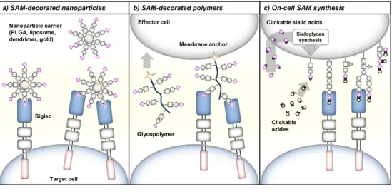
of SAMs to Siglecs can be achieved via three different glycoengineering approaches (Figure 4).
Introduction
42
SAM-decorated nanoparticles
Sialosides can be crosslinked to various types of nanoparticle such as liposomes, gold nanoparticles or dendrimer-/poly(lactic-co-glycolic acid) (PLGA)-based nanoparticles, allowing their multivalent presentation. Recently, SAM-decorated nanoparticles for delivery of cargo to Siglec expressing cells have been developed (Figure 4a). The group of Paulson has generated BPC-Neu5Ac-coated liposomes loaded with the cytotoxic agent doxorubicin to target and kill Siglec-2 expressing B cell lymphoma cells in vivo.65 Moreover, liposomes decorated with SAMs specific
for Siglec-1 or Siglec-7 enabled targeting of antigens to antigen-presenting cells to activate antigen-specific T cells.66,67 Besides their function in cargo delivery, SAM-decorated nanoparticles can be applied to stimulate Siglec signaling.46 Paulson
and coworkers reported that multivalent presentation of SAMs with high affinity for Siglec-2 on liposome nanoparticles triggered immunosuppressive signaling in B cells.68 The liposomes were crosslinked with antigens to target the B cell receptor
and BPA-NeuGc or BPC-Neu5Ac to engage mouse or human Siglec-2. B cell receptor signaling was strongly inhibited by these liposomes leading to B cell apoptosis and antigen-specific tolerance induction. In a similar approach,
liposomes decorated with 3′-BPA-NeuGc that target Siglec-G on mouse B cell subsets were able to inhibit B cell receptor signaling and induced tolerance towards selected antigens.69 The suppression of B cell receptor signaling was dependent
on SHP-1 indicating that immune inhibitory signaling of Siglec-G was directly
stimulated by the 3′-BPA-NeuGc liposom


![Bis(2,3 dimethylanilinium) diaquabis[dihydrogendiphosphato(2−)]cobaltate(II)](data:image/gif;base64,R0lGODlhAQABAIAAAP///wAAACH5BAEAAAAALAAAAAABAAEAAAICRAEAOw==)