155
In the name of GodShiraz E-Medical Journal
Vol. 12, No. 3, July 2011http://semj.sums.ac.ir/vol12/jul2011/89040.htm
Distal Radius Fracture, a Comparison Between Closed Reduction
and Long Arm Cast Vs. Closed Reduction and Percutaneous Pinning
and Short Arm Cast.
Mardani Kivi M*, Asadi K*, Hashemi Motlagh K**, Shakiba M±.
*Assistant Professor, Department of Orthopedic surgery, **General Practitioner, ± MS of Epidemiology, Guilan University of Medical Sciences, Rasht, Iran.
Correspondence: Dr. M. Mardani Kivi, Department of Orthopedics, Poursina Hospital, Rasht, Iran, Telephone: +98(131) 323-8373, Fax: +98(131) 323-8373, Email: dr_mohsen_mardani@yahoo.com
Received for Publication: October 10, 2010, Accepted for Publication: April 30, 2011.
Abstract:
Background: Distal radius fracture represent approximately one-sixth of all fractures treated in emergency departments. According to high incidence rate, different mechanisms of injury and new treatments for this fracture, it is becoming one of the most challenging of all kinds of fractures.
Objective: to compare treatment outcome of traditional cast immobilization versus modern percutaneous pinning procedure in patients with distal radius fractures.
Methods: In this randomized clinical trial study, 198 patients with "displaced but stable distal radius fracture without joint incongruity", were split into two groups and each group was treated by one of the following standard protocols: A- Closed reduction + Long arm cast; B- Closed reduction and Percutaneous pinning and Short arm cast. The patients were followed up after operation for three months from the point of view of: 1- satisfaction(based on Saito chart) 2-Loss of Reduction 3- Finger stiffness 4- Pin tract infection and 5-The mean of post operation follow up visits. For statistical analysis the Fisher's exact test and chi-square test were used by SPSS software16
Results: In group A, six cases of loss of reduction were detected in the first week who were treated by re-reduction and P.C pinning procedure; But no cases of loss of reduction were diagnosed in group B. Satisfaction percentage for Excellent value was 81.8% in group A and 93.9% in group B (p= 0.131). Finger stiffness incidence rate in group B was meaningfully lower than group A (p=0.039). Pin tract infection incidence rate was 15.1% in group B; all of them were treated by pin removal and oral antibiotic therapy. The mean of post operation visits was 4.4 in group B and 3.6 in group A out of five sessions (p<0.0001).
Conclusion: It seems that closed reduction and P.C pinning is a safer and less complicated procedure, especially in decreasing finger stiffness in these fractures.
Keywords: Distal radius fractures, Percutaneous pinning, Treatment outcome Key Messages: Distal radius fractures; Percutaneous pinning fixation Vs. Cast
156
Introduction:
The optimal treatment of distal radius fractures has changed dramatically over the last two decades. Although Cast im-mobilization was almost the only univer-sal treatment, today it is progressing to operative interventions.
Distal radius fracture consists approx-imately of one-sixth of all fractures treated in Emergency departments.(1-3)
Although most injured people are elderly, but recent researches revealed that there is an increasing incidence rate of this fracture in all age range.(4,5) More
impor-tantly, studies suggest that there are two different mechanisms of injury: one, an insufficiency fracture in elderly patients due to osteoporosis, and the other is a traumatic injury in young males second-ary to motor-vehicle accidents.(6-9) The
differences in these injuries and corres-ponding groups may account for some of the discrepancies in treatments. De-creased bone mineral density, Female gender, Ethnicity, Heredity and early menopause are the risk factors for this injury.(10-12)
Although closed reduction and casting is the main treatment in children (13), there
are several different interventions for treating adults, including: Open reduction and internal fixation, Pin and Plaster, Ex-ternal fixation, Percutaneous pinning Fix-ation, and the combination of the men-tioned procedures.(1,4,14-17)
In this study, patients with displaced but stable distal radius fractures with con-gruous joint (step<2mm) have been ran-domly treated with one of these proce-dures: (A: Closed reduction and Long arm cast, B: Percutaneous pinning and
Short arm cast) and treatment outcome has been documented over a period of three months.
Material and Methods:
This study is a randomized clinical trial.
198 musculoskeletally mature patients between 16-75 years of age with dis-placed but stable distal radius fracture with congruous joint with less than 2mm joint gap [type I of Fernandez classifica-tion (18) ] who had been admitted to
Rasht emergency hospital, were included. All other patients with open physis, open fracture, dorsal comminution, dorsal tilt more than 20 degree, history of previous wrist or forearm fractures, congenital or other forearm or other anomalies, pre-vious history of wrist operation, history of psychiatric problems, and fractures in other parts of injured upper limb were excluded. After taking consent, patients were divided into two groups randomly. After general anesthesia, in the first group, patients were operated with closed reduction and long arm cast. The second group of patients was operated with closed reduction, percutaneous pin-ning with smooth without threaded 1.5mm or 2mm pin and then immobilized with short arm cast by the same ortho-pedist. The pin was shortened, curved and then remained out of the skin and the splint of near pin was removed for monitoring pin tract infection. Procedures and outcomes were done by ignoring left or right limb dominancy.
For all patients, AP-view and Lateral-view of wrist radiographs were taken and the patients were discharged if they had been qualified by four reduction criteria
157
(1- radial shortening less than 5mm, 2- radial inclination more than 15 degree, 3- volar tilt between 0-15 degree, and 4- joint gap less than 2mm). Otherwise, they were taken under re-reduction and excluded from the study.
All patients were asked to attend clinic in 1st, 3rd, 6th, 8th, and 12th weeks after
in-tervention for follow up. The control radi-ographs were taken in 1st, 3rdand 6th
weeks, and if they didn't have acceptable reduction (according to four mentioned criteria), another intervention for reduc-tion was done and if they had acceptable reduction, the splints were opened in 6th or 8th weeks and pins were removed as
outpatient and wrist physiotherapy was started.
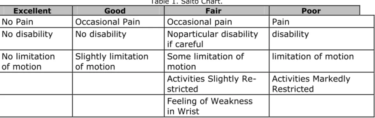
The following factors were evaluated in follow up visits: the mean of post opera-tional visits in each group, loss of reduc-tion pin tract infecreduc-tion frequency, and subjective satisfaction of every patient according to Saito chart (20) (Table1).
A questionnaire for gathering necessary information was sent out and completed for each individual from the beginning of treatment and during follow up visits. At last for statistical analysis the Fisher's exact test and chi-square test were used by SPSS software 16.
Table 1. Saito Chart.
Excellent Good Fair Poor
No Pain Occasional Pain Occasional pain Pain No disability No disability Noparticular disability
if careful
disability No limitation
of motion Slightly limitation of motion Some limitation of motion limitation of motion
Activities Slightly
Re-stricted Activities Markedly Restricted
Feeling of Weakness
in Wrist
Findings:
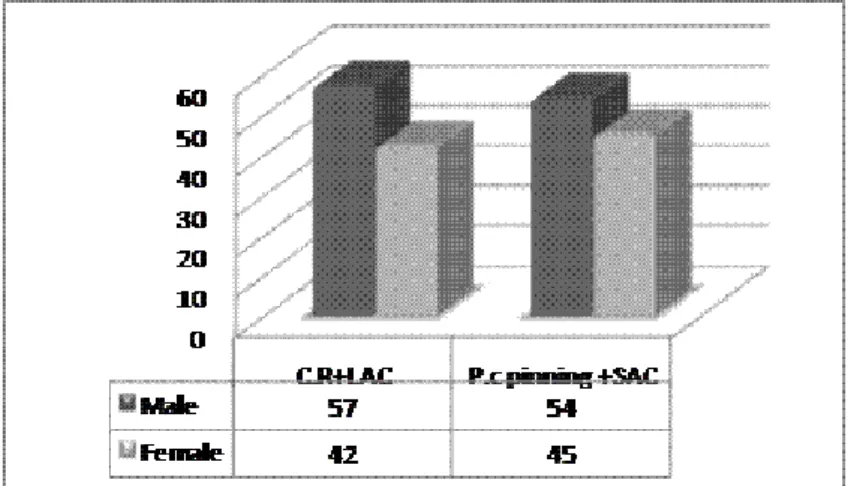
The mean age of all cases was 50.8 ± 15. The majority of the patients were between 50-70 years old (40.9%). The mean age of the first group was 49.15 and it was 52.45 for the second group (p= 0.313). 111 cases (56.1%) were male and 87 of patients were female (p= 0.804).Gender frequency in both group is shown in (Figure 1).
The mean of all patients' follow up visits is 4.04 from 5 sessions which is shown in (Table 2).
Data analysis revealed that the quantity of follow ups in percutaneous pinning group is significantly more than cast im-mobilization group (p<0.0001).
We have discovered that 30 cases of all 198 patients suffered from finger stiff-ness after three months of intervention, as is shown in (Figure 2).
It is revealed that finger stiffness is sta-tistically lowered in percutaneous pinning fixation group rather than the other group (p=0.039)
We have found 15 cases (15.1%) with pin tract infection in P.C pinning fixation
158
group who all were treated thoroughly with pin removal and oral anti-biotic therapy.
In cast immobilization group, there were 6 patients who had lost reduction during the first week. For all of them Re-reduction and P.C pinning fixation were
performed. It is revealed that we have better satisfaction issues in P.C pinning Fixation group rather than cast immobili-zation group, but it didn't reach the sta-tistical significance (p=0.131). Subjective patient satisfaction is shown in (Table 3).
Figure 1. Gender frequency
Table 2. The mean of patients' follow ups.
Group No. Mean of vis-its Standard Deviation T value Statistical evaluation C.R + LAC 99 6/3 86/0 5/4 0001/0 P< P.C Pining + SAC 99 4/4 71/0
159
Table 3. Subjective satisfaction frequencyGroup Satisfaction C.R + LAC P.C Pining + SAC Total No. Percentage No. Percentage No. Percentage Excellent 81 81.8 93 93.9 174 87.9
Good & Fair
18 18.2 6 6.1 24 12.1 Total 99 100 99 100 198 100
Discussion:
Incidence rate of distal radius fracture increases with aging, which is associated with all of the risk factors for osteoporo sis.(4,8,10 and 21) Likewise, peak of incidence
was between 50-70 years of age in this study. Patient satisfaction has been stu-died in several series. "Rodriguez" in Spain and "Chen" et al. in Taiwan stated that almost 90% of their patients were satisfied about their procedures.(23,24)
"Kreder" et al. in a 2 year prospective study on 113 patients suggested that external fixation and P.C pinning fixation had better radiological and functional results compared to traditional cast im-mobilization (25); even though, "Stofelen"
in a randomized clinical trial showed that there is no relationship between func-tional results in patients treated with casting and those who had P.C pinning procedures.(26) There exists a trend for
better functional and subjective satisfac-tion in more than 90% of cases treated with P.C pinning fixation in our study, but it didn't reach statistical significance (p= 0.131). Advantages of P.C pinning fixa-tion of distal radius fracture are that it is a quicker and less technically demanding technique compared to more complex forms of fixation.(27) Additionally, there is
less soft tissue disruption than open re-duction and it can be used to supplement cast immobilization.(27) Disadvantages
include the complications of pin site in-fection, potentially less accurate fracture reduction than open techniques and po-tentially less stable fixation compared to plating techniques.(27) Pin tract infection
has been reported from 6% to 38% in other studies (25, 28); likewise, this
para-meter was 15.1% in our study. In anoth-er study of "Kredanoth-er" et al, they found patients with displaced intra-articular distal radius fractures undergoing P.C pinning fixation had a more rapid return of function and had a better functional outcome.(29) "Fuji" et al. stated that P.C
pinning fixation is a simple and minimal-ly-invasive procedure, which is useful in preventing re-displacement of frag-ments.(20) However a few opponents of
this technique claim that these fractures tend to collapse even after pin remov-al.(30) But "Kurup" in a retrospective
study over three years found that distal radius fractures treated by P.C pinning fixation did not suffer significant loss of reduction of fracture position after pin removals and this remains true regard-less of age, sex, and fracture type or du-ration of pin fixation.(31) In our study,
there was no case of re-displacement in P.C pinning fixation group, and we found six patients with re-displaced fracture fragments in cast immobilization group. Also it was revealed that P.C fixation sig-nificantly decreases finger stiffness in these fractures (p=0.039)
160
"Rosental" et al. in a prospective follow up of 18 cases with dorsally angulated distal radius fracture found that intra-focal pinning significantly provides better maintenance of volar tilt and ulnar va-riance during 11 weeks post intervention when compared with closed reduction and cast treatment alone.(32) Also it has
been shown that with the use of intra-focal pinning, maintenance of radial length is the most important factor in providing superior functional outcomes when compared with maintenance of radial tilt or palmar tilt.(33)
For any orthopedic surgeons, follow-ups and post interventional visits have impor-tant roles in any treatment procedures. There are various clinical and demo-graphic factors affecting poor follow up rates. Compared with patients who com-plained about follow up, those who lost to follow up had lower physical and mental health scores on the SF-36 forms, more often were treated non-operatively, and more likely had not surpassed secondary education.(34-36) Likewise, we found that
there were better follow up rates in P.C pinning fixation group (mean of vis-its=4.4) than patients who were treated non-operatively (mean of visits= 3.6)
Conclusion:
It seems that Percutaneous pinning fixa-tion is a safer and less complicative in-tervention with less lost-to-follow up rates than traditional non operative cast immobilization treatment.
Conflicts of interest:
The authors did not receive grants or outside funding in support of their Re-search or preparation of this manuscript. They did not receive payments or other
benefits or a commitment or agreement to provide such benefits from a commer-cial entity. No commercommer-cial entity paid or directed, or agreed to pay or direct, any benefits to any research fund, founda-tion, educational institufounda-tion, or other cha-ritable or nonprofit organization with which the authors are affiliated or asso-ciated.
References:
1.Golden GN. Treatment and programs of Colles' fractures. Lancet. 1963; 1:511–4.
2. Love C. Acute treatment for Colles' frac-ture: the case for a radical review. Curr Or-thop. 2000; 14:290-3.
3. Koval KJ, Harrast JJ, Anglen JO, et al. Fractures of the distal part of the radius. The evolution of practice over time. Where is the evidence? J Bone Joint Surg Am. 2008; 90 (9):1855-61.
4. Liporace FA, Adams MR, Capo JT, et al. Distal Radius Fractures. J Orthop Trauma 2009; 23: 739-48.
5. O'Neill TW, Cooper C, Finn JD, et al. Inci-dence of distal forearm fracture in British men and women. Osteoporosis Int. 2001; 12: 555-8.
6. Swiontkowski MF. Increasing rates of fo-rearm fractures in children. JAMA. 2003; 290 (24): 3193.
7. Solgaard S, Petersen VS. Epidemiology of distal radius fractures. Acta Orthop Scand. 1985; 56 (5): 391-3.
8. Lill CA, Goldhahn J, Albrecht A, et al. Im-pact of bone density on distal radius fracture patterns and comparison between five dif-ferent fracture classifications. J Orthop Trauma. 2003; 17: 271-8.
9. Hollevoet N, Verdonk R. Outcome of distal radius fractures in relation to bone mineral density. Acta Orthop Belg. 2003; 69: 510-4.
10. Mensforth RP, Latimer BM. Hamann-Todd collection aging studies: osteoporosis fracture syndrome. AmJ Phys Anthropol. 1989; 80 (4): 461-79.
11. Lester GE, Anderson JJ, Tylavsky FA, et al. Update on the use of distal radial bone density measurements in prediction of hip and colles' fracture risk. J Orthop Res. 1990; 8 (2): 220-6.
12. Mallmin H, ljunghall S, Naessen T. Colles' fracture associated with reduced bone
161
mineral content. Photon densitometry in 74patients with mathched controls. Acta Or-thop Scand. 1992; 63 (5): 552-4.
13. Miller BS, Taylor B, Widmann RF, et al. Cast Immobilization Versus Percutaneous Pin Fixation of Displaced Distal Radius Fractures in Children. J Pediatr Orthop 2005; 25: 490-4.
14. Raia F, Catalano L. What’s new in Distal Radius Fracture Treatment for 2007.Current opinion in Orthopaedics. 2007; 18 (4): 328-33
15. Lewis T, Yen D. Percutaneous 3 Kir-schner Wire Fixation Including the Distal radioulnar Joint for Treatment of Pilon Frac-tures of the Distal Radius—Technical Note. J Trauma. 2010; 68:485–9.
16. Mah ET, Atkinson RN. Percutaneous Kirschner wire stabilization following closed reduction of Colles’ fractures. J Hand Surg Br. 1992; 17: 55–62.
17. Axelrod TS, McMurtry RY. Open reduc-tion and internal fixareduc-tion of comminuted, intraarticular fractures of the distal radius. J Hand Surg Am. 1990; 15A: 1–11.
18. Fernandez DL, Jupiter JB. Fractures of the distal radius: operative treatment. Intr Course Lect. 1993; 42: 73-88.
19. Graham TJ. Surgical correction of malu-nited fractures of the distal radius. J Am Acad Orthop Surg.1997; 5: 270.
20. Fujii K, Henmi T, Kanematsu Y, et al. Fractures of the distal end of radius in elder-ly patients: A Comparative study of anatom-ical and functional study. Journal of Ortho-paedic Surgery 2002; 10 (1): 9-15.
21. Arora R,Gabl M, Gschwentner M, et al. A Comparative Study of Clinical and Radiologic Outcomes of Unstable Colles Type Distal Radius Fractures in Patients Older than 70 Years: Non operative Treatment Versus Vo-lar Locking Plating. J Orthop Trauma. 2009; 23: 237-42.
22. Cummings SR, Black DM, Rubin SM. Life time risks of hip, Colles', or vertebral frac-ture and coronary heart disease among white post menopausal women. Arch Intern Med. 1989; 149: 2445-8.
23. Chen C, Juhn R, Ko J. treatment of Distal Radius Fractures with percutaneous pinning and Pin-in-Plaster. American Association for HAND Surgery. 2008; 3: 245-50.
24. Rodriguz-Merchan C. Plaster cast versus percutaneous pin fixation for comminuted fractures of the distal radius in patients
be-tween 46 and 65 years of age. Journal of Orthopaedic Trauma 1997; 11 (3): 212-7
25. Kreder H, Agel J, McKee M, et al. A Randomized, controlled Trial of Distal Radius fractures with Metaphyseal Displacement but without Joint Incongruity: Closed reduction and Casting versus Closed Reduction, span-ning External Fixation and optional percuta-neous K-wires. J Orthop Trauma. 2006; 20 (2): 115-21.
26. Stoffelen DVC, Broos PL. Closed reduc-tion versus kapandji-pinning for extra-articular distal radius fractures. J Hand Surg (Br). 1999; 24B: 89-91.
27. Deakin DE, Deshmukh SC. Dorsally an-gulated fractures of the distal radius. J Trauma. 2010; 12: 21-29.
28. Hargreaves DG, Drew SJ, Eckersky R. Kirschner wire pin tract infection rate. A ran-domized controlled trial between percutane-ous and buried wires. J Hand Surg Br. 2004; 29: 374-6.
29. Kreder HJ, Hanel DP, Agel J, et al. Indi-rect reduction and percutaneous fixation versus open reduction and internal fixation of displaced intra-articular fractures of the distal radius. J Bone Joint Surg (Br). 2005; 87B: 829-36.
30. Brady O, Rice J, Nicholson P, et al. The unstable distal radial fracture one year post Kapandji intra-focal pinning. Injury. 1999; 30 (4): 251-5.
31. Kurup HV, Mandalia VM, Shaju KA, et al. Late collapse of distal radius fractures after K-wire removal: is it significant? J Orthopaed Traumatol. 2008; 9: 69-72.
32. Rosenthal AH, Chung KC. Intrafocal pin-ning of distal radius fractures: a simplified approach. Ann Plast Surg. 2002; 48: 593-9.
33. Trumble TE, Wagner W, Hanel DP, et al. Intrafocal (Kapandji) pinning of distal radius fractures with and without external fixation. J Hand Surg (Am). 1998; 23: 381-94.
34. Murnaghan ML, Buckley RE. Lost but not forgotten: patients lost to follow-up in a trauma database. Can J Surg. 2002; 45:191-5.
35. Murray DW, Britton AR, Bulstrode CJ. Loss to follow-up matters. J Bone Joint Surg Br. 1997; 79: 254-7.
36. Tejwani NC, Takemoto RC, Nayak G, et al. Who is Lost to Follow-up? A Study of Pa-tients with Distal Radius Fractures. Clin Or-thop Relat Res. 2010; 468: 599-604.