for removing embedded fishhooks are described in this article.
Patient Evaluation
Most fishhook injuries are penetrating soft tissue injuries to the hand, face, head or upper extremity but can involve any body part. These injuries usually do not involve deeper tissue structures because of the linear forces applied along the fishing line to the fishhook that drive the point parallel to the skin and keep it from deep penetration.
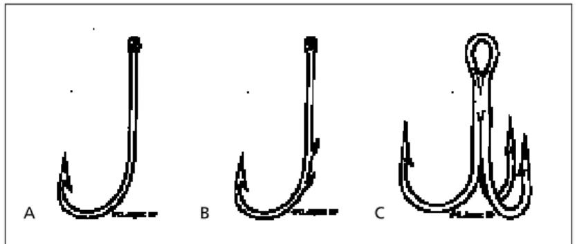
Many different types and sizes of fishhooks are available (Figure 1). When examining the hook, it is important to note if the fishhook is single, multiple or treble, whether the hook is barbed, and the number and location of the barbs—these details will help determine the best removal technique. Often, persons will know the type of hook they were using and may be able to provide a sample for inspection. Occasionally, more serious tissue trauma occurs from fishhook injury. While not rou-tinely performed, radiographs may aid in determining the type of fishhook and the depth of penetration in difficult cases.1
Neu-rologic and vascular status, proximal and
dis-A
ngling is a popular sport worldwide, and fishhook in-juries are common in recre-ational and commercial fish-ing settfish-ings. Persons with fishhook injuries may not present to the office or emergency department because removal of embedded fishhooks can usually be accomplished in the field. However, some embedded fishhooks cannot be removed in this manner and require evaluation of the injury and exploration of the wound for the presence of a foreign body. Four techniquesFishing is a common recreational sport. While serious injuries are uncommon, pene-trating tissue trauma involving fishhooks frequently occurs. Most of these injuries are minor and can be treated in the office without difficulty. All fishhook injuries require careful evaluation of surrounding tissue before attempting removal. Ocular involve-ment should prompt immediate referral to an ophthalmologist. The four most com-mon techniques of fishhook removal and injury management are described in this arti-cle. The choice of the method for fishhook removal depends on the type of fishhook embedded, the location of the injury and the depth of tissue penetration. Occasion-ally, more than one removal technique may be required for removal of the fishhook. The retrograde technique is the simplest but least successful removal method, while the traditional advance and cut method is most effective for removing fishhooks that are embedded close to the skin surface. The advance and cut technique is almost always successful, even for removal of large fishhooks. The string-yank method can be used in the field and can often be performed without anesthesia. Wound care follow-ing successful removal involves extraction of foreign bodies from the wound and the application of a simple dressing. Prophylactic antibiotics are generally not indicated. Tetanus status should be assessed and toxoid administered if needed. (Am Fam Physi-cian 2001;63:2231-6.)
Fishhook Removal
MATTHEW GAMMONS, M.D., and EDWARD JACKSON, M.D.
Michigan State University College of Human Medicine, East Lansing, Michigan
OFFICE PROCEDURES
FIGURE 1. Types of fishhooks. (A) Simple-single barbed fishhook. (B)
Multiple-barbed fishhook. (C) Treble fishhook.
A B C
ILLUSTRATIONS BY CHARLES BOYTER
This article is one in a series of “Office Procedures” articles coordinated by Thomas J. Zuber, M.D., Assistant Professor, Depart-ment of Family and Community Medi-cine, Emory Univer-sity School of Medicine, Atlana.
tal to the wound, should be assessed. Any fishhook injury that may involve deeper structures such as bone, tendons, vessels or nerves requires careful evaluation before attempting removal.
Cases of penetrating eye trauma secondary to fishhook injury have been reported in the literature.2-5One such injury even included
intracranial trauma.3 Fishhooks that
pene-trate the orbital area or are embedded in a location in which removal may injure the eye should be covered with a metal patch or cup and the patient should be sent immediately for ophthalmologic consultation.6Permanent
vision damage may occur with removal of the fishhook although minimal vision deficit was evident on initial presentation.
Principles of Removal
Four primary techniques have been described for the removal of fishhooks: retro-grade, string-yank, needle cover, and advance and cut. Each method and some modifications to these techniques are described in detail in this article. The method selected to remove an
because they result in the least amount of tis-sue trauma. The more invasive procedures, such as the needle cover and advance and cut techniques, are reserved for more difficult fishhook removal.7Sometimes multiple
tech-niques must be attempted before the fish-hook is successfully removed.
Most removal methods require the admin-istration of a local anesthetic or a nerve block. Superficially embedded hooks may not require anesthesia if they can be backed out or removed easily by the string-yank method. Local care typically involves cleaning the site with povidone-iodine or hexachloro-phene solution before attempting removal of the fishhook. Saline irrigation may be required. Fishhooks with more than one point (i.e., treble fishhooks) should have the uninvolved points taped or cut to avoid im-bedding these during the removal procedure. A local anesthesia should be administered before attempting removal of any barbed fishhook. All items attached to the hook (i.e., fish line, bait and the body of the lure itself) should be removed. The physician and bystanders should take care not to be struck by the hook on removal. Eye protection should be worn, especially when performing the string-yank method.
Retrograde Technique
Retrograde technique is the simplest of the removal techniques but has the lowest success rate. It works well for barbless and superfi-cially embedded hooks. Downward pressure is applied to the shank of the hook. This maneuver helps rotate the hook deeper and disengage the barb, if present, from the tissue. The hook can then be backed out of the skin along the path of entry (Figure 2). Any resis-The Authors
MATTHEW GAMMONS, M.D., is a resident physician in the Department of Family Prac-tice at Saginaw Cooperative Hospitals, Saginaw, Mich., and a clinical instructor in the Department of Family Practice at Michigan State University College of Human Medi-cine, East Lansing, Mich. Dr. Gammons received his medical degree from Ross Univer-sity School of Medicine, Portsmouth, Commonwealth of Dominica, West Indies. EDWARD JACKSON, M.D., is the associate director of family practice, Saginaw Coop-erative Hospitals, and an associate professor in the Department of Family Practice, Michigan State University College of Human Medicine. Dr. Jackson received his med-ical degree from Jefferson Medmed-ical College of Thomas Jefferson University, Philadel-phia. He completed a residency in family practice at Saginaw Cooperative Hospitals.
Address correspondence to Matthew Gammons, M.D., 1000 Houghton Ave., Sagi-naw, MI 48603. Reprints are not available from the authors.
tance or catching of the barb during the pro-cedure should alert the physician to stop and consider other removal methods.
String-Yank Technique
The string-yank technique is a highly effec-tive modification of the retrograde technique and is also referred to as the “stream”technique. It is commonly performed in the field and is believed to be the least traumatic because it cre-ates no new wounds and rarely requires anes-thesia.8It may be used to remove any size
fish-hook but generally works best when removing fishhooks of small and medium size. This tech-nique also works well for deeply embedded fishhooks, but cannot be performed on parts of the body that are not fixed (e.g., earlobe).9
Physicians should be familiar with the concepts of this method because improper technique could cause further tissue damage.
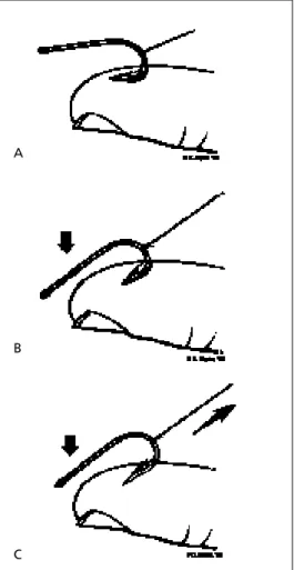
A string, such as fishing line, umbilical tape or silk suture, should be wrapped around the mid-point of the bend in the fishhook with the free ends of the string held tightly (Figure 3). A bet-ter grip on the string can be achieved by wrap-ping the ends around a tongue depressor.1The
involved skin area should be well stabilized against a flat surface as the shank of the fishhook is depressed against the skin. Continue to depress the eye and/or distal portion of the shank of the hook, taking care to keep the shank parallel to the underlying skin. A firm, quick jerk is then applied parallel to the shank while continuing to exert pressure on the eye of the fishhook. The fishhook may come out with sig-nificant velocity so the physician and bystanders should remain out of the line of flight. A com-mercial fishhook removal device, based on this technique, is available. (Minto Research and Development Inc., Redding, Calif.)10
Needle Cover Technique
The needle cover technique requires dex-terity on the part of the physician. It works well for the removal of large hooks with sin-gle barbs but is most effective when the point of the fishhook is superficially embedded
Fishhook Removal
FIGURE 2. Retrograde technique. Downward pressure is applied to the shank of the fishhook while it is backed out along the point of entry.
FIGURE 3. String-yank method. (A) Wrap a string around the midpoint of the bend in the fishhook. (B)Depress the shank of the fish-hook against the skin. (C)Firmly and quickly pull on the string while continuing to apply pressure to the shank.
A
B
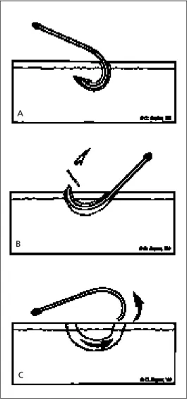
and can be easily covered by the needle. After skin preparation and administration of local anesthesia, an 18-gauge or larger needle is advanced along the entrance wound of the fishhook (Figure 4). The direction of inser-tion should be parallel to the shank. The bevel should point toward the inside of the curve of the fishhook, enabling the needle opening to engage the barb. It is important to have the bevel pointed in the correct direc-tion so that the longer edge of the needle matches the angle of the fishhook point. The physician should advance the fishhook to disengage the barb, then pull and twist it so
that the point enters the lumen of the needle. The physician can then back out the fishhook (the same way as in the retrograde tech-nique), taking care to move the needle along the track with the fishhook.
A modification of this technique involves sliding a no. 11 scalpel blade along the wound to the point of the fishhook. The fishhook may then be backed out because the incision
FIGURE 4. Needle cover method. (A)Advance an 18-gauge or larger-gauge needle along the fishhook until the needle opening covers the point. (B) The fishhook and needle are then removed at the same time.
A
B
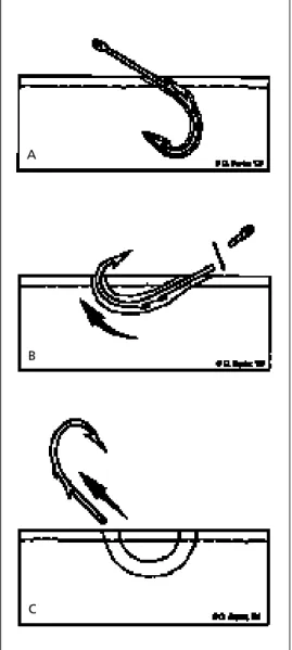
FIGURE 5. Advance and cut method: single-barbed fishhook. (A) The fishhook is advanced through the skin. (B) The barb is then cut off and (C) the remaining hook is backed out through the entry wound.
A
B
allows room for the point. This modification may also be used in combination with the needle cover technique for more difficult fish-hook injuries.
Advance and Cut Technique
One advantage of this traditional method of fishhook removal is that it is almost always successful, even when removing larger fish-hooks; however, additional trauma to the sur-rounding tissue is a disadvantage. The ad-vance and cut technique is most effective when the point of the fishhook is located near the surface of the skin.9It involves two
meth-ods of removal: one for single-point fish-hooks (Figure 5)and one for multiple-barbed fishhooks (Figure 6). Infiltration with a local anesthetic is performed over the area where the fishhook has penetrated the skin. Using pliers or needle drivers, the point of the fish-hook (including the entire barb) is advanced through the skin. The point is then cut free with the pliers or another cutting tool, allow-ing the rest of the fishhook to be backed out with little resistance.
For multiple-barbed fishhooks, the area should be anesthetized and the fishhook advanced. Instead of removing the point, the eye of the fishhook is removed. The physician can then continue to pull the fishhook in the same direction as the point was advanced.
Post-Removal Wound Care
After removal of the fishhook, the wound should be explored for possible foreign bodies (e.g., bait). It is usually sufficient to leave the wound open, then apply an antibiotic oint-ment and a simple dressing. Tetanus toxoid should be administered to persons for whom more than five years has elapsed since their last tetanus booster. Well-conducted, con-trolled studies do not exist that support the need for systemic antibiotics in these cases; they are generally not indicated.7Prophylactic
antibiotic therapy may be considered for per-sons who are immunosuppressed or have poor wound healing (e.g., patients with
dia-Fishhook Removal
Although it may produce additional tissue trauma, the major advantage of the advance and cut technique when removing an embedded fishhook is that it is almost always successful.
FIGURE 6. Advance and cut method: multiple-barbed fishhook. (A) The fishhook is advanced through the skin. (B)The eye of the fishhook is then cut off and (C)the remaining portion of the fishhook is pulled through the exit wound created by advancing the point.
A
B
betes mellitus or peripheral vascular disease). Prophylactic antibiotic therapy may also be considered for deeper wounds that involve the tendons, cartilage or bone. Follow-up care should be performed to ensure adequate heal-ing and the absence of infection.
REFERENCES
1. Lantsberg L, Blintsovsky E, Hoda J. How to extract an indwelling fishhook. Am Fam Physician 1992; 45:2589-90.
2. Aiello LP, Iwamoto M, Taylor HR. Perforating ocular fishhook injury. Arch Opthalmol 1992;110:1316-7. 3. Swanson JL, Augustine JA. Penetrating intracranial trauma from a fishhook. Ann Emerg Med 1992;21: 568-71.
4. White MF, Owens SD, Dooley CD, Kimble JA, With-erspoon CD. Fishing related eye injuries: a report of 27 cases. Invest Opthalmol Vis Sci 1990; 31:21. 5. Deramo VA, Maus M, Cohen E, Jeffers J. Removal
of a fishhook in the eyelid and cornea using a ver-tical eyelid-splitting technique. Arch Ophthalmol 1999;117:541-2.
6. Morris JA, Swiontkowski MF, Merrmann HJ. Wilderness trauma emergencies. In: Auerbach PS, ed. Wilderness medicine: management of wilder-ness and environmental emergencies. 3d ed. St. Louis: Mosby, 1995:342-62.
7. Doser C, Cooper WL, Ediger WM, Magen NA, Mildbrand CS, Schulte CD. Fishhook injuries: a prospective evaluation. Am J Emerg Med 1991;9: 413-5.
8. Haynes JH. Fishhook removal. In: Pfenninger JL, Fowler GC, eds. Procedures for primary care physi-cians. St. Louis: Mosby,1994:128-32.
9. Diekema DS, Quan L. Fishhook removal. In: Hen-retig FM, King C, eds. Textbook of pediatric emer-gency procedures. Baltimore: Williams & Wilkins, 1997:1223-7.
10. Rudnitsky GS, Barnett RC. Soft tissue foreign body removal. In: Roberts JR, Hedges JR, eds. Clinical procedures in emergency medicine. 3d ed. Phila-delphia: Saunders, 1998:623-4.