Robert Brown MD, Anthony F DiMarco MD, Jeannette D Hoit PhD CCC-SLP,
and Eric Garshick MD MOH
Introduction
Physiologic Effects of Spinal-Cord Injury on the Respiratory System Mortality in Chronic Spinal-Cord Injury
Dyspnea Interventions
Respiratory Muscle Training Abdominal Binder
Ventilator-Assisted Speech Respiratory Muscle Pacing Assisted Cough
Mechanical Ventilation Summary
Respiratory dysfunction is a major cause of morbidity and mortality in spinal cord injury (SCI), which causes impairment of respiratory muscles, reduced vital capacity, ineffective cough, reduc-tion in lung and chest wall compliance, and excess oxygen cost of breathing due to distorreduc-tion of the respiratory system. Severely affected individuals may require assisted ventilation, which can cause problems with speech production. Appropriate candidates can sometimes be liberated from me-chanical ventilation by phrenic-nerve pacing and pacing of the external intercostal muscles. Partial recovery of respiratory-muscle performance occurs spontaneously. The eventual vital capacity depends on the extent of spontaneous recovery, years since injury, smoking, a history of chest injury or surgery, and maximum inspiratory pressure. Also, respiratory-muscle training and abdominal binders improve performance of the respiratory muscles. For patients on long-term ventilation, speech production is difficult. Often, practitioners are reluctant to deflate the tracheostomy tube cuff to allow speech production. Yet cuff-deflation can be done safely. Standard ventilator settings produce poor speech quality. Recent studies demonstrated vast improvement with long inspiratory time and positive end-expiratory pressure. Abdominal binders improve speech quality in patients
Robert Brown MD is affiliated with the Pulmonary and Critical Care Unit, Department of Medicine, Massachusetts General Hospital, and with Harvard Medical School, Boston, Massachusetts. Anthony DiMarco MD is affiliated with Rammelkamp Research Center, MetroHealth Medical Center, and with the Department of Physiology and Biophysics, Case Western Reserve University, Cleveland, Ohio. Jeannette D Hoit PhD CCC-SLP is affiliated with the Department of Speech, Language, and Hearing Sciences, University of Arizona, Tucson, Arizona. Eric Garshick MD MOH is affiliated with the Pulmonary and Critical Care Medicine Section, Medical Service, Veterans Affairs Boston Healthcare System, and with Channing Laboratory, Department of Medicine, Brigham and Women’s Hospital, and with Harvard Medical School, Boston, Massa-chusetts.
Robert Brown MD presented a version of this paper at the 37th RESPIRATORY
CAREJournal Conference, “Neuromuscular Disease in Respiratory and Crit-ical Care Medicine,” held March 17–19, 2006, in Ixtapa, Mexico. Dr Garshick’s work is supported by the National Institutes of Health/ National Institute of Child Health and Human Development (project grant R01 HD42141).
Correspondence: Robert Brown MD, Pulmonary and Critical Care Unit, Bulfinch 148, Massachusetts General Hospital, 55 Fruit Street, Boston MA 02114. E-mail: rbrown5@partners.org.
with phrenic-nerve pacers. Recent data show that the level and completeness of injury and older age at the time of injury may not be related directly to mortality in SCI, which suggests that the care of SCI has improved. The data indicate that independent predictors of all-cause mortality include diabetes mellitus, heart disease, cigarette smoking, and percent-of-predicted forced expiratory volume in the first second. An important clinical problem in SCI is weak cough, which causes retention of secretions during infections. Methods for secretion clearance include chest physical therapy, spontaneous cough, suctioning, cough assistance by forced compression of the abdomen (“quad cough”), and mechanical insufflation-exsufflation. Recently described but not yet available for general use is activation of the abdominal muscles via an epidural electrode placed at spinal cord level T9-L1.Key words: spinal cord injury, respiratory failure, diaphragm, rib cage, airway clearance.
[Respir Care 2006;51(8):853– 868. © 2006 Daedalus Enterprises]
Introduction
It is estimated that in the United States each year there are about 11,000 new cases of spinal cord injury (SCI)1
and that there are currently about 250,000 persons alive with SCI. Because of improvements in medical care and survival, the prevalence of people living with SCI has increased, and it is predicted that there will be greater and greater numbers of older patients with SCI. Currently the average age at injury is 37.6 years, and about 80% of those affected are male. The racial distribution appears to be changing. Between 1973 and 1979, 76.9% were white and 14.2% were African American. Since 2000, 62.9% of those injured have been white and 22% have been African Amer-ican. The cause of this apparent trend is unclear, but it may be due to actual race-specific incidence rates rather than changing location of centers collecting data or changing referral patterns to those centers.1
The American Spinal Injury Association has promul-gated standards (revised in 2002) for the classification of level of injury and extent of impairment in SCI.2 In
tetraplegia there is injury to one of the 8 cervical seg-ments of the spinal cord, whereas with paraplegia the lesions involve thoracic, lumbar, or sacral regions of the cord. The most caudal segment of the spinal cord with normal motor function defines the motor level of injury. In the thoracic region, where there are no key muscles to test, sensory level is used to estimate the extent of motor impairment. The higher and more complete the motor level of injury, the greater the respiratory muscle impairment.
Respiratory dysfunction and related diseases, such as pneumonia, which can be complicated by septicemia or pulmonary emboli, are common causes of death in SCI. From the point of view of respiratory dysfunction, it is instructive to consider SCI in 2 phases: (1) the initial phase immediately following the injury and the year thereafter and (2) the later, chronic phase during the rest of the life of the affected individual.
Physiologic Effects of Spinal-Cord Injury on the Respiratory System
Injuries above the level of the phrenic motoneurons (C3, 4, and 5) cause virtually complete paralysis of both the muscles of inhalation and exhalation and dependence on mechanical ventilation or phrenic-nerve stimulation. At lower levels of injury, the prospect of breathing without mechanical assistance is improved. Scanlon et al3observed
that within a month of injury there appears to occur a reduction in lung compliance that does not change during the year thereafter. This observation casts doubt on the previously held notion that reduced lung compliance in SCI is due to chronic lung injury from repeated infec-tions.4 –10 The cause of the early reduction in lung
com-pliance is unclear but has been ascribed partially to re-duced lung volume and partially to changes in the mechanical properties of the lung from alterations in sur-factant, which can occur rapidly with ventilation at low lung volume.3,9,11–13Chest-wall compliance is also
prob-ably reduced in tetraplegia.3,14 This issue is complex
be-cause the abdominal compartment of the chest wall is highly compliant in SCI, but the rib cage compartment may be stiff because of muscle spasticity or abnormalities that develop in rib articulations with the spine and ster-num, due to poor inspiratory-muscle performance that pre-vents stretching to the predicted total lung capacity.15–17
In tetraplegia, distortion of the respiratory system causes inefficient ventilation (ie, energy cost that is high for the ventilation achieved).18 –22This inefficiency contributes to
the risk of respiratory muscle fatigue, in particular when loads are imposed on the muscles, as with pneumonia or airways obstruction. During spontaneous breathing, distor-tion occurs such that the upper anterior rib cage moves inward during inhalation.18,21,23This is caused by the lack
of activity in the external intercostal muscles when the diaphragm contracts and by the high compliance of the abdominal wall, which diminishes the extent to which the diaphragm (acting through the zone of apposition and the
insertion of the diaphragm to the rib cage) can contribute to rib-cage expansion (see Appendix).18
During inspiratory resistive-loaded breathing in the able-bodied, the lower transverse dimension of the rib cage increases and functional residual capacity (FRC) decreases because of active expiration below the relaxed volume of the respiratory system. Since the optimal length of the diaphragm for tension development is below FRC, and since contractions at the optimal length of the diaphragm for tension development are most efficient,24 –26the active
expiration increases diaphragm length to one that is ad-vantageous for tension development and efficiency. Dur-ing inspiratory resistive-loaded inhalation in tetraplegia, there occurs a decrease in the lower transverse dimension of the rib cage.27This distortion has a related oxygen cost
but does not contribute to minute ventilation, and thus causes inefficiency. In addition, there are 2 reasons for inefficiency of diaphragm performance during resistive-loaded breathing in tetraplegia. Unlike in the able-bodied, FRC does not decrease in tetraplegia27so the diaphragm
does not operate at or near the optimal length of the dia-phragm for tension development. Also, because of the highly compliant abdomen, the diaphragm shortens much more during inhalation in tetraplegia than in the able-bod-ied, which causes the diaphragm to operate over a disad-vantageous length. It appears that the distortion is less severe in chronic than in acute SCI, but there have not been studies to compare the efficiency of ventilation in those 2 groups.
Because of the changes in compliance, chest-wall dis-tortion, and impairment in both muscles of inhalation and exhalation in neurologically complete cervical SCI below C2, there occurs a reduction in vital capacity (VC) to 20 –50% of predicted, inefficiency in ventilation, and mark-edly impaired cough. For those who require long-term assisted ventilation there is the additional problem of speech production.
Partial recovery of respiratory-muscle performance may occur over the year following injury. For example, Blue-chardt et al28found improvement with time in forced
ex-piratory volume in the first second (FEV1) and FVC in 12
subjects with motor complete injuries (8 cervical, one C7-T1, and 3 thoracic). They did not measure the role of specific respiratory muscles or of changes in the compli-ance of the components of the respiratory system in the observed improvement. Between 90 days and 210 days, FEV1improved from 1.82 L to 2.54 L, and FVC increased
from 2.03 L to 2.69 L. Such progress has been attributed to improvement in diaphragm performance,28 –31reflex
ac-tivity in the intercostal muscles,32 and enhanced
perfor-mance of the accessory muscles of the neck.33 In that
regard, Axen et al34followed 36 tetraplegic subjects for
about 10 months following injury. VC improved from 45% of predicted to 58% of predicted, with the greatest rate of
improvement occurring during the first 3 months. Corre-sponding to the improvement in VC was improvement in shoulder and upper-arm muscles, with some segmental innervation in common with the diaphragm (C3-C5). The data suggested that the spontaneous improvement in VC was mediated, at least in part, by corresponding improve-ment in the neural supply to the diaphragm. In a related study, Brown et al35 made measurements of total lung
capacity and its subdivisions and of respiratory-muscle performance directly (including transdiaphragmatic pres-sure) in 5 neurologically complete acute tetraplegics within 47 days of injury and during the year thereafter. Increases occurred in mean inspiratory capacity (1.84 L to 2.71 L) and expiratory reserve volume (0.11 L to 0.27 L). Expi-ratory and inspiExpi-ratory muscle performance (including that of the diaphragm) also improved with time. The improve-ment in transdiaphragmatic pressure was consistent with the conclusion of Axen et al,34that after acute cervical SCI
there occurs some spontaneous recovery of diaphragm in-nervation.
With regard to pulmonary function (such as FEV1and
FVC), the eventual outcome depends on the level and completeness of the injury, the extent of the spontaneous recovery noted above, and other factors. The American Thoracic Society (ATS) has suggested testing standards for the able-bodied.36These standards require subjects to
reproducibly inhale to total lung capacity and expire force-fully, completely, and without hesitation for at least 6 sec-onds.
To determine whether individuals with SCI could meet these standards, 278 individuals with chronic SCI were tested at least 1 year after injury.37It appeared that
expi-ratory muscle weakness caused a relatively long transition time between maximum inhalation and maximum exhala-tion. Participants with the weakest respiratory muscles, who had the highest and more complete injuries, were unable to successfully meet ATS testing standards. Using 1994 ATS testing criteria,3683% (230/278) had at least 3
acceptable expiratory efforts, and in 78% (217/278) FVC and FEV1met reproducibility standards (ie, the 2 highest
values were within 200 mL of each other). In the 48 sub-jects who did not meet acceptability standards, the per-cent-of-predicted FVC, FEV1, peak expiratory pressure,
and peak inspiratory pressure were lower, and a greater proportion had complete cervical injuries.
The investigators examined the nature of the delay at the beginning of a forced expiratory maneuver in these subjects. The delay (referred to as excessive back-extrap-olated volume by ATS36) was just slightly more than that
observed in the able-bodied (7.4% of the FVC, versus
ⱕ5% in the able-bodied), the expiratory efforts were oth-erwise technically acceptable, and the testing was repro-ducible. Individuals unable to sustain an expiratory effort for 6 seconds were able to achieve a flow plateau of at
least 0.5 seconds at residual volume. Only 65% of indi-viduals with cervical motor complete SCI were able to produce spirometry values that were acceptable and repro-ducible by the ATS standards intended for the able-bodied. That value increased to 88% if the ATS standards were modified for SCI to allow excessive back-extrapolated vol-ume and an effort less than 6 seconds if there was a plateau of at least 0.5 second at residual volume. Based on the data from the study, it was recommended that laboratories that test individuals with SCI should adopt the modified testing standards for persons with SCI, since the data obtained thereby are highly reproducible and since otherwise it might be considered erroneously that these persons cannot be tested reliably.
Factors other than level and completeness of injury and extent of spontaneous recovery may influence FVC and FEV1in SCI. Early assessments of pulmonary
func-tion in SCI included relatively few subjects and focused mainly on the relationship between level and complete-ness of injury and the resultant reduction in pulmonary function.38 – 41Studies that included fewer subjects whose
smoking history was unknown and who often were from rehabilitation centers reported mean FEV1and FVC
val-ues of approximately 40 –50% of predicted in complete cervical injury.40,42,43
More recently, investigators have begun to address fac-tors in addition to SCI level in 3 cross-sectional studies of larger cohorts that included persons tested years after SCI, while otherwise healthy. These studies include 216 per-sons tested at the Bronx Veterans Affairs hospital,44239
persons tested in Los Angeles,45and a cohort of 339
vet-erans and nonvetvet-erans tested at the Boston Vetvet-erans Af-fairs Medical Center.46In the latter study, after adjusting
for age and stature, lower FEV1 values were related to
increasing years since injury, lifetime cigarette smoking (pack years), a history of chest injury or operation, a his-tory of physician-diagnosed asthma, self-report of wheeze (using a respiratory symptom questionnaire), and maxi-mum inspiratory pressure. In the study from the Bronx, smoking resulted in a reduction in FEV1/FVC in both
tetraplegics and paraplegics. In the Los Angeles cohort the effect of smoking was variable, but, as in the Boston co-hort, decrements in FEV1 were associated with greater
years since injury. In the Boston study, the adjusted mean FEV1and FVC in C4-C5 complete motor SCI were 56% and 53% of predicted values, respectively, and, in C6-C8 complete motor SCI the values were 65% and 64% of predicted, respectively. The Bronx cohort had a mean FVC of 59% in nonsmoking tetraplegics. A mean FVC of 60% was observed in the Los Angeles SCI cohort. These more recent studies provide evidence that pulmonary function attributable to SCI level and extent of injury is better than recognized previously. When pulmonary function is less,
it is likely that factors other than the injury itself are re-sponsible.
Mortality in Chronic Spinal-Cord Injury
Following SCI, the mortality rate is higher than in the able-bodied,47–54 and the most common causes of death
are related to respiratory illnesses.1,48,50,51,53,54 Impaired
muscles of inhalation prevent deep breaths, and some pa-tients with cervical-level SCI may not sigh,55even to the
extent that they can, leading to atelectasis and related gas-exchange and lung-compliance abnormalities. Dysfunc-tional muscles of exhalation cause impaired cough and secretion clearance (with associated atelectasis), increase in airways resistance, and persistence of infection when it occurs.
Retrospective assessments have indicated a correla-tion between excess mortality and neurological level of injury, older age at injury, and injury in earlier calendar years.1,47– 49,53,54 The latter has been thought to reflect
recent improvement in medical care of SCI. A limitation of the earlier retrospective studies is that, other than those factors noted above, it was difficult to assess additional risk factors for mortality.
New information is available from a recent prospective study of the role of both respiratory and nonrespiratory factors on mortality in 361 men with chronic SCI.56
Thir-ty-seven deaths occurred over a median of 55.6 months. After adjusting for age, the data identified potentially treat-able factors related to SCI mortality. Independent predic-tors of all-cause mortality were diabetes mellitus, a history of heart disease, current or recent (within 7 years) cigarette smoking, and percent-of-predicted FEV1at entry into the
study. Unlike in prior retrospective studies, level and com-pleteness of SCI, older age at injury, and injury in earlier calendar years were not directly related to mortality. This suggests that care of those with high-level SCI has im-proved in recent decades. However, it is possible that these factors will emerge as significant as the cohort is followed longer and more deaths occur. The results indicate that, as in the able-bodied, as persons with SCI survive longer, comorbid diseases and personal behavior, such as smok-ing, are increasingly important in determining mortality. Compared to the United States rates, overall mortality was 47% greater. When both the underlying and contributing causes of death were considered, diseases of the respira-tory and circularespira-tory systems contributed to 24.3% and 40.5% of the deaths, respectively. Whether interventions such as smoking cessation, treatment of cardiovascular disease and diabetes mellitus and improvement in FEV1 with respiratory-muscle training (see below) affect mor-tality in SCI has not been established.
Dyspnea
It is often assumed that the prevalence of breathlessness due to a specific task is greater at higher levels of SCI. However, this may not be the case, because the perception of breathlessness may be abnormal in tetraplegia. For ex-ample, in tetraplegia, steady-state hypercapnia produces less breathlessness than in able-bodied controls.57
There are now limited data available on the prevalence and predictors of breathlessness in neurologically com-plete SCI. In one study,58 those with cervical and high
thoracic SCI reported breathlessness with “activity” more frequently than those with low paraplegia (45% vs 25%, respectively). Yet, in that study, the need to “stop to catch their breath” was similar among those with cervical, high, and low paraplegia, which makes the impact of injury level on breathlessness unclear. Also, with the exception of a 12% prevalence of dyspnea when the subjects were dressing, the data obtained were not task-specific, so spe-cific activities that cause shortness of breath were not eval-uated. The issue is complex to study, because, in incom-plete SCI, motor function may range from nearly normal (many such persons can walk without much difficulty) to nearly neurologically complete (with almost no function below the level of the lesion).
Ayas et al59provided additional information related to
breathlessness, specifically during activities related to mov-ing about. The subjects analyzed had neurologically com-plete SCI and, to get around, used hand-driven wheel-chairs⬎50% of the time. In that study, the prevalence of breathlessness was greater when the level of injury was higher. Obesity, cigarette-smoking status (current, former, never), age, and years since SCI were not factors related to breathlessness while moving about, so the finding was attributable only to SCI level.
Further information about breathlessness in SCI was provided by Grandas et al,60who studied dyspnea during
activities of daily living in SCI, categorized by level of mobility. The activities of daily living included talking, eating, dressing or undressing, and leaving the house. Those who needed a motorized wheelchair to get around and who were thereby deemed to be most neurologically impaired were compared to others who used a hand-propelled wheel-chair, walked with an aid such as a crutch or cane, or walked without assistance. Overall, dyspnea during activ-ities of daily living was most common, among motorized-wheelchair users (23.5%). In them, 17.7% noted dyspnea while talking for more than a few minutes. In the others, dyspnea while talking was uncommon, and the prevalence of dyspnea varied with activity (range 3.4 –14.3%). They more commonly experienced dyspnea while dressing or undressing or dyspnea limiting their ability to leave the house. Percent-of-predicted FEV1did not predict dyspnea
in that study, so the higher prevalence of dyspnea in the
motorized-wheelchair users was not due to worse pulmo-nary function.
The mechanisms for the increased breathlessness with a higher level of injury are not clear. In the able-bodied, for a given exercise task, those with the least exercise capacity have the greatest breathlessness. This may explain the ef-fect of level of injury on breathlessness in SCI, since, in those with higher-level injury there is decreased exercise capacity, which is limited by muscle paralysis, decreased stroke volume, and chronotropic response to exercise, due to loss of supraspinal control over the sympathetic nervous system, and diminished cardiac preload from low plasma volume and peripheral venous pooling.59,61– 64Afferent
in-formation from inflation of the lungs relieves breathless-ness, even when mechanoreceptor activity from the chest wall is absent, such as in compete tetraplegia.65 During
exercise, tetraplegics have smaller tidal volume (VT) than
those with lower levels of injury.66The diminished lung
inflation may thereby contribute to the increased breath-lessness in tetraplegia. The sensation of breathbreath-lessness is also affected by mechanoreceptor information from the chest wall. For example, in heart-lung transplantation, al-though vagus-nerve innervation of the lung is interrupted, breathlessness from hypercapnia is reduced when VT is
increased. This relief must be due to mechanoreceptor information from chest wall muscles, joints, and tendons. In neurologically complete tetraplegia, perception of such mechanoreceptor information is absent, so breathlessness may be greater.
In motorized-wheelchair users, the relatively high rate of breathlessness during talking may be related to diffi-culty interrupting breathing to manipulate phrasing and speech loudness, because their breathing is already greatly impaired.60,67The motorized-wheelchair users who did not
experience dyspnea during talking probably used an adap-tive breathing strategy. This has not been studied, but if a successful adaptive strategy is used, training those who do get breathless while talking could provide relief.
It appears that, as in the able-bodied and in COPD,68
exercise diminishes breathlessness in SCI. Wien et al69
studied breathlessness in 183 subjects with neurologically motor complete or incomplete cervical or high thoracic (T6 and above) SCI who, to get around, used hand-pro-pelled wheelchairs more than 50% of the time. Spirometry and maximum inspiratory and expiratory pressure were measured. Those with complete injuries had a greater prev-alence of breathlessness than the others. Wheelchair ath-letes were defined as those who responded “yes” to the question, “Are you a wheelchair athlete?” The distribution of level of SCI was not different between wheelchair ath-letes and nonathath-letes. Current cigarette smoking (⬎20 per day) was a significant risk factor for breathlessness, inde-pendent of pulmonary function and SCI level and com-pleteness. Athletes were significantly less likely to answer
“yes” to the questions about experiencing breathlessness, and they had significantly higher percent-of-predicted FEV1
(82.7% [n⫽47] vs 74.6% [n⫽132], p⫽0.001) and peak inspiratory pressure (104.2 ⫾ 40 cm H2O [n ⫽ 46] vs
87.2 ⫾ 31.3 cm H2O [n ⫽ 127], p ⫽ 0.01). Statistical
analysis of the data showed that the contribution of FEV1
to breathlessness was explained by smoking and a history of obstructive lung disease, and when maximum inspira-tory pressure was included in the prediction model, the athletes still reported less breathlessness. Thus, the data showed that the mechanism of decreased breathlessness in wheelchair athletes is not better pulmonary function or respiratory-muscle performance. As in others who exer-cise regularly, the decrease in breathlessness is probably a fitness effect. Large-scale prospective studies to test whether smoking cessation and fitness programs reduce breathlessness in SCI have not been performed. It is not clear that exercise programs would be successful over the long-term, since few Americans are motivated to perform exercise regularly.
Interventions Respiratory Muscle Training
The problems of poor respiratory-muscle performance, distortion of the respiratory system, impaired cough, and speech production in ventilated persons with SCI have at least partial solutions that will be addressed in the ensuing paragraphs.
In the able-bodied it has been demonstrated that, like other skeletal muscles, the muscles of ventilation can be trained for both strength and endurance.70Typically,
vig-orous and forceful efforts are required to produce a sig-nificant effect. Even marathon runners, who breathe vig-orously but not forcefully, have respiratory muscle strength that is not enhanced.71Respiratory muscle weakness and
inefficiency of breathing predispose tetraplegics to fatigue of the respiratory muscles,27,72but substantial training
ef-forts can enhance performance.72–75
For example, the subjects studied by Gross et al72
in-spired for 30 min daily, 6 days a week, against a resistance just sufficient to produce electromyographic evidence of fatigue. Uijl et al75 trained their subjects at 70% of the
endurance capacity of their respiratory muscles twice daily for 6 weeks. Rutchik et al74trained their subjects over 8
weeks, by having them breathe for 15 min twice daily against progressively higher resistances, The subjects stud-ied by Kogan et al73trained weekdays for 2.5– 4.5 months
by inhaling through a narrow orifice and developing with each breath a target of 80% of their maximum inspiratory muscle pressure. In each of the various studies cited, per-formance enhancement occurred. For example, in the study by Gross et al72there was improvement in both strength
and endurance. Uijl’s subjects experienced an increase in respiratory-muscle endurance and peak oxygen consump-tion achieved during arm-crank exercise.75In the study by
Rutchik et al74 there were significant increases in peak
inspiratory pressure (from 66.2 cm H2O to 78.5 cm H2O),
in FVC (from 2.81 L to 3.07 L) and in total lung capacity (from 5.17 L to 5.71 L). The results from Kogan et al73
were similar and, in addition, thickening of the diaphragm occurred, which indicates hypertrophy as a result of the training.
During the often-long period of liberating patients with SCI from assisted ventilation, the spontaneous recovery of respiratory muscle function noted above is likely to con-tribute to success. It is likely that simultaneous respiratory-muscle training would help, but the best training regimen has not been established. In principle, regimens that target both endurance and strength would seem most desirable. It is also not known whether trained subjects will continue the training, with the associated expectation of deteriora-tion once the training is abandoned. Although the subjects studied by Kogan et al benefited from the training, none chose to continue training after the end of the study.
Abdominal Binder
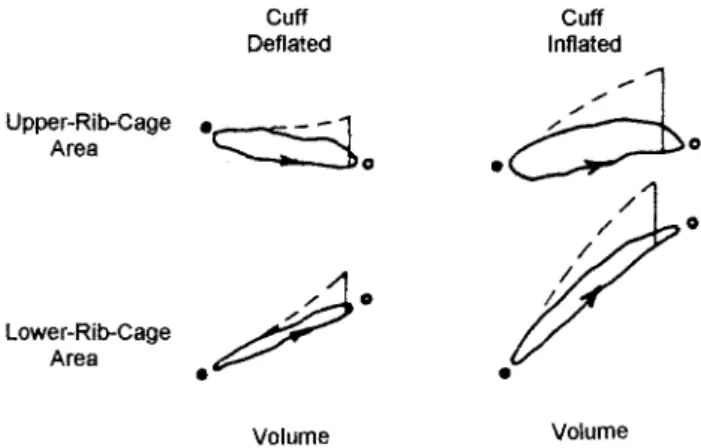
Using inductance pneumography belts during spontane-ous breathing in 6 tetraplegic subjects, Urmey et al18 mon-Fig. 1. Data obtained during tidal breathing (solid lines) and during relaxation (dashed lines) from a representative subject with ab-dominal cuff deflated versus inflated. The vertical axis represents the cross-sectional area of the (upper or lower) rib cage. The hor-izontal axis represents the change in lung volume. The vertical lines were obtained when, at or near end-tidal inspiration, the subject relaxed against an occluded valve. With the cuff deflated, the upper-rib-cage area was less at end-inspiration than at func-tional residual capacity. With the cuff inflated, the lower-rib-cage area increased more per unit change in lung volume than it did with the cuff deflated. This is indicated by the increase in slope of the plot of volume versus lower-rib-cage area. Also, as a result of cuff inflation, upper-rib-cage area increased during tidal inspira-tion, whereas it had decreased with the cuff deflated. (From Ref-erence 18, with permission.)
itored upper-rib-cage and lower-rib-cage displacements and compared them to the relaxation (undistorted) character-istic of the rib cage. Subjects were studied without and with extrinsic abdominal support, which covered the ab-dominal wall and did not overlie the lower lateral rib cage. (Note, however, that abdominal binders that do overlie the lower lateral rib cage are not as effective.) Extrinsic ab-dominal support increased abab-dominal pressure and, as con-firmed by percussion, increased the zone of apposition of the diaphragm to the rib cage.
In addition, the results of axial forcing of the lower rib cage were studied by applying a 4.5-kg cephalad force to the costal margin to mimic the action of the muscular insertion of the diaphragm to the rib cage. Figure 1 shows a typical plot of the area of the upper-rib-cage and the area of the lower-rib-cage as functions of lung volume. During spontaneous breathing, upper-rib-cage area was smaller at end-tidal inhalation than at FRC in all the subjects who demonstrated distortion from the relaxed configuration. With support of the abdominal wall, the upper-rib-cage area increased in all but one subject during tidal inhalation, which shows that the support diminished distortion. In none of the subjects did upper-rib-cage area superimpose the relaxation characteristic, so some distortion remained. Cephalad forcing of the lower-rib-cage at the costal mar-gin caused the upper rib cage to move outward, which shows that the lower rib cage and upper rib cage are cou-pled well. In summary, the study showed that, in tetraple-gia, over the tidal-breathing range, increased abdominal pressure and decreased abdominal compliance due to the presence of extrinsic abdominal support diminishes rib-cage distortion. As inferred from the force-balance analy-sis by Loring and Mead,76the diminished distortion is due
to (1) an increase in the area of diaphragmatic apposition to the rib cage and (2) an increase in the insertional com-ponent of diaphragm action on the rib cage (see Appen-dix).
McCool et al77 subsequently showed that abdominal
binding influences rib-cage motion over the entire range of inspiratory capacity in tetraplegia. In able-bodied controls, total lung capacity decreased because the binder impeded diaphragm decent. In tetraplegia, binding led to an in-crease in total lung capacity and in rib-cage dimensions, which showed that expansion of the paralyzed rib cage dominated the disadvantage of impeded diaphragm de-scent. Abdominal binders are in common use in tetraple-gia, but the beneficial effects need to be weighed against the simultaneous reduction in FRC and the related poten-tial effects on gas exchange.
Ventilator-Assisted Speech
Because the respiratory system provides the aerome-chanical drive to the larynx and upper-airway structures
for speech production, SCI can impair speech. Speech is often affected by injuries to the cervical cord (and is typ-ically normal with lower injuries). When cervical injury spares diaphragm function, common speech signs include low loudness (due to impaired alveolar pressure-genera-tion capability) and short phrases (due to small VT).78In
such cases, an abdominal binder may improve speech.79
When cervical injury impairs diaphragm function, a ven-tilator is required and speech is substantially altered. The specific nature of ventilator-assisted speech depends on the type of ventilator used. Discussion here will focus on invasive positive-pressure ventilation and ventilation with a phrenic-nerve pacer. More detailed accounts of these forms of ventilator-assisted speech are available else-where.80 – 82
Speech produced with invasive positive-pressure venti-lation delivered via tracheostomy is substantially different from normal speech, primarily because the ventilator-de-livered air enters below the larynx. If the tracheostomy tube cuff is inflated (and the tube is unfenestrated), the patient will not be able to speak at all. Therefore, the first and most critical step toward allowing a patient to speak is to deflate the cuff. This can be done safely by increasing the ventilator-delivered VT to compensate for air loss
through the larynx. A typical error is to choose a VTthat
is too small to maintain a constant PaCO2. Commonly, we
have used VTas high as 1.2 L. This is not as large as first
appears. The inspiratory capacity of an adult of average size is about 3 L, so a 1.2-L breath does not bring the subject even close to total lung capacity. In addition, the deflated cuff allows gas to leak out of the mouth for speech production, so the effective VTis much less than 1.2 L.
Deflation of the cuff makes it possible for the patient to communicate relatively easily with family, friends, and hospital staff. Nevertheless, tracheostomy-ventilation speech is usually abnormal and is characterized by short phrases, long pauses, poor voice quality, and variable loud-ness.83These abnormal speech characteristics are closely
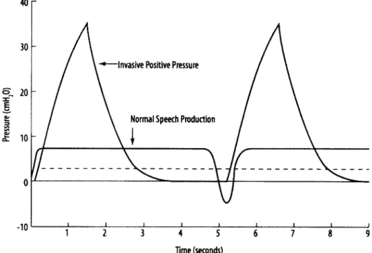
linked to the abnormal tracheal pressure associated with tracheostomy ventilation. As shown in Figure 2, tracheos-tomy-ventilation tracheal pressure (under a volume-con-trolled condition) rises quickly to a high peak, falls quickly, and remains below the minimum pressure required to vi-brate the vocal folds until the next inspiration. This is in stark contrast to the steady and low tracheal pressure as-sociated with normal speech production.
Fortunately, tracheal pressure can be modified with ven-tilator adjustments to improve speech. Perhaps the most simple and successful set of ventilator adjustments is the combination of prolonged inspiratory time and positive end-expiratory pressure (PEEP).80,84Prolonged inspiratory
time (or decreased inspiratory flow) increases the time that speech can be produced during inspiration, and PEEP in-creases the time that speech can be produced during
ex-piration. Thus, as shown in Figure 3, more speech can be produced per breath with each of these adjustments indi-vidually, and their effects are additive when they are com-bined. Although other ventilator adjustments can also im-prove speech,81,85the effectiveness of prolonged inspiratory
time and PEEP has the most empirical support. Whatever ventilator adjustments are implemented, speech may be further improved with behavioral therapy provided by a speech-language pathologist.
A common clinical approach to improving tracheosto-my-ventilation speech following cuff deflation is the in-sertion of a one-way inspiratory valve (ie, a “speaking valve”) between the tracheostomy tube and the ventilator
line. This approach carries high risks (including death) if the valve is inserted without the cuff being deflated. Ven-tilator adjustments such as those just described (prolonged inspiratory time and PEEP) are much safer than a one-way valve, and the speech can be just as good. For example, a comparison of tracheostomy ventilation with a one-way valve and with 15 cm H2O PEEP was found to generate
nearly identical tracheal-pressure waveforms (Fig. 4) and equally high-quality speech.84
Fig. 2. Phonation relative to pressure and time. The dashed line represents the minimum pressure required for phonation. See text for details. (From Reference 81, with permission.)
Fig. 3. Changes (from usual) in speaking rate with lengthened inspiratory time (TI), positive end-expiratory pressure (PEEP), and
lengthened TIplus PEEP. (From Reference 84, with permission.) Fig. 4. Tracheal pressure during speaking with a one-way valve
(top panel) versus with positive end-expiratory pressure (PEEP) set to 15 cm H2O (bottom panel). Note that the tracings are almost
indistinguishable. See text for details. (From Reference 84, with permission.)
Speech with phrenic-nerve-pacer ventilation also can be abnormal, and is usually characterized by short phrases, low auditory volume, and long inspiratory pauses.86 As
with tracheostomy-ventilation speech, adjustments to the pacer may improve speech, although this has not been proven. Speech improvements have been found with the use of an abdominal binder,87and include louder speech,
longer phrases, and better voice quality. These improve-ments are probably linked to the VTincrease caused by the
binder (ie, inward positioning of the abdominal wall by the binder stretches the diaphragm, increases its contractile force, and, as described above and in the Appendix, re-duces distortion of the rib cage). Other means of increas-ing VT, such as glossopharyngeal breathing, also hold
po-tential for improving speech in patients with phrenic-nerve pacers.
Respiratory Muscle Pacing
New techniques have provided the capacity to restore inspiratory-muscle function in SCI. Conventional bilateral phrenic-nerve pacing can allow patients with ventilator-dependent tetraplegia complete freedom from mechanical ventilation. Compared to mechanical ventilation, phrenic-nerve pacing provides better patient mobility, better com-fort level, and lower health-care costs.88,89Although this
has not been studied formally, patients often claim that their breathing feels more natural with phrenic-nerve pac-ing than with mechanical ventilation. Why this should be so is not entirely clear. Perhaps it is because of elimination of the noise associated with the mechanical ventilator and the tubing that tugs on the neck. Also, with conventional mechanical ventilation via tracheostomy tube, the inspired gas does not pass through the nose, whereas during inha-lation from phrenic-nerve stimuinha-lation, the negative pres-sure generated by diaphragm contraction causes inspired gas to pass through the nose and thereby restores the sense of smell. This may contribute substantially to the sense that the breath is more natural. Phrenic-nerve pacing usu-ally does not allow removal of the tracheostomy tube, because most patients develop obstructive sleep apnea. This is because the pacing of the diaphragm is not syn-chronous with respirophasic activation of oropharyngeal muscles that prevent airway collapse during normal breath-ing durbreath-ing sleep. It would seem possible to avoid this problem by using CPAP during sleep in phrenic-nerve paced patients, but this has not been studied.
With any method of phrenic-nerve pacing, intact phren-ic-nerve function must be assured. This is usually accom-plished by electrical stimulation of the phrenic nerves in the neck and recording the resultant compound action po-tential on the surface of the rib cage overlying the dia-phragm. An alternative method involves magnetic stimu-lation of the phrenic motoneurons. For electrode placement,
the technique of phrenic-nerve pacing generally requires a thoracotomy,90 –92which is associated with substantial
mor-bidity, high cost, and hospitalization. Also, the procedure carries a small risk of phrenic-nerve injury. Recent studies, however, suggest that phrenic-nerve stimulation can be achieved less invasively by placement of intramuscular electrodes directly into the diaphragm via laparoscopic surgery. Laparoscopy is less invasive, has cosmetic bene-fits, and can be performed as an out-patient procedure or with a single overnight observation stay, with lower over-all cost. Also, laparoscopic placement of intramuscular electrodes does not require manipulation of the phrenic nerves and therefore carries less risk of phrenic-nerve in-jury.
Animal experiments first suggested that each hemidia-phragm could be activated with intramuscular diahemidia-phragm electrodes.93Electrodes were placed on the ventral surface
of the diaphragm via a midline abdominal incision. To evaluate the efficacy of this method, transdiaphragmatic pressure generated by a single intramuscular electrode po-sitioned near the point of entrance of the phrenic nerve (motor point) was compared to transdiaphragmatic pres-sure generated by direct phrenic-nerve stimulation with cuff electrodes. The study demonstrated that intramuscular electrodes positioned within 1–2 cm of the motor point resulted in transdiaphragmatic pressure similar to the pres-sure obtained with direct phrenic-nerve stimulation (Fig. 5). Clinical trials of this technique were initially hindered by lack of the necessary tools to safely place the electrodes within the diaphragm and methods to accurately locate the diaphragm motor points. In early animal studies, laparo-scopic placement of intramuscular diaphragm electrodes often resulted in the inadvertent placement of electrodes
Fig. 5. Comparison of transdiaphragmatic pressure generated dur-ing phrenic-nerve stimulation (PS) and intramuscular diaphragm stimulation (DS) in anesthetized dogs. Supramaximal amplitudes were applied over a 10 – 40 Hz range during airway occlusion. Each number represents results from a separate animal. (From Reference 93, with permission.)
through the diaphragm and into the thoracic cavity, be-cause the laparoscope delivering the electrode approached the diaphragm almost perpendicular to the plane of the diaphragm. Therefore, an insertion instrument was designed that allowed electrode placement in the same plane as the diaphragm.94 –96With this instrument, both in animal and
clinical studies, there were not any instances of electrodes traversing the diaphragm and entering the thoracic cavity. Since uniform activation of the diaphragm requires place-ment of intramuscular electrodes near the motor points of the phrenic nerves into each hemidiaphragm, and the phrenic nerves are not visible on the abdominal surface of the diaphragm, a mapping procedure was developed to precisely determine the location of the motor points.97,98
This procedure entails evaluating several test sites in the general region of the motor points (based on previous anatomic studies) with a suction electrode that can be re-versibly applied to the diaphragm.
After extensive animal testing, studies were performed in an initial group of 5 ventilator-dependent tetraplegic subjects.99,100 Two intramuscular electrodes were placed
laparoscopically in each hemidiaphragm, to activate the diaphragm, with the goal of full-time ventilatory support. Two weeks following placement of the diaphragm elec-trodes, inspired-volume generation was evaluated with each of the 4 electrodes individually.101In each instance,
max-imum or near-maxmax-imum amplitude (24 –25 mA) was re-quired to achieve maximum inspired-volume-generation. Since patients on long-term mechanical ventilation suffer from diaphragm atrophy, a period of gradually increasing stimulation (reconditioning period) was necessary to re-store diaphragm function. Stimulation of both electrodes within each hemidiaphragm generally resulted in greater inspired volumes than did stimulation of one electrode alone, so stimulation was applied with all 4 electrodes. During the reconditioning period, attempts were made to gradually reduce the stimulus frequency to the lowest value that resulted in adequate ventilation. Respiratory rate was set at 10 –12 breaths/min and inspiratory time (train dura-tion) at 1.1 s. Initially, pacing was provided for 5–10 min
each hour for 5– 6 h/d, and gradually increased, as toler-ated. When continuous pacing for 6 – 8 hours was achieved, the number of hours per day was gradually increased.
Figure 6 shows the maximum changes in inspired vol-ume that resulted from separate left and right hemidia-phragm stimulation and bilateral stimulation, at various intervals during the reconditioning period, superimposed, from one subject. Similar results were observed in the other subjects.100 The inspired volumes achieved during
long-term pacing were in the range 800 –950 mL. Each of the subjects achieved substantial independence from me-chanical ventilatory support (ie, full-time ventilatory sup-port in 3 subjects and up to 20 h/d in the remaining sub-ject). End-tidal PCO2 during long-term pacing was 30 –
32 mm Hg. Intramuscular diaphragm pacing has since been performed on 7 additional subjects, each of whom also achieved either full-time or near-full-time indepen-dence from mechanical ventilation.100
For patients who do not have intact phrenic nerves, conventional or intramuscular diaphragm pacing is not an option. However, studies of animals with surgical phrenicectomy found that the application of electrical cur-rent on the epidural surface of the spinal cord in the upper thoracic region results in substantial inspired-volume gen-eration.101 The maximum inspired volumes were
gener-ated by stimulation at the T2-T3 region on the ventral surface (Fig. 7). Stimulation of the intercostal muscles at this level in combination with bilateral phrenic-nerve stim-ulation resulted in inspired-volume generation that approx-imated the inspiratory capacity. These studies suggested
Fig. 6. Superimposed maximum changes in inspired volume from separate left, separate right, and bilateral diaphragm stimulation, over the course of the reconditioning period of one subject. Su-pramaximal stimulus variables (24 mA or 25 mA, 50 Hz) were employed. (From Reference 100, with permission.)
Fig. 7. Effects of electrical stimulation applied at different spinal cord regions before phrenicotomy (circles) and after phrenicotomy (dots) in a dog anesthetized with pentobarbital. Inspired volume is expressed as a percentage of maximum. Inspired-volume gener-ation was greatest in the vicinity of the T2-T3 spinal-cord region and decreased progressively at spinal levels cephalad and caudad to that region. Stimulation was applied with supramaximal stimu-lus amplitude and stimustimu-lus frequency of 50 Hz. (From Reference 101, with permission.)
that near-maximal, coordinated activation of the inspira-tory intercostal muscles could be accomplished by electri-cal stimulation with a single electrode.101Because the
mech-anism of intercostal activation involves direct stimulation of thoracic ventral roots, this technique is referred to as ventral root stimulation.
An initial clinical trial of ventral root stimulation was undertaken to provide ventilatory support in ventilator-dependent tetraplegic patients who were not candidates for phrenic-nerve pacing.102 An epidural disc electrode was
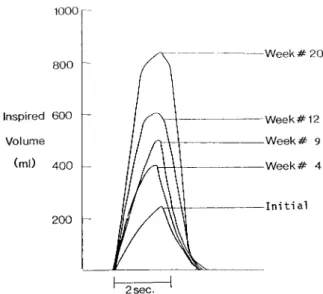
positioned on the ventral surface at the T2-T3 level of the spinal cord, through a cervical hemilaminectomy. Initial ventral root stimulation resulted in very small inspired volumes (150 –240 mL), most likely secondary to muscle atrophy. Following a reconditioning program, there were substantial increases in maximum inspired volumes (to 470 – 850 mL). Figure 8 shows an example of the changes in inspired volume over the course of the conditioning program. The maximum time that patients could be main-tained off mechanical ventilation, however, was a few hours. This study demonstrated that intercostal muscle pac-ing alone does not result in sufficient inspired-volume pro-duction to support ventilation for prolonged periods.
There are a number of factors that may have accounted for the different outcome of the clinical trial, compared to the previous animal studies.103 An important factor that
may have decreased inspired-volume production in the human trial is the reduction in rib-cage and lung
compli-ance in patients with tetraplegia.3,11It is also possible that
the intercostal muscles were not activated to the same extent in humans. The differences in shape of the thorax between species may also be important. During inspira-tion, there is greater expansion of the anteroposterior di-ameter in the quadruped thorax. These differences may provide the intercostal muscles of the dog with a mechan-ical advantage over humans.
Though not successful in maintaining long-term venti-latory support, ventral root stimulation did result in sub-stantial inspired-volume production. The method was ap-plied in a second clinical trial of 4 patients with intact unilateral phrenic-nerve function.104 Unilateral
phrenic-nerve pacing does not provide VT sufficient to support
ventilation. It was thought that intercostal pacing com-bined with unilateral phrenic-nerve pacing could provide long-term ventilatory support. Spinal-cord electrodes were positioned at the T2-T3 spinal-cord level. A phrenic-nerve electrode was placed unilaterally, via the thoracic approach, to pace the diaphragm. With reconditioning, the maximum inspired volumes resulting from combined intercostal and diaphragm stimulation gradually increased to between 550 mL and 1,310 mL. With the lower stimulus frequen-cies employed with long-term pacing, inspired volumes during long-term pacing ranged between 350 mL and 850 mL. Each patient was able to achieve full-time or near-full-time ventilatory support. End-tidal PCO2 was in
the range of 33–35 mm Hg during long-term pacing. As with diaphragm pacing, the subjects reported improved sense of smell, mobility, and overall sense of well being. In fact, one patient achieved employment as a result. This technique was associated with some adverse effects, in-cluding mild hand flexion.102 Also, with high stimulus
currents, one of the subjects experienced symptoms sug-gestive of autonomic dysfunction, including headache, flushing, and diaphoresis. Reducing the stimulus current eliminated the symptoms.
Perhaps the single most important impairment of the respiratory system that occurs in SCI is ineffective cough due to dysfunction of the abdominal and internal intercos-tal muscles. To address this problem, the technique of direct stimulation of the spinal cord by epidural electrodes has now been extended to produce vigorous contraction of the abdominal muscles.105 In one subject, during
com-bined stimulation at the T9 and L1 levels, peak airway pressure and peak expiratory flow were 132 cm H2O and
7.4 L/s, respectively. These are within the range of pre-dicted values for an able-bodied person. The subject is able to trigger the stimulation on his own and, as a result, does not require assistance for airway clearance. The tech-nique may have a substantial impact on the outcome of respiratory-tract infections in SCI.
In summary, the successful development of new tech-niques and approaches to restore inspiratory and
expira-Fig. 8. Maximum inspired volumes with intercostal stimulation at various times during the reconditioning period of one patient. As a result of gradually increasing the pacing duration, there were pro-gressive increments in inspired volume. Because inspiratory time was fixed, increases in inspired volume were achieved by increases in inspiratory flow. The improvement in inspired volume is attrib-utable to the training effect of long-term stimulation and reversal of intercostal muscle atrophy. (From Reference 102, with permis-sion.)
tory muscle activation has expanded the options for pa-tients with SCI. Whether these methods will reduce the need for mechanical ventilation, reduce the incidence of respiratory complications, and significantly impact the mor-bidity and mortality of high SCI remains to be determined.
Assisted Cough
Impaired cough leads to accumulation of secretions that arise from respiratory infections or that are due to the presence of a tracheostomy tube. Several methods are avail-able to deal with the secretions. Each may be performed in association with conventional chest physical therapy.
Spinal-Cord Stimulation. The new technique described above, involving stimulation of the spinal cord at T9 and L1, is still being developed105but shows great promise.
Spontaneous Cough. For mild and nonviscous secre-tions, spontaneous coughs may suffice. Often, the effec-tiveness is enhanced if the seated subject lurches forward. This maneuver raises intra-abdominal pressure, which is transmitted to the thorax, enhancing expiratory flow.
Quad Cough. A related maneuver known as a “quad cough” is also often effective. The subject is placed in the supine or slightly upright posture and is straddled by the therapist whose hands are placed under the left and right costal margins. Then the subject inhales to total lung ca-pacity and, upon a signal, begins to cough. The therapist enhances the cough effort by vigorous pressure applied to the abdomen, in the rhythm of a cough, and the process is continued to low lung volume. The forceful application of pressure to the abdomen greatly enhances expiratory flow and mobilization of secretions. Indeed, maximum expira-tory flow and the phenomenon of expiraexpira-tory flow limita-tion that is essential to an effective cough can be achieved when the maneuver is done properly (unpublished data from Robert Brown [author], and Steven M Scharf MD, University of Maryland, Baltimore, Maryland). Contrain-dications to performing quad coughs are the presence of an inferior vena cava filter or abdominal aortic aneurysm or prosthesis.
Suction Catheter. Other methods to enhance cough uti-lize negative pressure applied at the mouth or orifice of the endotracheal tube or tracheostomy tube. The most com-mon device is a standard suction catheter, which is con-venient when there is a tracheal tube in place. When there is not, nasotracheal or orotracheal suctioning can be done, but this is uncomfortable and can lead to trauma to the nose, pharynx, and larynx. The usefulness of the standard suction catheter is limited, because it barely reaches the right main bronchus and does not enter the left main
bron-chus. Versions of the catheter that can be turned to enter the proximal portion of the left main bronchus are not in common use and are of unproven efficacy. In addition, each use of a suction catheter introduces bacteria to the tra-chea and large bronchi, with the associated risk of infection.
Mechanical In-exsufflation. The latter limitations can be overcome with the use of a mechanical insufflation-exsufflation device (CoughAssist, JH Emerson, Cambridge, Massachusetts). This device is particularly effective when used attached to a tracheal tube. When applied to the mouth, the negative-pressure phase can be uncomfortable or cause collapse of the pharyngeal airway, particularly in patients with dysfunction of the oropharyngeal muscles, such as due to amyotrophic lateral sclerosis. The device can be used frequently. For secretion clearance in patients with neu-romuscular disease, one of us (Brown) has used it as often as a patient with excessive secretions would ordinarily cough. For example, use every 5 minutes for an hour may be very effective in clearing retained secretions. The clinical effec-tiveness of the device has been demonstrated.106,107
Bronchoscopy. Perhaps the most-effective method of secretion clearance is bronchoscopy, and this should be used when other methods have been inadequate. The ex-perience of one of us (Brown) has been that performing quad coughs during the bronchoscopy mobilizes secre-tions from airways that are not bronchoscopically visible and thereby enhances secretion clearance. During the quad cough, the bronchoscope must be poised to suction the secretions that are “milked” into the visible airways, lest those secretions migrate distally and out of vision during the subsequent inhalation.
Mechanical Ventilation
The care of patients with high-level SCI is made more complex by the need for mechanical ventilation. Although most patients are ventilated via tracheostomy tube, Bach and colleagues found that patients with SCI and other patients with neuromuscular disorders can be successfully decannulated and treated with noninvasive ventila-tion.108 –111 Patient-ventilator interfaces for such
ventila-tion are reviewed elsewhere in this issue of RESPIRATORY
CARE.112 Patients seem to prefer noninvasive ventilation
for reasons of appearance, comfort, swallowing, and speech.113Other advantages appear to be reduction in
se-cretions because the tracheostomy tube is not present to irritate the trachea, fewer hospitalizations, and lower cost of care.114More attention should be paid to the option of
noninvasive ventilation, especially given the considerable literature supporting this approach.
Mechanically ventilated patients typically prefer large VT. The reason is not established, but it may be related to
relief of the dyspnea that is associated with small tidal breaths.65Large breaths also have the benefit of
prevent-ing small-airway narrowprevent-ing or closure by stretchprevent-ing air-way smooth muscle, and by reducing surface tension by expanding the surface area of pulmonary surfactant. Breath sizes as large as 1.0 L (often with PEEP of 5 cm H2O) are
common and do not cause ventilator-associated lung dam-age in the absence of acute lung injury from other causes. The common result is low PaCO2, but there are not any
known deleterious long-term consequences of this.
Summary
Respiratory dysfunction is a major cause of morbidity and mortality in SCI. In the last several decades, much has been learned about the mechanisms of respiratory dys-function, and this has led to useful interventions. Respira-tory-muscle training improves VC. A multicenter effort to assess the effect of respiratory-muscle training on prob-lems such as dyspnea and weak cough is warranted. Res-piratory-muscle pacing has shown promise in liberating patients from mechanical ventilation and in improving cough. Several techniques are now available to clear se-cretions, including mechanical in-exsufflation, which ap-pears to be underutilized in both the acute and chronic settings. For those who require assisted ventilation and have tracheotomies, the quality of speech production can be improved dramatically with simple adjustments of the ventilator settings. Noninvasive methods to provide as-sisted ventilation are also effective, yet underutilized. With further research, there will perhaps be enough progress to substantially reduce the morbidity and mortality from re-spiratory dysfunction in SCI.
REFERENCES
1. Spinal cord injury: facts and figures at a glance. National Spinal Cord Injury Center Statistical Center, Birmingham, Alabama. J Spi-nal Cord Med 2006;29(4):379–80.
2. ASIA impairment scale, clinical syndromes, and standard neuro-logical classification of spinal cord injury. http://www.asia-spinalinjury.org/publications/index.html.
3. Scanlon PD, Loring SH, Pichurko BM, McCool FD, Slutsky AS, Sarkarati M, Brown R. Respiratory mechanics in acute quadriplegia: lung and chest wall compliance and dimensional changes during re-spiratory maneuvers. Am Rev Respir Dis 1989;139(3):615–620. 4. Sharp JT, Sweany SK, VanLith P. Physiologic observations in
dif-fuse pulmonary fibrosis and granulomatosis. Am Rev Respir Dis 1966;94(3):316–331.
5. Gibson GJ, Pride NB. Lung distensibility: the static pressure-vol-ume curve of the lungs and its use in clinical assessment. Br J Dis Chest 1976;70(3):143–184.
6. Gibson GJ, Pride NB, Davis JN, Loh LC. Pulmonary mechanics in patients with respiratory muscle weakness. Am Rev Respir Dis 1977;115(3):389–395.
7. Bergofsky EH. Respiratory insufficiency in mechanical and neuro-muscular disorders of the thorax. In: Fishman AP, editor. Pulmo-nary diseases and disorders. New York: McGraw-Hill; 1980:1559.
8. Bergofsky EH. Respiratory failure in disorders of the thoracic cage. Am Rev Respir Dis 1979;119(4):643–669.
9. Bergofsky EH. Mechanism for respiratory insufficiency after cer-vical cord injury: a source of alveolar hypoventilation. Ann Intern Med 1964;61:435–447.
10. Stone DJ, Keltz H. The effect of respiratory muscle dysfunction on pulmonary function: studies in patients with spinal cord injuries. Am Rev Respir Dis 1963 Nov;88:621–629.
11. DeTroyer A, Heilporn A. Respiratory mechanics in quadriplegia: the respiratory function of the intercostal muscles. Am Rev Respir Dis 1980;122(4):591–600.
12. Young SL, Tierney DF, Clements JA. Mechanism of compliance changes in excised rat lungs at low transpulmonary pressure. J Appl Physiol 1970;29(6):780–785.
13. Grinton SF, Hyatt RE, Rohrback MS, Deschamps C. Causes of pressure volume curve shifts in chest restricted dogs. Fed Proc 1987;46:1108.
14. DeTroyer A, Pride NB. The respiratory system in neuromuscular disease. In: Roussos C, Macklem PT, editors. The thorax. (Lung biology in health and disease, vol 85). New York: Marcel Dekker; 1985:1089–1121.
15. Goldman JM, Williams SJ, Denison DM. The rib cage and abdom-inal components of respiratory system compliance in tetraplegic patients. Eur Respir J 1988;1(3):242–247.
16. Estenne M, DeTroyer A. The effect of tetraplegia on chest wall statics. Am Rev Respir Dis 1986;134(1):121–124.
17. Goldman JM, Rose LS, Morgan MD, Denison DM. Measurement of abdominal well compliance in normal subjects and tetraplegic patients. Thorax 1986;41(7):513–518.
18. Urmey W, Loring S, Mead J, Slutsky AS, Sarkarati M, Rossier A, Brown R. Upper and lower rib cage deformation during breathing in quadriplegics. J Appl Physiol 1986;60(2):618–622.
19. Derenne J, Macklem PT, Roussos C. The respiratory muscles: me-chanics control and pathophysiology. Am Rev Respir Dis 1978; 118(1):119–133.
20. Fugl-Meyer AR, Grimby G. Rib cage and abdominal volume ven-tilation partitioning in tetraplegic patients. Scand J Rehab 1971; 3(4):161–167.
21. Mortola JP, Sant’Ambrogio G. Motion of the rib cage and the abdo-men in tetraplegic patients. Clin Sci Mol Med 1978;54(1):25–32. 22. Moulton A, Silver JR. Chest movements in patients with traumatic
injuries of the cervical cord. Clin Sci 1970;39(3):407–422. 23. Danon J, Druz WS, Goldberg NB, Sharp JT. Function of the
iso-lated paced diaphragm and the cervical accessory muscles in C1 quadriplegics. Am Rev Respir Dis 1979;119(6):909–919. 24. Braun NM, Arora NS, Rochester DF. Force-length relationship of
the normal human diaphragm. J Appl Physiol 1982;53(2):405–412. 25. Kim MJ, Druz WS, Danon J, Machnach W, Sharp JT. Mechanics of
the canine diaphragm. J Appl Physiol 1976;41(3):369–382. 26. de Haan A, de Jong J, van Doorn JE, Huijing PA, Woittiez RD,
Westra HG. Muscle economy of isometric contractions as function of stimulation time and relative muscle length. Pfluegers Arch 1986; 407(4):445–450.
27. Manning H, McCool FD, Scharf SM, Garshick E, Brown R. Oxy-gen cost of resistive-loaded breathing in quadriplegia. J Appl Physiol 1992;73(3):825–831.
28. Bluechardt MH, Wiens M, Thomas SG, Plyley MJ. Repeated mea-surement of pulmonary function following spinal cord injury. Para-plegia 1992;30(11):768–774.
29. Oo T, Watt JW, Soni BM, Sett PK. Delayed diaphragm recovery in 12 patients after high cervical spinal cord injury: a retrospective review of the diaphragm status of 107 patients ventilated after acute spinal cord injury. Spinal Cord 1999;37(2):117–122.
30. McKinley WO. Late return of diaphragm function in a ventilator-dependent patient with a high cervical tetraplegia: case report, and interactive review. Spinal Cord 1996;34(10):626–629.
31. Lieberman J, Corkill G, Nayak NN, French BN, Taylor RG. Serial phrenic nerve conduction studies in candidates for diaphragmatic pacing. Arch Phys Med Rehab 1985;61(11):528–531.
32. Silver JR, Lehr RP. Electromyographic investigation of the dia-phragm and intercostal muscles in tetraplegics. J Neurol Neurosurg Psychiatry 1981;44(9):837–842.
33. Frisbie JH, Brown R. Waist and neck enlargement after quadriple-gia. J Am Paraplegia Soc 1994;17(4):177–178.
34. Axen K, Pineda H, Shunfenthal I, Haas F. Diaphragmatic function following cervical cord injury: neurally mediated improvement. Arch Phys Med Rehabil 1985;66(4):219–222.
35. Brown R, Loring SH, Pichurko BM, Scanlon PD, Slutsky AS, Sarkarati M, et al. The role of respiratory muscles in the recovery of respiratory function in acute tetraplegia. J Spinal Cord Med. In press 2006.
36. Standardization of spirometry, 1994 Update. American Thoracic Society Am J Respir Crit Care Med 1995;152(3):1107–1136. 37. Kelley A, Garshick E, Gross ER, Lieberman SL, Tun CG, Brown
R. Spirometry testing standards in spinal cord injury. Chest 2003; 123(3):725–730.
38. Fugl-Meyer AR. Effects of respiratory muscle paralysis in tetraplegic and paraplegic patients. Scand J Rehabil Med 1971;3(4):141–150. 39. Fugl-Meyer AR, Grimby G. Ventilatory function in tetraplegic
pa-tients. Scand J Rehabil Med 1971;3(4):151–160.
40. Kokkola K, Moller K, Lehtonen T. Pulmonary function in tetraple-gic and parapletetraple-gic patients. Ann Clin Res 1975;7(2):76–79. 41. Ohry A, Molho M, Rozin R. Alterations of pulmonary function in
spinal cord injured patients. Paraplegia 1975;13(2):101–108. 42. Forner JV. Lung volumes and mechanics of breathing in
tetraple-gics. Paraplegia 1980;18(4):258–266.
43. Haas F, Axen K, Pineda H, Gandino D, Haas A. Temporal pulmo-nary function changes in cervical cord injury. Arch Phys Med Re-hab 1985;66(3):139–144.
44. Almenoff PL, Spungen AM, Lesser M, Bauman WA. Pulmonary function survey in spinal cord injury: influences of smoking and level and completeness of injury. Lung 1995;173(5):297–306. 45. Linn WS, Spungen AM, Gong H Jr, Bauman WA, Adkins RH, Waters
RL. Smoking and obstructive lung dysfunction in persons with chronic spinal cord injury. J Spinal Cord Med 2003;26(1):28–35.
46. Jain NB, Brown R, Tun CG, Gagnon D, Garshick E. Determinants of FEV1, FVC, and FEV1/FVC in chronic spinal cord injury. Arch Phys Med Rehabil. In press 2006.
47. Samsa GP, Patrick CH, Feussner JR. Long-term survival of veterans with traumatic spinal cord injury. Arch Neurol 1993;50(9):909–914. 48. DeVivo MJ, Stover SL. Long-term survival and causes of death. In:
Stover SL, DeLisa JA, Whiteneck GG, editors. Spinal cord injury: clinical outcomes from the models systems. Gaithersburg MD: As-pen Publishers; 1995:289–316.
49. Mesard L, Carmody A, Mannarino E, Ruge D. Survival after spinal cord trauma: a life table analysis. Arch Neurol 1978;35(2):78–83. 50. Soden RJ, Walsh J, Middleton JW, Craven ML, Rutkawki SB, Yeo JD. Causes of death after spinal cord injury. Spinal Cord 2000; 38(10):604–610.
51. Hartkopp A, Bronnum-Hansen H, Seidenschnur AM, Biering-So-rensen F. Survival and cause of death after traumatic spinal cord injury: a long-term epidemiological survey from Denmark. Spinal Cord 1997;35(2):76–85.
52. Yeo JD, Walsh J, Rutkowski S, Soden R, Craven M, Middleton J. Mortality following spinal cord injury. Spinal Cord 1998;36(5): 329–336.
53. Frankel HL, Coll JR, Charlifue SW, Whiteneck GG, Gardner BP, Jamous MA, et al. Long-term survival in spinal cord injury; a fifty year investigation. Spinal Cord 1998;36(4):266–274.
54. Whiteneck GG, Charlifue SW, Frankel HL, Fraser MH, Gardner BP, Gerhart KA, et al. Mortality, morbidity, and psychosocial out-comes of persons spinal cord injured more than 20 years ago. Paraplegia 1992;30(9):617–630.
55. McKinley AC, Auchicloss JH Jr, Gilbert R, Nicholas JJ. Pulmonary function, ventilatory control, and respiratory complications in quad-riplegic subjects. Am Rev Respir Dis 1969;100(4):526–532. 56. Garshick E, Kelley A, Cohen SA, Garrison A, Tung CG, Gagnon
D, Brown R. A prospective assessment of mortality in chronic spinal cord injury. Spinal Cord 2005;43(7):408–416.
57. Lieberman SL, Mourad I, Brown R, Schwartzstein RM. Spinal cord injury diminishes both the ventilatory response and air hunger due to steady state hypercapnia (abstract). Am Rev Respir Dis 1993; 147:A550.
58. Spungen AM, Grimm DR, Lesser M, Bauman WA, Almenoff PL. Self-reported prevalence of pulmonary symptoms in subjects with spinal cord injury. Spinal Cord 1997;35(10):652–657.
59. Ayas NT, Garshick E, Lieberman SL, Wien MF, Tun C, Brown R. Breathlessness in spinal cord injury depends on injury level. J Spi-nal Cord Med 1999;22(2):97–101.
60. Grandas NF, Jain NB, Denckla JB, Brown R, Tun CG, Gallagher ME, Garshick E. Dyspnea during daily activities in chronic spinal cord injury. Arch Phys Med Rehab 2005;86(8):1631–1635. 61. Burkett LN, Chisum J, Stone W, Fernhall B. Exercise capacity of
untrained spinal cord injured individuals and the relationship of peak oxygen uptake to the level of injury. Paraplegia 1990;28(8): 512–521.
62. Irizawa M, Yamasaki M, Muraki S, Komura T, Seki K, Kikuchi K. Relationship between heart rate and oxygen uptake during sub-maximal arm cranking in paraplegics and quadriplegics. Ann Physio Anthrop 1994;13(5):275–280.
63. Coutts KD, Rhodes EC, McKenzie DC. Maximal exercise responses of tetraplegics and paraplegics. J Appl Physiol 1983;55(2):479–482. 64. Figoni S. Exercise responses and quadriplegia. Med Sci Sports
Exerc 1993;25(4):433–441.
65. Manning HL, Shea SA, Schwartzstein RM, Lansing RW, Brown R, Banzett RB. Reduced tidal volume increases “air hunger” at fixed PCO2in ventilated quadriplegics. Respir Physiol 1992;90(1):19–30. 66. Van loan MD, McCluer S, Loftin JM, Boileau RA. Comparison of physiological responses to maximal arm exercise among able-bod-ied, paraplegics and quadriplegics. Paraplegia 1987;25(5):397–405. 67. Bailey ER, Hoit JD. Speaking and breathing in high respiratory
drive. J Speech Lang Hear Res 2002;45(1):89–99.
68. Carrieri-Kohlman V, Gormley JM, Douglas MK, Paul SM, Stul-barg MS. Exercise training reduces dyspnea and the distress and anxiety associated with it: monitoring alone may be as effective as coaching. Chest 1996;110(6):1526–1535.
69. Wien MF, Garshick E, Tun CG, Lieberman SL, Kelley A, Brown R. Breathlessness and exercise in spinal cord injury. J Spinal Cord Med 1999;22(4):297–302.
70. Leith DR, Bradley M. Ventilatory muscle strength and endurance training. J Appl Physiol 1976;41(4):508–516.
71. Loke J, Mahler DA, Virgulto JA. Respiratory muscle fatigue after marathon running. J Appl Physiol 1982;52(4):821–824.
72. Gross D, Ladd HW, Riley EJ, Macklem PT, Grassino A. The effect of training on strength and endurance of the diaphragm in quadri-plegia. Am J Med 1980;68(1):27–35.
73. Kogan I, McCool FD, Liberman SL, Garshick E, Shannon K, Fris-bee JH, Brown R. Diaphragm hypertrophy during inspiratory mus-cle training in tetraplegia (abstract). Am J Respir Crit Care Med. 1996;153(4):A25.