Page | 149
Finger Force Sensor Instrumentation Design
Jose Agraz
12, Alexander Grunfeld
12, Daniel Muse
3, Robert Pozos
31Department of Bioengineering, University of California, Los Angeles, CA
2Cedars Sinai Medical Center, West Hollywood, CA
3Department of Biology, San Diego State University, CA
Abstract: Carpal tunnel syndrome (CTS) refers to a class of injuries characterized by compression of the median nerve in the carpal tunnel. CTS injuries cause pain, weakness, and numbness in the hands and are common among video gamers and workers that maintain their wrists in ergonomically unfavorable positions. Current CTS qualification methods are well documented, whereas the condition of the hand muscles is often overlooked. At present, there is no simple, inexpensive, and portable instrument that quantifies finger force values pre and post clinical intervention. Our design measures finger force using force sensors placed on the surface of individual keys on an ergonomically designed keyboard, which allows finger force measurements for all five fingers, individually or as a group. In this work we; 1) Show a clear difference in finger force between an uninjured and injured hand and further correlated it to forearm electromyography (EMG), and 2) Use the system in a pilot study to evaluate the apparatus usefulness through the testing of the significance of finger force before and after fatigue. Finger force signals from the subjects' index finger were collected and force peaks extracted from the raw data. The resulting data was analyzed using the variance (ANOVA) test. Although, there was no statistical significance between the finger force applied, the system proved accurate, rugged, and flexible to accommodate fine tuning changes on hardware and software, with a minimum of dead time during the pilot study. Thus, this tool offers a great platform for further finger force studies.
Keywords: LabVIEW, Instrumentation, Finger, Carpal Tunnel Syndrome, Force, Control.
Introduction
Repetitive activities performed over an extended period of time with constant excessive load or effort and poor body mechanics are causes of Repetitive Stress Injury (RSI). When RSI occurs at the wrist and/or fingers, persons complain of numbness, tingling, and pain in the area of the thumb, index, and middle fingers and in some cases, a burning sensation, weakness, and clumsiness [1]. The pain often increases at night and can radiate to the forearm, upper arm, and neck [2]. As the injury progresses, there is a decrease in hand muscle strength and reduced finger range of motion. RSI symptoms maybe manifested by carpal tunnel syndrome (CTS), defined as the narrowing of the tunnel formed by the wrist (carpal) bones through which the median nerve travels. The compression of the median nerve influences its sensory and motor innervations to the fingers (except for the little finger) causing the aforementioned symptoms. The U.S. Department of Labor (DoL) concluded that CTS is the "Chief occupational hazard", affecting eight million Americans and accounting for 41% of all work-related injuries. It is estimated that 25% of all computer operators have CTS and by the year 2014, the DoL estimates over 50% of the workforce may be affected. Approximately 20,000 medical procedures are performed every year to correct various aspects of CTS. However, only 23% of all CTS patients are able to return to their previous professions after surgery [3].
Presently, the diagnosis of CTS remains controversial [4-6]. Neither hand maneuvers nor NCV instrumentation mimic real world situations, and their usefulness as a diagnostic tool is questionable [7]. Atcheson, Ward and Lowe evaluated 297 patients, of which 38% were diagnosed with CTS and studied for any underlying pathologies that may contribute to the diagnosis of CTS. They concluded that a person with CTS is one who complains only of pain in their upper limb [8]. There are subjective criteria for diagnosis CTS: 1) The national institutes of occupational safety and health (NIOSH) criteria for diagnosis of work-related CTS [9], 2) Examiner's global assessment of CTS made by the examining physician, paired with clinical CTS criteria, and a distribution of the median nerve spreading of neuropathic symptoms [10]. The diagnosis of CTS is difficult [7], however, through the years researchers have designed different methods to quantify CTS. Examples include: hand maneuvers, nerve conduction velocity (NCV), and finger force measuring instrumentation.
A. Hand maneuvers
Hand maneuvers were the first type of CTS diagnostic tools, such as the Phalen [11], Tinel [12], and Durkan test [13] (a slight variation of the Tinel test). These tests maneuver the patient's hand into specific positions to elicit particular
Page | 150 symptoms. Although the Phalen's test has become obsolete, the Tinel's test is still used. Phalen's test is performed by having the patient hold the wrist in a flexed position for 60 seconds, then CTS may be diagnosed if the patient feels any hand pain or paralysis. To perform Tinel's test, the clinician taps the patient on the area surrounding the median nerve, the test is positive if the patient reports a tingling sensation in the fingers. However, Both hand maneuvers tests vary in specificity and sensitivity, making the tests an inefficient diagnostic tool [14].
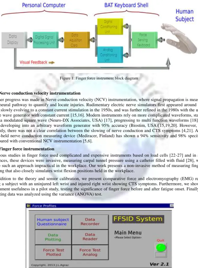
Figure 1: Finger force instrument block diagram
B. Nerve conduction velocity instrumentation
Further progress was made in Nerve conduction velocity (NCV) instrumentation, where signal propagation is measured in a neural pathway to quantify and locate injuries. Rudimentary electric nerve simulators first appeared around 1750 [15], slowly evolving to a constant current stimulation in the 1950s, and was further refined in the 1980s with the use of a sine wave generator with constant current [15,16]. Modern instruments rely on more complicated waveforms, starting with a modulated square wave (Neuro-DX Associates, USA) [17], progressing to multi function waveforms [18], and later developing into an arbitrary waveform generator with 95% accuracy (Biostim, USA)[15,19,20]. However, until recently, there was not a clear correlation between the slowing of nerve conduction and CTS symptoms [4,21]. A new hand-held nerve conduction measuring device (Mediracer, Finland) has shown a 94% sensitivity and 98% specificity compared with conventional NCV instrumentation [5,6].
C. Finger force instrumentation
Previous studies in finger force used complicated and expensive instruments based on load cells [22-27] and in some instances, these devices were invasive, measuring carpal tunnel pressure using a catheter filled with fluid [28], which made such an approach impractical in the workplace. Our work presents a non-invasive method of measuring fingertip loading that also closely simulates wrist flexion positions held in the workplace.
In addition to the theory and sensor calibration, we present comparative force and electromyography (EMG) results using a subject with an uninjured left wrist and injured right wrist showing CTS symptoms. Furthermore, we show the instrument usefulness in a pilot study, testing the significance of finger force before and after fatigue onset. Finally, the resulting data was analyzed using the variance (ANOVA) test.
Page | 151 Methodology
The system measures finger force by using five resistive-based technology force sensors placed on the surface of individual keys to measure finger force, for all five fingers, individually or in a group (Fig 1).
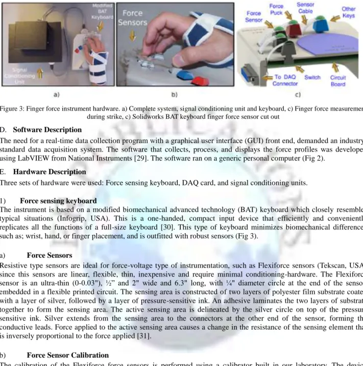
Figure 3: Finger force instrument hardware. a) Complete system, signal conditioning unit and keyboard, c) Finger force measurement during strike, c) Solidworks BAT keyboard finger force sensor cut out
D. Software Description
The need for a real-time data collection program with a graphical user interface (GUI) front end, demanded an industry-standard data acquisition system. The software that collects, process, and displays the force profiles was developed using LabVIEW from National Instruments [29]. The software ran on a generic personal computer (Fig 2).
E. Hardware Description
Three sets of hardware were used: Force sensing keyboard, DAQ card, and signal conditioning units. 1) Force sensing keyboard
The instrument is based on a modified biomechanical advanced technology (BAT) keyboard which closely resembles typical situations (Infogrip, USA). This is a one-handed, compact input device that efficiently and conveniently replicates all the functions of a full-size keyboard [30]. This type of keyboard minimizes biomechanical differences such as; wrist, hand, or finger placement, and is outfitted with robust sensors (Fig 3).
a) Force Sensors
Resistive type sensors are ideal for force-voltage type of instrumentation, such as Flexiforce sensors (Tekscan, USA) since this sensors are linear, flexible, thin, inexpensive and require minimal conditioning-hardware. The Flexiforce sensor is an ultra-thin (0-0.03"), ½‖ and 2" wide and 6.3" long, with ¼" diameter circle at the end of the sensor, embedded in a flexible printed circuit. The sensing area is constructed of two layers of polyester film substrate coated with a layer of silver, followed by a layer of pressure-sensitive ink. An adhesive laminates the two layers of substrate together to form the sensing area. The active sensing area is delineated by the silver circle on top of the pressure sensitive ink. Silver extends from the sensing area to the connectors at the other end of the sensor, forming the conductive leads. Force applied to the active sensing area causes a change in the resistance of the sensing element that is inversely proportional to the force applied [31].
b) Force Sensor Calibration
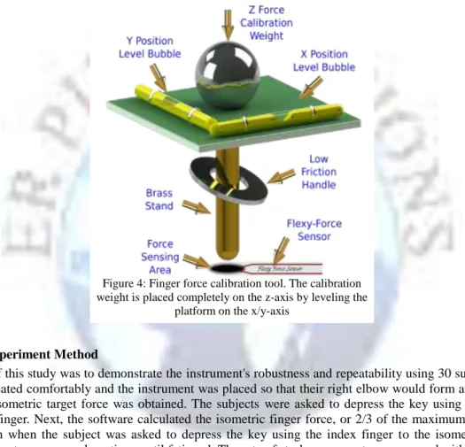
The calibration of the Flexiforce force sensors is performed using a calibrator built in our laboratory. The device consists of a rectangular platform with two bubble levels (x & y axis) attached to the platform surface. In addition, attached to the platform underside is a 4" brass bolt. The bolt tip is placed over each key cap (Fig 4). Weights are placed on top of the platform and corresponding voltages are recorded. The calibrator is positioned on top of the keycap puck. The Flexiforce sensor only measures straight-down (z-axis) force, therefore, both bubble levels must be at center to minimize stray x/y-axis forces that may introduce errors to the force measurement. Stainless steel weights were placed incrementally according to weight onto the rectangular platform and the corresponding voltages were recorded. Experiments using this calibration tool were repeated a minimum of three times on different days. The output voltage data was then converted to Newtons (N) using Newton's formula for force, followed by a calculation of the line slope and storage of this data as a calibration coefficient
F. NI Data Acquisition Cards
The Data Acquisition (DAQ) PI-MIO E card is a jumperless, switchless data acquisition board that uses the DAQ-STC as the system timing control (National Instruments, USA). DAQ-STC is the backbone of the sensor system and the timing control application specific integrated circuit (ASIC). The DAQ runs at a maximum speed of 250KHz, collecting data using ten analog channels, five force channels and five switch channels. These channels sample at a 1KHz
Page | 152 samples/second. The DAQ is set up as a non-referenced single-ended (NRSE) using one analog channel input line. The output connects to the positive input of the Programmable Gain Instrumentation Amplifier (PGIA) and the negative input of the PGIA connects to the analog input sense (AISENSE) connection.
G. Injured/uninjured Hand Experiment Method
The objective of this study was to demonstrate the instrument's practicality in diagnosing CTS. A human subject having been diagnosed with CTS symptoms on the right wrist was selected from the subject pool. She was a healthy female of 40 years old with an uninjured left wrist and an injured right wrist showing CTS symptoms, where the injury was caused from a fall a month previously. In order to further correlate finger force to muscle function, we also collected EMG signals from the right and left forearms using the Biomonitor ME6000 (Mega, Finland) at a 1KHz sampling rate. The subject was seated comfortably and the instrument was placed so that their right/left elbow would form a right angle (Fig 3). Then, the subject was asked to press on the keyboard keys sequentially using all five fingers for 60 seconds. First tapping the keys using the uninjured left hand and then using the injured right hand. Finger force and EMG data were stored in digital form and plotted using a Matlab (Mathworks, USA) program developed in our laboratory.
H. Fatigue Experiment Method
The objective of this study was to demonstrate the instrument's robustness and repeatability using 30 subjects. First, the subjects were seated comfortably and the instrument was placed so that their right elbow would form a right angle (Fig 3)). Then, the isometric target force was obtained. The subjects were asked to depress the key using maximum force with the index finger. Next, the software calculated the isometric finger force, or 2/3 of the maximum force. The data collection began when the subject was asked to depress the key using the index finger to the isometric target force shown on a computer screen and continue until fatigued. The rate of stroke was one tap per second with rhythm set by a metronome. The first step for the analysis of force data was performed by a force peak search analysis program developed in our laboratory [29]. This program reads the force data recorded and saves the maximum force value for each repetition. Then, the maximum force values were stored in American Standard Code for Information Interchange (ASCII) form for further analysis. We hypothesized that because of the index finger fatigue, the force level applied during the last 25 repetitions would be significantly less than the force applied during the first 25 repetitions. To test this hypothesis, an analysis of the variance (ANOVA) test was performed on the subject force averages using a statistical package for the social sciences (SPSS) 7.5 (IBM, USA). The sources of variance used in the analysis were the first 25 repetition force averages and the last 25 repetition force averages of all 30 subjects. The sources of variance were analyzed to determine whether fatigue (last 25 repetitions) has any significant influence on the force applied.
I. Recruitment of Subjects
The recruitment of human subjects was approved by San Diego State University's Human Use Committee. All 30 subjects pool was composed of 28 males and 2 females, ranging in ages from 21 to 40 years, were San Diego State University electrical engineering students. They were each asked to read and sign a consent form and participated in the pilot test that lasted an average of 15 minutes.
Figure 4: Finger force calibration tool. The calibration weight is placed completely on the z-axis by leveling the
Page | 153 Results
A. Force Sensors Calibration
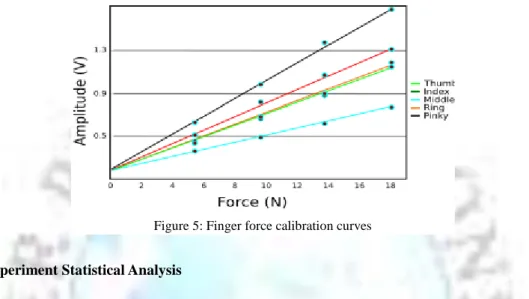
The calibration results show a linear response for all five force sensors (Fig 5) with a slopes varying from 1/40 to 1/16. The slopes were stored in the software and used as calibration coefficients. No significant change in calibration slope coefficients was found throughout the studies.
B. Injured Hand Experiment
Following isometric force measurements, the comparison between the uninjured hand and injured hand yielded a large decrease in force and EMG signal amplitude (Fig 6). The thumb and index fingers showed the smallest decreased in force by -75%, while the pinky the highest decrease of -87%. However, the subject was not able to tap on the keyboard using the middle finger on the left hand. The average forearm EMG signal decreased by -56%, table I
C. Fatigue Experiment Statistical Analysis
The first 25 repetitions of the force applied for all 30 subjects showed an µ=1/4N and a σ=0.15. For the last 25 repetitions of the force applied by the same 30 subjects are showed an µ=1/4N and a σ=0.13. The result of the analyses using ANOVA showed that the (last 25 repetitions) was found not to be statistically significant in the ANOVA test. Significance was defined as having a p level less than ½N, but the ANOVA test had a Sig of 0.152N.
Figure 6: Human subject finger force and corresponding forearm EMG signal profiles. Subject had an injured right wrist showing CTS symptoms, resulting in a 2/3 decrease in finger force and forearm EMG signal amplitude. a) Uninjured left wrist finger force profiles, b) Injured right wrist finger force profiles, c) Uninjured left wrist forearm EMG signal, d) Injured right wrist forearm EMG
signal
Page | 154 Table 1: Uninjured/Injured hand force test
Discussion
We have designed a finger force measuring instrument and completed two studies demonstrating the instrument's practicality, robustness, and repeatability. The first study proofs practicality by comparing an uninjured and an injured hand showing CTS symptoms, the result shows a clear decrease in finger force and EMG signal amplitude for the injured hand. The second study proofs robustness and repeatability by collecting finger force data from 30 subjects for 14 days without the system showing changes in force calibration slope coefficients, or instrument failure. We also hypothesized that finger fatigue would influence the finger force level applied by the 30 subjects, but our results do not reveal statistically significant findings. We attribute the result to hand positioning and the differing learning abilities of the subject while taping on the keyboard. Future experiments should aim to develop a full-size keyboard, we predict that our data may more closely resemble the daily force profiles produced while at the workplace and be a better tool with which to analyze the force profiles of human subjects.
Furthermore, contrary to previous load cell based instruments [22-27], our instrument design is simple, inexpensive, and highly portable. Moreover, our instrument's force sensors have a larger bandwidth than load cells and a closer mechanical connection to the finger, allowing for more detailed finger force profiles. However, sheer finger forces may produce force sensor delamination over months of use, we suggest placing the sensors underneath the keycaps to prevent sheer finger forces from reaching the sensors. This modification should be done with care, as hasty modifications may result in inaccurate force readings. Throughout the studies, we encountered three different types of finger force bell shaped curve profiles; smooth, noisy, and smooth with a leading inflection. The later had been reported previously [22]. We foresee that further studies on force shape profiles may lead to better understanding of finger force mechanics. Finally, the development of a complete software package system that performs both data acquisition and real-time ANOVA analysis would be an improvement over our current software. Overall, we developed an instrument capable of measuring the time-dependency of finger force while also maintaining a high degree of flexibility to hardware and software adjustments.
Conclusions
This paper describes the design and development of a simple, inexpensive, and portable finger force measuring instrument for five fingers. We show the instrument's practicality by comparing finger forces and forearm EMG's from an uninjured and injured hand. The data collected showed a decrease in force by 75% for the thumb and index, and -87% for the pinky, while the forearm average EMG signal decreased by -56%. We also show the instrument's robustness and repeatability by performing a finger force fatigue study with 30 human subjects. We hypothesized that because of the index finger fatigue, the force level applied during the last 25 repetitions would be significantly less than the force applied during the first 25 repetitions. While there was not a statistical significance difference in finger force, the calibration slope coefficients remain the same throughout the experiment without system failures. Furthermore, the system proved accurate, rugged, and flexible for a multiple of future finger force studies.
Acknowledgment
The authors would like to thank Patricia Giarraputo, Jesse Yonkovich, Kathleen Burgess, and Carol Greentree for their scientific and editorial input, and Julie Markey for her assistance with the statistical analysis.
References
[1]. M. Dawson, "Entrapment neuropathies of the upper extremities,'' New England Journal of Medicine, no. 329, pp. 2013--2018, 1993.
[2]. P. Kotwal and M. Varshney, "Carpal tunnel syndrome: Controversies and consensus,'' Pb Journal of Orthopaedics, vol. 11, no. 1, 2009.
Page | 155 [4]. L. Robinson and M. Kliot, "Stop using arbitrary grading schemes in carpal tunnel syndrome,'' 2008.
[5]. T. U, K. M. Ryhanen, Raatikainen, Honkala, and L. V, "A handheld nerve conduction measuring device in carpal tunnel syndrome,'' Acta Neurol Scand, vol. 115, pp. 390--7, 2007.
[6]. T. P. Green, M. Kallio, M. R. A. Clarke, P. Pathak, V. Lesonen, and U. Tolonen, ―Carpal tunnel syndrome diagnosis: validation of a clinic-based nerve conduction measurement device,'' J. Biomedical Science and Engineering, vol. 4, pp. 282--288, 2011.
[7]. Atroshi, I. Gummesson, C. Johnsson, R. Ornstein, E. Ranstam, and I. Rosen, "Prevalence of carpal tunnel syndrome in a general population,'' JAMA, vol. 282, no. 2, pp. 153--158, 1999.
[8]. Manganotti, "Task-related coherence and task-related spectral power changes during sequential finger movements,'' Electroencephalography and Clinical Neurophysiology, vol. 109, pp. 50--62, 1998.
[9]. NIOSH, "Musculoskeletal disorders and workplace factors - a critical review of epidemiologic evidence for work-related musculoskeletal disorders of the neck, upper extremity, and low back,'' NIOSH, no. 97-141, 1997.
[10]. F. D. Burke, J. Ellis, H. McKenna, and M. J. Bradley, "Primary care management of carpal tunnel syndrome,'' Postgrad Med J, vol. 79, pp. 433—437, 2003.
[11]. S. RA and S. C, "When is the phalen's test of diagnostic value: an electrophysiologic analysis?'' J Clin Neurophysiol, vol. 26, p. 132–3, 2009.
[12]. J. Tinel, "Le signe du fourmillement dans les lésions des nerfs périphériques,'' Presse médicale, vol. 47, pp. 388--389, 1915. [13]. J. Durkan, "A new diagnostic test for carpal tunnel syndrome,'' The Journal of Bone and Joint Surgery, vol. 73, p. 535–538,
1991.
[14]. I. Ibrahim, W. Khan, N. Goddard, and P. Smitham, "Carpal tunnel syndrome: A review of the recent literature,'' Open Orthop J, vol. 6, pp. 69--76, 2012.
[15]. J. J. Katims, "Method for transcutaneous electrical stimulation,'' Patent US 4\,305\,402 A, 12 15, 1981.
[16]. J. Dufresne, E. King-Smith, and W. ReMine, "Output limited electrical stimulator for biological tissue,'' Patent US4\,690\,145 A, 09 17, 1987.
[17]. J. L. Hedgecock, "Stair step voltage actuated measurement method and apparatus,'' Patent US6\,830\,550 B2, 12 14, 2004. [18]. E. Herbst, "Multi-functional electrical stimulation system,'' Patent US 6\,029\,090 A, 02 22, 2000.
[19]. C. Davis, L. Lowenstein, E. M. L. Brubaker, and K. Kenton, "Measuring urinary sensation with current perception threshold: A comparison between method of limits and method of levels,'' Obstetrics and Gynecology International, vol. 2012, 2012. [20]. N. Inc, "Clinical and research update (2010 - 2011),'' Neurotron, Tech. Rep., 2011.
[21]. C. L, T. JA, C. BA, L. LM, Hollingworth, and H. PJ, "The relationship between electrodiagnostic findings and patient symptoms and function in carpal tunnel syndrome,'' Arch Phys Med Rehabil, vol. 88, pp. 19--24, 2007.
[22]. D. Remple, J. Dennerlein, C. Mote, and T. Armstrong, "A method of measuring fingertip loading during keyboard use,'' J Biomechanics, vol. 27, no. 8, pp. 1101--1104, 1994.
[23]. B. Martin, T. Armstrong, J. Foulke, N. Sivakumaran, E. Klinenberg, E. Serina, and D. Rempel, "Keyboard reaction force and finger flexor electromyograms during computer keyboard work,'' Human Factors, vol. 38, pp. 654—664, 1996.
[24]. E. Serina, C. Mote, and D. Remple, "Force response of the fingertip pulp to repeated compression-effects of loading rate, loading angle and anthropometry,'' J Biomechanics, vol. 30, no. 10, pp. 1035—1040, 1997.
[25]. D. Rempel, P. J. Keir, W. P. Smutz, and A. Hargens, "Effects of static fingertip loading on carpal tunnel pressure,'' Journal of Orthopaedic Research, vol. 15, pp. 422--426, 1997.
[26]. J. T. Dennerlein, E. Diao, T. D. Mote, and D. M. Rempel, "The effect of keyboard keyswitch make force on applied force and ® nger ¯ exor muscle activity,'' Ergonomics, vol. 40, no. 8, pp. 800--808, 1997.
[27]. M. Gerard, T. Armstrong, A. Franzblau, B. Martin, and D. Rempel, "The effects of keyswitch stiffness on typing force, finger electromyography, and subjective discomfort,'' American Industrial Hygiene Association Journal, vol. 60, no. 6, pp. 762--769, 1999.
[28]. D. Rempel, E. Serina, E. Klinenberg, B. J. Martin, T. J. Armstrong, J. A. Foulke, and S. Natarajan, "In vivo finger flexor tendon force while tapping on a keyswitch,'' Journal of Orthopaedic Research, vol. 17, pp. 178--184, 1998.
[29]. J. Agraz, "Labview based control software for finger force sensor instrumentation design,'' IEEE, pp. 86--91, 2013. [30]. Infogrip, "Bat keyboard by inforgrip for the right hand,'' inforgrip, Tech. Rep., 2009.