627
SOME
COMPLICATIONS
OF COLLES'
FRACTURE
AND
THEIR TREATMENT
By
W. H. STEPHENSON, F.R.C.S.From theDepartment ofSurgery, Postgraduate Medical SchoolofLondon,HammersmithHospital
Though Colles' fractureisa common
injury
itscomplications and their treatment are seldom
dealt with in surgical papers. In this paper it is
proposed to discuss the
following complications:
I. Redisplacement.
2. Mal-union.
3. Laxity of the inferior radio-ulnarjoint.
4. Joint stiffness and adhesions.
5. Traumatic arthritisof thewristjoint. 6. Pain overthe ulnar aspectofthewrist. 7. Late ruptureofthe extensor
pollicis
longus
tendon.8. Sudeck's
atrophy (post-traumatic
osteo-dystrophy).
9. Injuries of themediannerve. io. Prolonged absence from work. I. Redisplacement
This is the commonest
complication
of im-portance. Between I5 and 20 per cent. of allColles' fractures for which reduction has been
required
show somedegree
ofredisplacement.
The three main causes for this are
(a)
faulty
im-mobilization, (b)
gross comminution and(c)
com-pression of bone on the dorsal aspect of the fracture.
Immobilization of thejoints
immediately
aboveand below the site of a fracture is a cardinal
principle in treatment, but thestandard fixation of a colles' fracture, whether by plaster castorother
forms of splintage, makes no attempt to fix the elbow joint andso controlrotation oftheforearm.
Itisimperative, therefore, that whatever
splintage
isused it should beappliedwithconsiderablecare and should be observed at frequentintervals,
especiallyduringthe firsttwoweeks. Ithas been stated by Lambrinudi, andrepeated
by
Charnley
(I950), thatfixation with the forearm inpronation
and wrist in ulnar deviation is the most reliablepositionforholding reduction. Immobilization in
other positions has been tried, such as the Cotton Loder (full palmar-flexion of the wrist and ulnar deviation with extreme pronation ofthe forearm), but no position is completely successful in
pre-venting redisplacement.
Gross comminution may be seen in all age groups but is more commonly observed in the elderly. Comminutionmeansinstability, and even with the best support redisplacement is therefore
alwaysapossibility.
Compressionofboneonthedorsalaspect of the
fracture is the directresult of the causative force. In some instances it is so well marked that after
good reduction has been obtained a distinct gap maybeseendorsallybetween the fracture surfaces inthe lateral radiograph.
Treatment
Minordegreesofmalalignmentmaybeaccepted as they are compatible with good function. In
elderly subjects evengross redisplacementmay be
accepted and it may be justifiable to allow the
primary displacement to remain unreduced.
Success fromsimple manipulationcanbeexpected up to 14 days from the initial injury, after which time the use of the Thomas' wrench offers more
chance of success. Beyond the third week it is better to acceptthe position and treat the case as oneofmal-union, becauseforcible attempts at re-duction are usually unsuccessful and may do harm. 2. Mal-Union
Mal-union isalsoacommoncomplication. The
severity and typeof deformity vary considerably. The normalforwardinclination of the plane of the articular surface of the radius is 20°, but this is lost in nearly 20 per cent. of patients. The articular surface may be at right angles to the shaft or even grossly tilted backwards. This is the commonest form of mal-union occurring alone.
628 POSTGRADUATE MEDICAL JOURNAL Decemtiber 1951
...Q
...
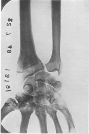
FIG. i.-Mal-united Colles' fracture with inferior radio-ulnar dislocation and traumatic arthritis. The patient, a man aged 68, had sustained the fracture many years previously. The wrist was painless, thegrip goodand the rangeofmnovement almost full.
It may be accompanied by shortening of the radius,orby radial and dorsaldisplacementofthe
lower fragmentgiving prominence to the headof the ulna and broadening of the wrist. In these
circumstances some distortion ofthe anatomy of the inferior radio-ulnar joint is inevitable and
subluxation or even dislocation of this joint may
be found. Treatment
Inmanypatientswith mal-unionthe functional result is satisfactory even when the deformity is gross (Fig. i). Conservative treatment is
in-dicated whenthedeformityisslight,the disability only moderate and the patient elderly. At the
outset a course of active exercises and hot wax
baths mayregainadequate movement and power.
Operation is seldom indicated. As a general
rule its aim should be directed solely to the im-provement of function, and it is therefore rarely
advised for mal-union when function is adequate. However, intheyounger age groupsandespecially
in women with gross dorsal tilting ofthe lower
radial fragment, anosteotomythrough thefractuie sitemaybeperformedtorestorenormalalignment
even though wrist movements have not been seriously restricted.
In thesepatientsthe object of the operation is to delay late traumatic arthritis and to improve the appearance of the wrist. The osteotomy is per-formed through a dorsal approach at the level of
the old fracture. Itis importanttofillwith abone graft the dorsal defectcreated whenalignment has
beenrestoredlestdisplacementshould recur. The
wrist is then immobilized in plaster until union has taken place.
When in additionto dorsaltilting there is con-siderable shorteningof the radius and some radial
displacement, surgical interference may be re-quired on account of pain, stiffness and an
un-December 1951 S'1'EPHENSON: SomeComlplications of Col/es' Fractture 629 sightly deformity. Here much of the disability is
due to the derangement of the inferior
radio-ulnarjoint, andexcision ofthelower IA in. of the ulna gives a satisfactory result. Following this operation (vide infra), the prominence of the ulnar head is abolished, pronation andsupination
are considerably improved or fully restored and
discomfortis relieved. Theresultsfollowingmore
complicated operations are uncertain and often
disappointing.
Very occasionally in cominuted fractures a projecting fragment of bone on the anterior or posterior aspect of the radius interferes with
tendon action and causes local pain from a trau-matic teno-synovitis (Fig. 2). Excision of the
offending fragment is seldom required as the symptoms donot asarule persist.
3. Laxityof the Inferior Radio-UlnarJoint
It has been demonstrated by Lippman
(I937)
by experiments on the cadaver, that severance of
the triangular fibro-cartilage produces a very small degree of abnormal laxity, but when in
additionthe dorsal radio-ulnar ligament is divided, a dislocating radio-ulnar joint results. These
findings have been confirmed by the writer in the
dissecting room. Lippman has come to the
con-clusion that when aColles' fracture with displace-ment issustained, the dorsal ligament is damaged and its subsequent failure to heal is the main cause of the residual laxity, the associated lesion
being either a rupture of the triangular fibro-cartilageor afracture ofthestyloid process of the ulna.
Although
the condition frequently occurs withmal-union, it is quite often present when the
fracture has united in excellent position. Minor degrees oflaxityaresometimes followedeventually
by arthritis of the joint while major degrees
constitute adislocation. Clinical Features
There may be no symptoms. Pain over the
ulnar sideofthe wrist, local tenderness, limitation of supination and pronation, prominence of the head of the ulna and abnormal laxity form the full clinical picture. The radiographs reveal a
dis-location when present and occasionally arthritic
changes.
Treatment
When disability exists, a moulded leather wrist strap is the best form of conservative treatment.
Manipulation in anattempt to increase pronation andsupination does not meet with success and is,
indeed, dangerous, several instances of fracture of theshaftof the ulnahavingbeenreported(Patrick,
x6).
Excision of-the
distal Ii in. of the ulna isthe best operative procedure and gives good
results (Boyd and Stone,
I944)
(Figs.3a
and 3b). It is performed through a dorsi-medial incision; the bone is resected extra-periosteally to avoid new bone formation, and it is not necessary to remove the triangular fibro-cartilage. Active exercisesshould be commenced adayortwoafter the procedure. As aresultof the operation, little orno residual weakness of the wrist ensues.4. JointStiffness and Adhesions
The majorityofpatiepts legain full movements of the wrist a few weeks after immobilization has been disconitinued. Residual stiffness maybe due
either to intra-articular adhesions following a fracture involving the radio-carpal
joint,
or to extra-articular adhesions following traumatic oedema with organization of the serofibrinous exudate into adhesions. Stiffness due tothe lattercause is seen in particular when more than one attempt atreductionhasbeenrequired. Itshould also be remembered that persistent stiffness is sometimesanearlysymptomof traumaticarthritis.
.:::..:: .;..:..::..:...:... ..:..:.. ...::. ~~~~~~~~~~~... . .... ::. .... .:..,:.e ... ... .i *~ ~ ~ ..~ .!:...-r *.:.t.:S.::!.:.r< ... ::.::...?, 'S'.:8}: ~~~~~~~~~~~...:::;
Fie.. 2.-Mal-united Colles' fracture showingY a smIll spur ofbone on the anterior aspect of the fracture
site. This projection caused persistent local dis-comfortespecially at work. Excision of the pro-jectionwrasrequired.
630 POSTGRADUATE MEDICAL JOURNAL December i95
.1
CO
aFIG. 3 (a).-Colles fracture with inferior radio-ulnar subluxation.
The patient, a managed 32,complained of pain over the ulnar aspect of the wrist, weakness of grip and inabilitytorotate the forearm.
Treatment
Active use of theshoulder, elbow and hand from the outset is an essential feature of the routine treatment of a Colles' fracture and plays a large part in the dispersal of oedema, thus materially helping to prevent subsequent stiffness of the wrist
joint.
Active use, hot wax baths and exercises super-vised by the physiotherapist should be instituted, and if after a lapse ofseveral weeks a satisfactory return of movement has not been regained, re-course may be had to manipulation under a
general anaestheticand followed byfurther active
exercises. Manipulation may be repeated if necessary.
5. Traumatic Arthritis of the WristJoint This condition is an infrequent sequel and by
comparison arises much more commonly after
fracture of the carpal scaphoid. There isno clear
C
C
Cl
I
FIG. 3 (b).-The same wrist after operation. Full painless function was regained.
explanation for this. Interruption of the con-tinuity of the articular cartilageby the fracture line alone issufficient to initiate arthriticchanges, and
when, as in comminuted fractures, it is often im-possible to restore completely the anatomical
alignment of the articularsurface, arthritis occurs more rapidly.
Major degrees of mal-union without
recogniz-able interference with the articular cartilage by thefracture are occasionallyfollowed by arthritis.
This is seen chiefly in those patients who make
constant demands on their wrists at work (e.g.
STEPHENSON: SomeComplications of Colles' Fracture to raised pressure on the articular surfaces and
continuedstresses ontheligaments.
It should be borne in mind that the so-called
traumaticarthritis maybe, in fact, anaggravation or reactivation, caused by the trauma, of a
pre-existing chronicarthritis. ClinicalFeatures
The symptoms may arise very soon after the fracture or be delayed for years. The signs are
those ofan osteoarthritis inany
joint,
and in the early stages the diagnosis is basedmainly
on theclinical features, as the radiographic changes are
minimal. Even when the arthritis is
advanced,
osteophyticlipping isnot awell-marked feature in the radiographs and is best looked for at the tip ofthe
radialstyloid
process.Treatment
Mild cases benefit by suitable physiotherapy.
When thearthritisiswell
established
thetreatmentdepends upon the severity of the symptoms, the occupation and age of the patient. A moulded
leather wristsupport is adequatefor the
majority
of patients and arthrodesis of the wrist joint is
onlyrarely indicated.
6. Painoverthe Ulnar Aspect of the Wrist
Thisisafairlycommoncomplaint after fixation
has beendiscarded. Althoughthe symptoms may
bepresentwhenthereis non-union ofafractureof theulnarstyloidprocess, in thewriter's experience
it is morefrequently foundwhenno such fracture has been sustained. Thepairi is then attributable
to an unhealed sprain or partial rupture of the
internal lateral ligament of the wrist, and it may wellbethat even in the presence ofnon-union of the ulnar styloid process, the discomfort is ligamentous inorigin. Apointoflocaltenderness
isconsistently found closeto the proximal attach-mentofthisligament.
Treatment
The discomfort is almost always of temporary
durationand responds to diathermy.
7. Late Rupture of the Extensor Pollicis
Longus Tendon
This is an uncommon but well-recognized complication. Formerly it was thought that the rupturewasdueto aprocess ofattrition causedby roughness of the tendon groove on the dorsum of
the lower end of theradius. However, in a sub-stantial proportion ofthe reported cases, no re-duction
of*
the fracture was required and noroughening of the groove found subsequently at
operation. At thepresent time it is held that an
avascular necrosis of the tendon occurs following
injury to the meso-tendon with haematoma forma-tion andsubsequent fibrosis. Operative findings tend to support this view (Trevor, 195o). The
lesion isfoundnearthelower borderofthedorsal carpalligament. Atoperation in recent ruptures, bruising, yellowish discoloration and fraying of the tendon ends may be seen. Onlyoccasionallyis rougheningof the tendon groovefound.
Clinical Features
Femalespreponderate andthe patient isusually
over 50 years of age. The youngest patient
re-corded is a girl aged I4 years (McMaster, 1932).
Local pain andswellingmay precedethe rupture,
which is sometimes heralded by a sudden snap. In other patients, inability to extend the
inter-phalangeal joint of the thumb is the first
com-plaint.
Physicalexamination reveals inabilityto extend
the interphalangeal joint, slight limitation of
abduction ofthe thumb and absence ofthe
sub-cutaneousbow-stringwhich is formed by the intact
tendon.
Treatment
Satisfactory results have been obtained from a
variety of operations, but the result following direct end-to-end suture is always unpredictable.
Among the many operations performed are:
(a) Transferenceof the extensorindicisproprius
to the distal end of the tendon. This operation
gives gratifying results"and causes minimal
inter-ferencewith the function ofthe index finger, the
only disability being a suggestion ofweakness of
extensionandatendencytoradial deviation ofthe digit at the metacarpo-phalangeal joint. As the
extensor indicis tendon lies on the ulnar side of
the tendon of the extensor communis digitorum,
the unopposedaction of thelatter tendon leadsto theslightradial deviation.
(b) Sutureof thedistal end of thetendontothe
extensor brevis pollicis or to both the extensor brevispollicis andabductorpollicis longus.
(c)A freetendon graft.
(d) Transference of extensor carpi *radialis
longior or brevior into the distal end of the tendon.
(e) Restoration ofthe continuity of the tendon
by using nylon suture material, fibrous tissue
ultimately bridging the gap between the tendon
ends(Trevor, 1950).
8. Sudeck's Atrophy (Post-Traumatic Osteo-Dystrophy)
Sudeck's atrophymayfollow aColles' fracture, but is a relatively more common complication of minor injuries of the wrist. Its
aetiology
is notfully understood. It is believed that the
injury
F
632 POSTGRADUATE MEDICAL JOURNAL December '1I
initiates an abnormal reflex arc, with pain
im-pulses forming the afferent side and sympathetic impulses, both vasoconstrictor and vasodilator, the efferent.
ClinicalFeatures
Attention is first drawn to the dystrophy a few weeks after the
injury
by pain, with stiffness of thew.rist and fingers which is at first due to muscle spasm. The pain is materially aggravated by movement. Vasomotor changes take the form of acyanotic, moist, glossy skin.In radiographs the degree of decalcification is far greater than that of a simple disuse atrophy. There is at first a patchy decalcification of the bones of the wristand hand which is seenespecially close to the joints; later the decalcification be-comes diffuse, leadingto a glassy appearapce. In very severe and protracted types there may be
fibrous ankylosisof the carpaljoints. Treatment
Treatment is unsatisfactory, but wax
ba.ths,
active use, encouragement and the passage of time may effect a cure. Immobilization, deep X-ray therapy, injections of local anaesthetic into the
cervico-dorsalsympatheticchain andpreganglionic cervico-dorsal sympathectomy have been tried with varying success. Sympathectomy has little or no effect on the bone changes but relieves the pain temporarily and may abolish the vasomotor phenomena.
Prognosis
Full recovery is unlikely. Only when the dystrophy is mild can a good recovery be
an-ticipated, and when severe, a substantial residual
disability is to be expected (Klser Sven, I947-8).
9. Injuries of the Median Nerve
An incomplete lesion of the median nerve, sustained at the time of injury or following
re-duction,isbyno meansuncommon. ltis duetoa
contusion ofthe nerve. Clinicallyitis found that the sensory supply of the nerve ispartially affected;
the motor side almost invariably remains intact. The patient complains of numbness of one or more digits in the median distribution and on examination there isimpairment but notcomplete
loss of sensation in the affected skin area. As the loss of sensation is only of temporary duration,
no treatment is required.
More serious injuries of the nerve due to anterior displacement of bone spicules have been described but are rare. Lesions of thesuperficial
branch of the radial nerve may also occur.
Io. Prolonged Absence from Work
This isfrequentlyduetouncomfortableprimary
splintage andto failure of the surgeon to insist on active use of the limb atonce. In some modern industrial centres workers often lose less than a day off work owing to the immediate provision of suitable employment.
BIBLIOGRAPHY
BOYD, H. B., and STONE, M. M. (I944),J. Bone Jt. Surg., 26,313.
CHARNLEY, J. (I950), 'Closed Treatmentof Common Fractures,'
Edinburgh: Livingstone.
KLSER SVEN (1947-48),ActaOrthopaed. Scand.,I7,253.
LIPPMAN, R. K.(I937),Arch.Surg., 35, 772.
McMASTER, P. E.(I932),J.BoneYt.Surg., I4,93.
PATRICK, J.(1946),Ibid., 28, 737.