Dottorato in Scienze molecolari e Biomolecolari (XXIX ciclo).
Dipartimento STEBICEF (Scienze e Tecnologie Biologiche Chimiche e Farmaceutiche) Settore scientifico disciplinare CHIM/08 (Chimica farmaceutica)
DEVELOPMENT AND OPTIMISATION OF COMPUTATIONAL
TOOLS FOR DRUG DISCOVERY
DOCTOR PhD COORDINATOR
UGO PERRICONE CH.MO PROF. PATRIZIA DIANA
SUPERVISOR SUPERVISOR
Development and optimisation of
computational tools for drug discovery
To my whole family and especially to Anna for supporting me in every moment of this PhD, without their help it would be more and more difficult
To Prof. Thierry Langer for having accepted me as student in his group, for his precious friendship and for everything he taught me
To Marcus Wieder for his priceless friendship and for being the most important guide during my PhD, with the hope to return him everything he taught me
To my supervisors for supporting me in every moment
To Laura and Marta, two angels passed away too early in this life, I will always carry your memory in my hearth!
INDEX
1"INTRODUCTION"..."1" 2"" AIMS"OF"THE"WORK"..."15" 3" CHEMOMETRICS"AND"DRUG"DESIGN"..."16" 3.1!Conf<VLKA:"A"structure<based"revisitation"of"the"Virtual"Lock<and<Key"Approach."..."24! 3.1.1 Introduction!...!25!3.1.2 Material and methods!...!25!
3.1.3 Results and discussion!...!26!
3.1.4 Conclusions!...!37!
4." DYNAMIC"APPROACH"TO"VIRTUAL"SCREENING"..."38" 4.1!Comparing"pharmacophore"models"derived"from"crystal"structures"and"from"molecular" dynamics"simulations"..."42! 4.1.1 Introduction!...!43!
4.1.2 Materials and methods!...!44!
4.1.3 Results and discussion!...!45!
4.1.4 Conclusions!...!53!
4.2!Evaluating"the"stability"of"pharmacophore"features"using"molecular"dynamics"simulations" " "..."55! 4.2.1 Introduction!...!56!
4.2.2 Materials and methods!...!57!
4.2.3 Results and discussion!...!58!
4.2.4 Conclusions!...!64!
4.3!"Pharmacophore"models"derived"from"molecular"dynamics"simulations:"A"case"study"..."65! 4.3.1 Introduction!...!66!
4.3.2 Materials and methods!...!67!
4.3.3 Results and discussion!...!68!
4.3.4 Conclusions!...!75!
4.4!A"dynamic"–"shared"Pharmacophore"approach"to"improve"early"enrichment"in"virtual" screening."A"case"study"on"PPAR"alpha"..."76! 4.4.1 Introduction!...!77!
4.4.2 Materials and methods!...!78!
4.4.3 Results and discussion!...!81!
4.4.4 Conclusions!...!91!
5" COMPUTATIONAL" CHEMISTRY" IN" POLYPHARMACOLOGY" AND" DRUG" REPURPOSING"..."93" 5.1!The"repurposing"of"old"drugs"or"unsuccessful"lead"compounds"by"in"silico"approaches:" new"advances"and"perspectives."..."93! 5.2!Drugs"polypharmacology"by"in"silico"methods:"new"opportunities"in"drug"discovery."..."94! BIBLIOGRAPHY"..."95" ! "
! !
‘If we were to name the most powerful assumption of all, which leads one on and on in an attempt to understand life, it is that all things are made of atoms, and that everything that living things do can be understood in terms of the jigglings and wigglings of atoms.’
1
INTRODUCTION
The most important issue in Medicinal Chemistry is without any doubt the drug design part, often referred to as rational drug design or simply rational design. It represents the process of finding new drugs based on the knowledge of a biological target or all the biochemical steps in which the target is involved [1]. Most commonly, The aim of a drug discovery process is to find an organic small molecule responsible for modulating the biochemical patterns of a cell process. The activation or inhibition of a biomolecule function, such as of a protein or of a nucleic acid, results in turn in a therapeutic benefit to the patient. In its basic sense, rational drug discovery involves the design of molecules that, showing a highly complementary chemistry to a specific target, can interact with it, starting a cascade of biochemical responses. In addition to organic small molecules new classes of drugs become everyday increasingly important as, for example, biopharmaceuticals and especially therapeutic antibodies. In order to test and validate these protein-based therapeutics, different techniques for improving the affinity, selectivity, and stability of them have also been developed [2].
In the drug design process, prediction of binding affinity is nowadays the most improved task and, at the same time, the most reliable. However, there are many other properties, such as bioavailability, metabolic half-life, and side effects that must be optimized prior to get a safe and efficacious drug. These pharmacokinetic parameters are yet difficult to predict through rational design techniques. Nevertheless, today, more attention has been focused on selecting candidate molecules presenting physicochemical properties that can lead to fewer complications during development and hence can help in the pathway from lead compound to marketed drug [3]. Furthermore, in silico methods, used prior to in vitro experiments, have shown a huge benefit in predicting possible ADME (Absorption, Distribution, Metabolism, and Excretion) properties for the potential candidates as well as their toxicological profiles [4]. In contrast to traditional methods of drug discovery based on testing candidate drugs through in vitro and in vivo assays, and connecting the retrieved effects to treatments, rational drug design is based on an initial hypothesis that a desired effect is due to the modulation of a precise biological target, specifically tuned by a structurally complementary
involvement of the target in the studied biochemical disease pathway. This can be sometimes confirmed by the association between target mutations and disease states [5]. The second is the druggability of the chosen target. This relies on the target capability of binding to a small molecule for the modulation of its activity [6]. In a rational drug discovery protocol, the research of small molecules potentially capable to bind to a specific target begins with a screening of libraries containing probable drug candidates. This process can be assessed as “wet screening” or may be done through the computational means searching for drug and lead-likeness of compounds [7]. Several methods are available to estimate drug-likeness such as Lipinski's Rule of Five and a range of scoring methods such as lipophilic efficiency [8].
The optimisation process of a drug design protocol is characterised by a huge number of properties that must be simultaneously tuned. For this reason, it is of common use to adopt some multi-object optimization techniques [9]. Finally, despite all the efforts made in the last years to optimise drug discovery protocols, a successful drug design campaign seems to be mostly reliant on serendipity and bounded rationality [10].
In the last years, the application of computational techniques in drug discovery and development process has gained in popularity, implementation, and appreciation. Different terms have been applied to this area, the most common used are computer-aided drug design (CADD), molecular modelling and in silico drug design. The success behind CADD application is due to its capability of increasing the hit rate of novel drug compounds when compared to the classical HTS approach. Compared to the latter, in silico methods allow the use of combinatorial chemistry and a much more targeted search, thanks to publicly available databases growth. The main scope of molecular modelling is to explain the molecular basis of therapeutic activity of some molecules and predict possible derivatives that would improve activity [11, 12]. In a drug discovery campaign, computational techniques are usually used for three major purposes:
(1) Filter large compound libraries into smaller sets of predicted active compounds that can be tested experimentally leveraging chemical and biological information about ligands and/or targets to identify and optimize new drugs;
optimize drug metabolism and pharmacokinetics properties such as absorption, distribution, metabolism, excretion, and the potential for toxicity (ADMET);
(3) Help in the rational design of novel compounds, either by modifying starting molecules or by tying together fragments into novel chemotypes.
Fast expansion in this area has been made possible thanks to advances in computational software and hardware, and increasing database of publicly available ligand molecules and target protein structures. One of the most important advantages in the use of in silico methods is the reduction of chemical space size and, thereby, the possibility to focus on more promising candidates for lead discovery and optimization. The main goal of virtual screening is therefore to eliminate compounds with undesirable properties and enrich the set of molecules with desirable properties. In another words, in silico modelling is used to significantly minimize time and resource requirements of chemical synthesis and biological testing. As shown in Fig. 1.1, in silico methods become nowadays more and more important as a first step of the entire workflow for the drug discovery process, avoiding possible false positive or false negative results in the search of possible hits to develop. In the last years, in fact, there has been a rapid growth of virtual screening usage, as confirmed by the increase in the number of citations matching keywords “virtual screening”. By using the SCOPUS database [13], it is possible to check that the articles explicitly reporting the keyword “virtual screening” steeply increase about the year 2000 reaching a number of articles 20 times higher in 2015.
Fig.1.1 Virtual screening workflow adopted prior to in vitro assays
!
D.V. Green of GlaxoSmithKline in a review published in 2003 concluded with: “The future is bright. The future is virtual” [14]. Already in 2003, it was estimated that computer modelling and simulations would account for ~ 10% of pharmaceutical R&D expenditures and that they will have rose to 20% by 2016 [15]. In these days,
PriceWaterhouseCoopers has published “Pharma2020”, the latest market research about the state of the art and the future of computational chemistry within the pharmaceutical companies [16]. In Fig. 1.2, the comparison between the state of the art and future predictions is reported.
State of the art Oncoming future 2020 prediction
Fig.1.2 comparison between state of the art and future predictions in CADD usage in pharma industries
Referred to CADD there are two major types of drug design. The first is referred to as ligand-based approach [17], and the second, structure-based [18] Fig. 1.3.
Fig.1.3 Ligand-based and Structure-based approaches in drug discovery
Ligand-based drug design is usually adopted when there is no 3D structural knowledge of the target studied. The use of molecules known to be active on the biological target of interest is the starting point used for such an approach. This kind of design strategy is also called indirect drug design because, starting from known active compounds on a specific protein, it tries to find the essential chemical features useful for interacting with that target. Once all the structural information has been collected it is in fact possible to search for chemical similarity between known and new molecules. One of the most applied ligand-based approaches is based on the indirect building of a pseudo receptor derived from a pharmacophore model that defines the minimum necessary structural characteristics a molecule must possess in order to bind to the target. In other words, a model of the biological target binding pocket may be built based on the knowledge of what binds to it, and this model in
21]. A pharmacophore model can be considered as an abstraction of molecular features necessary for the molecular recognition between a ligand and a biologic macromolecule. The IUPAC defined it as “an ensemble of steric and electronic features that is necessary to ensure the optimal supramolecular interactions with a specific biological target and to trigger (or block) its biological response” [22]. The pharmacophore features include hydrophobic centroids, positive or negative ionisable sites, hydrogen bond acceptors or donors and aromatic rings (Fig.1.4). These pharmacophore features may be located on the ligand or may be project points presumed to be located in the receptor [23].
Fig.1.4 Pharmacophore model generated with Ligandscout software. In yellow hydrophobic features are represented, hydrogen-bond acceptors are signed in red and hydrogen-bond donors in green. Blue rings stands for aromatic features
Another common ligand-based method relies on cheminformatics. In this case, ligand structural information is converted into molecular descriptors, and, through statistical analysis, one can predict possible target for a new molecule. This kind of prediction is based on the structural similarity between the new molecule and a known set of compounds. Such an approach has been developed and applied to the search of new potential drugs [24, 25]. Ligand-based drug design can be also exploited to search for a quantitative structure-activity relationship (QSAR). In this approach, one can determine the statistical correlation between calculated properties of molecules, expressed as molecular descriptors, and their experimental biological activity. Once found the most robust model, the information can be exploited to predict the activity of new analogues [26, 27]. A QSAR model has the form of:
In the last years, a more complete approach has been developed: 3D QSAR. This term refers to the application of force field calculations based on three-dimensional structure of molecules. It exploits the calculation of non-covalent empirical potentials between atom couples, such as the Lennard-Jones potential, rather than using experimental constants to define the interatomic interactions. Some of the parameters analysed are the steric fields (shape of the molecule), the hydrophobic regions (water-soluble surfaces), and the electrostatic fields [28–30].
Structure-based drug design exploits the knowledge of the three dimensional structure of the biological target, obtained through methods such as X-ray crystallography or NMR spectroscopy [31, 32]. The lack of target 3D structure can be overtaken by means of a homology model of the target, using the experimental structures of similar proteins. In this case, the studied protein will be folded according to the amino acid sequence homology with other proteins having known folding structures [33, 34]. In case of low homology levels, it is possible to assess folding prediction through the use of protein threading. In this technique, also known as fold recognition, each amino acid in the target sequence is assigned to a position in a template structure, and an evaluation of how well the target fits the template is done. After the best-fit template is selected, the structural model of the sequence is built [35, 36]. Starting from the knowledge of the biological target structure, candidate drugs can be optimally designed by medicinal chemists, predicting their binding affinity and selectivity. The two main structure based techniques are the 3D pharmacophore modelling [21, 37, 38] and molecular docking [39, 40]. Pharmacophore modelling is more and more preferred to docking for several reasons. First of all, it is more universal. In fact, pharmacophores represent chemical functions, applicable not only to a specific bounded molecule, but also to unknown ones. Secondly, it is very efficient because the computational resources needed for the pharmacophore modelling are really poor. For this reason, it is very suitable for large libraries virtual screening. In the end, it also allows researchers to tune it on the fly adding and removing features or adjusting their tolerance in order to optimise both the sensitivity and selectivity of the screening.
Molecular docking is usually applied to deeply evaluate the interaction between a small molecule and a protein at the atomic level. This helps to study the behaviour of
small molecules in the binding site of target proteins and to deepen biochemical paths. The docking protocol consists of two main parts: firstly, the prediction of the ligand position, conformation and orientation within the binding site (usually referred to as pose) then, on a determined pose, the evaluation of the binding affinity. These two steps rely on what is defined as searching algorithm (for the pose research) and scoring function, for the binding affinity calculations [39, 41].
Different types of molecular docking have been developed in the last years. The two main approaches exploit ligand flexibility or receptor and ligand flexibility respectively. In the former, ligand conformations may be generated prior to docking or within the receptor binding cavity [42]. To select proper energetically conformations of ligands, knowledge-based [43] or force field-based methods are used [44].
The above mentioned in silico approaches used in drug design can be roughly further classified based on the purpose of their application [45]. One of the most used applications of CADD is the virtual screening. It consists in the search of new ligands as potential drugs for a specific target by searching large databases of 3D structures of small molecules that can well fit into the binding pocket of a protein or on a pharmacophore model. A second strategy is the de novo design of ligands. In this case, molecules are designed starting from the essential interaction pattern within the binding pocket by assembling molecular fragments that can satisfy those interactions. The strength of such an approach is that molecules created are not present in any database, but are new entities [46]. The last approach consists in the optimisation of already existing molecules to maximise the efficacy or to minimise side effects while maintaining the essential features to interact with the chosen target [47].
In the last years a new way of using CADD has been more and more adopted. It is based on the integration between screening techniques with simulation ones as, for example, Molecular Dynamics (MD).
MD in drug design has demonstrated to give a huge impact in the improvement of drug design strategies. The knowledge of molecular motions can be fundamental for understanding compatibility between two different molecules. Thanks to the modern techniques, the initial idea of a frozen receptor that can accommodate a small molecule without mutating its conformation –also known as “Lock-and key” model
undergoes some conformational changes based on the ligand to bind [49, 50]. In Figure 1.5 the general process of molecular dynamics calculations is reported.
Fig.1.5 Simplified Scheme of molecular dynamics calculations
The first step of MD is the availability of a 3D target structure. This can be obtained throughout X-ray crystallography, Nuclear Magnetic Resonance (NMR), or by homology-modelling. The 3D coordinates of the receptor structure will be used as starting point for the integration of the equation of motion. For this calculations, Energy, expressed as forces between atoms, is calculated exploiting Force Field (FF) parameters according to the formulas reported in Figure 1.6 [51]. The FF contains all the information useful for the calculation of the total energy of the molecules, including bonded and non-bonded terms relatives to atoms within the simulation. In FF parameters, the bonded part of measurement contains chemical bonds stretching and atomic angles variations modelled as simple virtual springs. Dihedral angles are instead represented by sinusoidal functions that approximate the energy differences between eclipsed and staggered conformations. The non-bonded terms are represented by van der Waals interactions, for the neutral species, Lennard-Jones 6-12 potential, and using Coulomb’s law for the charged interactions [52].
Fig.1.6 Example of empirical Force Field parameters
Even though current force fields present some weaknesses because of several approximations and simplifications, MD simulations play today a very important role in drug discovery because they are the only way to study receptor motions. Just a single protein conformation, for example, tells little about protein dynamics. The static models can be valuable to study the structure of a protein, but drug binding or molecular recognition in general are dynamic processes otherwise not comprehensible if not through the use of MD. The molecular recognition process involves in fact different possible arrangements of both ligand and protein and not their unique and static conformation.
Following the receptor theory, ligands can bind and stabilize only a subset of the different conformations of a receptor and this can cause an induced shift of all the receptor conformations towards the most appropriate to bind the ligands [49]. Moreover, once bound to the protein, the ligand can induce some rearrangements in the binding pocket that are not reproducible in its absence [53]. In the last few years, several approaches have been adopted to simulate the flexibility and dynamicity of the receptors to adopt in virtual screening campaigns. For example, recently, Lexa et al. published a review where it is possible to study all the different approaches adopted in order to take into account protein flexibility for molecular docking [54]. Herein, some of the mentioned methods are presented. One of the most common
the Lennard–Jones repulsion term between the receptor and the ligand allowing some minimal backbone movement and side-chain flexibility. Movements are then followed by a rigid-body protein relaxation protocol [55]. In the relaxation methods, the docked complex is taken as a starting point for focusing on protein flexibility by modelling induced-fit effects. The main limitation of this approach is that the dynamic simulation can be only assessed on an all-atom structure: it cannot be performed if the protein is not explicitly represented (e.g. docking grid). Monte Carlo (MC) or MD simulations are actually the most adopted techniques to perform complex relaxation and interactions study. Such a kind of refinement is usually performed after the docking process is finished and the best pose for docking is chosen and it allows other investigations such as solvent effects, examination of the kinetic stability, and prediction of ΔGbind [56, 57]. The last two methods present the limitation that there is not a real view to the conformational modification of the target during the binding process with the ligand. For these reasons other new algorithms have been proposed, for example the induced fit docking method. In the latter, the docking simulation is run considering ligand and protein side chains as flexible to explore new conformational space. The main limitation of such an approach is that its computational requirements are a limitation feature, especially on large-scale virtual screening studies. Furthermore, the only conformational space of the protein is relative to side chains rotamers, it is in fact based on the use of side chain conformation libraries [58, 59]. Most published methods for flexible protein– ligand docking are based on a limited number of receptors and are usually applied to small molecule libraries, that make the evaluation of the methods difficult. The use of a large test set is in fact vital in the performance assessment of a new screening method especially when one wants to measure performance across a range of different targets. Moreover, the use of a dynamical approach to docking is more resource and time-intensive than semi-flexible docking.
The use of multiple receptor conformations for docking however may sometimes decrease the selectivity of the screening process increasing, for example, the false positive rate. The use of multiple conformations may also lead to the creation of a ligand optimal for an average receptor structure that is not experimentally accessible, a so-called ‘paradoxical inhibitor’. To avoid this kind of risks it is possible to take into consideration only receptor conformations that are present in low-energy landscape of the protein. This issue has driven many researchers to focus on the
choice of the optimal method for the selection of the possible receptor conformation to adopt in the screening process.
The dynamic approach has also been adopted in pharmacophore based virtual screening. In these cases, structure-based pharmacophore features are generated starting from protein-ligand complexes taken from molecular dynamics. Recently, a dynamic pharmacophore approach has been proposed by Choudury et al. In their work, some snapshots are extracted from MD and structure-based pharmacophore models are generated within the protein-ligand complexes chosen. The built models are then compared with the docking approach using known active and inactive compounds [60].
Another way to study dynamic pharmacophore, starts from MD to cluster trajectory frames based on the root mean square deviation (RMSD) of the protein-ligand system or the most populated conformations of the receptor [61, 62]. The RMSD for frame x is reported in Equation 2. The procedure is repeated for every frame in the simulation trajectory.
Eq.2
where N refers to the number of atoms in the analysed selection;
tref is the reference time, (typically the first frame is adopted as the reference and it refers to time t=0);
r' is the position of the selected atoms in frame x after it has been superimposed on the reference frame, where frame x is recorded at time tx.
One of the limitations of these approach is represented by the dismissing of the dynamic information from the MD simulations and the consideration of only some coordinates chosen by the operator. Such a method is in fact strongly biased by the ability of the MD simulation to represent the configurational space and the operator capability to select the most representative frames out of the whole simulation [63, 64]. Moreover, the ligand binding process could be related to a unique receptor conformation, maybe not representative in the dynamic trajectory and this could be missed in the clustering approach. In this case the use of dynamic pharmacophore represent a real thread for the virtual screening campaign [65, 66].
always seems to be the correct choice of protein conformation to adopt to generate docking grid or pharmacophore models.
For molecular docking, one should run a number of virtual screening experiments equal to the number of obtained coordinate sets. Unfortunately, the choice of significant structures is no obvious because it is not possible to detect a priori which coordinate set will give good results in virtual screening. From a virtual screening point of view, in fact, every protein conformation that results in a differently ranked molecule list contains potentially important information.
For the pharmacophore approach, the models generated from the MD trajectory (one pharmacophore model for each coordinate set) are equal to the number of screening runs and also in this case every model carrying out new information could be crucial in the realisation of a screening campaign. Comparing the two methods, dynamic structure-based pharmacophore models present less variability compared to the dynamic docking approach based on the coordinate of the amino acid side chains. Pharmacophore feature space is very limited compared to configuration space of the protein side chains coordinates. The geometry tolerance of the pharmacophore features allows to find the same models for slightly different protein configurations. A possible evident solution for reducing bias in the dynamic approach to virtual screening could be the development of a protocol capable to really explore all the coordinates generated during the MD simulation without having to choose some structures or conformations. Obviously such an approach results to be very time and resources consuming and it is strongly related to the number of atoms to simulate and to the libraries to screen.
2
!
AIMS OF THE WORK AND OUTLOOKS
!The aim of my PhD project was the development, optimisation, and implementation of new in silico virtual screening protocols.
Specifically, this thesis manuscript is divided into three main parts, presenting some of the papers published during my doctoral work.
The first one, here named CHEMOMETRIC PROTOCOLS IN DRUG DISCOVERY, is about the optimisation and application of an in house developed chemometric protocol. This part has been entirely developed at the University of Palermo - STEBICEF Department - under the guide of my supervisors. During the development of this part I have personally worked on the tuning and optimisation of the algorithm and on the docking campaigns to obtain molecule conformaitons. The second part, THE APPLICATION OF MOLECULAR DYNAMICS TO VIRTUAL SCREENING, presents a new approach to virtual screening, in particular the attention is focused on different approaches to the application of protein flexibility and dynamics to virtual screening.
This part, has been carried out in cooperation with the University of Vienna - Department of Pharmaceutical Chemistry. For these works I have worked in the development of the general workflow, to a lesser extent to the programming (coding) part of the applications used and I mainly focused on the realisation of the screening campaigns and results interpretation.
The third and last part, COMPUTATIONAL CHEMISTRY IN POLY-PHARMACOLOGY AND DRUG REPURPOSING, concerns the study of the in silico methods applied to two main topics of the drug discovery process, such as the drug repurposing and the polypharmacology. In this part I will briefly describe what published in two reviews dealing to the above mentioned topics.
In conclusion during this doctoral project, I have demonstrated how the use of in silico tools can be useful in the drug discovery process. The Chemometric protocols developed and optimised represent in fact a helpful strategy to use for target fishing. Whereas, the application of molecular dynamics to virtual screening, especially for pharmacophore modelling, is a new way to deepen crucial features to be adopted in the search of new putative active compounds.
3
CHEMOMETRICS AND DRUG DESIGN
Some of the in silico methods such as molecular docking and pharmacophore modelling could be considered as the modern virtual application of the elderly lock-and-key model based on the structural complementarity between a ligand molecule and a receptor [48, 67, 68].
In the recent years, these methods have demonstrated to give an important boost to the pharmaceutical research. On one hand there has been an increase of the computational approaches reliability. On the other hand, however, the putative leads discovered through the computational methods, once synthesized and tested in vitro
can sometimes disappoint the researchers’ expectations. Such a problem causes a waste of a huge amount of time and resources. Moreover, some of the discarded compounds can be instead potentially good candidates to develop. Such a kind of issue is always referred as a false positive and false negative ratio capability of a virtual screening technique. Another interesting aspect is that compounds that sometimes are discarded for a target, can be interesting on others, as suggested in several works [69, 70]. For instance, two main aspects known as “polypharmacology” and “drug repurposing”, are known to have shifted researchers’ efforts to constantly try to characterize drug-biological target associations [71, 72]. The structural knowledge of targets and ligands has allowed to use chemical and sequence similarities among molecules and receptors to identify putative drugs to be addressed towards different targets in [73, 74]. For this reason, in the first stage of a drug discovery campaign could be useful to test early candidates towards a panel of different biological targets [75]. The possible correlation between ligand and target structures is a well-known issue, but unfortunately today it is still not possible to unambiguously interpret it.
In computational chemistry, the molecular structure can be identified and categorized by molecular descriptors. Molecular descriptors have been successfully adopted by several disciplines, such as chemistry, pharmaceutical sciences, environmental protection policy, and health researches, as well as in quality control. These parameters can be considered as the translation of a chemical property (i.e. chemical structure) into numbers. This kind of conversion, allows treating chemical properties from a mathematical point of view, expanding the exploration panorama that can be
"The molecular descriptor is the final result of a logic and mathematical procedure which transforms chemical information encoded within a symbolic representation of a molecule into a useful number or the result of some standardized experiment."
Following this definition, molecular descriptors can be categorized into two main groups: theoretical molecular descriptors, directly connected to the symbolic representation of the molecule, and physico-chemical properties or experimental measurements, such as logP or molar refractivity.
In molecular modelling, theoretical molecular descriptors are usually adopted. This group can be further considered as a collection of smaller groups:
•! 0D-descriptors (i.e. constitutional descriptors, count descriptors); •! 1D-descriptors (i.e. list of structural fragments, fingerprints); •! 2D-descriptors (i.e. graph invariants);
•! 3D-descriptors (such as, for example, 3D-MoRSE descriptors, WHIM descriptors, GETAWAY descriptors, quantum-chemical descriptors, size, steric, surface and volume descriptors);
•! 4D-descriptors (such as those derived from GRID or CoMFA methods, Volsurf).
The above classification is taken from the book “The handbook of molecular descriptors” by Roberto Todeschini [77].
The use of a single molecular descriptor is not enough to predict a biological target for a molecule. However, the use of a carefully selected set of molecular descriptors can be a very powerful translator that can reveal important information about necessary structural features of a molecule to interact with a specific receptor. For example, topological descriptors based on a multiple bioactive reference structures have been employed in similarity-based virtual screening, showing to be potentially more effective than fingerprints, scaffold-hopping or ligand topological pharmacophores [78, 79].
Recently, the use of molecular similarity approach, has been more and more adopted for the discovery of some potential lead compounds [80]. Nonetheless, it is important to point out that a chemical similarity between two molecules, expressed as similar molecular descriptors’ chemical space, is not always synonym of same biological
In the last years, the research group I worked with for my PhD has developed an approach based on the use of the molecular descriptors as the mean through which building biological lock models for different targets in order to identify new putative drug molecules. This indirect approach starts with the calculation of molecular descriptors for known inhibitors of a selected target. Based on the molecular descriptors values, the idea is to build a target profile, here called lock model, which is created on the structural features of its specific binders. The research of new candidates is based on the possibility of finding new molecules responding to the structural requisites for the target profile previously created [24].
All the chemical structures have been collected from the BindingDB, a Public database including chemical structures classified by biological activity [83].
The first key step of this in house method, called Virtual Lock-and-Key Approach (VLKA), was the random choice of 47 biological targets, form now indicated as Tn presenting known inhibitors with measured biological activity available in the BindingDB.
Starting from these structures, known inhibitors were chosen from BindingDB and CODESSA PRO software [84] was used in order to calculate a set of molecular descriptors. This software is able to calculate about 1000 molecular descriptors, from 0D to 3D. As mentioned before, the aim of the protocol is to build a lock model for each biological target (Tn) starting from a target profile traced by molecular descriptors value of its known inhibitors. In order to choose a compound selection for the lock model constitution, a biological data cut-off was adopted (Ki, IC50, EC50) [24]. For the creation of the protocol, 173 molecular descriptors were chosen in order to have not blanks for all the selected compounds constituting the lock set. For the calculation of 3D-molecular descriptors, global minimum conformations from in vacuo minimisation were selected. Mean (m) and standard deviation (s) of the molecular descriptors values (Xi,j) for each biological target (Tn) were calculated (Fig. 3.1A). The hypothesis behind the protocol is that the value of each molecular descriptor of a suitable inhibitor should be close to the same molecular descriptor mean (m) calculated for all the inhibitors of the same biological target. Starting from that, every molecular descriptor value [Xi,j(Tn)] of the compounds, included in the Lock set, was converted into a numerical coefficient in relation the closeness to m (Fig. 3.1B), as reported in Eq. (3.1):
if Xi,j(Tn) > µ ± σ, !α = 0; (eq.3.1) if (µ-½σ) < Xi,j(Tn)< (µ+½σ), !α = 1; if –σ < Xi,j(Tn) <-½σ, !α = 0.5; if +½σ < Xi,j(Tn) <+σ, !α = 0.5.
where: X represents the molecular descriptor value; i is related to the structure;
j is related to the molecular descriptor; Tn represents the biological target.
Basically, each biological target needs specific chemical-physical properties to be activated, so, it is wise to assume that some molecular descriptors could express better than the others the key structural requirements for the specific biological target. Starting from this consideration, the molecular descriptors values were weighed for each Tn on the basis of the α coefficients determined for the lock set, by considering the sum of the α value for each descriptor (Dj) for all compounds, belonging to the specific biological target (Fig. 3.1C). The following step was to normalize these values by defining the !Dj coefficients (Fig. 3.1D) as reported in Eq. (3.2).
Fig. 3.1 Virtual lock-and-key approach flow chart. A: Calculation of Mean (m) and standard deviation (s) of the molecular descriptors values (Xi,j) for each biological target (Tn); B: Conversion of each molecular descriptor value [Xi,j(Tn)] in a coefficient; C: Molecular descriptors weighing by a coefficient for each biological target (Tn); D: Normalization step by defining the uDj coefficients; E: Partial scores 4 calculation; F: Total score V calculation
(eq. 3.2)
where: i, j, and Tn are defined in Eq. (3.1);
represents the higher α sum of all molecular descriptors belonging to specific biological target.
The αi,j(Tn) and ωDj coefficients were used to calculate the affinity of all the 7352 compounds under investigation for each biological target. Thus according to Eq. (3.4) the partial score φ was calculated, and the total score Φ was defined as sum of
φ Eq. (3.3) (Fig. 3.1 E-F).
(eq. 3.3)
(eq. 3.4)
where: "i,j represents the partial score;
# represents the total score; i, j, and Tn are defined in Eq. (3.1)
All the calculated scores, for all the structures for each biological target were converted in rankings.
At the end, the Φ scores rank all the 7352 database compounds with respect to the 47 biological target. Inhibitors related to each biological target should occupy the higher rankings. To verify this hypothesis the enrichment score (E%), considered as the percentage of correct classification, was calculated according to eq. (3.5):
(eq. 3.5)
where: W represents hypothetical lowest rankings; B represents hypothetical highest rankings;
P represents the obtained rankings.
Two different E% scores: E%1 related to the “lock set”, and E%2 for the “total set” of a biological target were calculated. The E%1 reached an average value of 80.4% and for many targets values up to 95%, and the E%2 reached an average value of 79.0% (Fig. 3.2).
Fig.3.2E% for the lock and total set!
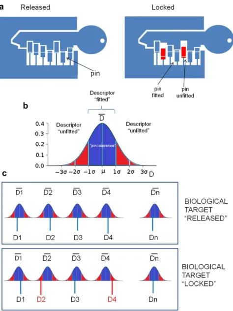
As previously mentioned, the core of VLKA protocol consists in setting-up a “lock model” for a biological target, starting from the respectively known inhibitors. In this scenario, molecular descriptors could be considered as pins of a lock (receptor binding pocket) to be released by a key (molecule) (Fig. 3.3a). Considering this assumption, a new molecule could be considered an inhibitor of a biological target if the values of its molecular descriptors fall in the calculated range values for the set of known inhibitors for the same target.
Briefly, for each structure, the range of molecular descriptors constituting a “lock pin” were defined considering the mean value of them (D mean) and the standard deviation (s) as tolerance (Fig. 3.3b). When the molecular descriptors values of a molecule fall into these defined ranges the lock can be released and the structure can be considered as a potential inhibitor (Fig. 3.3c).
To be released, a real lock needs that all pins must fit the lock structure whereas, in this protocol, the higher is the number of fit pins, the higher will be the affinity to the considered biological target.
In the VLKA, the biological target lock pins are represented by a sequence of 173 molecular descriptors.
Fig. 3.3 From the lock to biological target. a) How a real lock works; b) The range of “lock pin” molecular descriptors values (mean m ` s) can be considered the “pin tolerance”; c) When all the molecular descriptors values fall into the “pin tolerances” the biological target “releases”.
The affinity score of a molecule against a specific target is then evaluated as the number of the molecular descriptors “fitted” (Fig. 3.3).
Moreover, as mentioned before, not all the molecular descriptors have the same weight in the lock constitution: some of them are really representative for the lock while some of them can be omitted. So it was necessary to prioritize some descriptors among others. Using this approach, it was possible to rank molecules of the training set based on their affinity against the protein set. What was expected from these assumptions was that inhibitors of a specific biological target should be retrieved in the higher ranking positions for that target.
3.1 Conf-VLKA: A structure-based revisitation of the
Virtual Lock-and-Key Approach
3.1.1 Introduction
Starting from the in house application VLKA, in the attempt to deepen the ligand conformation influence on the protocol, we decided to test the same previous algorithm of scoring and ranking starting from the docked conformation of ligands. Docking calculation was used for two different purposes: to retrieve docking scores, first (in order to test the algorithm for target assignation and possible off target application), then to provide the docked pose of ligands into the relative targets to be used in the VLKA method. The docked ligand poses were in fact used to re-calculate the 142 3D-descriptors (Conf-VLKA), out of the total 173 descriptors originally used in the VLKA. In fact, the remaining 31 descriptors out of the initial 173 set were 1D and 2D, and they did not need to be re-calculated because not influenced by ligands conformation. The original VLKA results, based on molecular descriptors obtained with in vacuo optimized structures [24], were then compared to the new approach in the attempt to evaluate the likely influence of 3D ligand conformation on the protocol prediction capability. The comparison between the two methods was also made, by analysing docking results in scoring and ranking molecules, for the different targets [85].
3.1.2 Material and Methods
Target choice
Being the most important issue of the approach the comparison of the new protocol with the previous one, we decided to maintain the same targets and ligands of the original method [24].
VLKA algorithm: scoring and ranking
For the algorithm details please refer to the previous paragraph VIRTUAL LOCK AND KEY APPLICATION [24].
Ligand Structure similarity evaluation
In order to check the structural diversity of ligands for each target set, preventing the enrichment of redundant molecular analogues, we set up a topological evaluation of the whole database. For each target, ligand structures were submitted to calculation of radial fingerprint [86], molprint2D fingerprint [87] and MACCS keys [88] and then analysed in terms of Tanimoto distance [89] using similarity matrix on
CANVAS [90, 91]. The Tanimoto similarity cut-off value usually chosen as index of similarity is above 0.75 [92].
3D biological structures selection and optimization
To carry out this comparative approach, the 3D structures of the biological targets included in the VLKA have been downloaded from the RCSB Protein Databank (PDB) [93], complexed with co-crystalized ligands. The selected structures were submitted to the optimization and refinement process using Protein Preparation Wizard utility of Maestro Schrödinger suite. During this process bond orders were assigned, the missing hydrogens were added, the disulfide bonds were eventually assigned, the water molecules were deleted, the protonation of aminoacids were determined. At the end, the hydrogen bonds of the proteins were optimized, and restrained minimization was carried out on heavy atoms converging to RMSD equal to 0.30 Å, and on the hydrogen atoms.
Docking and descriptors calculation
The ligands co-crystalized within PDB structures were extracted and docked using Glide XP high performance docking procedure [94–96], as a test for pose prediction quality of the searching docking algorithm. The 7352 compounds of the VLKA were submitted to the docking and scoring procedure versus the own target, and then versus the entire biological targets dataset. The best ligand pose for each compound was selected according to the XP Glide Score. Once docked, 3D molecular descriptors for the best pose structures were re-calculated as in the original work [24].
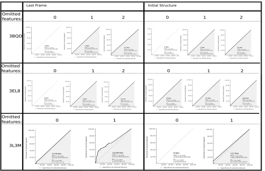
3.1.3 Results and Discussion
The aim of this work was to explore the VLKA protocol capability using docked conformation of ligands. The original approach was based on molecular features of known inhibitors expressed as 1D, 2D, and 3D descriptors calculated on in vacuo
conformation of molecules. The new method was based on the 3D descriptors calculation on the best docked conformations of each compound. This last approach (Conf-VLKA), in our opinion, could give a new interesting point of view due to the fact the original descriptor matrix consisted of only 31 1D/2D descriptors over 173, the total descriptors used [24]. So it is plausible to observe a variation in most of the values due to the change of 3D conformation of molecules. Consequently, the
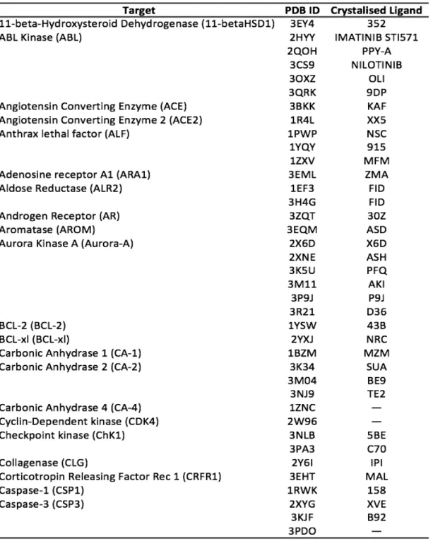
“locks” and the pin tolerance could result different from the original VLKA. To set up the study, biological targets were taken from RCSB Protein Data Bank. 43 out of 47 biological targets were retrieved into the PDB because of a lack for some 3D structures such as CAMK2 (Calmoduline Kinase 2), CB2 (Cannabinoid Receptor 2), Ghrelin Receptor (GHSR), and Diacylglycerol acyltransferase (DGAT-1). Even though it is common practice to re-build protein structures by means of homology modelling, when these ones are not available in databases, this procedure, starting from the primary sequence, allows obtaining calculated structures and hence less reliable structures respect to experimental ones. So, finally we decided to discard targets for which 3D-structures were unavailable. For many targets, multiple structures were retrieved, and for some targets (CA-4, CDK4, CT), no bound ligand was available. All the 3D biological structures, taken into account, are reported in Table 3.1.
Table 3.1. cont.
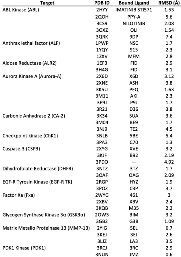
According to the Glide docking procedure, target grids were calculated on the 3D coordinates of the crystallised ligand within the PDB crystal. For those targets not bearing any ligand, we decided to exploit the PDBsum database information to calculate target grids [36], on the residues identified to be part of the binding pocket. Cognate docking was applied to the PDB dataset to test the docking searching algorithm capability. The root mean square deviation (RMSD) between the re-docked pose of the ligand and the original co-crystalized one was the accuracy parameter chosen. Generally, the lowest is the RMSD the most accurate is the docking algorithm, this also allowing us to choose the more suitable target structure (Table 3.2).
For systems presenting more than one PDB available, the one presenting the lowest RMSD value was chosen. The cut-off value for choosing a system was set as < 2.0 Å. For a few systems we had to choose the lowest value that was anyway quite higher than 2.0 Å (Table 3.3).
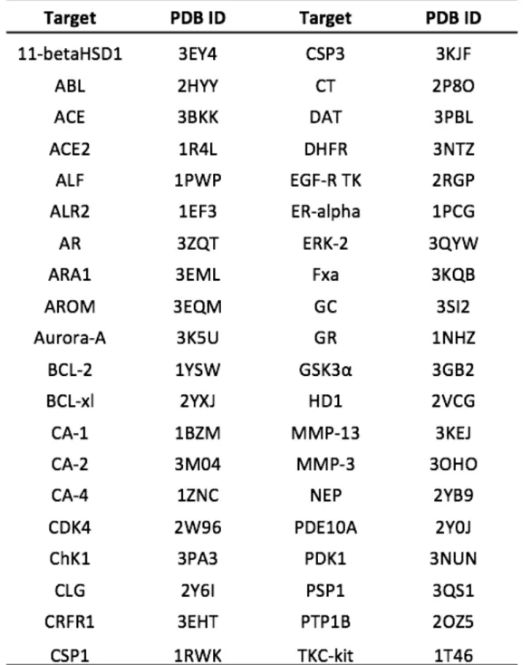
Table 3.3. 3D PDB structures selected
The next step was the application of the docking on the 7352 VLKA compounds. For each target, docking calculations were performed on specific target compounds (lock set and total set), and on the rest of the entire VLKA dataset. For three targets (Asparaginyl Endopeptidase, AE; Aldosterone Syntase, CYP11B2, Delta Opioid Receptor, DOR), no docked pose was generated for the majority of the compounds, so we decided to exclude them from the analysis because not significant. The best pose for each molecule was chosen according the Glide Score and visual inspection in order to avoid atomic clashes. The docked conformation of molecules was then
submitted to the VLKA algorithm. As previously explained, in the VLKA, the structural affinity of a compound towards a specific target is expressed by a score (Φ), calculated on the weighted average values of molecular descriptors. Based on this parameter each compound is ranked versus each receptor creating the E% score for every target analysed; the highest is the score, the highest is the probability that the ligand is correctly assigned to its target. At this step of our study, we replaced the
Φ scores with the docking scores, and recalculated the E% scores. The application of docking protocol gave us results for 40 out of 47 targets included in the original VLKA. The simple use of docking algorithm for target assignment of molecules pointed out that E%, both in case of lock set (E%1) and test set (E%2), are lower than the E% scores of the original VLKA, with a mean value of 60.0%. Just for few targets, the E% scores exceed the 80.0% (Figure 3.4). These results reflect that the use of docking scores did not revealed suitable for this kind of approach, maybe because the docking score itself does not take into account the structural features of compounds for target assignation of molecule, but simply evaluate the energetic profile of the ligand-protein interaction. One of the reasons why this approach gave to us a lower capability compared to the original one could be due to the docking scoring function. In fact, it does not work the same on all the targets. For this reason we wanted to use docking only to consider the molecules poses to recalculate 3D descriptors instead of using docking score.
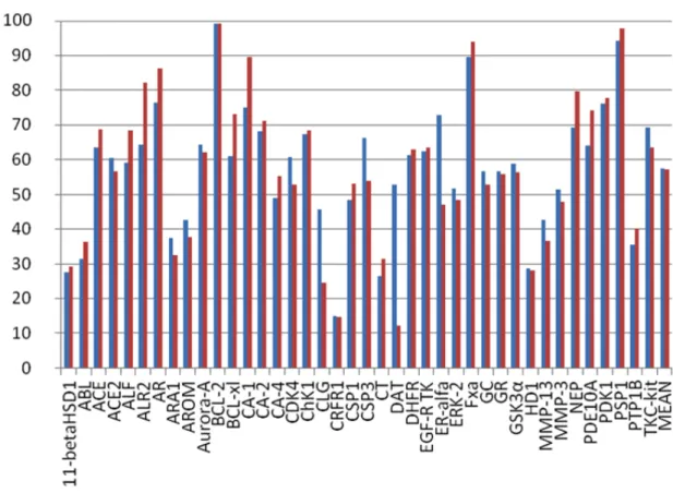
Figure 3.4. E%1 (blue) and E%2 (red) related to docking scores
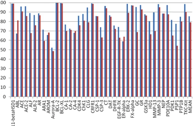
New 3D descriptors values (calculated on the docked conformation of ligands) were inserted into the matrix of the 7352 compounds and the latter was submitted to VLKA algorithm (Conf-VLKA). As last step, the E% scores were calculated again on the new scoring and ranking results. The average E%1 related to the lock set showed a value of 86%, quite greater than the average E%2 related to test set of inhibitors (79.1%). In some cases (11-betaHSD1, BCL2, BCL-xl, CRFR1) E%1 and E%2 showed a more evident rise, while other targets such as ALF, GC, MMP-13, PDK-1, E%2 resulted in a moderate predictive ability (>60%).
Fig. 3.5E1% (blue) and E%2 (red) related to 3D descriptors calculated from docking poses
In order to avoid analogue redundancies in the ligands set used in this protocol, we wanted to assess structural similarity evaluation of compounds. For each target, ligand structures were submitted to fingerprint calculations as described in the methods section. For the ligand sets analysed, no values higher than 0.75 between the structures belonging to the same target set.
The aim of this paper is to compare three different approaches: the original VLKA, where molecular descriptors are calculated on in vacuo optimized structures, with two more approaches, one based on docking scores and the other exploiting docked conformation of ligands for 3D molecular descriptors calculation.
In the original VLKA approach, the average E%1 achieved the 80.4%, and the E%2 hit the 78.9%. In some cases (11betaHSD1, AE, BCL-2, CRFR1, DGAT-1, PSP1) E%2 yielded a high level of predictive capability (98%). For other biological targets (ALF, BCL-xl, CT, DHFR, DOR, GC, GSK3α, PDK-1) E%2 showed lower values, but despite this, E% values confirmed a quite good predictive capability (>60.0). Only for ALF, this value dropped to 53.1%. In the cases of ABL, ARA1, AROM, AURORA-A, CA-4, CDK4, CSP1, DAT, ER-alpha, the obtained data resulted
on docking score, the average E%1 and E%2 values were lower than the first above mentioned approach, both near to the 60%. Just few target showed prediction capability >80% (BCL2, CA-1, and PSP1).
In the last approach, the conf-VLKA, the average E%1 was 86%, and for many targets it rose up at 98%. The average E%2 was 79.1%, just greater than E%2 in the original approach. In conclusion, we found that the use of the simple docking score for target fishing is not always reliable, maybe because of a caveat of docking scores which, is known, are not fully related to the protein-ligand binding energy. Docking is much more interesting when used to explore ligand conformations inside the binding pocket. In fact, in the last approach, the use of docked ligand conformations to recalculate the 3D descriptors and the locks, slightly enhanced the E%1 and E%2 compared to the original approach (ΔE%1=+6%, ΔE%2=+0.2%). Even though the average accuracy of the prediction is similar to the previous one, the most interesting data is that for certain targets there was a rise of the E%. For BCL-xl target an increase of 11% for the E%1 and a 18%. E%2 were observed. For ER-alpha the value of E%1 rose up from 30% to 58% and the variation of the E%2 was only of the 3%. The best results were observed for MMP-13 (ΔE%1=+41%, ΔE%2=+18%) and CDK-4 (ΔE%1=+ 9%, ΔE%2=+6%). Also the PTP-1B target showed a significant variation of the E% values with a ΔE%1=+25%, ΔE%2=+14%.
In the light of these considerations, the best results and the strongest variation between the old approach and the Conf-VLKA occur for dataset compounds with a high degree of branching considered as number of rotamers. This could be justified by the fact that the most branched is the molecule the most it will be sensible to conformation variations and the best it will be represented by 3D descriptors, as demonstrated by Good et al. in 2004 [97, 98].
3.1.4 Conclusions
In this paper, we modified the previous in house developed VLKA protocol in order to analyse the ligand conformational effect on the protocol capability, in particular, calculating 3D molecular descriptors on the docked conformation of ligands. Our VLKA protocol was designed to predict the possible biological target for new molecules starting from the structural information contained in molecular descriptors calculated on a set of known inhibitors. This first protocol was able to correctly predict biological target for the whole dataset with a good degree of reliability (80%) [24], and revealed experimentally useful to optimize the biological activity of some pyrimidine derivatives [99, 100]. Applying the structure based approach to VLKA we observed that, the use of the simple docking scores instead of molecular descriptors, revealed not satisfactory results, instead, the Conf-VLKA showed slightly better results (86%) than the first VLKA protocol for certain target sets, for others no interesting variations were observed. On the light of these considerations, it seems like the conf-VLKA approach works slightly better, compared to the previous protocol, when applied to targets whose ligands present a highly branched structure. According to what already found by Good et al., in a work on the effect of chemical structure complexity on molecular descriptors weight for ligand-based virtual screening [97, 98]. Another issue to be addressed is that probably the performance of the Conf-VLKA is connected to the docking algorithm that works better on some proteins more than others. VLKA and Conf-VLKA revealed different strength points. While VLKA revealed really fast and immediate to apply, Conf-VLKA, although need more computational time, on some proteins revealed a small rise in performance, especially for systems in which compounds have a great number of torsional bonds and branching. Nevertheless, both approaches (VLKA and Conf-VLKA) are totally user-defined, so that it is suitable for the use of in vacuo
descriptors calculation or the descriptors calculation based on binding conformation of ligands. This work is a first preliminary study on the ligand conformational effect on the VLKA protocol capability. We are now working on the same protocol using induced fit docking in order to take into account the target flexibility induced by ligands.
4.
DYNAMIC APPROACH TO VIRTUAL SCREENING
Proteins are constitutionally flexible molecules. They exert their biological function undergoing various conformational changes more or less wide. This aspect covers a huge importance for the exploration of protein – ligand interactions [50, 101]. The receptor and ligand flexibility and the induced conformational changes should be considered to correctly estimate the binding mode and the thermodynamics behind the binding process [102]. Unfortunately, drug design and virtual screening campaigns often neglect these aspects, using a static representation of the protein target. Several approaches have been introduced in computational chemistry software to take into consideration protein flexibility [103, 104]. The most representative are: side-chain flexibility [105], soft docking, induced fit [106, 107] and conformational ensemble-based docking [108, 109]. A correct incorporation of protein dynamics for drug design is still a challenging task. It has been shown in many cases that including protein flexibility leads to higher rates of false positives, since a larger number of putative ligands can be accommodated into different conformations of the binding pocket [110, 111].
Frequently, virtual screening protocols are set up on a conformational ensemble of proteins in order to include protein flexibility. Such an approach is based on behalf of proteins existing as an ensemble of substates of activation represented by different conformations [112, 113]. The main step of this approach is the generation of protein conformational ensembles prior to docking and the subsequent binding simulation of small molecules within the protein binding pocket of different size/shape [114, 115]. However, the approach strongly depends on the sampling quality chosen. One of the biggest limitations in using static X-ray or NMR receptor structures is that the available experimental conformations may not be sufficient to represent suitable conformations of the binding site for correct prediction of accommodation of new ligands [116, 117]. Despite the various methods adopted to sample protein flexibility, it is still difficult to collect suitable receptor conformations to be used prior to virtual screening processes [118, 119].
Often, protein conformations are collected starting from MD simulations [120–123]. One of the recently developed method to use MD prior to virtual screening is presented in the Relaxed Complex Scheme (RCS) approach [124]. Another MD-based approach is MD-based on the sampling of the Receptor Conformation Ensemble,
appropriate for accommodating ligands, which are chemically and structurally diverse and thus unbiased toward a particular class of ligands [125]. Recently, this approach was successfully employed for ligand profiling of drug metabolizing enzymes sulfotransferases. In their work, Martiny et al. adopted docking on RCE generated by MD simulations, combined with hierarchical conformational clustering of different binding site conformations [126].
Another interesting approach, adopted by Rueda et al. is to explore collective movement-based conformational changes [127]. In his work, Rueda exploits cross-docking on the ensemble structures generated by MD. In the last years, receptor flexibility has been also assessed using a potential grid representing the receptor deformed through selected collective movements and global structural changes following ligand binding [128]. However, considering a large number of modelled conformations may sometimes lead to less predictive VS results compared to those obtained by using the best performing crystal or NMR structures, due to the possible generation of non-native protein-ligand conformations [129, 130].
Applying these concepts to structure-based pharmacophore screening, it is important to point out that the pharmacophore model is sensitive to the atomic coordinates of the protein-ligand complex from which it was derived The first issue is closely linked to the source of the coordinates for the protein-ligand complex, whose coordinates are usually taken from the Protein Data Bank (PDB) [93]. Very often, the protein structures solved by X-ray crystallography may be affected by errors such as crystal contacts and solvent effects; for this reason, the reliability of protein-ligand coordinates has been frequently questioned [131, 132]. Proteins and small molecules are inherently dynamic and undergo a wide range of motions, ranging from the vibrations of individual bonds to collective, large structural movements. The crystal structure of the protein-ligand complex represents only a single snapshot of a dynamic system, providing neither information about the conformational flexibility of the ligand, nor about motions of the residues in and near the binding pocket [133, 134]. Thus, pharmacophore models derived from such structures might include features that are artefacts, caused either by crystal packing effects or by the single set of coordinates of the structure. Moreover, these PDB-derived pharmacophore models could contain too few or too many features resulting in a limited use. Increased number of pharmacophore features are normally accompanied by a loss of
chemical features are not suitable for database screening [135]. In this regard, the most important issue becomes the choice of reliable criteria to prioritize them. In the last years, several efforts have been made to integrate the natural dynamic behaviour of proteins in pharmacophore models. One proposed approach was based on the multi-complex pharmacophore models. Here, the models were derived from multiple crystal structures of the same protein in contact with different small molecules. Protein-ligand interaction patterns were extracted from the available structures and merged in pharmacophore maps [135, 136]. This approach, however, is limited to proteins for with multiple crystal structures are available and which have the same binding mode: it does not really consider the dynamics of the ligand-protein complex.
One very general way to avoid dependence on a single set of coordinates is the use of molecular dynamics (MD) simulations to generate multiple sets of coordinates and use these as the basis for pharmacophore models. MD simulations have proven invaluable to understand the dynamics of biomolecules [137–139]. Several approaches have been proposed to generate trajectories of protein-ligand complexes, subsequently clustered to extract representative structures as reliable pharmacophore models. [140, 141]. Most recently, Choudhury et al. presented a new way to build pharmacophore models from MD simulation. For each structure saved during MD simulations, one pharmacophore was generated and then ranked based on docking and screening results [60].
In the next chapters of this PhD thesis, I will present the chronological pathway of the study carried out to explore the use of a new approach to the pharmacophore screening: The Dynamic Pharmacophore.
In Chapter 4.1 I will present the results of a method based on the application of the MD prior to the pharmacophore modelling [142]. In Chapter 4.2 I will describe an approach based on the most frequent pharmacophore features retrieved from MD simulations and then adopted as pharmacophore model [143]. In Chapter 4.3 the application of the “most frequent features” method to the study of the IGF-1R (insulin-like growth factor-1 receptor) kinase domain is reported [144].
Finally, chapter 4.4 will concern an innovative approach to the dynamic pharmacophore model, based on a different starting point compared to those discussed in the previous chapters. Several PDB crystal structures were explored,
containing different ligands, to check the occurrence of a common interaction pattern and maintained during the MD simulations[145].
![Fig. 3.1 Virtual lock-and-key approach flow chart. A: Calculation of Mean (m) and standard deviation (s) of the molecular descriptors values (Xi,j) for each biological target (Tn); B: Conversion of each molecular descriptor value [Xi,j(Tn)] in](https://thumb-us.123doks.com/thumbv2/123dok_us/9722456.2853825/25.892.181.849.57.496/calculation-deviation-molecular-descriptors-biological-conversion-molecular-descriptor.webp)