Article
Piwi Is a Key Regulator of Both Somatic and Germline
Stem Cells in the
Drosophila
Testis
Graphical Abstract
Highlights
d
Piwi is required cell autonomously for germline and somatic
stem cell maintenance
d
Reducing Piwi in testicular somatic cells causes germline
stem cell expansion
d
Piwi nuclear localization in somatic cells is required for
differentiation
d
Piwi regulates the somatic expression of Fasciclin 3 in
gonadal development
Authors
Jacob Gonzalez, Hongying Qi, Na Liu,
Haifan Lin
Correspondence
haifan.lin@yale.edu
In Brief
Gonzalez et al. report a cell-autonomous
function of Piwi in somatic stem cell
self-renewal and show that over-expansion of
germline stem cells in
Drosophila
is
suppressed by Piwi in the soma.
Gonzalez et al., 2015, Cell Reports12, 150–161 July 7, 2015ª2015 The Authors
Cell Reports
Article
Piwi Is a Key Regulator of Both Somatic
and Germline Stem Cells in the
Drosophila
Testis
Jacob Gonzalez,1Hongying Qi,1Na Liu,1and Haifan Lin1,2,*
1Yale Stem Cell Center and Department of Cell Biology, Yale School of Medicine, New Haven, CT 06519, USA
2Shanghai Institute of Advanced Immunochemical Studies and School of Life Science and Technology, ShanghaiTech University, Shanghai, China
*Correspondence:haifan.lin@yale.edu http://dx.doi.org/10.1016/j.celrep.2015.06.004
This is an open access article under the CC BY-NC-ND license (http://creativecommons.org/licenses/by-nc-nd/4.0/).
SUMMARY
The Piwi-piRNA pathway is well known for its
germ-line function, yet its somatic role remains elusive.
We show here that Piwi is required autonomously
not only for germline stem cell (GSC) but also for
so-matic cyst stem cell (CySC) maintenance in the
Drosophila
testis. Reducing Piwi activity in the testis
caused defects in CySC differentiation.
Accompa-nying this, GSC daughters expanded beyond the
vicinity of the hub but failed to differentiate further.
Moreover, Piwi deficient in nuclear localization
caused similar defects in somatic and germ cell
differentiation, which was rescued by somatic Piwi
expression. To explore the underlying molecular
mechanism, we identified Piwi-bound piRNAs that
uniquely map to a gene key for gonadal development,
Fasciclin 3, and demonstrate that Piwi regulates its
expression in somatic cyst cells. Our work reveals
the cell-autonomous function of Piwi in both somatic
and germline stem cell types, with somatic function
possibly via its epigenetic mechanism.
INTRODUCTION
Adult stem cells have the unique ability to self-renew and differen-tiate into specific cell lineages. The balance between self-renewal and differentiation is controlled by both intra- and extracellular mechanisms (Lin, 2008). Studies on extracellular mechanisms in model systems have revealed the crucial role of the microenvi-ronment of stem cells, termed the stem cell niche, in stem cell self-renewal (Morrison and Spradling, 2008). Studies of the niche in most tissues, however, have been hampered by the difficulty in defining its location, structure, and function.
TheDrosophilatestis provides a genetically tractable model for studying adult stem cells and their respective niches, in part due to its well-defined spatial organization of stem cells and their microenvironment (de Cuevas and Matunis, 2011). At the most anterior tip of the testis, two stem cell populations can be found: germline stem cells (GSCs) and somatic cyst stem cells (CySCs). Both types of stem cells share a single niche that is composed of a group of somatic cells called hub cells.
The stem cells divide asymmetrically, such that the daughter stem cell maintains its contact with the hub while the other daughter moves away and initiates differentiation. The immedi-ate daughters produced by GSCs and CySCs are referred to as gonialblasts (GBs) and somatic cyst cells, respectively. As a GB migrates away from the niche, it undergoes four rounds of incomplete mitosis to produce a germline cyst containing 16 in-terconnected spermatogonia, followed by spermatocyte growth. Unlike GBs, somatic cyst cells are post-mitotic cells whose sole function is to support germline cysts through their path to mature sperm (Kiger et al., 2000). Although recent work has provided insight into the crosstalk between somatic cyst cells and germ cells, the mechanisms remain poorly understood.
Piwi was initially discovered as a gene required for GSC main-tenance in theDrosophilaovary (Lin and Spradling, 1997). It is the founding member of the evolutionary conserved Argonaute pro-tein family (Cox et al., 1998), which is composed of Argonaute (Ago) and Piwi subfamilies. The Ago subfamily binds to small interfering RNAs (siRNAs) and microRNAs (miRNAs) that ubiqui-tously exist in many tissues, whereas the Piwi subfamily binds to yet another class of small non-coding RNAs known as Piwi-inter-acting RNAs (piRNAs) that are generally regarded to function only in the germline (Juliano et al., 2011). A number of reports have shown that the Piwi subfamily is essential for transposon repression and genomic stability (Carmell et al., 2007; Sienski et al., 2012). Recently, high-throughput sequence analysis of piRNAs inDrosophila, murine testes, andXenopuseggs has re-vealed that a significant portion of piRNAs uniquely map to the 30 UTRs of specific genes, suggesting that Piwi activities may be extended to gene-coding regions (Robine et al., 2009; Saito et al., 2009). Furthermore, the Piwi-piRNA mechanism has been shown to regulate mRNAs at the post-transcriptional level (Rouget et al., 2010; Watanabe et al., 2015). All these advances, however, have underscored the germline-specific function of Piwi. Although Piwi and other piRNA components inDrosophila have been demonstrated to be involved in epigenetic program-ming in somatic cells (Brower-Toland et al., 2007; Huang et al., 2013; Yin and Lin, 2007) and in somatic signaling that maintains GSCs in the ovary (Cox et al., 1998; Qi et al., 2011), it remains un-clear whether Piwi or the piRNA pathway has a developmental and/or physiological function in somatic tissue.
To further explore the function of Piwi in somatic and germline tissues, we extended our analysis to theDrosophilatestis. Here, we report that Piwi is required cell autonomously not only for
GSC maintenance but also for CySC maintenance. These ana-lyses clearly demonstrate the function of a Piwi subfamily protein in somatic stem cells. In addition, we show that compromising Piwi function in the somatic cyst cell lineage causes an accumu-lation of early germ cells. This supports an important interaction between the somatic and germline stem cell lineages. Interest-ingly, reducing Piwi activity in hub cells did not affect stem cell maintenance or differentiation. Moreover, the nuclear localiza-tion of Piwi in cyst cells is required for somatic and germ cell differentiation, suggesting that Piwi may exert its function through an epigenetic mechanism. Finally, we show that Piwi ex-erts its somatic function at least by regulating the expression of Fasciclin 3(Fas3), a gene important for somatic cell development in the gonad (Li et al., 2003), viaFas3-targeting piRNAs.
RESULTS
piwiMutants Display Defects in Both Somatic and Germ
Cell Lineages
Previous work has shown that piwi mutants display severe gonadal defects in both sexes (Cox et al., 1998; Lin and Spra-dling, 1997). To further characterize thepiwimutant testicular
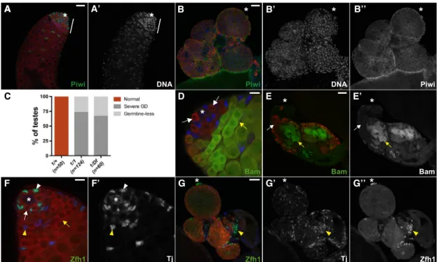
phenotype, we focused on a loss-of-function mutant allele, piwi1. In contrast to the piwi1/piwi+ control (Figures 1A and 1A0), we frequently observed an accumulation of early germ cells in homozygous piwi1 mutant adult testes (73%, n = 124), as indicated by Vasa (a germline marker) and DNA staining (Figures 1B–1B00). In addition, a minor population (27%) ofpiwi1mutant animals was completely devoid of germ cells (germline-less) (Figure 1C). Interestingly, these persisting early germ cells expressed variable levels of Bam-GFP, which is normally repressed in GSCs/GBs but induced in transit-amplifying sper-matogonia (Figures 1D–1E0). This finding implicates that piwi mutation results in defect in differentiation of amplifying sper-matogonia. Contrary to GSCs, somatic cells are typically found in clusters and randomly distributed away from the germ cells, as judged by somatic lineage markers Zfh1 and Traffic jam (Tj, Figures 1F–1G00). We ruled out the possibility that these pheno-typic consequences were due to background mutations, becausepiwi1/Df(2L)BSC mutants displayed pleiotropic defects similar to homozygouspiwi1mutants (Figure 1C). We also exam-ined heteroallelic mutants that carried a hypomorphic allele, known aspiwi2, in combination withpiwi1. Interestingly, piwi1/ piwi2mutant animals rarely displayed an accumulation of early
A A’ B B’ C D E E’ F F’ A G G’ G’’ B’’
Figure 1. piwiMutants Display Defects in Both Somatic and Germ Cell Lineages
(A) Sibling control (piwi1
/+). Piwi (green) is expressed in somatic and germ cell lineages. Note that DNA-bright cells are restricted to the apical end of the testis (white bar).
(B) Apiwi1
/piwi1
testis accumulates DNA-bright, Vasa-positive germ cells (red). Piwi expression is negligible in all cells (green, B00). (C) Frequency of testes from 0- to 5-day-old adult flies that display a specified phenotype (germline defect [GD]) in a given genotype. (D)piwi1
/+; Bam-GFP/+ testis. Bam (green) accumulates in spermatogonia (yellow arrow), but not in GSCs/GBs (white arrows).
(E)piwi1/piwi1; Bam-GFP/+ testis. Bam-negative and positive germ cells (white and yellow arrow, respectively, in E0) accumulate throughout the testis. (F) Sibling control (piwi1
/+). Zfh1 (green) accumulates at high levels in CySCs (white arrowhead) and Tj (blue, F0) accumulates in CySCs and early cyst cells. (G)piwi1
/piwi1
testes typically have persisting Tj (blue, G0) and Zfh1-expressing somatic cells (green, G00) that are segregated from germ cells (yellow arrowhead). Tj marks the hub, CySCs, and early cyst cells (blue in A–G, except B and E). Vasa marks germ cells (red in A–G). DAPI marks DNA (blue in A and B). Asterisk denotes the hub and the apical end of testes inpiwi1
/CyO and piwi1
/piwi1
, respectively. Scale bars represent 25mm (A, B, E, and G) and 10mm (D and F). See also Figure S1.
germ cells (18%, n = 107;Figures S1C–S1D0). Instead, the major-ity (51%) of mutant testes appeared unperturbed despite Piwi deficiency (Figures S1B and S1C), likely due to the hypomorphic nature ofpiwi2allele. Similar to homozygouspiwi1mutants, a minor population (31%) of mutant testes was germline-less ( Fig-ures S1C, S1E, and S1E0). Moreover, somatic cells were main-tained in the absence of germ cells but typically formed irregular clusters. These results suggest that hypomorphic mutations of piwican alter the penetrance of germ cell differentiation defects in male gonads. Taken together, these data indicate that Piwi functions are essential for the earliest steps of spermatogenesis.
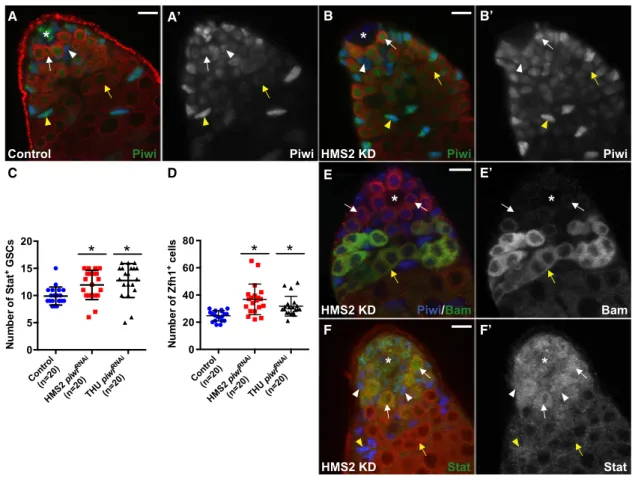
Piwi Expression in Hub Cells Does Not Appear to Be Essential for Stem Cell Maintenance or Differentiation
To begin to dissect the cell-type-specific function of Piwi, we examined the expression of Piwi protein in adult testes. As re-ported previously (Cox et al., 2000), Piwi is a nuclear protein that is expressed in multiple somatic and germline cell types in
the testes (Figures 2A and 2A0). Previous work in our lab demon-strated that in the Drosophila ovary, Piwi exerts its niche signaling functions in the terminal filament and cap cells to main-tain GSCs (Cox et al., 1998; Szakmary et al., 2005). Interestingly, Piwi expression appears the highest in hub cells (Figures 2A and 2A0)—the equivalent of cap cells in theDrosophilaovary. Hub cells have been shown to be niche-signaling cells essential for GSC maintenance in the testis (Kiger et al., 2001). We therefore examined whether Piwi expression in hub cells is required for GSC self-renewal.
We knocked down Piwi expression in hub cells using trans-genic lines, in each of which a UAS-piwi RNAi construct (HMS2 or THU; see Experimental Procedures) was driven by Unpaired (Upd)-Gal4. TheUpd-Gal4driver promotes expression of UAS constructs specifically in hub cells of the embryonic and adult gonad (Le Bras and Van Doren, 2006) (Figures S2A and S2A0). Both piwi RNAi constructs in combination with Upd-Gal4were capable of efficiently knocking down Piwi protein in
D E’
A A’ B B’
C E
F’ F
Figure 2. Knockdown of Piwi in the Hub Does Not Affect Stem Cell Maintenance or Differentiation
(A, B, and E) Control testes (Upd-Gal4;gfpRNAi
) (A) andHMS2 piwiRNAi
testes (Upd-Gal4;HMS2 piwiRNAi
) (B and E) from 7-day-old flies. (A) Piwi (green, A0) is expressed at high levels in the hub, GSCs (white arrow), and CySCs (white arrowhead). (B) Piwi expression (green, B0) is reduced in hub cells, but levels remain unaffected in GSCs (white arrow) and CySCs (white arrowhead), including their daughter cells (yellow arrow and arrowhead, respectively).
(C and D) Quantification of Stat-positive GSCs (C) and Zfh1-positive cells (D) in testes with Piwi KD in hub cells (*p < 0.005).
(E) Bam (green, E0) marks spermatogonia (yellow arrow), but not GSCs/GBs (white arrows), despite reduced levels of Piwi (blue) in hub cells.
(F) 21-day-old flies. Stat (green, F0) accumulates in GSCs (arrow) and CySCs (arrowhead), but not their daughters (yellow arrow and arrowhead, respectively), despite Piwi KD in hub cells.
Arrowheads and arrows mark the somatic and germ cells, respectively. Tj marks the hub, CySCs and early cyst cells (blue). Vasa marks germ cells (red). Asterisk denotes the hub. Scale bars represent 10mm (A–D). Error bars represent SD for each genotype.
hub cells (Figures 2B and 2B0). Despite efficient Piwi reduction in the hub, spermatogenesis remained unperturbed. In 7-day-old males, resident stem cells were found adjacent to the hub and their differentiated progeny at the basal end of testes (Figures 2B–2E0). In addition, the expression of Bam, a gene that is neces-sary and sufficient for germ cell differentiation (Go¨nczy et al., 1997), is suppressed in GSCs and GBs but induced in transit-amplifying spermatogonia (Figures 2E and 2E0). These data indi-cate that germ cell differentiation progressed normally when the Piwi level is greatly reduced in hub cells.
To closely examine whether Piwi reduction in the hub affects stem cell maintenance, we counted the number of Stat-positive GSCs and Zfh1-positive CySCs adjacent to the hub under Upd-Gal4-knockdown conditions. The average number of both GSCs and CySCs moderately increased 1.2- to 1.5-fold relative to that of controls (Figures 2C and 2D), indicating that stem cells popu-lations are maintained despite Piwi knockdown (KD) in the hub. In 21-day-old males, KD testes were still morphologically normal and undergo spermatogenesis (87% of HMS2, n = 46; 100% of THU, n = 55) (Figure 2F). Moreover, the transcriptional activator of JAK-STAT signaling, pStat, accumulated in germline and somatic cells adjacent to the hub (Figures 2F and 2F0) (Kiger et al., 2001), indicating that resident stem cells were able to respond to the self-renewal signals emanating from the niche. Taken together, these results indicate that Piwi expression in hub cells is not required to support spermatogenesis.
Piwi Is Required Autonomously for GSC and CySC Maintenance
We next investigated the biological function of Piwi in GSCs and CySCs. To test whether Piwi is required autonomously in GSC maintenance, we generated negatively marked (GFP) piwi mutant clones in adult testes using flippase/flippase-recogni-tion-target-mediated mitotic recombination. We counted the number ofpiwimutant GSC clones over a time course post-clonal induction (pci). Marked control GSC clones were observed adjacent to the hub in 43% (n = 28), 36% (n = 50), 30% (n = 30), and 30% (n = 33) of testes examined at 2, 4, 8, and 16 days pci, respectively, indicating that control GSCs are maintained (Figures S2D and S2D0). In contrast, GSCs homozy-gous mutant forpiwi1were observed in 49% (n = 68), 27% (n = 64), 20% (n = 79), and 3.5% (n = 86) of testes examined over the same period of time, indicating that mutant GSC clones are gradually lost over time (Figures S2E and S2F). In addition,piwi mutant germline clones that are located away from the hub appear to progress normally through differentiation without any apparent defects (Figures S2E and S2E0). These results indicate that Piwi is required autonomously for GSC maintenance but may be dispensable at later developmental stages.
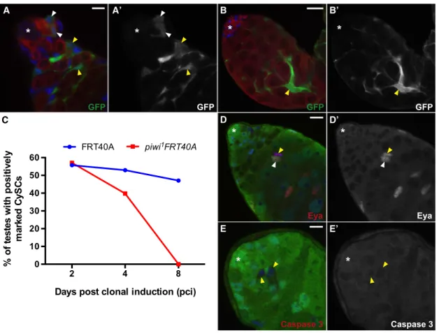
To address whether Piwi is required for CySC maintenance, we generated GFP-positive piwi mutant clones by using a mosaic technique known as MARCM (mosaic analysis with a repressible cell marker). Marked control CySC clones were observed in 56% (n = 61), 53% (n = 87), and 47% (n = 102) of testes examined at 2, 4, and 8 days pci, respectively (Figure 3C). In contrast, CySCs homozygous mutant for piwi1 were decreased by 4 days pci (40%, n = 83), and nopiwi1 mutant CySCs were identified 8 days pci (n = 91) onward (Figures 3A–
3C). Interestingly, markedpiwimutant cyst cells maintained their close association with germ cells (90%, n = 50 at 8 days pci), suggesting that CySCs lacking Piwi do not die but rather differ-entiate. In support of this hypothesis, mutant cyst cell clones expressEyes absent(Eya), a marker for late cyst cell differentia-tion (Figures 3D and 3D0) (Fabrizio et al., 2003), but do not ex-press activated caspase-3, a marker for cells undergoing active apoptosis (Figures 3E and 3E0). Together, these results indicate that Piwi is required autonomously for CySC maintenance in adult testes.
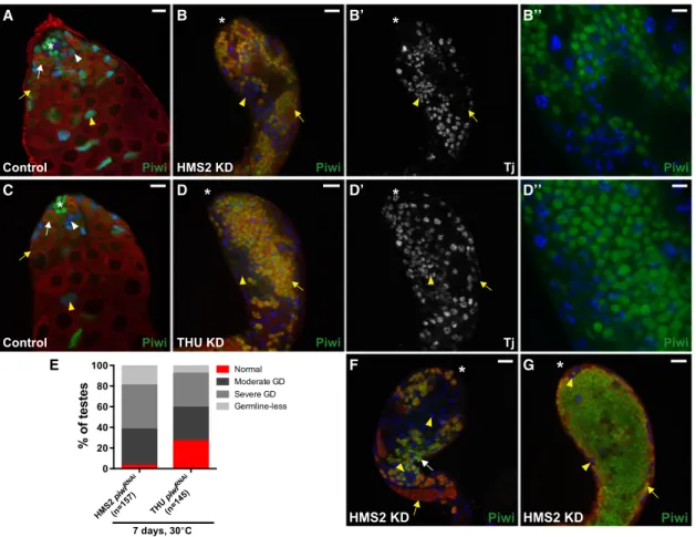
To further validate the intrinsic requirement for Piwi in CySCs, we used a temperature-sensitive allele ofGal80, a Gal4 inhibitor, to temporally control the expression ofpiwiRNAi constructs dur-ing adulthood (Lee and Luo, 1999). Gal80tsinhibits Gal4 activity
at 18C but becomes inactive at 30C. Thus,piwiRNAioccurred in CySCs and their daughters only when adult males were shifted to 30C, as driven byTraffic jam (Tj)-Gal4(Figure S2B and S2B0). As expected, testes of sibling controls maintained at either 18C or 30C were indistinguishable from those in wild-type (Figures 4A and 4B0). In contrast, testes from two differentpiwiRNAifly strains (HMS2 and THU) that were shifted to 30C for 20 days displayed a 2- to 3-fold reduction in the number of Zfh1-positive cells, a marker for CySCs and their immediate daughters (Leatherman and Dinardo, 2008), as compared with sibling controls (Figures 4C and 4D). These data also indicate that Piwi is required auton-omously for CySC maintenance in adult testes.
Interestingly, Piwi-depleted adult cyst cells ectopically express Zfh1 far from the hub in a small fraction of testes examined (16% of HMS2, n = 73; 19% of THU, n = 52). However, the majority of KD testes maintained the organizational pattern of somatic line-age markers Zfh1, Tj, and Eya relative to sibling controls (81%– 84%). Moreover, Piwi-depleted adult cyst cells intermingled normally with germ cells in 94%–98% of testes examined (Figures S4K and S4K0). Consistent with our previous findings (Figure 3), these data indicate that Piwi is not absolutely required for cyst cell differentiation in adult testes.
Piwi Is Also Required for Testicular Formation prior to Spermatogenesis
To further investigate the function of Piwi in the entire gonadal somatic lineage,piwiRNAi constructs were expressed starting in the precursors of CySCs before testicular formation using Tj-Gal4, which expresses Gal4 specifically in all somatic gonadal cells from embryonic to adult stages (Li et al., 2003) (Figures S2B and S2B0).Tjdeficiency severely affects gonadogenesis, resulting in testes that contain only a few groups of pre-sper-matocyte germ cells yet are filled with somatic cells that are segregated from the germ cells (Li et al., 2003). Similar to this phenotype, testes fromTj-Gal4-driven HMS2 and THUpiwiRNAi flies typically become filled with Tj-positive somatic cells and Vasa-positive early germ cells (Figure 5). The association be-tween cyst cells and germ cells was disrupted; the majority of Piwi-depleted cyst cells were located near the surface of the gonad, and the interior was filled with Piwi-positive germ cells in 38% (HM2, n = 157) and 44% (THU, n = 145) of testes examined (Figures 5B0, 5D0, and 5G). In 7-day-oldpiwiRNAiflies, Zfh1-positive cells were found far away from the hub in 73% (HM2, n = 44) and 79% (THU, n = 92) of testes examined (Figures
S3B0, S3D0, and S3F). We frequently observed Tj-positive cells proliferating far from the hub, as indicated by drastic increase in both EdU (Figures S3G–S3J0) and PH3 staining (Figures S3K–S3L0). Only at a low frequency were we able to identify Eya-positive somatic cells, a marker for late cyst cell differentia-tion (Figures S3E–S3F0). This Tj-like phenotype indicates that Piwi has an important role in testicular morphogenesis.
Piwi Function in CySCs and Early Cyst Cells Non-autonomously Promotes Germ Cell Differentiation
It has been previously shown that defects in germline encyst-ment by the CySC lineage can block the progression of sper-matogonial cells, ultimately leading to the accumulation of early-stage germ cells (Lim and Fuller, 2012; Sarkar et al., 2007). Given that Piwi is required for adult CySC maintenance (Figures 3and4), we examined whether the loss of Piwi in the CySC lineage blocks early germ cell differentiation. Indeed, testes frompiwiRNAi flies displayed an accumulation of early
germ cells far from the hub, as judged by Piwi expression (Figures 5F and 5G). Moreover, analysis of various markers for germ cell differentiation supports that persisting germ cells display defective differentiation (Figures S4A–S4F0). For clarity, we classified piwiRNAiphenotypes into four categories based on the severity of germline defects: normal, moderate, severe, or germline-less. In 7-day-old piwiRNAi flies, early germ cells accumulated and occupied the bulk of the lumen in 43% (HMS2, n = 157) and 33% (THU, n = 145) of testes examined ( Fig-ures 5E and 5G). Among these testes, variable Bam expression and abnormal fusomes were observed in 100% of them (Figures S4A–S4D0). Within 1 week, the frequency modestly increased to 45% (HMS2, n = 112) and 37% (THU, n = 148), indicating that the severity of such defects may progress overtime. In addition, moderate germ cell differentiation defects were observed in 32%–35% of testes examined, such that distinct clusters of Piwi-positive germ cells were found far from the hub, but sper-matocytes were still present (Figures 5E and 5F). Similar to
A A’ B B’
C D D’
E E’
Figure 3. Piwi Is Required Autonomously for CySC Maintenance in Adult Testes
(A and B) MARCMpiwi1
mutant clones can be identified by GFP (green). (A) Representative image at 2 days pci. Tj-positive CySCs mutant forpiwi1
(white arrowheads) can be found next to the hub. Differentiatedpiwi1
mutant clones associate normally with Vasa-positive germ cells (yellow arrowheads). (B) Representative image at 8 days pci.piwi1
CySCs cannot be found in the niche; rather, late cyst cells lackingpiwi1
(yellow arrowhead) are found far from hub. (C) The percentage of control andpiwi1
mutant CySC clones maintained at the niche post clonal induction (pci).
(D and E)piwi1mutant clones can be identified by lack of GFP (green). (D) Representative image at 4 days pci.piwi1mutant clones found distal to the hub (yellow arrowhead) express Eya (red, D0), similar to unmarked clones (white arrowhead). (E) Representative image at 4 days pci.piwi1
mutant clones (yellow arrowheads) do not accumulate activated Caspase 3 (red, E).
Tj marks the hub, CySCs, and early cyst cells (blue in A–E). Vasa marks germ cells (red in A and B). Asterisk denotes the hub. Scale bars represent 7.5mm (A), 25mm (B), and 10mm (D and E). See alsoFigures S2andS6.
piwi1mutant animals, germ cells were completely absent in 8%– 19% of testes examined (Figure 5E). Together, these data indi-cate that Piwi functions in the CySC lineage non-autonomously control germ cell differentiation and possibly GSC maintenance. To confirm the above conclusion, we examined testes from flies carrying piwiRNAi in combination with Gal80ts shifted to
30C for 10 or more days. Indeed, KD testes displayed various degrees of defects in germ cell differentiation, as judged by Piwi expression (Figures S4G–S4L). For example, at 15 days, moderate germ cell differentiation defects were evident in 36% of testes examined (HMS2, n = 69) (Figure S4I and S4L), whereas 6% displayed severe germ cell differentiation defects (Figures S4K and S4L). Within the subsequent 5 days, the frequency of testes displaying severe germ cell differentiation defects increased substantially to 28% (Figure S4L), indicating that the severity of such defects progress overtime. Moreover, 10% and 5% of testes examined at 15 and 20 days, respectively, dis-played complete loss of germ cells (Figures S4J and S4L). Similar results were obtained from THU piwiRNAi testes (Figure S4L). These data confirm that Piwi functions are required in adult CySCs and early cyst cells to promote germ cell differentiation.
Expression of Piwi in CySCs and Early Cyst Cells
Rescues Differentiation Defects Found inpiwi1Mutants
To further validate that Piwi is required in CySCs and early cyst cells to promote germ cell differentiation, we examined whether Piwi expression is sufficient to rescue thepiwimutant pheno-type. A wild-type Piwi construct was expressed specifically in hub cells, cyst cells, or germ cells in thepiwi1mutant back-ground (Figures S2A–S2C0). Expectedly, Piwi expression in CySCs and early cyst cells, but not in the hub or germline, was sufficient to rescue both somatic and germ cell differentiation defects found inpiwi1mutant testes (Figure S5). 95% of rescued testes displayed a wild-type-like morphology (n = 146;Figures
S5B and S5C), as judged by Nomarski microscopy, DAPI stain-ing, and the organizational pattern of Zfh1 and Eya3-expressing cells intermingling with germ cells (Figures S5D–S5E0). Further-more, sibling controls that lacked wild-type Piwi displayed defects resembling homozygous piwi1 mutants (Figures S5A, S5A0, and S5C). Together, these results indicate that Piwi expression in the CySC lineage is sufficient to control germ cell differentiation.
Piwi-Mediated Somatic Induction of Germ Cell Differentiation Is Independent of the EGFR Signaling Pathway
Since epidermal growth factor receptor (EGFR) signaling in so-matic cyst cells has been shown to regulate germline differenti-ation (Sarkar et al., 2007), we investigated whether Piwi functions through EGFR signaling. We examined the expression of phos-phorylated extracellular signal-regulated kinase (pERK), an active form of mitogen-activated protein kinase (MAPK), in Piwi KD somatic cyst cells. In control testes, pERK is expressed in most, but not all, somatic cyst cells (Figures S6A and S6C). Simi-larly, pERK expression is maintained in Piwi-deficient cyst cells despite the presence of germline defects (Figures S6B, S6B0, S6D, and S6D0). Moreover, the maintenance of pERK expression is comparable in mutant cyst cells clones and wild-type clones (Figures S6E–S6E00). Together, these data suggest that somatic Piwi function regulates germ cell differentiation independent of the EGFR/MAPK signaling pathway. These results are consistent with ovarian Piwi analysis (Ma et al., 2014).
Piwi Nuclear Localization in Cyst Cells Is Required for Somatic and Germ Cell Differentiation
Piwi proteins are known to regulate gene expression at epige-netic and post-transcriptional levels (Peng and Lin, 2013). To explore the mechanism underlying the spermatogenic function
A A’
B B’
C C’
D Figure 4. Knockdown of Piwi in Adult CySCs Causes Defects in Somatic Stem Cell Main-tenance
(A) Sibling control (18C,Tj-Gal4/+;HMS2 piwiRNAi / Gal80ts
). Zfh1-positive somatic cells (blue, A0) are found adjacent to the hub. Note that Piwi expression remains unaffected in CySCs and early cyst cells (white and yellow arrowhead, respectively). (B) Sibling control (+/+; HMS2 piwiRNAi/Gal80ts) at 20 days post-temperature shift (pts). Same as (A). (C) Piwi KD in CySCs results in decrease of Zfh1-positive cells (blue, white arrowhead) adjacent to the hub. Piwi-positive germ cells accumulate away from the hub (green, yellow arrow), while GSCs are typi-cally maintained adjacent to the hub (white arrow). (D) Quantification of Zfh1-positive somatic cells in aged-matched sibling control (blue circles) and HMS2/THU piwiRNAi
flies (red squares) maintained at 18C or shifted to 30C for 20 days, respectively (*p < 0.0001). Arrowheads and arrows mark the somatic and germ cells, respectively. Zfh1 marks CySCs and early cyst cells (blue). Vasa marks germ cells (red). Asterisk denotes the hub. Scale bars represent 10 mm (A–C). Error bars represent SD for each genotype. See alsoFigures S4–S6.
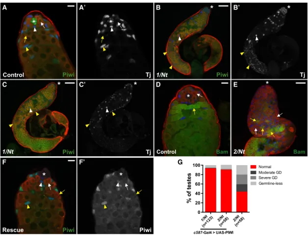
of Piwi, we took advantage of a mutation,piwiNt, that mislocal-izes Piwi to the cytoplasm ofDrosophilaovaries (Klenov et al., 2011). We verified that nuclear localization of PiwiNtprotein is lost in all Piwi-expressing cells from male flies carrying heteroal-lelic combinations ofpiwiNtwith piwi1 or piwi2 mutant alleles (Figures S7A–S7B0). Interestingly, as piwiNt mutant animals were aged to 10–15 days old, the entire testes became filled with Tj-positive cells, suggesting that cyst cells do not differen-tiate properly (Figures 6A–6C0). Moreover, Tj-positive cells were found in large clusters segregated away from germ cells in 18% (piwi1/piwiNt, n = 90) or 17% (piwi2/piwiNt, n = 124) of testes examined (Figures 6B–6C0), which is strikingly similar to the phenotypes described inpiwi1mutant andpiwiRNAianimals. Thus, these data suggest that Piwi nuclear localization, likely
re-flecting its function in epigenetic regulation, is required for cyst cell differentiation and possibly to facilitate somatic and germ cell interactions.
In the germline, DNA-bright early germ cells occupied the bulk of the lumen in piwiNt mutants (Figures S7F and S7F0). More specifically, 34% (piwi1/piwiNt, n = 90) or 37% (piwi2/piwiNt, n = 124) of mutant testes examined displayed severe germ cell differentiation defects (Figure S7C). Persisting germ cells ex-pressed variable levels of Bam-GFP, indicative of defective germ cell differentiation (Figure 6E). In addition, 32% of testes examined displayed moderate germ cell differentiation defects (Figures S7C and S7D). Moreover, 8%–10% of mutant testes were devoid of germ cells (Figures S7C and S7E). These data indicate that Piwi nuclear localization in somatic and/or germ
A B B’ B’’
C D D’ D’’
E F G
Figure 5. Knockdown of Piwi in Early Somatic Gonadal Cells Disrupts Cyst Cell and Germ Cell Development
(A) Control testes (Tj-Gal4;gfpRNAi
). In the presence ofgfpRNAi, Piwi expression (green) remains unaffected in somatic (blue, arrowheads) and germ cell (red, arrows) lineages.
(B)HM2 piwiRNAi(Tj-Gal4; HMS2 piwiRNAi/+). Surface view of KD testis shows the accumulation of Tj-positive somatic cells (blue, arrowheads) and Vasa-positive germ cells (red, arrows) far from the apical end. Persisting somatic cells are segregated from germ cells (blue, B’). Note that Piwi protein is undetectable in Tj-positive cells but remains unaffected in germ cells. (B00) is a magnification of (B).
(C) Sibling control (THU piwiRNAi
/+). Same as (A). (D)THU piwiRNAi
(Tj-Gal4/THU piwiRNAi
). Same as (B). (D00) is a magnification of (D).
(E) Frequency of testes from 7-day-old flies that display a specific phenotype (germline defect [GD]) in a given genotype.
(F and G) Piwi KD testes displaying a moderate (F) and severe (G) germline defect phenotype, respectively. Note that the majority of Piwi-positive germ cells are found in the lumen of the testis, whereas Piwi-negative somatic cells are located along the periphery.
Arrowheads and arrows mark the somatic and germ cells, respectively. Tj marks the hub, CySCs, and early cyst cells (blue). Vasa marks germ cells (red). Asterisk denotes the hub (A and C) or apical end of testes (B and D). Scale bars represent 10mm (B00and D00) and 25mm (A–G). See alsoFigures S3–S6.
cells is required for germ cell differentiation and possibly GSC maintenance.
The pleotropic defects observed in piwiNt mutants closely resemble testes fromTj-Gal4driven piwiRNAi flies (cf.Figures S7and5). This raised the possibility that differentiation defects may be due to the loss ofsomaticPiwi function. To test this hy-pothesis, wild-type Piwi was expressed specifically in cyst cells of thepiwiNtmutant background (Figures S2C and S2C0). As ex-pected, rescue of thepiwiNtphenotype was observed in 98% (piwi1/piwiNt, n = 63;piwi2/piwiNt, n = 107) of testes examined from 10- to 15-day-old flies. Moreover, rescue ofpiwiNt pheno-types was still observed in 93% (piwi1/piwiNt, n = 123) or 90% (piwi2/piwiNt, n = 58) of testes examined from 20- to 25-day-old
flies (Figures 6F and 6G), indicating that the rescued phenotype was maintained through adulthood. In contrast, testes from sib-ling controls that do not contain wild-type Piwi display pleio-tropic defects similar topiwiNtflies (piwi2, n = 58;Figure 6G). Together, these data indicate that Piwi nuclear localization in somatic cyst cells is sufficient for somatic and germ cell differentiation.
Piwi and piRNAs Regulate Fas3 in Somatic Cyst Cells
To explore the molecular mechanism underlying Piwi function in the testis, piRNAs bound to Piwi were isolated by immunopre-cipitation using an anti-Piwi antibody (Figure 7A), followed by deep sequencing (seeSupplemental Experimental Procedures).
A A’ B B’
C C’ D E
F F’ G
Figure 6. Piwi Nuclear Localization in Cyst Cells Is Required for Somatic and Germ Cell Differentiation
(A–C) Sibling control(piwiNt
/+)(A) andpiwi1
/piwiNt
testis (B and C) from 10- to 15-day-old flies. (A) Piwi protein (green) localizes in the nuclei of somatic and germ cells (arrows and arrowheads). (B) Surface view of mutant testis shows accumulation of Tj-positive somatic cells far from apical end (blue, B0). Small clusters of germ cells (red) are typically found near surface of the gonad. (C) Cross section of mutant testis shows accumulation of Vasa-positive germ cells (red) in the lumen. Majority of Tj-positive somatic cells are located along the periphery (C0, yellow arrowheads); however, Tj-positive cells can be found associated with germ cells (white arrowhead).
(D) Sibling control (piwiNt
/+; Bam-GFP/+).
(E)piwi2/piwiNt; Bam-GFP/+. Bam-GFP (green) accumulates in most excess germ cells (yellow arrows), but large grouping of germ cells that do not express Bam protein are present (white arrows).
(F) Rescued testis (c587-Gal4; piwi1
/piwiNt
; UAS-PIWI/+) from 20- to 25-day-old flies. Forced expression of wild-type Piwi in cyst cells rescues somatic and germ cell differentiation defects found inpiwiNt
mutant testes.
(G) Frequency of testes that display a specified phenotype (GD: germline defect) in a given genotype from 20- to 25-day-old flies.
In panels A–F0, arrowheads and arrows mark the somatic and germ cells, respectively. Tj marks the hub, CySCs, and early cyst cells (blue). Vasa marks germ cells (red). Asterisk denotes the hub (A and D) or the apical end of the testis (B and C). Scale bars represent 10mm (A, D, and F) and 50mm (B, C, and E). See also Figure S7.
The length of Piwi-associated piRNAs in the testis peaks at 26 nt (Figure 7B), similar to Piwi-associated piRNAs in ovaries ( Bren-necke et al., 2007) and from whole flies (Yin and Lin, 2007). While the majority of piRNAs associated with Piwi in ovaries and whole flies are derived from retrotransposons (Brennecke et al., 2007),
only 33% of the testicular piRNAs are derived from transposable elements in testes (Figure 7C). Interestingly, many Piwi-associ-ated piRNAs in the testis were derived from protein-coding genes (49%), includingTj(Figures 7C and 7D). Particularly, we found that the first intron of Fas3, a gene that encodes for an
A B C
D E
F G
H I
J
Figure 7. Piwi and piRNAs RegulateFas3in Somatic Cyst Cells
(A) Small RNAs associated with Piwi in adult testes were immunoprecipitated (IP) using anti-Piwi antibody. RNA molecules extracted from the immunoprecip-itated complexes were visualized by32
P-ATP labeling on denaturing acrylamide gel. Piwi-bound small RNAs are visualized (red arrow). (B) Size profile of all mapped small RNAs, excluding miRNAs and abundant cellular RNAs, such as rRNA and tRNA.
(C) Pie chart for the annotation of all mapped small RNAs, excluding abundant cellular RNAs and miRNAs. (D) Summary of piRNA targeting for selected genes. Up to one mismatch was allowed.
(E) piRNA distribution along theFas3gene. Red and blue signals represent sense and antisense piRNAs, respectively. Up to one mismatch was allowed. The UTR, CDS, and intron of Fas3 are shown.
(F) Control testes (Tj-Gal4;gfpRNAi). Fas3 is expressed in hub cells (*), but not in cyst cells (arrowheads). (G)Tj-Gal4/+; HMS2 piwiRNAi
/+. Piwi-depleted cyst cells ectopically express Fas3 far from the hub (arrowhead). Note that Fas3 is still maintained in hub cells (*). (H) Sibling control (piwiNt
/+). Same as (F). (I)piwi1
/piwiNt
testis. Same (G).
(J) Model for Piwi function in the testicular stem cell niche. Piwi is required cell autonomously for GSC and CySC maintenance. Piwi in the hub might regulate stem cell numbers (dotted-dashed lines). Piwi in cyst cells (CC) non-autonomously controls germ cell differentiation (arrow). Piwi and possibly piRNAs regulateFas3 in cyst cells.
adhesion molecule, generates piRNAs that are antisense to, and therefore may target, the Fas3 mRNA (Figures 7D and 7E). How-ever, no anti-sense piRNA was found to target the key compo-nents of the EGFR signaling pathway (Figure 7D), consistent with our observation that somatic Piwi signaling to the germline is independent of the pathway (Figure S6).
Fas3 has been shown to mediate cell sorting (Chiba et al., 1995; Elkins et al., 1990), yet piwi mutants display failure of cell mixing between somatic and germ cell lineages (Figures 1 and5). We therefore examined whether Fas3 is regulated by Piwi in the testis. We took advantage of piwiRNAi to knock down Piwi in somatic cyst cells and assessed Fas3 protein levels in these cells. In wild-type testis, Fas3 is expressed in the hub but is undetectable in cyst cells (Figure 7F). Remarkably, the majority of KD testes (65%, n = 54) displayed ectopic Fas3 expression in cyst cells found far from the hub (Figure 7G). This indicates that Piwi suppresses Fas3 expression in somatic cyst cells, presum-ably via theFas3-targeting piRNAs. Moreover, 40% of piwiNt mutant testes also displayed ectopic Fas3 expression in cyst cells (n = 147;Figure 7I). This further suggests that Piwi nuclear localization may be required for repression of Fas3 expression.
DISCUSSION
Over the past decade, much effort has been focused on identi-fying the molecular activities of Piwi in the germline ofDrosophila and vertebrate model organisms, yet its somatic function re-mains elusive. The work presented here highlights the essential function of Piwi as a cell-autonomous regulator for the self-renewal of a somatic stem cell type and its parallel function for GSCs (Figure 7J). In addition, it reveals that somatic Piwi function regulates Fas3 expression, likely through the Piwi-piRNA pathway. This might be a mechanism to ensure the coordinated division, differentiation, and interaction of two stem cell lineages within an organ for proper organogenesis.
Piwi Appears to Be Dispensable in Hub Cells for Stem Cell Maintenance and Differentiation
Previously, we have reported that Piwi functions in the niche cells of the Drosophila ovary to promote GSC maintenance (Cox et al., 1998; Szakmary et al., 2005). Surprisingly, we found that Piwi reduction in hub cells did not affect the maintenance of resident stem cells or their differentiation. We cannot rule out the possibility that Piwi KD is insufficient to disrupt its function. However, this is unlikely, considering thatUpd-Gal4begins its expression in the embryonic gonad (Le Bras and Van Doren, 2006); thus, the hub has experienced reduced levels of Piwi as soon as it is formed. Moreover, restoring wild-type Piwi solely in the hub was insufficient for rescuing differentiation defects found in mutant testes. Thus, it is quite possible that Piwi func-tions are not essential in the hub for stem cell maintenance or differentiation.
Cell-Autonomous Function of Piwi in Stem Cell Maintenance
Our data indicate that Piwi is required autonomously for stem cell maintenance. Using clonal analysis, we show thatpiwi1mutant GSCs and CySCs cannot be maintained at the hub. Furthermore,
temporal-specific knockdown of Piwi in adult CySCs causes a significant reduction in somatic stem cell populations. Thus, this study clearly demonstrates the cell-autonomous role of Piwi in adult stem cells. Our data complement previous studies with respect to GSCs, because in theDrosophilaovarian system, Piwi was known to be required autonomously for GSC self-renewal (Ma et al., 2014). Moreover, this cell-autonomous function is particularly novel with respect to CySCs, since it rep-resents the clear demonstration of a developmental function of Piwi in a somatic stem cell type. The somatic function of Piwi proteins has been recently reported inHydra, a basal eukaryote (Juliano et al., 2014). A number of reports have also correlated Piwi proteins with cancer cell proliferation in various somatic tis-sues (Ross et al., 2014), yet the causal relationship or underlying mechanism has not been demonstrated. Given the highly conserved molecular mechanism mediated by Piwi proteins dur-ing evolution, the cell-autonomous function of Piwi in stem cells may also be conserved for human somatic stem cells, with impli-cations in cancer.
Cell-Autonomous Function of Piwi in Somatic Cyst Cell Development
This study revealed a critical role for Piwi in regulating four distinct aspects of somatic cyst cell development. First, when Piwi was knocked down starting from somatic gonadal cells, the testis displays defects in cyst cell development that closely resemble the testicular phenotype ofTj, a gene in the Piwi-piRNA pathway (Li et al., 2003; Saito et al., 2009). As theTjphenotype is due to its function in gonadogenesis, our results indicate that Piwi functions are critical in somatic gonadal cells prior to sper-matogenesis. Second, Piwi is required in CySCs for their mainte-nance, as mosaic analysis (Figure 3) andpiwiRNAi (Figure 4) result in loss of somatic stem cell populations. Third, Piwi regu-lates cyst cell differentiation, as Piwi-depleted cyst cells retain stem cell characteristics far from the hub (Figure S3). Fourth, Piwi may promote the intermingling of cyst cells and germ cells, as Piwi-depleted cyst cells typically did not mix with germ cells (Figure 5). Our analysis suggests that the latter two Piwi functions may be restricted to the developing male gonad and might be interrelated. Thus, Piwi may have different biological roles during different stages of testicular development and spermatogenesis.
Piwi Acts in Somatic Cyst Cells to Control Germ Cell Differentiation via an EGFR-Independent Pathway
Several reports have demonstrated that defects in germline encystment causes spermatogenic arrest (Sarkar et al., 2007). Interestingly, loss of Piwi functions in cyst cells during develop-ment and adulthood results in the accumulation of mitotically active germ cells. Moreover, Piwi expression in somatic cyst cells, but not in the hub or germline, was capable of rescuing so-matic and germ cell differentiation defects ofpiwimutants. Our analysis suggest that Piwi-mediated somatic induction of germ cell differentiation appears to be independent of EGFR signaling, similar to fly ovaries (Ma et al., 2014). Recently, Piwi was shown to be required in ovarian escort cells, the functional equivalent of somatic cyst cells inDrosophilatestes, to promote ovarian germ-line cyst differentiation (Jin et al., 2013; Ma et al., 2014), even though testicular somatic cyst cells are replenished via stem
cell division whereas ovarian escort cells are replaced by self-duplication (Kirilly et al., 2011). These results suggest that Piwi is a critical somatic factor capable of controlling germ cell behavior in both sexes.
Piwi Appears to Epigenetically Regulate Cyst Cells to Instruct Germ Cell Differentiation
Among the threeDrosophilaPiwi proteins, Piwi is the only nu-clear protein, the only protein that is expressed in both somatic and germ cell lineages, and the only protein implicated in epige-netic regulation (Cox et al., 2000; Huang et al., 2013; Yin and Lin, 2007). The differentiation defects observed in approximately two-thirds of testes from piwiNt mutants indicate the crucial requirement of Piwi nuclear localization for somatic and germ cell differentiation. Since the known mechanism of Piwi in the nu-cleus is epigenetic regulation, our findings suggest that Piwi achieves its function in the somatic cells likely via epigenetic regulation. This is in contrast to its role during oogenesis, which does not require the nuclear localization of Piwi (Klenov et al., 2011). However, the analysis byKlenov et al. (2011)only covered 1- to 5-day-old female flies, which might be too soon to reveal similar oogenic defects.
The pleiotropic defects observed inpiwiNtmutants were nearly identical to the phenotypic consequence ofTj-Gal4driven Piwi knockdown, indicating that the epigenetic regulation of Piwi may occur in early somatic gonadal cells during gonadogenesis. As Piwi expression in somatic gonadal cells of the piwiNt mutant background was sufficient to restore spermatogenesis to wild-type-like conditions, this study uncovers the nuclear, and thus likely epigenetic, function of Piwi in gonadogenesis and spermatogenesis.
The Piwi-piRNA Association in Fly Testes
Piwi proteins associate with a variety of piRNAs inDrosophila and mammalian testes, including transposon and non-trans-poson piRNAs (Nishida et al., 2007; Watanabe et al., 2015). In Drosophilaovarian somatic cells, piRNAs derived from the 30 UTR ofTraffic jamhave been proposed to targetFas3, suggest-ing a connection between Piwi and silencsuggest-ing of theFas3gene (Saito et al., 2009). Our work further demonstrates that Piwi is associated withFas3-targeting piRNAs and that Piwi regulates Fas3 in somatic cyst cells in vivo, which in turn might be important for gonadogenesis and spermatogenesis. It has been suggested that Piwi achieves epigenetic programming of euchromatic genes by forming Piwi-piRNA complexes that bind to the nascent transcripts of target genes, which in turn re-cruits epigenetic factors such as heterochromatin protein 1a and Su(var)3-9 histone methyltransferase (Brower-Toland et al., 2007; Huang et al., 2013). Because the nuclear localization of Piwi is required to suppress Fas3 expression, we speculate that this suppression may be achieved by epigenetic regulation of the Fas3 gene via the nuclear Piwi-piRNA complex targeting of theFas3nascent transcript that is still tethered to the gene. This targeting would require antisense piRNAs and is quite spe-cific, as we were unable to find an antisense piRNA that maps to genes involved in EGFR signaling (Figure 7D). This is consistent with our observation that Piwi deficiency does not affect the EGFR signaling pathway. Given the well-conserved molecular
mechanism of Piwi, its known function in somatic cells in lower eukaryotes, and the correlation between ectopic expression of Piwi proteins and human cancers (Juliano et al., 2014; Rajase-thupathy et al., 2012; Ross et al., 2014), it is tempting to specu-late that the biological functions of Piwi reported here both in the soma and in the germline may be conserved during evolution. EXPERIMENTAL PROCEDURES
RNAi ofpiwi
ForpiwiRNAi, transgenic flies carrying three different RNAi constructs (HMS1, HMS2, and THU) were expressed specifically in somatic gonadal cells during gonadogenesis throughout adulthood usingTraffic (Tj)-Gal4(Li et al., 2003) (Figure S3B). Testes fromTj-Gal4-driven HMS2 and THUpiwiRNAi
flies dis-played aTj-like phenotype (seeResults), indicating that Piwi has an earlier role in gonadogenesis. However, testes fromTj-Gal4-driven HMS1 flies were typically devoid of Zfh1-positive somatic cells, indicating that somatic expres-sion of HMS1piwiRNAi
causes cell lethality. As HMS1 driven byUpd-Gal4also resulted in lethality (more severe than the strongpiwimutant phenotype), HMS1 likely has a side genetic effect beyond reducing piwiexpression. Thus, we used HMS2 and THU, but not HMS1, flies forpiwiRNAi.
SUPPLEMENTAL INFORMATION
Supplemental Information includes Supplemental Experimental Procedures and seven figures and can be found with this article online athttp://dx.doi. org/10.1016/j.celrep.2015.06.004.
AUTHOR CONTRIBUTIONS
J.G. and H.L. planned the experiments. J.G. performed the experiments. H.Q. performed Piwi-IP and prepared RNA for deep sequencing. N.L. performed bioinformatic analysis. J.G. and H.L. analyzed data and wrote the manuscript. ACKNOWLEDGMENTS
We thank E. Bach, S. DiNardo, D. McKearin, T. Xie, and the Bloomington DrosophilaStock Center for flies; R. Lehmann, M. Siomi, D. Godt, and the Developmental Studies Hybridoma Bank for antibodies; and J. Peng and V. Gangaraju for valuable comments on the manuscript. This work was sup-ported by NIH grant DP1CA174416 and the Mathers Foundation.
Received: June 18, 2013 Revised: February 17, 2015 Accepted: May 30, 2015 Published: June 25, 2015 REFERENCES
Brennecke, J., Aravin, A.A., Stark, A., Dus, M., Kellis, M., Sachidanandam, R., and Hannon, G.J. (2007). Discrete small RNA-generating loci as master regu-lators of transposon activity in Drosophila. Cell128, 1089–1103.
Brower-Toland, B., Findley, S.D., Jiang, L., Liu, L., Yin, H., Dus, M., Zhou, P., Elgin, S.C., and Lin, H. (2007). Drosophila PIWI associates with chromatin and interacts directly with HP1a. Genes Dev.21, 2300–2311.
Carmell, M.A., Girard, A., van de Kant, H.J., Bourc’his, D., Bestor, T.H., de Rooij, D.G., and Hannon, G.J. (2007). MIWI2 is essential for spermatogenesis and repression of transposons in the mouse male germline. Dev. Cell12, 503–514.
Chiba, A., Snow, P., Keshishian, H., and Hotta, Y. (1995). Fasciclin III as a synaptic target recognition molecule in Drosophila. Nature374, 166–168. Cox, D.N., Chao, A., Baker, J., Chang, L., Qiao, D., and Lin, H. (1998). A novel class of evolutionarily conserved genes defined by piwi are essential for stem cell self-renewal. Genes Dev.12, 3715–3727.
Cox, D.N., Chao, A., and Lin, H. (2000). piwi encodes a nucleoplasmic factor whose activity modulates the number and division rate of germline stem cells. Development127, 503–514.
de Cuevas, M., and Matunis, E.L. (2011). The stem cell niche: lessons from the Drosophila testis. Development138, 2861–2869.
Elkins, T., Hortsch, M., Bieber, A.J., Snow, P.M., and Goodman, C.S. (1990). Drosophila fasciclin I is a novel homophilic adhesion molecule that along with fasciclin III can mediate cell sorting. J. Cell Biol.110, 1825–1832. Fabrizio, J.J., Boyle, M., and DiNardo, S. (2003). A somatic role for eyes absent (eya) and sine oculis (so) in Drosophila spermatocyte development. Dev. Biol. 258, 117–128.
Go¨nczy, P., Matunis, E., and DiNardo, S. (1997). bag-of-marbles and benign gonial cell neoplasm act in the germline to restrict proliferation during Drosophila spermatogenesis. Development124, 4361–4371.
Huang, X.A., Yin, H., Sweeney, S., Raha, D., Snyder, M., and Lin, H. (2013). A major epigenetic programming mechanism guided by piRNAs. Dev. Cell24, 502–516.
Jin, Z., Flynt, A.S., and Lai, E.C. (2013). Drosophila piwi mutants exhibit germline stem cell tumors that are sustained by elevated Dpp signaling. Curr. Biol.23, 1442–1448.
Juliano, C., Wang, J., and Lin, H. (2011). Uniting germline and stem cells: the function of Piwi proteins and the piRNA pathway in diverse organisms. Annu. Rev. Genet.45, 447–469.
Juliano, C.E., Reich, A., Liu, N., Go¨tzfried, J., Zhong, M., Uman, S., Reenan, R.A., Wessel, G.M., Steele, R.E., and Lin, H. (2014). PIWI proteins and PIWI-interacting RNAs function in Hydra somatic stem cells. Proc. Natl. Acad. Sci. USA111, 337–342.
Kiger, A.A., White-Cooper, H., and Fuller, M.T. (2000). Somatic support cells restrict germline stem cell self-renewal and promote differentiation. Nature 407, 750–754.
Kiger, A.A., Jones, D.L., Schulz, C., Rogers, M.B., and Fuller, M.T. (2001). Stem cell self-renewal specified by JAK-STAT activation in response to a support cell cue. Science294, 2542–2545.
Kirilly, D., Wang, S., and Xie, T. (2011). Self-maintained escort cells form a germline stem cell differentiation niche. Development138, 5087–5097. Klenov, M.S., Sokolova, O.A., Yakushev, E.Y., Stolyarenko, A.D., Mikhaleva, E.A., Lavrov, S.A., and Gvozdev, V.A. (2011). Separation of stem cell mainte-nance and transposon silencing functions of Piwi protein. Proc. Natl. Acad. Sci. USA108, 18760–18765.
Le Bras, S., and Van Doren, M. (2006). Development of the male germline stem cell niche in Drosophila. Dev. Biol.294, 92–103.
Leatherman, J.L., and Dinardo, S. (2008). Zfh-1 controls somatic stem cell self-renewal in the Drosophila testis and nonautonomously influences germline stem cell self-renewal. Cell Stem Cell3, 44–54.
Lee, T., and Luo, L. (1999). Mosaic analysis with a repressible cell marker for studies of gene function in neuronal morphogenesis. Neuron22, 451–461. Li, M.A., Alls, J.D., Avancini, R.M., Koo, K., and Godt, D. (2003). The large Maf factor Traffic Jam controls gonad morphogenesis in Drosophila. Nat. Cell Biol. 5, 994–1000.
Lim, J.G., and Fuller, M.T. (2012). Somatic cell lineage is required for differen-tiation and not maintenance of germline stem cells in Drosophila testes. Proc. Natl. Acad. Sci. USA109, 18477–18481.
Lin, H. (2008). Cell biology of stem cells: an enigma of asymmetry and self-renewal. J. Cell Biol.180, 257–260.
Lin, H., and Spradling, A.C. (1997). A novel group of pumilio mutations affects the asymmetric division of germline stem cells in the Drosophila ovary. Devel-opment124, 2463–2476.
Ma, X., Wang, S., Do, T., Song, X., Inaba, M., Nishimoto, Y., Liu, L.P., Gao, Y., Mao, Y., Li, H., et al. (2014). Piwi is required in multiple cell types to control germline stem cell lineage development in the Drosophila ovary. PLoS ONE 9, e90267.
Morrison, S.J., and Spradling, A.C. (2008). Stem cells and niches: mechanisms that promote stem cell maintenance throughout life. Cell132, 598–611. Nishida, K.M., Saito, K., Mori, T., Kawamura, Y., Nagami-Okada, T., Inagaki, S., Siomi, H., and Siomi, M.C. (2007). Gene silencing mechanisms mediated by Aubergine piRNA complexes in Drosophila male gonad. RNA13, 1911– 1922.
Peng, J.C., and Lin, H. (2013). Beyond transposons: the epigenetic and somatic functions of the Piwi-piRNA mechanism. Curr. Opin. Cell Biol.25, 190–194.
Qi, H., Watanabe, T., Ku, H.Y., Liu, N., Zhong, M., and Lin, H. (2011). The Yb body, a major site for Piwi-associated RNA biogenesis and a gateway for Piwi expression and transport to the nucleus in somatic cells. J. Biol. Chem. 286, 3789–3797.
Rajasethupathy, P., Antonov, I., Sheridan, R., Frey, S., Sander, C., Tuschl, T., and Kandel, E.R. (2012). A role for neuronal piRNAs in the epigenetic control of memory-related synaptic plasticity. Cell149, 693–707.
Robine, N., Lau, N.C., Balla, S., Jin, Z., Okamura, K., Kuramochi-Miyagawa, S., Blower, M.D., and Lai, E.C. (2009). A broadly conserved pathway generates 30UTR-directed primary piRNAs. Curr Biol.19, 2066–2076.
Ross, R.J., Weiner, M.M., and Lin, H. (2014). PIWI proteins and PIWI-interact-ing RNAs in the soma. Nature505, 353–359.
Rouget, C., Papin, C., Boureux, A., Meunier, A.C., Franco, B., Robine, N., Lai, E.C., Pelisson, A., and Simonelig, M. (2010). Maternal mRNA deadenylation and decay by the piRNA pathway in the early Drosophila embryo. Nature 467, 1128–1132.
Saito, K., Inagaki, S., Mituyama, T., Kawamura, Y., Ono, Y., Sakota, E., Kotani, H., Asai, K., Siomi, H., and Siomi, M.C. (2009). A regulatory circuit for piwi by the large Maf gene traffic jam in Drosophila. Nature461, 1296–1299. Sarkar, A., Parikh, N., Hearn, S.A., Fuller, M.T., Tazuke, S.I., and Schulz, C. (2007). Antagonistic roles of Rac and Rho in organizing the germ cell microen-vironment. Curr Biol.17, 1253–1258.
Sienski, G., Do¨nertas, D., and Brennecke, J. (2012). Transcriptional silencing of transposons by Piwi and maelstrom and its impact on chromatin state and gene expression. Cell151, 964–980.
Szakmary, A., Cox, D.N., Wang, Z., and Lin, H. (2005). Regulatory relationship among piwi, pumilio, and bag-of-marbles in Drosophila germline stem cell self-renewal and differentiation. Curr Biol.15, 171–178.
Watanabe, T., Cheng, E.C., Zhong, M., and Lin, H. (2015). Retrotransposons and pseudogenes regulate mRNAs and lncRNAs via the piRNA pathway in the germline. Genome Res.25, 368–380.
Yin, H., and Lin, H. (2007). An epigenetic activation role of Piwi and a Piwi-associated piRNA in Drosophila melanogaster. Nature450, 304–308.