The Role of Hydrogen Sulfide in Normal and
Aberrant Late Lung Development
Inaugural Dissertation
submitted to the
Faculty of Medicine
in partial fulfillment of the requirements
for the PhD-Degree
of the Faculties of Veterinary Medicine and Medicine
of the Justus Liebig University Giessen
by
Madurga Hernández, Alicia
of
Valencia, Spain
2
From the Max Planck Institute for Heart and Lung Research,
Bad Nauheim
Director / Chairman: Prof. Dr. Norbert Weissman of the Faculty of Medicine of
the Justus Liebig University Giessen
First Supervisor and Committee Member: Prof. Dr. Werner Seeger
Second Supervisor and Committee Member: Prof. Dr. Martin Diener
Committee Members: Prof. Dr. Christine Wrenzycki, Prof. Dr. Maik Gollasch
3
1
Declaration
I declare that I have completed this dissertation single-handedly without the unauthorized help of a second party and only with the assistance acknowledged therein. I have appropriately acknowledged and referenced all text passages that are derived literally from or are based on the content of published or unpublished work of others, and all information that relates to verbal communications. I have abided by the principles of good scientific conduct laid down in the charter of the Justus Liebig University of Giessen in carrying out the investigations described in the dissertation.
4
“The basic mechanisms that regulate lung development and the link with human lung disease are among the most challenging and exciting areas of scientific inquiry”.
5
2
Abstract
Bronchopulmonary dysplasia (BPD) is a chronic lung disease characterized by arrested alveolarization, a complication of premature birth. The gasotransmitter hydrogen sulfide (H2S)
is emerging as a mediator of lung physiology and disease. In this study, the impact of systemic application of H2S on post-natal alveolarization was evaluated in a mouse BPD model.
Exposure of newborn mice to 85% O2 for 10 days decreased the total number of alveoli in the
lung by 56% and increased mean alveolar septal wall thickness by 29%, assessed by stereological analysis. Systemically administration of GYY4137, the slow-release H2S donor,
for 10 days improved lung alveolarization in mouse pups breathing 85% O2, compared with
vehicle-treated littermates. Though without effect on lung oxidative status, systemic H2S
administration reduced leukocyte infiltration into alveolar airspaces caused by hyperoxia, and normalized lung interleukin (IL)-10 levels that were else diminished by 85% O2. The
rapid-release H2S donor NaHS was used to treat primary mouse alveolar type II (ATII) cells; NaHS
had no impact on cell viability but stimulated ATII cell migration. Glibenclamide attenuated the impact of NaHS on ATII cell migration, implicating ion channels; and was accompanied by activation of Akt, hinting at two possible mechanisms of H2S action. Although exposure of
ATII cells to 85% O2 produced substantial changes in gene expression, exposure to either
GYY4137 or NaHS had no impact on ATII cell gene expression, as determined by microarray suggesting that the effects observed were independent of changes in gene expression. H2S can
be generated endogenously by cystathionine -synthase (Cbs) and cystathionine -lyase (Cth). In this study it is demonstrated that the expression of Cbs and Cth in mouse lungs is dynamically regulated during lung alveolarization, and that alveolarization is impaired in Cbs-/- and Cth -/-mouse pups, where a 50% reduction in the total number of alveoli was observed, with no influence on mean alveolar septal wall thickness. Immunofluorescence staining and laser-capture micro dissection revealed that CBS and CTH expression was present in the lung vessels and in the airway epithelium. Vessel remodeling occurred in the absence of Cbs and Cth, since it led to a 100-500% increase in vessel muscularization of small- and medium- size lung vessels. Inhibition of CBS or CTH expression in endothelial cells, using either small interfering RNA or via pharmacological inhibition (propargylglycine) respectively, diminished angiogenic capacity resulting in a 50% decrease in number of tubes formed, and a 30-40% decrease in tube length. On the other hand, administration of GYY4137, promoted endothelial tube formation. These data support the further investigation of H2S as a candidate interventional strategy to
6
H2S-generating enzymes Cbs and Cth in lung alveolarization and pulmonary vascular
development and homeostasis.
3
Kurzfassung
Die bronchopulmonale Dysplasie (BPD) ist eine chronische Lungenerkrankung, charakterisiert durch eine gehemmte Alveolarisierung und tritt als Komplikation bei Frühgeburten auf. Der Gasotransmitter Wasserstoffsulfid (H2S) ist als ein Mediator in der Lungenphysiologie und in
Erkrankungen bekannt. In dieser Studie wurde die Wirkung einer systemischen Applikation von H2S auf die postnatale Alveolarisierung im BPD Modell evaluiert. Die Exposition
neugeborener Mäuse zu 85% O2 für 10 Tage verringerte die totale Anzahl der Alveoli in der
Lunge um 56% und erhöhte die durchschnittliche Wanddicke der alveolaren Septen um 29%, festgestellt durch stereologische Analysen. Die systemische Verabreichung von GYY4137, einem H2S Donor mit langsamer Freisetzung, für 10 Tage verbesserte die Alveolarisierung in
den Jungtieren, die 85% O2 ausgesetzt waren, im Vergleich zu den Jungtieren, die nur die
Trägersubstanz verabreicht bekamen. Ohne Effekte auf den oxidativen Status der Lunge reduzierte die systemische H2S Gabe die durch die Hyperoxie begründete
Leukozyten-Infiltration in den alveolären Luftraum und normalisierte die Interleukin (IL)-10 Level, die durch 85% O2 verringert werden. NaHS, ein H2S Donor mit schneller Freisetzung, wurde zur
Behandlung primärer alveolärer Typ II (ATII) Zellen verwendet. NaHS hatte keine Auswirkung auf die Lebensfähigkeit der Zellen, stimulierte jedoch die Zellmigration. Glibenclamide verringerte die Wirkung von NaHS auf die ATII Zellmigration, was eine Rolle von Ionenkanälen in diesem Prozess impliziert und wurde durch die Aktivierung von Akt begleitet, was Hinweise auf zwei mögliche Mechanismen der H2S Aktivität gibt. Obwohl die Exposition
von ATII Zellen zu 85% O2 erhebliche Veränderungen der Genexpression bewirkt, hatte weder
die Exposition zu GYY4137 noch zu NaHS eine Wirkung auf die Genexpression der ATII Zellen, wie durch Microarray Analysen festgestellt wurde. Dies lässt vermuten, dass die beobachteten Effekte unabhängig von Veränderungen in der Genexpression sind. H2S kann
endogen durch die Cystathionin β-Synthase (Cbs) und die Cystathionin -Lyase (Cth) generiert werden. In dieser Studie wurde gezeigt, dass die Expression von Cbs und Cth in der Lunge der Maus während der Alveolarisierung dynamisch reguliert wird und dass die Alveolarisierung in Cbs-/- und Cth-/- Jungtieren der Maus beeinträchtigt ist. Hier wurde eine Reduzierung der totalen
7 der alveolaren Septen wurde nicht festgestellt. Immunofluoreszenzfärbungen und Lasermikrodissektionen zeigten, dass eine Expression von CBS und CTH in den Lungengefäßen und dem Epithelium der Atemwege stattfand. Bei Abwesenheit von Cbs und Cth wurde ein Blutgefäß-Remodeling festgestellt, was zu einer 100 – 500%-igen Zunahme der Muskularisierung der kleinen und mittleren Blutgefäße führte. Die Inhibierung der CBS oder CTH Expression in Endothelzellen, entweder unter Verwendung von small interfering RNA oder einer pharmakologisch wirksamen Substanz (Propargylglycin) verminderte die angiogene Kapazität mit dem Resultat einer um 50% verminderten Anzahl der formierten tubes und einer Reduzierung der Länge der formierten tubes um 30 – 40%. Die Verabreichung von GYY4137 förderte die endotheliale tube Formierung. Diese Daten unterstützen eine weitere Untersuchung des H2S als einen Kandidaten für Interventionsstrategien, um der beeinträchtigten
Alveolarisierung, die mit BPD assoziiert ist, zu begegnen. Weiterhin kann vermutet werden, dass die H2S-generierenden Enzyme Cbs und Cth in der Alveolarisierung und der pulmonalen
8
4
Index
1 Declaration... 3 2 Abstract ... 5 3 Kurzfassung ... 6 4 Index ... 8 5 List of figures ... 12 6 List of tables ... 13 7 List of abbreviations ... 14 8 Introduction ... 18 Lung development ... 18 Bronchopulmonary dysplasia ... 20 8.2.1 Introduction ... 20 8.2.2 Definition ... 208.2.3 Incidence and prevalence ... 21
8.2.4 Morbidity and mortality ... 21
8.2.5 Risk factors ... 21
8.2.6 Signs and symptoms ... 22
8.2.7 Pathophysiology ... 22 8.2.8 Diagnosis ... 22 8.2.9 Treatment ... 23 8.2.10 Complications ... 24 8.2.11 Prognosis ... 25 Hydrogen sulfide ... 25 8.3.1 Introduction to gasotransmitters ... 25
8.3.2 Nitric oxide and carbon monoxide ... 26
8.3.3 Introduction to hydrogen sulfide ... 27
8.3.4 Biochemistry of hydrogen sulfide under physiological conditions ... 27
9
8.3.6 Toxicology of hydrogen sulfide ... 28
8.3.7 Endogenous hydrogen sulfide synthesis, regulation and catabolism ... 29
8.3.8 Cbs-/- and Cth-/- mice ... 30
8.3.9 Endogenous levels of hydrogen sulfide in health and disease ... 31
8.3.10 Mechanism of action of hydrogen sulfide ... 32
8.3.10.1 Sulfhydration of proteins ... 32
8.3.10.2 Interaction with potassium channels... 32
8.3.10.3 Vasorelaxation ... 32
8.3.10.4 Oxygen sensitive responses ... 33
8.3.10.5 Anti-inflammatory effects ... 33
8.3.10.6 Cytoprotective effects ... 34
8.3.10.7 Antioxidant effects ... 34
8.3.11 Interaction with other gasotransmitters ... 35
8.3.12 Effects of hydrogen sulfide in the nervous system ... 35
8.3.13 Role of hydrogen sulfide in different lung diseases ... 36
9 Hypothesis and aims ... 39
10 Materials and methods ... 40
Equipment and software ... 40
Reagents ... 41
Cell lines ... 44
Primer list ... 44
Antibodies ... 45
siRNA ... 46
Mouse model of bronchopulmonary dysplasia ... 46
Lung processing ... 47
Design-based stereology ... 47
Bronchoalveolar lavage ... 49
Immunofluorescence staining of mouse lung sections ... 50
Determination of medial wall thickness index and degree of vessel muscularization ... 50
10
Endothelial tube-formation assay ... 51
Primary ATII cells and cell-lines ... 52
ATII cell isolation and staining for flow cytometry ... 52
ATII cell isolation and staining for immunofluorescence ... 52
RNA isolation and cDNA synthesis for RT-PCR analysis ... 53
Laser-capture micro dissection ... 53
RT-PCR analysis ... 53
Protein isolation for immunoblots ... 54
Protein expression analysis ... 54
Wound healing assay ... 54
ELISA ... 55
Glutathione assay... 55
Microarray analyses... 55
Transgenic mice... 56
Genotyping of mouse genomic DNA ... 56
Statistical analyses ... 56
11 Results ... 57
Systemic H2S administration partly restores normal alveolarization in an experimental ... animal model of BPD ... 57
11.1.1 Stereological analysis of lung structure in normally and aberrantly developing lungs . 57 11.1.2 Stereological analysis of the impact of H2S donor administration on normal and aberrant late lung development ... 57
11.1.3 Analysis of H2S donor administration on inflammatory cell infiltration in neonatal mouse pup lungs ... 60
11.1.4 Cytokine response to GYY4137 administration ... 62
11.1.5 Impact of GYY4137 on the oxidative status of the developing lung ... 63
11.1.6 Impact of NaHS on viability of primary mouse alveolar type II cells ... 64
11.1.7 Impact of NaHS and GYY4137 treatment on gene expression in primary mouse alveolar type II cells ... 65
11
11.1.9 Impact of NaHS on Akt signaling in primary mouse alveolar type II cells ... 70
The role of Cbs and Cth during late lung development ... 72
11.2.1 The expression of Cbs and Cth is dynamically regulated during alveolarization ... 72
11.2.2 The expression of both Cbs and Cth can be abrogated during post-natal lung development in mice... 73
11.2.3 Loss of Cbs or Cth impairs normal lung alveolarization in mice. Lung histology ... 74
11.2.4 Loss of Cbs or Cth impairs normal alveolarization in mice. Stereology analysis ... 75
11.2.5 Loss of Cbs or Cth affects lung development, transition from P7.5 to P14.5 ... 78
11.2.6 Both Cbs and Cth are preferentially expressed in specific lung compartments ... 79
11.2.7 Both Cbs and Cth localize to the airways and vessel walls in the lungs of mouse pups 81 11.2.8 Both Cbs and Cth contribute to the development or maintenance of normal ... pulmonary vasculature ... 84
11.2.9 Both Cbs and Cth participate in the angiogenesis of human pulmonary microvascular endothelial cells ... 88
12 Discussion ... 91
13 Acknowledgements ... 99
12
5
List of figures
Figure 1 Scheme of the stages of lung development. ... 19
Figure 2 Chest X-ray of a one month old female patient with BPD. ... 23
Figure 3 Potential pathways of H2S production and metabolism. ... 30
Figure 4 Anti-inflammatory effects of hydrogen sulfide. ... 34
Figure 5 Mechanism of action of H2S in chronic lung diseases. ... 37
Figure 6 Effect of lipopolysaccharide induced acute lung injury and effect of H2S inhalation on lung structure. ... 38
Figure 7 Example of stereological analysis of neonatal mouse lungs exposed to either 21% O2 or 85% O2 pressure,fixed via the airways, and stained with Richardson’s stain. ... 49
Figure 8 GYY4137 administration decreased impaired alveolar development in an experimental mouse model of bronchopulmonary dysplasia. ... 58
Figure 9 GYY4137 administration diminished leukocyte recruitment into the alveolar airspaces that was provoked by exposure to hyperoxia. ... 61
Figure 10 GYY4137 administration alters the pulmonary expression of inflammatory mediators in neonatal mouse lungs. ... 63
Figure 11 GYY4137 administration does not impact the oxidative status of neonatal mouse lung as assessed by glutathione oxidation. ... 64
Figure 12 H2S delivered exogenously by a chemical donor does not cause apoptosis or cell death of primary mouse alveolar type II cells. ... 65
Figure 13 Scheme representing the experimental settings of primary alveolar type II cells exposed to hydrogen sulfide donors. ... 66
Figure 14 NaHS promotes wound closure in monolayers of primary mouse alveolar type II cells. ... 69
Figure 15 H2S donors promote activation of Akt. ... 71
Figure 16 RT-PCR to assess changes of mRNA expression of Cbs and Cth, on different time points during normal late lung development. ... 72
Figure 17 Genotyping of mouse pups for Cbs and Cth. ... 73
Figure 18 Loss of Cbs and Cth expression in lungs of Cbs-/- and Cth-/- mice by immunoblot. ... 73
Figure 19 Representative lung histology of postnatal day P7.5 and P14.5 of wild type mice, Cbs -/-mice, and Cth-/- mice. ... 74
13
Figure 20 Aberrant late lung development in the Cbs-/- mice and Cth-/- mice assessed via stereology
methods. ... 75
Figure 21 Aberrant late lung development in the Cbs-/- mice and Cth-/- mice assessed via stereology methods, evolution from P7.5 to P14.5. ... 78
Figure 22 Cbs and Cth are mostly expressed in the airway and vascular compartment. ... 80
Figure 23 Localization of CBS in the airways, vessel walls and alveolar epithelium. ... 82
Figure 24 Localization of CTH in the airways, vessel walls and alveolar epithelium. ... 83
Figure 25 Decreased vascular supply in the Cbs-/- mice and the Cth-/- mice. ... 85
Figure 26 Loss of Cbs or Cth promotes increased muscularization of small pulmonary vessels... 86
Figure 27 Loss of Cbs or Cth leads to an increase in an index of medial wall thickness in small pulmonary vessels. ... 87
Figure 28 Inhibition of CBS expression alters tube formation in vitro. ... 89
Figure 29 CTH and H2S modulate endothelial tube formation in vitro. ... 90
6
List of tables
Table 1 Comparison of the modes of action of gasotransmitters and neurotransmitters. ... 26Table 2 Primers for RT-PCR analysis ... 44
Table 3 Primers for genotyping ... 44
Table 4 Antibodies ... 45
Table 5 Structural parameters of developing mouse lungs during exposure to 21% O2 or 85% O2 assessed by stereological analysis (Madurga et al., 2014). ... 59
Table 6 Gene expression in primary mouse alveolar type II cells up-regulated by exposure to 85% O2 in vitro. ... 67
Table 7 Gene expression in primary mouse alveolar type II cells down-regulated by exposure to 85% O2in vitro. ... 68
Table 8 Stereological parameters of wild type mice, Cbs-/- mice and Cth-/- mice on postnatal day P7.5. ... 76
Table 9 Stereological parameters of wild type mice, Cbs-/- mice and Cth-/- mice on postnatal day P14.5 ... 77
14
7
List of abbreviations
% Per cent
°C Degree Celsius
αSMA α smooth muscle actin
µ Micro (10-6)
μg Microgram
μl Microliter
μM Micromolar
A Alveoli and ducts
Akt Protein kinase B
Alv air Alveolar air spaces
Alv epi Alveolar epithelium
ATII Primary alveolar type II
AV Annexin V
B Alveolar bridge
BAL Bronchoalveolar lavage
bp Base pair
Brg1 Brahma-related gene 1
BSA Bovine serum albumin
C Capillaries
CA Carbonic anhydrase
cAMP Cyclic adenosine monophosphate
CAT Cysteine aminotransferase
CBS Cystathionine-β synthase
cDNA Complementary DNA
CDO Cysteine dioxygenase
CE Coefficient of error
cGMP Cyclic guanosine monophosphate
CLY Cysteine lyase
CO Carbon monoxide
COPD Chronic obstructive pulmonary disease
CPAP Continuous positive airway pressure
15 Ct Cycle threshold CTH Cystathionine-γ lyase CV Coefficient of variation DAB 3,3-diaminobenzidine dH2O Distilled water
DNA Deoxyribonucleic acid
E Embryonic
EDTA Ethylendinitrilo-N, N, Nʹ, Nʹ-tetra-acetic-acid
eNOS Endothelial NOS
et al. Et alia
FBS Fetal bovine serum
FC Fold change
GD Gestational day
GSH Glutathione, reduced form
GSSG Glutathione, oxidized form
GYY4137 4-methoxyphenyl(morpholino)phosphinodithioate morpholinium salt
HMOX Heme oxygenase
H2O Water
HPAEC Human pulmonary artery endothelial cell
HRPO Horse radish peroxydase
H2S Hydrogen sulfide IF Immunofluorescence IHC Immunohistochemistry IL-1β Interleukin 1β IL-6 Interleukin 6 IL-10 Interleukin 10
iNOS Inducible NOS
i.p. Intraperitoneal
KATP ATP potassium
kDa kilo Dalton
MAPK Mitogen-activated protein kinases
mg Milligram
min Minute
16
MLE-12 Murine lung epithelial cell line
mM Millimolar
3-MPST 3-mercaptopyruvate sulfur transferase
mRNA Messenger RNA
MTT 3-(4,5-dimethylthiazol-2-yl)-2,5-diphenyltetrazolium bromide
MWT Index of medial wall thickness
N Number
NaCl Sodium chloride
NaHS Sodium hydrosulfide
Na3VO4 Sodium vanadate
NFκβ Nuclear factor κ-light-chain-enhancer of activated B cells
nM Nanomolar
NO Nitric oxide
Non-par Non parenchyma
NOS NO synthase
nNOS Neuronal NOS
Nrf2 Nuclear factor (erythroid-derived 2)-like 2
O2 Dimolecular oxygen
P Postnatal
PAG Propargylglycine
Par Parenchyma
PBS Phosphate-buffered saline
PCR Polymerase chain reaction
PI Propidium iodide
PLP Pyridoxal 5ʹ-phosphate
PPV Positive pressure ventilation
R-SH Thiol
S Surface area
Sv Surface density
SAM S-adenosyl methionine
Sep Septum
sGC Guanylyl cyclase
siRNA Small-interfering RNA
17
SUR Systematic uniform random
TNFα Tumor necrosis factor α
V Volume
Vv Volume density
vol/vol Volume per volume
vWF Von Willebrand factor
18
8
Introduction
Lung development
The pulmonary system develops from a series of complex events that occur during prenatal and early postnatal life. During lung development, the embryonic lung arises as a protrusion from the foregut endoderm and undergoes a process called “branching morphogenesis”, which begins a reproducible, bilaterally asymmetrical pattern of stereotypic branching into the surrounding splanchnic mesenchyme.
In the human lung, the branching is completed after 16 generations by week 16 post conception, and three lobes form on the right side and two on the left side of the lungs. These events require correct growth and differentiation of the epithelial and mesenchymal components of the immature lung to form the bronchial tree and alveoli; the pulmonary, bronchial, and lymphatic vasculatures; the nerves and the pleura.
In contrast to humans, the mouse has four lobes in the right side and one lobe on the left side. Moreover, the mouse only has 12 airway generations and alveolarization occurs postnatally. Classically, lung development has been classified into five stages: embryonic, pseudoglandular, canalicular, terminal saccular, and alveolar. Lung development is an early process that starts around the third week of the embryonic period and that can last until early adulthood. During the earliest lung development stage, the pseudoglandular stage (5-17 weeks of human pregnancy, embryonic (E) E9.5-E16.6 days in the mouse embryo), branching morphogenesis occurs in epithelial tubes lined with cuboidal epithelial cells. At this point, the structure is still too immature for adequate gas exchange. During the canalicular stage (16-25 weeks of human pregnancy, E16.6-E17.4 days in the mouse embryo), the respiratory tree continues growing in length and diameter, along with vascularization and angiogenesis. An increase in capillary number occurs, and the terminal bronchioles are divided into respiratory bronchioles and alveolar ducts. The terminal saccular stage (24 weeks to late fetal period in human, E17.4 to postnatal (P) day P5 in the mouse) is characterized by thinning of the interstitium. The alveolar epithelial cells are more clearly differentiated into type I and type II pneumocytes. The capillaries grow and form an intricate network during this stage and the lymphatic network becomes well developed. Maturation of surfactant synthesis and secretion is a crucial event to prevent lungs from collapsing and allow gas exchange, and near the end of this stage, the fetal lung can support gas exchange in premature newborns. During the alveolar stage (late fetal period to childhood in human, P5 to P30 in mouse) terminal sacs develop into mature alveolar
19 ducts and alveoli. Contrary to early lung development, this stage remains poorly understood. During this latter phase of development, important events take place for the maturation of the lung: the peak of secondary septation occurs, with an increase in the number and a decrease in the size of the alveoli, together with a thinning of the pulmonary septa and maturation of the lung microvasculature, increasing exponentially the surface area for gas exchange and minimizing the distance between air and blood. A neonatal lung is estimated to have about 50 million alveoli, still, this number increases six-fold to about 300 million by the age 7-8 years when alveolarization is largely complete. In the meantime, the adult alveolar capillary bed is able of accommodating the entire cardiac output of 5 L/min, rising five-fold to 25 L/min during maximal exercise (Madurga et al., 2013; Massaro & Massaro, 2007; Morrisey et al., 2013; Warburton, 2012; Warburton et al., 2010; Warburton et al., 2000).
Figure 1 Scheme of the stages of lung development.
Scheme of the five stages of lung development: embryonic, pseudoglandular, canalicular, terminal saccular, and alveolar. In mice, birth takes place after 20 days of embryonic development and pups are born in the saccular stage. In contrast, humans give birth around the 38th week of gestation, during the alveolar stage of lung maturation. GD, gestational day, P, postnatal day. With the permission of Dr. Gianni Carraro.
20
Bronchopulmonary dysplasia
8.2.1
Introduction
The development of the airways, through epithelial tube branching and later septation of terminal air sacs, occurs in close conjunction with the development of the pulmonary vasculature. Alterations to these developmental processes give rise to abnormal lung structure, deficiency of gas exchange, and newborn respiratory failure. Clinical examples of such perturbation of normal lung growth include bronchopulmonary dysplasia (BPD), cystic adenomatoid malformation of the lung, and hypoplasia of the lung (Smith et al., 2010; Warburton et al., 2010).
8.2.2
Definition
The term bronchopulmonary dysplasia was first described in 1967 by Northway and colleagues (Northway et al., 1967). The definition of BPD is complicated and has evolved over the years. Nowadays, BPD refers to a newborn at 35-37 weeks postmenstrual age treated with mechanical ventilation, continuous positive airway pressure (CPAP), or in need of oxygen concentrations of 30% at rest on oxygen saturation (sO2)of 90-96% or supplemental oxygen concentrations of
30% on sO2 of more than 96% (Ambalavanan & Carlo, 2004; Ehrenkranz et al., 2005; Jobe &
Bancalari, 2001; Jobe, 1999; Northway, 1992; Walsh et al., 2004).
The National Institute for Health in the USA has further divided the definition of BPD into:
-Mild: Requiring supplemental O2 at 28 weeks of age, but in air by 36 weeks corrected age.
-Moderate: Requiring <30% supplemented O2 at 36 weeks corrected age.
-Severe: Requiring >30% supplemental O2 and/or requiring CPAP or ventilation, at 36 weeks
21
8.2.3
Incidence and prevalence
The BPD disease is a complex multifactorial chronic lung disease of the premature newborn that affects thousands of infants each year with an incidence of 23% of infants with birth weights are below 1500 g. It is the single most important factor determining the length of hospital stay in newborns born at less than 29 weeks. The more premature the newborns are, the more severely affected they are. The prevalence is higher among very low birth weight and extremely preterm infants. Improved medical management of BPD has increased substantially the survival of these patients and also the pathophysiological picture of BPD, but the prevalence of BPD has also increased, especially in small infants who may have been exposed to in utero infection (Bose et al., 2011; Laughon et al., 2011; Shennan et al., 1988; Van Marter, 2009).
8.2.4
Morbidity and mortality
Improvement in clinical management has decreased substantially the mortality rates of BPD infants. The survival and morbidity among infants older than 24 weeks of gestational age have improved since the introduction of antenatal corticosteroids treatment and surfactant therapy, and nowadays infants with BPD usually have a milder disease. However, the survival rate remain 25-50% in the most preterm neonates of 23-24 weeks gestation, reflecting the lack of maturation in alveolarization and vascular development. Neonates with severe BPD remain at high risk for mortality and pulmonary morbidity during the first two years of life. Finally, factors such as gender affect the severity of this disease, since male infants usually have a more severe phenotype with worse neurodevelopmental outcome (Ambalavanan et al., 2009; Ambalavanan et al., 2008; Ambalavanan et al., 2011; Shennan et al., 1988).
8.2.5
Risk factors
The BPD disease may be caused by factors such as perinatal infection (chorioamnionitis), peripartum inflammation, volotrauma, borotrauma, postnatal steroid treatment, premature delivery, and prolonged mechanical ventilation, among others (Ambalavanan et al., 2009; Bose et al., 2011; Hartling et al., 2012; Jobe, 2005; Jobe & Ikegami, 2001; Kakkera et al., 2005; Kallapur & Jobe, 2006; Kunzmann, et al., 2013; Laughon et al., 2011; Rocha et al., 2010; Schelonka et al., 2005).
22
8.2.6
Signs and symptoms
Infants with BPD exhibit abnormalities during clinical examination, chest radiography, pulmonary function testing, and histopathologic studies. Initial abnormalities are consistent with respiratory distress syndrome which if they persist have a higher risk of evolving into BPD. The symptoms include the following: Tachypnea, tachycardia, increased respiratory effort (with nasal flaring, retractions and grunting), frequent desaturations and significant weight loss during the first ten days of life.
Infants with BPD are usually extremely immature and have a very low weight birth. Often the requirements of oxygen and ventilator support are higher during the first two weeks of life. At weeks 2-4, either oxygen supplementation, ventilator support, or both, are frequently increased to maintain adequate ventilation and oxygenation. A classic scenario is a 23-26 weeks gestation infant who over a period of 4-10 weeks progresses from mechanical ventilation, CPAP through to requiring additional oxygen (Bancalari et al., 1979; Stoelhorst et al., 2005).
8.2.7
Pathophysiology
The pathogenesis of BPD remains poorly understood. Premature birth and subsequent events likely interfere with alveolarization and vascularization, leading to a decrease in alveolar number, and overall to a reduced surface area for gas exchange.Today, BPDis characterized by arrested alveolarization in the saccular stage, lung inflammation with variable interstitial fibrosis and discontinuance of microvessel maturation (Abman, 2007; Blackwell et al., 2011; Madurga et al., 2013; Morrisey et al., 2013; O'Reilly et al., 2013).
8.2.8
Diagnosis
Today, the diagnosis of BPD is defined by oxygen need and postmenstrual age, but the pathological characteristics and mechanisms that underlie the disease remain to be better understood. Different tests and procedures are useful tools for the diagnosis and monitoring, BPD such as laboratory tests (blood gasometry tests), oxygenation monitoring, chest radiography, high-resolution chest computed tomography scanning, chest magnetic resonance imaging, lung biopsy, pulmonary function tests (airway resistance, lung compliance, and airway reactivity) and echocardiographic assessment (Ambalavanan & Carlo, 2004; Bancalari et al.,
23 1979; Jobe & Bancalari, 2001; Smith et al., 2005; Stoelhorst et al., 2005; Van Marter et al., 2000; Walsh et al., 2006; Walsh et al., 2004).
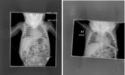
Figure 2 Chest X-ray of a one month old female patient with BPD.
Initial chest X-ray revealing minimal coarsening of the interstitial markings radiating from the hilia. One month later after prolonged incubation, the chest X-ray reveals bilateral, interstitial thickening and diffuse haziness in the lungs. Courtesy of Northeastern Ohio Universities College of Medicine-Canton Affiliated Hospitals.
8.2.9
Treatment
In most therapies, BPD infants are treated by surfactant replacement (Kresch & Clive, 1998; Mbuyamba et al., 1998), with oxygen supplementation, CPAP and mechanical ventilation (Northway et al., 1967). Different models and strategies of ventilation have been studied to potentially reduce lung injury and systematic reviews judge that optimal use of conventional ventilation may be as effective as high-frequency oscillatory ventilation in improving pulmonary outcomes. Many centers use “gentler ventilation” with more CPAP and less intubation to lower the rates of BPD. Infants with BPD that require mechanical ventilation, small tidal volumes are preferable to lower the risk of lung injury, although in the very preterm infants, higher tidal volumes are necessary to maintain effective ventilation (Morley et al., 2008; Nanan et al., 2008). For infants requiring oxygen and positive pressure ventilation (PPV), efforts are made to minimize oxygen toxicity, lung, and retina injury. Optimal ventilation levels include a pH level of 7.2-7.3, a partial pressure of carbon dioxide of 45-55 mm Hg, and a partial pressure of oxygen level of 50-70 mm Hg (with sO2 at 91-95 %) (Network et al., 2010).
24
Nonetheless, the normal oxygen requirement for a preterm infant is unknown, and repeated episodes of desaturation and hypoxia may appear in infants with BPD receiving mechanical ventilation, as a result of altered pulmonary mechanics, bronchospasm, excessive stimulation, decreased of respiratory drive and enforced exhalation efforts.
The use of other medications is also common, diuretics, bronchodilators and corticosteroids, which have many pharmacologically benefits but clinically significant adverse effects (Brion & Soll, 2001; Brundage et al., 1990; Halliday & Ehrenkranz, 2001a, 2001b, 2001c; Kersbergen et al., 2013; Papile et al., 1998). Patients with BPD have higher nutrition requirements, therefore nutritional strategies are important to manage these infants: Early administration of parenteral nutrition, supplementation with antioxidant vitamins (vitamin A), and caffeine treatment, avoid of fluid overload and early enteral feeding in small amounts (Darlow & Graham, 2007; Rocha et al., 2010; Schmidt et al., 2006). Postnatal growth failure is frequent in BPD patients, and may have extensive effects on long-term development outcomes. Therefore, strategies to optimize postnatal weight gain are necessary to improve pulmonary, retinal and neurologic development. Prenatal management of the pregnant mother to lower the risk of BPD includes treatment of maternal inflammatory conditions (chorioamnionitis) and treatment of maternal infection (Ureaplasma urealyticum) (Hartling et al., 2012; Schelonka et al., 2005).
Overall, present therapies palliate the symptoms, but there is a lack of better treatments that promote lung maturation, and prognostic factors to detect which neonates are more prone to develop BPD (Jobe, 2011; Madurga et al., 2013).
8.2.10
Complications
Patients with BPD have a higher risk of developing pulmonary hypertension and cor pulmonale, which affects at least one in six extremely low birth weight infants with moderate-severe BPD, which increases the morbidity and mortality rates. Infants with BPD are at high risk of respiratory infections in the first two years of life (respiratory syncytial virus) that can endanger the life of these babies. Infants with BPD are at higher risk of poor growth and abnormal neuro- and lung- development. Chronic pulmonary morbidities are common in BPD infants. Abnormal growth occurs in 50-60% of infants with BPD. Patients with BPD have more than double risk to suffer neurodevelopmental impairment, cerebral palsy and low intelligent quotient. Other risks include severe retinopathy of prematurity, hearing impairment, severe intraventricular hemorrhage and ventriculomegaly (Ambalavanan et al., 2008; Ambalavanan et al., 2011; Bader et al., 1987; Kirpalani et al., 2006; Massie et al., 2011; O’Reilly et al., 2013).
25
8.2.11
Prognosis
Most neonates with BPD survive but are at high risk for long-term pulmonary and neurologic sequelae. Persistent pulmonary hypertension or right ventricular hypertrophy are associated with a poor prognosis (Ambalavanan et al., 2008; Bader et al., 1987; Blayney et al. 1991; Kirpalani et al., 2006; Laughon et al., 2011; Massie et al., 2011; O'Reilly et al., 2013; Shennan et al., 1988; Smith et al., 2005).
Hydrogen sulfide
8.3.1
Introduction to gasotransmitters
The gasotransmitter family consists of three endogenous molecules of gases or gaseous signaling molecules: nitric oxide (NO), carbon monoxide (CO) and hydrogen sulfide (H2S).
The criteria defining gasotransmitters were proposed by Rui Wang in 2003 (Wang, 2003): (i) Gasotransmitters are small molecules of gas (ii) that are freely permeable to membranes, and can have endocrine, paracrine and autocrine effects. (iii) Gasotransmitters are endogenously and enzymatically generated, and their production is regulated. Moreover, (iv) gasotransmitters have specific functions at specific physiologically relevant concentrations, and (v) their functions can be mimicked by exogenously applied counterparts. Finally, (vi) the cellular effect of gasotransmitters may or may not be mediated by second messengers but should have specific and molecular targets. The gasotransmitters NO, CO, and H2S are different from classic
neurotransmitter and humoral factors while sharing some common characteristics, as it is documented in Table 1, first described in 1981 from clinical work with NO as a molecule that transmits information between cells in various parts of the body (Gillman & Lichtigfeld, 1981, 1983).
26
Table 1 Comparison of the modes of action of gasotransmitters and neurotransmitters.
Release Re-uptake Removal mechanism Revert direction Membrane
receptors Gasotransmitter Cytoplasm release No Nonenzymatic: oxidation, scavenging, methilation, etc. Bidirectional Not necessary Neurotransmitter Exocytotic vesicle
Yes Enzyme dependent Pre-
postsinaptic membrane (one direction)
Necessary
Under physiological conditions gasotransmitters are maintained at low levels, ensuring homeostasis of specific cells and organs, since the effects may not always be beneficial (gasotransmitters may cause inhibition of physiological cellular function). Nevertheless, these three gasotransmitters function in a regulatory capacity, controlling important physiologic functions including host defense against pathogens, vascular tone, apoptosis, neuromodulation, and energy metabolism (Kajimura et al., 2010).
8.3.2
Nitric oxide and carbon monoxide
The molecule NO was established as gasotransmitter in the brain and peripheral nervous system as a physiologic vasodilator, and NO is responsible of tumoricidal and bactericidal actions of macrophages. The physiologic role of NO as an endothelial derived relaxing factor is very well-known. The generation of NO is through the enzyme NO synthase (NOS), which has three different isoforms derived from different genes that convert arginine to citrulline and NO (Gadalla & Snyder, 2010). The neuronal NOS (nNOS) is localized in the brain and peripheral nervous system and in few non-neural tissues, whereas endothelial NOS (eNOS) generates NO that regulates blood vessels and inducible NOS (iNOS) is present ubiquitously throughout the body, but with highest abundance in inflammatory cells (macrophages). The nNOS and eNOS are calcium-calmodulin dependent constitutive enzymes, meanwhile iNOS is an inducible enzyme, produced in response to inflammatory stimuli, and is not dependent on calcium. The NO binds with high affinity to the heme group in the active site of soluble guanylyl cyclase (sGC), promoting the catalytic activity of the enzyme. Generation of cGMP leads to smooth muscle cell relaxation. The concentration of NO in tissues is likely to be in the 0.1 to 100 nM
27 range, but it has been noted of having a higher concentration up to 500 to 600 nM in arterioles (Kajimura et al., 2010).
The CO gasotransmitter is physiologically generated and can mediate neural activity in the brain and non-adrenergic non-cholinergic neurotransmission in the intestine. The CO molecule is enzymatically generated through two different isoforms of the enzyme heme oxygenase, heme oxygenase 1 (HMOX 1) and 2 (HMOX 2), with HMOX 1 being inducible, particularly activated through stressful and oxidative situations, and highly expressed in different tissues such as liver, kidney and spleen in the micromolar range. The HMOX 1 enzyme is responsible of degrading heme into biliverdin and CO (Kajimura et al., 2010; Nicholson & Calvert, 2010). In contrast, HMOX 2 is constitutively expressed and activated by calcium-calmodulin, much like nNOS and eNOS, and is mainly localized in the brain and the endothelial layer of blood vessels. The CO molecule also activates sGC but it is less potent than NO. Like eNOS and HMOX 2, CO is localized in the endothelial layer of blood vessels and behaves as an endothelial relaxing factor (Gadalla & Snyder, 2010).
8.3.3
Introduction to hydrogen sulfide
Hydrogen sulfide is an endogenous gas, considered to be the third gasotransmitter together with NO and CO, first described in 1989 as an endogenous “sulphide” in rat brain tissues and in normal human post-mortem brainstem that suggested endogenous production of H2S in the
brain (Nicholson & Calvert, 2010; Wang, 2010). In 1996 Abe and Kimura reported the role of H2S in human neuromodulation that was the beginning of research into H2S as a biological
signaling molecule (Gu & Zhu, 2011). Nowadays, it is acknowledged that H2S is produced
endogenously in mammals in the brain, blood vessels, liver, kidneys, lung, upper and lower gastrointestinal tract, reproductive organs, synovial joints, connective tissue, cochlea and adipose tissues. The H2S molecule is implicated in a series of physiological and pathological
processes in humans (Kabil et al., 2011; Predmore et al., 2012).
8.3.4
Biochemistry of hydrogen sulfide under physiological conditions
The H2S is small, lipid and water-soluble molecule and acts as a weak acid. The H2S molecule
can interact with receptors and can pass through biological membranes to employ its effects. At 37 ºC and pH 7.4 in aqueous solution, two thirds of H2S dissociates into protons and HS
28
form in extracellular fluids and plasma as HS- whereas within the cell (pH about 7.2) the
amounts of H2S and HS- are nearly equal (Nicholson & Calvert, 2010; Predmore et al., 2012).
8.3.5
Hydrogen sulfide donors
There are reagents that are able to release H2S, which have been very useful tools to study the
biological effects and drug development of H2S. The most frequently used H2S donor in
biological experiments is NaHS. Considered a fast release donor, NaHS can release H2S within
seconds in aqueous solutions (Li et al., 2008). On the other hand, GYY4137 is a slow-release H2S donor that mimics in a more physiological way the actions of H2S; the GYY4137
compound was synthesized by the Moore group on the basis of the structure of Lawesson’s compound, which releases H2S in organic solvents. The rate of H2S release from GYY4137
(1 mmol/L) is 4.17±0.5 nmol/25min in aqueous solution, and when incubated in aqueous buffer (pH 7.4, 37 ºC), the rate of H2S release climbs for 15 min and then plateaus at 75 min. Release
of H2S from GYY4137 is pH and temperature dependent, with less release at 4 ºC and more
release at pH 3.0 (Li et al., 2008). When administered intravenously or intraperitonealy, GYY4137 increases plasma H2S at 30 min and H2S plasma levels remain elevated over 180
min time course. In contrast, NaHS does not elevate plasma H2S at these time points (Li et al.,
2008). It has been proved that GYY4137 has vasorelaxant effects, antihypertensive and anti-inflammatory activity, showing that GYY4137 is a useful tool for studying the biological effects of H2S (Gu & Zhu, 2011; Lee et al., 2011; Li et al., 2008).
8.3.6
Toxicology of hydrogen sulfide
The presence of H2S in the environment is easily recognized by the peculiar smell of rotten
eggs. The H2S gas has been regarded as a toxic pollutant for decades, since small concentrations
of this gas can make the perceived quality of the air as unpleasant (1-3 ppm). With increasing concentrations, in an extremely dose-effect response and duration of exposure, H2S can cause
symptoms such as irritation of the eyes and the respiratory tract (20-50 ppm), headache, loss of appetite and decline in cognitive functions (200-500 ppm). When the concentration reaches around 1000 ppm H2S can be lethal due to saturation of the mitochondria and inhibition of
29
8.3.7
Endogenous hydrogen sulfide synthesis, regulation and catabolism
Cysteine is a semi-essential amino acid and one of the two sulfur-containing amino acids together with methionine. The source of cysteine is the diet, and partly also methionine processing. Cysteine is the major source of H2S in mammals and H2S is generated mainly
through three different enzymes (Figure 3): cystathionine β-synthase (Cbs), cystathionine γ-lyase (Cth, also called Cse and Cgl) and 3-mercaptopyruvate sulfur transferase (Mpst) (Nicholson & Calvert, 2010; Olson, 2011; Wang, 2010). In most peripheral tissues Cth levels are much higher than the levels of Cbs, while in the brain Cbs is the most predominant form (Nagahara, 2011; Robert et al., 2003). In lower amounts, Mpst has been found in the brain, mostly in neurons, and in the vascular endothelium (Gu & Zhu, 2011; Olson, 2011; Wang, 2010). Both Cbs and Cth are localized in the cytosol whereas Mpst exist both in the mitochondria and in the cytosol (Kajimura et al., 2010; Predmore et al., 2012). The Cbs enzyme condenses homocysteine with serine to generate the thiol ether cystathionine, during the condensation the hydroxyl group of serine is replaced with the thiolate of homocysteine. The human Cbs gene is located in chromosome 21 at 21q22.3. In humans and rats Cbs exists primarily as a homotetramer with a subunit molecular mass of 63 kDa. Each subunit also binds to the cofactors pyridoxal 5-phosphate (PLP), S-adenosyl methionine (SAM) and heme. The cofactor SAM is an allosteric activator of the Cbs enzyme while heme appears to be a redox sensor. The C-terminus of Cbs contains two domains of tandem repeats that seem to inhibit the enzymatic function, as the deletion of these domains activates Cbs. The Cbs enzyme can be sumoylated, inhibiting the catalytic activity of Cbs. The Cth enzyme can also form H2S from
cysteine, it hydrolyzes cystathionine into cysteine with ammonia and α-ketobutyrate as byproducts. The Cth enzyme converts cysteine to thiocysteine, pyruvate and ammonia, in a β-disulfide elimination reaction, with the thiocysteine then reacting with cysteine or other thiols to produce H2S and cysteine or the corresponding disulfide. The Cth enzyme is also dependent
on PLP, and it is selectively activated by calcium-calmodulin similar to the activation of eNOS, nNOS and HMOX 2 (Gadalla & Snyder, 2010). The Mpst enzyme catalyzes only sulfur transferases reactions from 3-mercaptopyruvate to various donors, and needs cysteine aminotransferase and further redox reactions with biological thiols such as glutathione to yield H2S (Gu & Zhu, 2011; Predmore et al., 2012; Wang, 2010).
30 Figure 3 Potential pathways of H2S production and metabolism.
Transsulfuration reactions that involve cystathionine β-synthase (Cbs), cystathionine γ-lyase (Cth) and 3-mercaptopyruvate sulfur transferase (Mpst) in the production and metabolism of H2S. CDO, cysteine dioxygenase; CAT, cysteine aminotransferase; CLY, cysteine lyase; CSD, cysteine sulfinate decarboxylase CA, carbonic anhydrase; R-SH, thiol; GSSG, glutathione oxidized form. (Olson, 2011).
The catabolic pathways of H2S are not fully understood, one catabolic pathway of H2S is
oxidation in the mitochondria and excretion in the urine, sulfate being the major end product of H2S catabolism. Another catabolism pathway is the thiol S-methyltransferase mediated
methylation of H2S to yield monomethylsulfide and dimethylsulfide. A third pathway involves
binding of H2S to methemoglobin to yield sulfhemoglobin. It has also been reported that H2S
can diffuse across the alveolar membrane (Kajimura et al., 2010).
8.3.8
Cbs
-/-and
Cth
-/-mice
The Cbs-/- mice were generated through insertion of a neomycin selection cassette that replaced a genomic fragment containing exons 3 and 4 of the Cbs gene (Watanabe et al., 1995). The Cbs-/- mice have a manifest phenotype and are broadly used as a mental retardation model, as a
model for hyperhomocysteinemia and as a thromboembolism model. The Cbs-/- mice display
several pathophysiologic features similar to hyperhomocysteinemic patients, including endothelial dysfunction and hepatic steatosis. The mice usually die during the weaning period between the 2-4 weeks of age, probably due to severe hepatic dysfunction and require sufficient
31 supplementation of cysteine for survival. The Cbs-/- mice have a severe retardation in body mass
that might be caused by taurine deficiency and an abnormal lipid metabolism. The Cbs-/- mice
also have cerebellar malformation and impaired learning ability. The adult Cbs-/- mice display
lung fibrosis and airspace enlargement in the absence of increased inflammatory cell infiltrates (Akahoshi et al., 2008; Hamelet et al., 2007; Namekata et al., 2004). The Cth-/- mutant mice were generated thorough exons 1 to 6 being replaced with a LacZ and neomycin selection cassette (Ishii et al., 2004). The Cth-/- mice have a milder phenotype compared to the Cbs -/-mice, the Cth-/- mice have a slight retard in growth, but homozygotes can reach adulthood and breed. The Cth-/- mice are normally used as a cystathionemia/cystathionuria model. The Cth -/-mice develop hypertension and have an impaired endothelium derived vasorelaxation capacity, are very sensitive to oxidative stress, and have lower taurine levels. When fed with low cysteine in diet, the Cth-/- mice exhibit acute skeletal muscle atrophy (Ishii et al., 2010; Ishii et al., 2004; Wang et al., 2013).
8.3.9
Endogenous levels of hydrogen sulfide in health and disease
A role for H2S in health and disease is supported by the correlations found between the
plasma/tissue levels of H2S/sulfane sulfur or H2S generating enzymes and the progression of
diseases such as insulin resistance, hypertension, hyperhomocysteinemia, diabetes, exacerbated cardiac injury following ischemia-reperfusion injury, cirrhosis, Alzheimer-disease, progression of adiposity, chronic kidney disease, gastrointestinal tract irritation, asthma and cancer among others (Gu & Zhu, 2011; Predmore et al., 2012). Like in the case of NO and CO, measuring the concentrations of a gas with a short half-life that can form complex with other molecules in biological samples, is still very challenging. Conventional methods for the determination of H2S fall into two categories: measuring “free” H2S or “labile” H2S. An example of the first
would be a polarographic sensor, and examples of the second would be colorimetric assays, gas-chromatography-mass spectrometry and high-performance liquid chromatography. Reported values of labile H2S in plasma and blood range between 20 and 30 µM, in contrast to
polarographic sensors with detection limit near 10 nM. In tissues from rat brain the values of free H2S varied from 14 nM to 70 µM (Kajimura et al., 2010).
32
8.3.10
Mechanism of action of hydrogen sulfide
The molecular targets of H2S include enzymes, proteins, transcription factors and membrane
ion channels (Predmore et al., 2012).
8.3.10.1
Sulfhydration of proteins
The H2S molecule can sulfhydrate different proteins, and sulfhydration can alter protein
function suggesting sulfhydration as an important physiologic signaling pathway. An example would be that H2S can sulfhydrate nuclear factor κ-light-chain-enhancer of activated B cells
(NFκB) and cause anti-apoptosis actions (Predmore et al., 2012).
8.3.10.2
Interaction with potassium channels
The H2S molecule can stimulate ATP potassium (KATP) channels. The KATP channels contain
nine cysteines with C43 that lies close to the surface, which is selectively influenced by oxidative insults. The mechanism of action of how H2S interacts with KATP channels is not
completely understood, but it has been reported that H2S can interact with the sulfonylurea
receptor subunit of the KATP channel with selective cysteine residues located at the extracellular
loop of the receptor subunit. The activation of KATP channels has been described as the
molecular basis of H2S-elicited cardiac protection, blood pressure lowering, or insulin release
inhibition (Wang, 2010).
8.3.10.3
Vasorelaxation
The H2S molecule can relax blood vessels, H2S resembles the principal properties of endothelial
derived relaxing factor, and it is localized in the endothelial layer of blood vessels. The H2S
vasorelaxation, reflects hyperpolarization mediated by the opening of KATP channels via the
sulfhydration at C43. Unlike NO and CO, H2S does not activate guanylate cyclase (Gu & Zhu,
33
8.3.10.4
Oxygen sensitive responses
The vessel wall produces H2S, and it has been proposed that H2S is an O2 sensor in both vascular
and non-vascular smooth muscle as well as a putative chemoreceptor (Olson et al., 2010). Catabolism of H2S is mainly through oxidation in the mitochondria; since oxidation depends
mainly on O2 availability, depending on the concentration of O2 it will affect the total amount
of biologically active H2S. In agreement with this theory, high O2 concentration correlates with
low H2S activity and a dilated pulmonary artery. Inversely, low O2 concentrations correlate
with higher H2S activity and artery contraction. Indeed, H2S has been found to have both
vasodilatory and vasoconstrictive effects depending on its concentration. It has also been reported that O2 can modulate the activity of both Cbs and Cth, which may have an additive
impact on the H2S mediated vascular response (Kajimura et al., 2010; Olson, 2011; Wang,
2010).
8.3.10.5
Anti-inflammatory effects
The H2S molecule is thought to have anti-inflammatory effects through action on KATP
channels, inhibition of activation of NFκB and p38 MAPK, scavenging of oxidants, up regulation of intracellular cAMP, and inhibition of caspase-3 cleavage. Considered a powerful inhibitor of leukocyte adherence to the vascular endothelium, H2S may obstruct inflammatory
processes by diminishing the tissue injury induced by neutrophils via induction of apoptosis and/or scavenging of neutrophils derived hypochlorous acid. Reactive oxygen species are mediators of NFκB activation and this process can be blocked by antioxidants such as GSH and cysteine. It has been described that H2S can down-regulate several pro inflammatory cytokines
including: NFκβ, TNFα, IL-1β, IL-6 and IL-8 (Chen et al., 2009; Predmore et al., 2012; Wallace et al., 2012; Zhang et al., 2013) and that it can activate anti-inflammatory chemokines such as IL-10 (Wang, 2010). Promotion of tissue repair by H2S is likely due to its vasodilatory
properties and activation of cyclooxygenase 2 expression and through promotion of angiogenesis. Another property of H2S is that can also act as an energy source substituting for
oxygen mitochondrial respiration, which may contribute significantly to protection and repair of tissue injury (Wallace et al., 2012). Other data support that H2S can also be
pro-inflammatory, mostly at high concentrations, promoting formation of pro-inflammatory cytokines and chemokines by up-regulation of NFκB of activated B cells or by activating the metabolism of substance P (Gadalla & Snyder, 2010; Wallace et al., 2012; Wang, 2010).
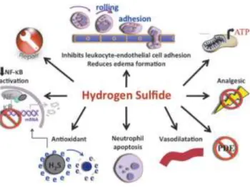
34 Figure 4 Anti-inflammatory effects of hydrogen sulfide.
Illustration of several mechanism of action involved in the anti-inflammatory effects of hydrogen sulfide that include inhibition of leukocyte-endothelial cell adhesion, vasodilation, neutrophil apoptosis, antioxidant, reduction of NFκB, inhibition of PDE, nociception, repair, and modulation of KATP channels (Wallace et al., 2012).
8.3.10.6
Cytoprotective effects
H2S can strongly influence the redox status through different mechanisms, such as increasing
GSH levels in the cytosol, the mitochondria, and nucleus of the cell, by increasing the GSH/GSSG ratio, activating the protective heat-shock proteins, activating the nuclear factor (erythroid-derived2)-like 2 (Nrf2) that results in activation of the antioxidant response elements of several antioxidant genes. By activating KATP channels, H2S can also have an anti-apoptotic,
anti-inflammatory and blood-pressure lowering effects. The cytoprotective effects of H2S also
involves activation of signaling pathways such as Akt pathways (Kimura et al., 2012; Predmore et al., 2012).
8.3.10.7
Antioxidant effects
The H2S molecule is capable of quenching free radicals, since HS- anions have reducing
chemical properties. The oxidation of HS- by biochemical relevant two-electron oxidants, yields hydrogen disulfide which also has oxidizing activity and is capable of regenerating H2S.
However, since the concentration of H2S and HS- is very low in blood and tissues, the
35 A major antioxidant protein in the human body is GSH, and the action of H2S influencing the
generation of GSH is often described in the literature: (i) H2S enhances the cellular glutamate
uptake, (ii) H2S- induced increase in the level of gamma-glutamylcysteine synthethase and
cysteine transporter activity in the cell, (iii) reduction of cystine into cysteine by H2S in the
extracellular space, and transport of cysteine into cells by the cysteine transporter, (iv) H2S
stimulation of the nuclear transcription factor Nrf2, which then up regulates GSH synthesis and transport, and (v) a decrease in the activity of the enzymes that catabolize GSH (Predmore et al., 2012; Wang, 2010).
8.3.11
Interaction with other gasotransmitters
There are many similarities between the three gases NO CO and H2S, at physiological
concentrations the three of them act as vasodilators, as cytoprotective and anti-inflammatory molecules. When it concerns cross talk between NO and H2S, it has been demonstrated that
H2S can relax SMCs through release of endothelium-derived hyperpolarizing factor and NO
from the endothelium. H2S can react with NO to form a nitrosothiol and reducing the
availability of NO to cause SMCs contraction, and through inhibition of eNOS as well as reduction in SMCs cAMP concentration (Kajimura et al., 2010; Li et al., 2009). Moreover, the group heme binds to the N-terminal portion of Cbs, comprising about 70 amino acids. In its ferrous state, this heme binds both CO and NO. The group heme has more affinity to CO than to Cbs, therefore CO can inhibit Cbs activity. The powerful influence of CO upon Cbs raises the possibility of cross talk between CO and H2S as messenger molecules (Gadalla & Snyder,
2010).
8.3.12
Effects of hydrogen sulfide in the nervous system
In the brain H2S is produced mainly in the astrocytes. H2S may have a neuro-protectant role,
since the first recognized sign of Cbs deficiency in humans is mental retardation and Alzheimer’s disease (Enokido et al., 2005; Olson, 2011; Robert et al., 2003). It is suggested that Cbs plays a crucial role in the development and maintenance of the central nervous system (Kimura et al., 2012; Robert et al., 2003). Cbs-deficient patients also suffer other symptoms such as seizures, abnormal electroencephalogram, extrapyramidal disturbances and psychiatric disorders (Gadalla & Snyder, 2010). In the nervous system H2S has been shown to have
36
protective effects, preventing the activation of neurotoxins, neuron degeneration, neuron apoptosis and gliosis in mice (Predmore et al., 2012).
8.3.13
Role of hydrogen sulfide in different lung diseases
The three H2S-generating enzymes are variably expressed in the lung depending on the species:
Cth and Mpst are widely expressed in bovine pulmonary arterial smooth muscle cells, while Cbs has only been found in bovine pulmonary arterial endothelial cells. In humans, both airway SMCs and primary fibroblast express Cth and Cbs. In contrast, in mice, both Cth and Cbs were found in pulmonary blood vessel SMCs and endothelial cells as well as in airway SMCs (Olson et al., 2010; Wang et al., 2011). The role of H2S in different chronic lung disease has been
recently reviewed by Chen et al. (Chen & Wang, 2012) and it is summarized in Figure 5. In chronic obstructive pulmonary disease (COPD), H2S has proven to be an anti-inflammatory and
bronchi dilatant agent, decreasing the expression of TNFα and IL-8 lung tissue of chronic cigarette smoke exposed rats treated with NaHS in comparison to control rats. Similar results were found by another group, using a tobacco smoke induced emphysema mouse model, where NaHS treatment ameliorated inflammation measured in bronchoalveolar lavage (BAL), reduced the increase in right ventricle systolic pressure, the thickness of pulmonary vascular walls and right ventricular hypertrophy. The serum levels of H2S COPD patients varied
depending on stage and tobacco exposure and further studies are needed to use it as a marker (Chen et al., 2005). In the case of asthma, lower levels of H2S were found in asthmatic patients,
related negatively to sputum counts and positively to forced expiratory volume capacities. In an experiment with ovalbumin treated rats, exogenous administration of NaHS increased peak expiratory flow, decreased goblet cell hyperplasia and collagen deposition score, decreased total cells in BAL and influx of eosinophils and neutrophils (Benetti et al., 2013; Chen & Wang, 2012; Chen et al., 2009). Moreover, the Cth-/- mice when challenged with ovalbumin, had
worsen airway inflammation and elevated cytokines such as IL-5 and IL-13 in BAL when compared to wild type littermates, and interestingly, recovery treatment with NaHS supplement rescued the Cth-/- mice from the aggravated pathological picture (Zhang et al., 2013). In
pulmonary fibrosis, NaHS has been reported to decrease pulmonary fibrosis caused by bleomycin, decreasing lung hydroxyproline content and malondialdehyde. The authors speculated about an anti-oxidative effect of NaHS against lipid peroxidation produced by ROS. Treatment with NaHS has been proven to arrest cultured human fibroblasts in the G1 phase of the cell cycle, and trigger them into apoptosis. Furthermore, treatment with H2S decreased
37 pSmad2/3 phosphorylation in A549 cultured cells stimulated with TGF-β1, and prevented epithelial to mesenchymal transition (Chen & Wang, 2012; Fang et al., 2009). Finally, H2S has
demonstrated a regenerative effect in two different models of acute lung injury, in a mouse model of lipopolysaccharide-induced lung injury, it improved lung structure in histological examination and prevented cytokine release in BAL fluid, clarified in Figure 6 (Faller et al., 2012). In a mechanical ventilation rat model, NaHS treatment improved inflammation and oxygenation in comparison to the control group (Aslami et al., 2010).
Figure 5 Mechanism of action of H2S in chronic lung diseases.
Several mechanism of action of H2S: Inhibition of airway smooth muscle cell proliferation, inhibition of bronchoconstriction, modulation of lung fibroblast differentiation, inhibition of epithelial to mesenchymal transition, modulation of hypoxic pulmonary vasoconstriction, neurogenic inflammation and inhibition of oxidative stress. Glutathione reduced form (GSH), glutathione oxidized form (GSSG), substance P (SP), calcitonin gene-related peptide (CGRP), transforming growth factor beta (TGF-β) (Chen & Wang, 2012).
38 Figure 6 Effect of lipopolysaccharide induced acute lung injury and effect of H2S inhalation on lung structure.
Control mice treated with phosphate buffer saline intranasal and room air (A), 80 ppm hydrogen sulfide (H2S) for 6 h (B), lipopolysaccharide (LPS) treated mice and room air (C) and LPS and 80 ppm H2S treated mice for 6 h (D). Sections stained with hematoxylin and eosin (Faller et al., 2012).
39
9
Hypothesis and aims
Bronchopulmonary dysplasia is a dangerous complication of the premature newborn. There is a lot of effort in the scientific community to unravel the disease mechanisms and identify therapies that could improve lung growth from these infants. It is thought that anti-inflammatory, cytoprotective, antioxidant and vascular protective approaches, which are meant to be properties of H2S, could be beneficial treating BPD.
In this context, it was also observed that the expression levels of the H2S generating enzymes
Cbs and Cth, changed over the course of normal mouse late lung development.
Thus it was hypothesized that:
In the BPD model H2S can be used as a potential treatment, due to cytoprotective,
anti-inflammatory, antioxidant and pulmonary vasodilatory properties and that Cbs and Cth play a role in normal late lung development.
Hence, the aims of the study were:
(a) Using a hyperoxia BPD mouse model to expose the animals either to vehicle or to a H2S
donor, and study the effect of the treatment on lung structure during normal and aberrant late lung development.
(b) In the case of improved in alveolarization in (a), to study the possible mechanisms of action that would impact lung development.
40
10
Materials and methods
Equipment and software
Equipment and software Manufacturer
Adobe Photoshop Software Adobe Systems, USA
Autoclave Systec, Germany
BD FACS CantoTM flow cytometer BD Biosciences, USA
Cell culture incubator HERAcell 150i Thermo Scientific, USA
Cell culture plates (6-, 24-, 96- well plates) Greiner Bio-One, Germany
Cell culture flasks (75 ml) Greiner Bio-One, Germany
Cell counter, Countess® Invitrogen, UK
Corning® Costar® cell culture plates Sigma-Aldrich, Germany
Culture-insert μ-dish© Ibidi® cells in focus, USA
Electrophoresis chambers Bio-Rad, USA
FACSDivaTM software BD Biosciences, USA
Filter Tip FT: 10, 20, 100, 200, 1000 Greiner Bio-One, Germany
Freezer -20 °C Bosch, Germany
Freezer -40 °C Kryotec, Germany
Freezer -80 °C Heraeus, Germany
Fridge +4 °C Bosch, Germany
Gel blotting paper 70 x 100 mm Bioscience, Germany
Hood with laminar flow Thermo Scientific, USA
Image J Software NIH, USA
Image QuantTM LAS4000 GE Healthcare, UK
Microscope (light, phase contrast, fluorescent,
Laser-capture micro dissection, confocal) Leica, Germany
Microtome (paraffin-, plastic-, cryo- sections) Leica, Germany
Mini spin centrifuge Eppendorf, Germany
Multifuge centrifuge, 3 s-R Heraeus, Germany
NanoDrop® ND 1000 Peqlab, Germany
NanoZoomer-XR C12000 Digital slide scanner Hamamatsu, Japan
PCR-thermo cycler: peqSTAR Peqlab, Germany
41
Petri dishes Eppendorf, Germany
Pipetboy Eppendorf, Germany
Pipetmans: P10, P20, P100, P200, P1000 Gilson, France
Precellys® 24 Homogenizer Peqlab, Germany
QWin software Leica, Germany
Single-use syringe Braun, Germany
Reaction tubes: 0.5, 1.5, 2 ml Eppendorf, Germany
Stepanizer© stereology software Tschanz, Switzerland
StepOnePlusTM Real-Time PCR Applied Biosystem, Germany
Test tubes: 15, 50 ml BD Biosciences, USA
Trans blot transfer membrane (0.2 μm) Bio-Rad, USA
Visiopharm NewCastTM computer-assisted
stereology system (VIS 4.5.3) Visiopharm, Denmark
VersaMax Micro plate reader Molecular Devices, Germany
Vortex machine Eppendorf, Germany
Vacuum centrifuge Eppendorf, Germany
Western blot chambers, Mini Trans-Blot Bio-Rad, USA
Western blot chambers, Mini-Protean 3 Cell Bio-Rad, USA
WIMASIS Software WimTube Wimasis GmbH, Deutschland
Reagents
Reagents Manufacturer
Acetone Roth, Germany
Acrylamide solution, Rotiphorese Gel 30 Roth, Germany
Agar-agar Merck, Germany
Agarose Invitrogen, UK
Accutase Invitrogen, UK
AlexaFluor® 488 Annexin V/Dead
Cell Apoptosis Kit Invitrogen, UK
BD BioCoatTM BD MatrigelTM plate Becton Dickinson, USA
Bioxytech© GSH/GSSG kit OxisResearch, USA
Bovine serum albumin Sigma-Aldrich, Germany