ABSTRACT
REBULI, MEGHAN ELIZABETH. Sex Specific Impact of Environmental Endocrine Disruptors on Neuroendocrine Development and Behavior. (Under the direction of Heather B. Patisaul.)
Endocrine disrupting compounds (EDCs) are exogenous chemicals that interfere with
the endocrine system and hormone action. EDC action, particularly on sex hormones,
indicates that they may participate in the aberrant organization or activation of sex
differences within the brain. Bisphenol A (BPA) is one such EDC and has previously
reported to affect sex-specific neuroendocrine physiology, estrogen signaling, and behavior.
The mechanisms underlying these brain and behavior outcomes are largely unknown and
defining them would allow for determination of their human relevance and potential
contribution to human disease, such as neuropsychiatric disorders. This work investigates
the potential for BPA to act on neuroendocrine physiology and behavior in three ways: (1)
neonatal alteration of estrogen receptor expression by prenatal exposure to BPA, (2) juvenile
and adult alteration of estrogen receptor expression by subchronic exposure to BPA, and (3)
juvenile and adult alteration of anxiety and activity behavior by perinatal exposure to BPA.
Sprague Dawley rats were gavaged on gestational days 6–21 for neonatal and perinatal
exposures, and further dosed until postnatal day 90 for subchronic exposures, with vehicle,
2.5 to 2700 μg/kg bw/day BPA, or 0.5, 5, or 10 μg/kg bw/day ethinyl estradiol. Estrogen
receptor expression in the hypothalamus and amygdala was investigated using in situ
hybridization, while behavior was evaluated using a battery of behavioral tests including the
primarily seen in subregions of the preoptic area of female rats. Finally, there were no
consistent effects of BPA on anxiety or activity behaviors, however study limitations limit
the ability make resolute conclusions on BPA’s potential to alter affective behaviors. These
results indicate that estrogen receptor expression in the neonatal, juvenile and adult brain can
be altered by BPA and may be one mechanism by which BPA can alter brain and behavior
© Copyright 2015 Meghan Elizabeth Rebuli
Sex Specific Impact of Environmental Endocrine Disruptors on Neuroendocrine Development and Behavior
by
Meghan Elizabeth Rebuli
A dissertation submitted to the Graduate Faculty of North Carolina State University
in partial fulfillment of the requirements for the degree of
Doctor of Philosophy
Zoology
Raleigh, North Carolina
2015
APPROVED BY:
_______________________________ ______________________________
Heather B. Patisaul John R. Godwin
DEDICATION
To my family. Thank you for your love, support, and motivation, which has inspired
BIOGRAPHY
Meghan Elizabeth Rebuli was born December 2, 1989 in Havelock, North Carolina.
She attended Havelock High School, where she was elected Student Body President,
participated as captain of the girls’ soccer team, was section leader in the marching band, and
won Homecoming Queen before graduating in 2007.
Her undergraduate institution was North Carolina State University, where she was
selected as a Park Scholar, studied abroad in Antigua, Guatemala, was a member of Alpha
Epsilon Delta, and participated as an undergraduate researcher. She graduated with a
Bachelor of Science in Biological Sciences, cum laude, with a concentration in Human
Biology, and a minor in Spanish in 2011.
After completion of her undergraduate career she continued on at North Carolina
State University to complete her doctoral studies in the Zoology graduate program
researching neuroendocrine development and behavior. During her graduate studies she
received two teaching awards for her efforts as a teaching assistant and presented her
research at multiple national and international conferences.
After graduation she plans to continue her research as a postdoctoral trainee at the
ACKNOWLEDGMENTS
I am sincerely and overwhelmingly grateful to my advisor, Dr. Heather B. Patisaul.
Her willingness to be my research mentor as an undergraduate allowed me to confirm and
solidify my interest in research. Her enthusiasm for neuroendocrinology inspired my pursuit
of a Ph.D. I will always be thankful for her encouragement, the ambition she instilled in me
to push towards scientific independence, and her ability to pursue what is best for her
students. I am excited for future collaborations and knowing that I will always be able to
consult with her on future endeavors.
This dissertation would not be possible without the research assistance and emotional
support from the graduate students, technicians, postdoc, and undergraduate students in my
lab, thank you all. I would also be lost without the understanding, inspiration and
reassurance of my family, immeasurable thanks.
Lastly, I would also like to thank my committee for their insight and encouragement
and the members of the Department of Biological Sciences and the Graduate School for their
TABLE OF CONTENTS
LIST OF TABLES ... ix
LIST OF FIGURES ...x
ABBREVIATIONS ... xii
CHAPTER 1—Effects of endocrine disrupting compounds on neurodevelopmental sex differences in perinatal and adolescent rodents: a review ...1
Abstract ...1
Background ...1
Objective ...1
Methods...1
Results ...2
Conclusion ...2
Key Words ...2
Introduction ...2
Methods...5
Results ...6
Hypothalamus ...6
Hippocampus ...9
Cortex, Cerebellum, and Mid-brain ...11
Whole Brain or Embryonic Brain Regions ...14
Sex differences ...16
Conclusion ...17
Acknowledgements ...19
Tables ...20
Figure ...31
CHAPTER 2— Prenatal Bisphenol A Exposure Alters Sex-Specific Estrogen Receptor Expression in the Neonatal Rat Hypothalamus and Amygdala ...32
Abstract ...32
Key Words ...33
Disclaimer ...33
Introduction ...33
Materials and Methods ...40
Animal care, prenatal BPA and EE exposure, brain collection and section preparation ...40
ISHH ...44
Statistics ...46
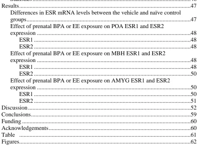
Results ...47
Differences in ESR mRNA levels between the vehicle and naïve control groups ...47
Effect of prenatal BPA or EE exposure on POA ESR1 and ESR2 expression ...48
ESR1 ...48
ESR2 ...48
Effect of prenatal BPA or EE exposure on MBH ESR1 and ESR2 expression ...48
ESR1 ...48
ESR2 ...50
Effect of prenatal BPA or EE exposure on AMYG ESR1 and ESR2 expression ...50 ESR1 ...50 ESR2 ...51 Discussion ...52 Conclusions ...59 Funding ...60 Acknowledgements ...60
Table ...61
Figures...62
CHAPTER 3— Investigation of the Effects of Subchronic Low Dose Oral Exposure to Bisphenol A (BPA) and Ethinyl Estradiol (EE) on Estrogen Receptor Expression in the Juvenile and Adult Female Rat Hypothalamus ...69
Abstract ...69
Key Words ...70
Disclaimer ...70
Introduction ...71
Materials and Methods ...76
Animal care, BPA and EE exposure, brain collection and section preparation ...76
In situ hybridization and histochemistry ...79
Image analysis and film quantification ...80
Statistics ...81
Results ...83
PND 21 Esr1 and Esr2 Expression in the POA ...83
Esr1 ...83
Esr2 ...83
PND 21 Esr1 and Esr2 Expression in the MBH ...83
Esr2 ...84
PND 90 Esr1 and Esr2 Expression in the POA ...84
Esr1 ...84
Esr2 ...84
PND 90 Esr1 and Esr2 Expression in the MBH ...85
Esr1 ...85
Esr2 ...86
Discussion ...86
Conclusions ...95
Funding ...95
Acknowledgements ...95
Figures...97
Supplementary Figures ...101
CHAPTER 4— Impact of Low Dose Oral Exposure to Bisphenol A (BPA) on Juvenile and Adult Rat Exploratory and Anxiety Behavior: A CLARITY-BPA Consortium Study ...106
Abstract ...106
Key Words ...107
Disclaimer ...107
Introduction ...108
Materials and Methods ...112
Animal husbandry and dose selection...113
Dose preparation and administration ...114
Weaning and transfer of subjects ...115
Behavioral Testing ...116
Elevated Plus Maze (EPM) ...118
Open Field (OF) ...118
Zero Maze (ZM) ...119
Summary of primary endpoints in the behavioral tasks ...119
Data decoding ...120
Statistical analysis of body weight and behavioral data ...120
Results ...123
Terminal body weight ...123
Juveniles ...124
Adults ...125
Estimating observed effect size of BPA and detection power ...126
Correlation between outcome measures ...127
Discussion ...128
Conclusions ...136
Funding ...137
Figures...138
Supplementary Tables ...145
Supplementary Figures ...156
CHAPTER 5—Conclusions ...158
LIST OF TABLES
CHAPTER 1— Effects of endocrine disrupting compounds on neurodevelopmental sex differences in perinatal and adolescent rodents: a review
Table 1.1 EDC Effects on the Hypothalamus ...20
Table 1.2 EDC Effects on the Hippocampus ...24
Table 1.3 EDC Effects on Cortex, Cerebellum, and Mid-brain ...26
Table 1.4 EDC Effects on Whole Brain and Embryonic Brain Regions ...29
CHAPTER 2— Prenatal Bisphenol A Exposure Alters Sex-Specific Estrogen Receptor Expression in the Neonatal Rat Hypothalamus and Amygdala Table 2.1 Summary of sex-specific ESR1 and ESR2 expression in ROIs examined ...61
CHAPTER 4— Impact of Low Dose Oral Exposure to Bisphenol A (BPA) on Juvenile and Adult Rat Exploratory and Anxiety Behavior: A CLARITY-BPA Consortium Study Supplementary Table 4.1 Animal studies examining the relationship between BPA exposure and anxiety-related behaviors ...145
Supplementary Table 4.2 Animal studies examining the relationship between BPA exposure and spontaneous motor activity ...151
Supplementary Table 4.3 Juvenile open field ANOVA p-values ...154
LIST OF FIGURES
CHAPTER 1— Effects of endocrine disrupting compounds on neurodevelopmental sex differences in perinatal and adolescent rodents: a review
Figure 1.1 The percentage of studies including sex ...31
CHAPTER 2— Prenatal Bisphenol A Exposure Alters Sex-Specific Estrogen Receptor Expression in the Neonatal Rat Hypothalamus and Amygdala Figure 2.1 ESR1 mRNA expression in the AVPV and MPOA ...62
Figure 2.2 ESR2 mRNA expression in the AVPV and MPOA ...63
Figure 2.3 ESR1 mRNA expression in the MBH ...64
Figure 2.4 ESR2 mRNA expression in the MBH ...65
Figure 2.5 ESR1 mRNA expression in the MePD and PLCo ...66
Figure 2.6 ESR1 mRNA expression in the PMCo and Ahi ...67
Figure 2.7 ESR2 mRNA expression in the MePD ...68
CHAPTER 3— Investigation of the Effects of Subchronic Low Dose Oral Exposure to Bisphenol A (BPA) and Ethinyl Estradiol (EE) on Estrogen Receptor Expression in the Juvenile and Adult Female Rat Hypothalamus Figure 3.1 Autoradiograms and atlas depictions of AVPV and MPOA ...97
Figure 3.2 Juvenile Esr1 mRNA expression in the AVPV ...98
Figure 3.3 Adult Esr2 mRNA expression in the AVPV ...99
Figure 3.4 Adult Esr2 mRNA expression in the MPOA ...100
Supplementary Figure 3.1 Autoradiograms and atlas depictions of VMNvl and ARC...101
Supplementary Figure 3.3 Juvenile Esr2 mRNA expression in the POA
and MBH ...103
Supplementary Figure 3.4 Adult Esr1 mRNA expression in the POA and MBH ...104
Supplementary Figure 3.5 Adult Esr2 mRNA expression in the POA and MBH ...105
CHAPTER 4— Impact of Low Dose Oral Exposure to Bisphenol A (BPA) on Juvenile and Adult Rat Exploratory and Anxiety Behavior: A CLARITY-BPA Consortium Study Figure 4.1 Methods Timeline ...138
Figure 4.2 Juvenile EPM ...139
Figure 4.3 Juvenile OF ...140
Figure 4.4 Adult EPM ...141
Figure 4.5 Adult OF ...142
Figure 4.6 Adult ZM ...143
Figure 4.7 Theoretical power analysis ...144
Supplementary Figure 4.1 Correlations matrices between adult anxiety-related behaviors ...156
ABBREVIATIONS
1 first test day
2 second test day
3V third ventricle
AXXXX Aroclor XXXX
AALAC Assessment and Accreditation of Laboratory Animal Care
AC anterior commissure
ADHD Attention Deficit Hyperactivity Disorder
Ahi amygdalo-hippocampal area
AhR arylhydrocarbon receptor
AMYG amygdala
ANOVA analysis of variance
ARC arcuate hypothalamic nucleus rARC rostral arcuate nucleus
cARC caudal arcuate nucleus ASD autism spectrum disorder
AVPV anteroventral periventricular nucleus
BPA bisphenol A
BPA-G BPA-glucuronide
bw body weight
CA1 Region I of hippocampus proper
CC corpus callosum
CEBS National Toxicology Program Chemical Effects in Biological Systems CLARITY Consortium Linking Academic and Regulatory Insights on BPA Toxicity CM central medial thalamic nucleus
CMC carboxymethylcellulose sodium salt
CPu Caudate Putamen
DES diethylstilbestrol
DG dentate gyrus
EAC endocrine active compound EDC endocrine disrupting compound
EE ethinyl estradiol
EPM elevated plus maze
ER estrogen receptor
ERα estrogen receptor alpha ERβ estrogen receptor beta
ERK extracellular signal-regulated kinase ESR estrogen receptor
ESR1/Esr1 estrogen receptor alpha ESR2/Esr2 estrogen receptor beta
f effect size
FAO Food and Agricultural Organization FDA Food and Drug Administration
GD gestational day
GH growth hormone
GLP good laboratory practice
GnRH gonadotropin releasing hormone HPA hypothalamic-pituitary-adrenal axis HPG hypothalamic-pituitary-gonadal axis
ic internal capsule
ISHH in situ hybridization histochemistry
LH luteinizing hormone
LHβ luteinizing hormone beta polypeptide LOAEL lowest-observed-adverse-effect level LOD limit of detection
LSD least significant difference
LV lateral ventricle
M male
MBH mediobasal hypothalamus
MePD medial amygdala
MPOA medial preoptic area
NCTR National Center for Toxicological Research NCSU North Carolina State University
NIEHS National Institute of Environmental Health Sciences NOAEL no-observed-adverse-effect level
NTP National Toxicology Program
OF open field
PBDE polybrominated diphenyl ether PCB polychlorinated biphenyl PFOA perfluorooctanoic acid
PLCo posterolateral cortical amygdaloid nucleus PMCo posteromedial cortical amygdaloid nucleus PND postnatal day
POA preoptic area
PRL prolactin
r empirical correlation
ROI region of interest
SD Sprague Dawley
SDN sexually dimorphic nucleus
TCDD 2,3,7,8 – tetrachlorodibenzo-p-dioxin VMN ventromedial hypothalamic nucleus
VMNvl ventrolateral division of the ventromedial hypothalamic nucleus
WHO World Health Organization
CHAPTER 1—Effects of endocrine disrupting compounds on neurodevelopmental sex differences in perinatal and adolescent rodents: a review
Authors: Meghan E. Rebuli1,2, Heather B. Patisaul1,2
Affiliations: 1Department of Biology, NCSU, Raleigh, North Carolina 27695; 2Keck Center
for Behavioral Biology, NCSU, Raleigh, North Carolina
Abstract
Background
Brain sex differences are found in nearly every region of the brain. They are
organized during gestation and early adolescence. EDCs interfere with hormone action and
are thought to interfere with the formation of sex differences. EDCs may be considered an
environmental factor contributing to the increased diagnosis of neuropsychiatric disorders
that emerge during adolescence (suggesting developmental etiology) and present with a sex
bias.
Objective
This review focuses on the available evidence for the ability of EDCs to impact the
emergence of brain sex differences, with a focus on effects detected at or before adolescence.
Methods
Studies were identified by searching PubMed using the keywords endocrine
disrupting compound, EDC, endocrine active compound, EAC, brain, neuro, and
development. Endpoints reviewed are brain gene, protein, hormone, and transcript
expression, as well as brain morphology. Studies on behavioral endpoints were not included.
Results
The hypothalamus was found to be particularly affected by estrogenic EDCs in a sex,
time, and exposure dependent manner. The hippocampus also appears vulnerable to
endocrine disruption by BPA and PCBs although there is little evidence from the available
data set to make any conclusions about sex-specific effects. Gestational EDC exposure can
alter fetal neurogenesis and gene expression. PCB, dioxin, and estrogenic EDC exposures,
were found to alter larger brain areas and organizational sex differences in the neonatal brain,
such as the cortex and cerebellum.
Conclusion
The developmental EDC exposure literature demonstrates evidence of altered
neurodevelopment as early as fetal life, with sex specific effects observed throughout the
brain even before puberty.
Key Words
Endocrine disrupting compounds, neurodevelopment, sex differences, developmental
exposure, rodent
Introduction
Brain sex differences are fundamental to understanding neurophysiology and
behavior (Becker et al., 2007; G.J. de Vries, 1984). These differences are present as early as
gestation and found in nearly every region of the brain, particularly in the hypothalamus (G.
J. De Vries, 2004; Ohtani-Kaneko, 2006). Many neuropathological and neuropsychiatric
disorders have a sex bias in their prevalence but it is unclear why (Paus, Keshavan, & Giedd,
and Autism spectrum disorder (ASD), emerge in childhood, suggesting that sex differences
in etiology and risk occur in development. Understanding how hormones and other factors
shape neural sexual dimorphisms, and sex-specific vulnerability to neuropsychiatric illness is
critical for understanding these disorders, yet sex differences are underreported in the
literature and available neurodevelopmental research is biased towards males (Woodruff,
Kibbe, Paller, Turek, & Woolley, 2014). One area of study in which brain sexual
differentiation has consistently been a central focus is endocrine disruption. Because
endocrine disrupting chemicals (EDCs) interfere with hormone action, it is hypothesized that
they may contribute to neurobehavioral effects by altering sexual differentiation. Accounting
for sex is thus an important aspect of EDC research and the outcomes are informative for the
broader neuroscience community. This review examines available evidence for the ability of
EDCs to impact the emergence of brain sexual dimorphisms, with a focus on effects detected
at or before puberty.
ADHD and ASD disproportionally affect boys while anxiety and depressive disorders
are more common in girls (CDC, 2012; Costello, Mustillo, Erkanli, Keeler, & Angold, 2003;
Ramtekkar, Reiersen, Todorov, & Todd, 2010; Tang & Pinsky, 2015). Rates of pediatric
psychiatric disorders, most notably ADHD and ASD, are rapidly rising, suggesting an
environmental component to their etiology. This sex bias indicates that sex hormones may
be involved, but potential mechanisms remain unclear. ASD, for example, has been
associated with prenatal androgen excess and attenuation of sex differences in some studies
studies have revealed that a complex mixture of gene by environment interactions likely
contribute to etiology(Merikangas, Nakamura, & Kessler, 2009).
EDCs may be one environmental factor contributing to greater prevalence of
adolescent neuropsychiatric disorders (J. S. Brown, Jr., 2009; de Cock, Maas, & van de Bor,
2012; Diamanti-Kandarakis et al., 2009; Schug, Janesick, Blumberg, & Heindel, 2011). The
Endocrine Society defines an EDC as an exogenous chemical substance or mixture that alters
the structure or function(s) of the endocrine system (Diamanti-Kandarakis et al., 2009). This
can occur through a range of mechanisms including direct agonism/antagonism of sex
hormone receptors (A.C. Gore & Dickerson, 2012) and abrogation of sex differences in
hormone receptor expression (Cao, Joyner, Mickens, Leyrer, & Patisaul, 2014; Cao,
Mickens, McCaffrey, Leyrer, & Patisaul, 2012; Cao et al., 2013). Thus, accounting for sex
and sexual dimorphisms in the developing (gestational through pubertal) brain is
fundamental to EDC research.
EDC effects on behavior, including sexually dimorphic behaviors that may be related
to onset of neuropsychiatric disorders, have been comprehensively reviewed (Leon-Olea et
al., 2014; Lephart, Setchell, Handa, & Lund, 2004; Palanza, Gioiosa, vom Saal, &
Parmigiani, 2008; Palanza, Morellini, Parmigiani, & vom Saal, 1999; Patisaul & Adewale,
2009; Schantz & Widholm, 2001), so this review focuses on molecular, cellular and
neuroanatomical neural endpoints including gene expression, neurogenesis, neural plasticity
and epigenetic changes. Rodent studies, with an emphasis on those reporting outcomes
stratified by sex, were identified and included. Effects reported prior to puberty were the
and ASD, clearly have fetal origins and emerge in infancy and early childhood. A better
understanding of how EDCs can affect sex differences in neurodevelopment can guide
knowledge on their role in human health and the onset of adolescent neuropsychiatric
disorders (Grandjean & Landrigan, 2006).
Methods
The review includes rodent studies (published by January 31, 2015) in which
exposure was gestational and/or neonatal and assessment was made prior to early adulthood
(before postnatal day (PND) 37). Studies were identified by searching PubMed using the
keywords: endocrine disrupting compound, EDC, endocrine active compound, EAC, brain,
neuro, and development. Effects in adults are available in numerous studies, but beyond the
scope of the present review. Thus, studies with older animals were included only if animals
younger than PND 37 were part of the experimental design. Studies on behavioral endpoints
were not included.
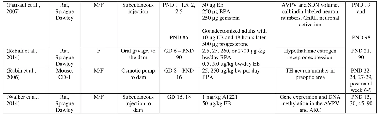
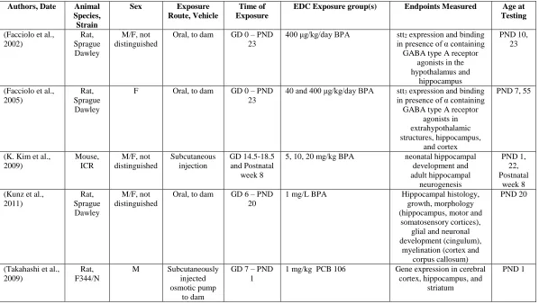
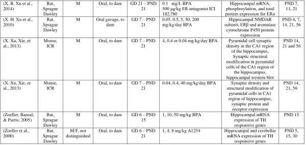
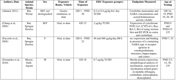
Details regarding the reviewed studies including the species, strain, sex, exposure
route, exposure window, dose, endpoint, and age at testing are summarized in four tables
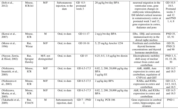
grouped by brain region of interest: hypothalamus (Table 1); hippocampus (Table 2); cortex,
cerebellum, and mid-brain(Table 3); and whole brain and embryonic brain regions (Table 4).
Estrogenic EDCs were hypothesized to be much more likely to influence the hypothalamus
(Table 1), while thyroid hormone-like EDCs are thought to be much more influential in the
hippocampus (Table 2). Tables 3 and 4 are studies that mostly concentrated on gene and
investigated larger regions, like cortex and cerebellum (Table 3) and whole brain or larger
embryonic brain regions (Table 4).
Results
Hypothalamus (Table 1)
The hypothalamus is the apical coordinator for homeostatic functions including
stress, emotion, reproduction, feeding, and the regulation of sex hormones production and
circulation (Campbell, 2005). In rodents, sexual dimorphisms within hypothalamic regions
are organized primarily during the perinatal period by aromatized testosterone (Lenz &
McCarthy, 2010) thus disruption of estrogen signaling is hypothesized to be a primary route
in which ECDs alter neuroendocrine development. Therefore it is not entirely surprising the
most of the available literature obtained for this review focused on three of the most
well-known estrogen-altering EDCs: Bisphenol A (BPA; exposures ranged from 2.5 μg/kg bw/day
to 50 mg/kg bw/day), polychlorinated biphenyls (PCBs) (exposures ranged from 1 mg/kg to
10 mg/kg), and the phytoestrogen, genistein (exposures ranged from 250 μg to 10 mg/kg
bw/day). Exposures were mostly gestational (7 studies), with several extending into the
neonatal period (6 studies) and the remaining focused only on neonatal exposure.
Of the 19 studies identified (Table 1), 15 included both sexes, reported outcomes
based upon sex, and reported sex-specific effects. Sex specific effects varied by region of
interest and compound investigated. For example, gestational exposure to the PCB mixture
Aroclor 1221 (A1221) via subcutaneous injection to pregnant rat dams (1mg/kg), altered the
expression of numerous estrogen-sensitive genes in the anteroventral periventricular nucleus
& Gore, 2014), while the arcuate nucleus (ARC) was found to be affected in males, but not
females (Walker et al., 2014). Affected genes included steroid hormone receptors (Ar, Thra,
Gper), neuropeptide receptors (Gnrh1 and Kiss1r), methyl transferase (Dnmt1), and clock
genes (Arntl and Per2) (Walker et al., 2014). Other studies have also identified the
developing AVPV as having sex-specific vulnerability to EDCs. Neonatal genistein
demasculinizes tyrosine hydroxylase immunoreactivity in the male AVPV (Patisaul, Fortino,
& Polston, 2006, 2007) and defeminized (reduced) kisspeptin fiber density in the female
AVPV and ARC (Losa et al., 2011). The AVPV is a highly sexually dimorphic region which
coordinates the luteinizing hormone (LH) surge in females and is thus essential for ovulation
(Simerly, 2002). These observed impacts on AVPV sexual differentiation are consistent with
the large body of literature reporting EDC-related effects on estrous cyclicity, fecundity, and
ovulation (Crain et al., 2008; Peretz et al., 2014).
Developmental EDC exposure also impacted neuropeptides and their receptors other
hypothalamic regions, somatostatin receptor expression (Facciolo, Alo, Madeo, Canonaco, &
Dessi-Fulgheri, 2002; Facciolo, Madeo, Alo, Canonaco, & Dessi-Fulgheri, 2005), progestin
receptor expression (Hays, Carpenter, & Petersen, 2002), and kisspeptin gene expression
(Dickerson et al., 2011; Navarro et al., 2009). In the periventricular and ventromedial
(VMN) nuclei, increased levels of somatostatin receptor 2 were found with BPA exposure at
PND 10, as well as increased levels of the same receptor at PND 23 in the periventricular
nucleus (Facciolo et al., 2002). Increased expression of somatostatin receptor 3 was also
found in the VMN and ARC at PND 7 after BPA exposure (Facciolo et al., 2005). In the
not found to be altered by dioxin, however it was found to alter GAD 67 levels, reducing the
naturally occurring sex difference in the rostral portion of the POA (Hays et al., 2002). In
whole hypothalamus, BPA decreased kisspeptin mRNA expression in both females and
males at PND 30 (Navarro et al., 2009). Also in the POA, A1221 reduced kisspeptin receptor
expression, specifically in males, at PND 1 (Dickerson et al., 2011). These examples
illustrate the variety of ways EDCs can affect hypothalamic development, either not affecting
sex differences by altering both sexes, reducing sex differences, or only affecting one sex
(Ceccarelli, Della Seta, Fiorenzani, Farabollini, & Aloisi, 2007; Dickerson et al., 2011; Hays
et al., 2002; Navarro et al., 2009).
The developing hypothalamus has sex-specific vulnerability to EDCs with the POA
and mediobasal hypothalamus (MBH) being the most intensely studied and robustly affected.
Estrogen receptor and aromatase expression are highly sexually dimorphic and altered by
developmental EDC exposure; thus potentially serving as a central mechanism by which
other aspects of hypothalamic sexual differentiation are altered by EDCs. Exposures
encompassing both the gestational and postnatal period seemed to be the most effective in
altering steroid receptor expression, while exposures limited to later postnatal development
seemed to result in more transient effects.
Because the hypothalamus is critically important for the regulation of mood and
emotion, as well as sociality (Engel et al., 2010; Eskenazi et al., 2013; Hoffman, Webster,
Weisskopf, Weinberg, & Vieira, 2010; Miodovnik et al., 2011), the animal outcomes
reviewed here are consistent with epidemiological data in children associating BPA and
et al., 2013; Eubig, Aguiar, & Schantz, 2010; Sagiv et al., 2010). Interestingly, no immune
related endpoints have been investigated to date in young animals, even though some
immune cells like microglia (Wood, 2011; Wu et al., 2013) and neutrophils (Molero et al.,
2002; Shindo, Moore, Flake, & Negishi, 2013) have been found to express estrogen receptors
and the immune system is influenced by the hypothalamus. Also, there were a lack of
phthalate, polybrominated diphenyl ethers (PBDE) and perfluorooctanoic acid (PFOA)
studies, even though these ubiquitous chemicals have been associated with mood and anxiety
disorders in humans such as ASD and ADHD (Crinnion, 2010; Gascon et al., 2011; Hoffman
et al., 2010; S. M. Kim et al., 2010).
Hippocampus (Table 2)
The hippocampus is fundamental to learning and memory. Hippocampal cell
proliferation and differentiation, as well as circuit development, are developmentally
regulated by sex and thyroid hormones (Bourguignon et al., 2013); making it a potential
target for endocrine disruption. In stark contrast to the studies on the hypothalamus, where
the majority of studies reported outcomes based on sex, none of the hippocampal studies
investigated sex-specific outcomes.
For the scope of this review, data was available from only two EDCs: BPA
(exposures ranged from 40 µg/kg/day to 200 mg/kg/day) and PCBs (exposures ranged from
1mg/kg to 8 mg/kg). In all studies exposures began in gestation, and in all but two (K. Kim
et al., 2009) extended into the postnatal period. Endpoints investigated included
gene expression. BPA was found to influence the formation of the dentate gyrus, causing it
to delineate earlier in PND 1 mice (sex was not distinguished) compared to controls (K. Kim
et al., 2009). BPA was also found to reduce synaptic density, enlarge the synaptic cleft, and
shorten the active zone in PND 14 and 21 male mice in region 1 of the hippocampus proper
(CA1) (X. Xu, Xie, et al., 2013). PCB mixture A1254 increased RC3/neurogranin mRNA
expression in the dentate gyrus (DG) at PND 15 (sex not stated) compared to controls
(Zoeller, Dowling, & Vas, 2000). Overall, evidence for disruptive effects in the
hippocampus is strongest for the PCBs (Morreale de Escobar, Obregon, & Escobar del Rey,
2004). Impacted processes include early delineation of the dentate gyrus, altered synapses in
the CA1 and altered neurogenesis, all consistent with reports on children exposed to PCBs
exhibiting cognitive, memory, and learning deficits (Jacobson & Jacobson, 1996; Stewart et
al., 2008).
Although BPA and PCBs can clearly alter hippocampal neurodevelopment, results
were not reported by sex in most cases but instead conducted in males only (6 studies),
females only (1 study), or with a group of animals where sex was not defined/distinguished
in analysis (4 studies). Thus it is not possible to ascertain which sex may be more vulnerable
to effects of EDCs in this region. This lack of focus on sexual dimorphisms in the
hippocampus is reflective of the hippocampal neuroscience literature as a whole which,
despite the identification of numerous structural and functional sex differences, has
traditionally has focused on one sex within a single study (Barth, Villringer, & Sacher, 2015;
Huang & Woolley, 2012; Shors, Chua, & Falduto, 2001; Woodruff et al., 2014).
deficits and other mental impairments, remains an area ripe for future investigation.
Moreover, identifying specifically when the developing hippocampus is most susceptible to
EDC exposures (most available studies exposed animals from mid-gestation to weaning),
needs further assessment. Ideally, these studies would also include both sexes.
Cortex, Cerebellum, and Mid-brain (Table 3)
Compared to the hypothalamus and hippocampus, relatively few studies have
examined developmental EDC related effects elsewhere in the brain, but we identified 3
relevant PCB studies (exposures ranging from 1-25 mg/kg), 7 BPA studies (exposures
ranging from 2 - 20,000 µg/kg) and 4 dioxin studies (2,3,7,8-tetrachlorodibenzo-p-dioxin
(TCDD; a class of highly persistent environmental compounds formed in industrial processes
such as waste combustion and the primary component of Agent Orange; exposures ranging
from 0.2 – 2 µg/kg bw/day). Seven of 14 studies differentiated results by sex, but three
failed to identify which sex was used. All but one study used an exposure window that began
in gestation and five used a longer perinatal exposure window.
Evidence for sex-specific vulnerability within this literature was uneven with one
study finding that females were more sensitive to PCB exposure than males, showing
decreased forebrain thyroxine concentrations as well as decreased deiodination, but only in
females (Morse, Wehler, Wesseling, Koeman, & Brouwer, 1996), . Other studies found that
both males and females were affected by TCDD or BPA. For example, dioxin was found to
lower Bcl-2 expression in the female cortex and cerebellum at PND 0, but not in male. In
contrast, dioxin was found to increase Bcl-xl expression at PND 0 in the cortex and
expression of genes such as S100b, Slc6a4, Htr1b, Maoa, Scla3, and Gap43, at PND 1, 4, 8
in sex and spatiotemporal manner in the cortex, thalamus, and pons (Itoh, Yaoi, & Fushiki,
2012). One study investigating BPA-related effects on extracellular signal-regulated kinase
(ERK) 1/2 signaling in the cerebellum via intracerebellar injection, reported that BPA and
ethinyl estradiol (EE) increased ERK cell numbers and signaling at low and high, but not
intermediate exposures (Zsarnovszky, Le, Wang, & Belcher, 2005). This effect was not
sex-specific, which is not entirely surprising given that the cerebellum is not considered a highly
sexually dimorphic structure. Sex specific effects were observed as early as the late
gestational period (Itoh et al., 2012; Morse et al., 1996; Nishizawa, Imanishi, & Manabe,
2005; Nishizawa et al., 2003; Nishizawa, Morita, Sugimoto, Imanishi, & Manabe, 2005)
indicating that maternal exposure to EDCs can have a significant effect on fetal brain
development, even in regions not typically considered to be sexually dimorphic
Collectively, the TCDD studies found decreased expression of myelin basic protein
(Fernandez et al., 2010), increased expression of apoptotic genes (Chang et al., 2005), and
inhibited acetylcholinesterase activity (Ahmed, 2011), suggesting impaired cerebellar
development and function. These studies help reveal the neurologic sources of motor and
learning disabilities seen in dioxin-exposed children (Vreugdenhil, Lanting, Mulder,
Boersma, & Weisglas-Kuperus, 2002). An extensive literature outside the scope of this
review has identified interactions with the aryl hydrocarbon receptor (AhR) as the primary
mode of action for TCDD neurotoxicity (Mimura & Fujii-Kuriyama, 2003; Patel, Kim,
Peters, & Perdew, 2006; Tijet et al., 2006). Dioxin is known to bind to AhR with high
et al., 1999). AhR is also thought to aid in the metabolism of xenobiotic compounds,
especially polycyclic aromatic hydrocarbons. Interestingly none of the dioxin studies
identified for this review examined effects on AhR activity or expression. By contrast,
increased expression of AhR was reported in the BPA studies in both males and females, all
investigating similar gestational periods of exposure in the ICR mouse (outbred strain
selected for high fertility and used in cancer research) (Nishizawa, Imanishi, et al., 2005;
Nishizawa et al., 2003; Nishizawa, Morita, et al., 2005). Although there was no report of
birth defects in late gestation (when the animals were evaluated), like abnormal liver or
kidney function, cleft palate, reduced litter size, etc. typically seen with altered AhR function
in dioxin exposure, increased AhR expression could potentially result in altered neural
differentiation, resulting in inappropriate catecholaminergic differentiation of Neuro2a cells
(Akahoshi, Yoshimura, & Ishihara-Sugano, 2006).
The reviewed PCB studies primarily investigated thyroid hormone related endpoints
in the forebrain and cerebellum, including brain thyroid hormone concentration and
metabolism (Morse et al., 1996) as well as thyroid hormone responsive gene expression
(Takahashi et al., 2009; Zoeller et al., 2000). Thyroid hormone is crucial for
neurodevelopment and alteration of gene expression and hormone metabolism may
contribute to the cognitive disorders seen in PCB exposed children (Jacobson & Jacobson,
1996; Stewart et al., 2008; Vreugdenhil et al., 2002).
In general these studies found spatiotemporal, sex, and exposure related effects on
gene expression related to neurogenesis, aryl hydrocarbon receptor expression and related
al., 2005; Nishizawa et al., 2003; Nishizawa, Morita, et al., 2005). Altered neuronal
differentiation, along with altered receptor signaling (Zsarnovszky et al., 2005) and changes
in neuronal migration (Itoh et al., 2012) could be a mechanism by which EDCs, including
BPA, alter hallmark behaviors of ADHD, ASD and other psychosocial disorders (de Cock et
al., 2012; Eubig et al., 2010), as reported in children (J. M. Braun et al., 2011; Harley et al.,
2013) and numerous animal studies (Grandjean & Landrigan, 2014; Landrigan, Lambertini,
& Birnbaum, 2012; Wolstenholme, Rissman, & Connelly, 2011).
Whole Brain and Embryonic Brain Regions (Table 4)
Because it is logistically difficult to differentiate brain regions in the pre-term rodent
brain, some analyses have focused on whole brain (4 of 9 studies) or large subregions
including the ventricular zone, cortical plate, telencephalon or diencephalon (5 of 9 studies).
Because sex differences emerge during embryonic development, examination of the fetal
brain for EDC effects yields critical insight as to the mechanisms by which EDCs manipulate
sexual differentiation, and the critical windows during which the developing brain is must
vulnerable. Only 2 studies differentiated results by sex, and, of the remaining, only 1
reported the sex examined. In one of the studies that differentiated results by sex, a sex
difference in the expression of Gper and Esrrg not present in controls was induced in whole
brain gene expression on GD 18.5 in BPA exposed animals, with males having higher
concentrations than females (Wolstenholme et al., 2012). These two genes are estrogen
receptor related genes and may indicate that males are more sensitive to BPA exposure.
Expression of thyroid hormone responsive, estrogen receptor related, myelination
common endpoints investigated, with BPA (4 studies with exposures ranging from 20 μg/kg
to 5 mg/kg), PCBs (3 studies with exposures ranging from 20 μg/kg – 6 mg/kg), and dioxin
exposure (2 studies with exposure ranging from 0.7-20 μg/kg TCDD). Expression of
myelination (Fernandez et al., 2010), organo- and neuro-genesis (Fujita et al., 2006; Komada
et al., 2012), and thyroid hormone related genes (Gauger et al., 2004; Morse et al., 1993), as
well as neuronal differentiation and migration (Itoh et al., 2012) and cell cycle exit
(Nakamura et al., 2012; Naveau et al., 2014) were all found to be altered by EDC exposure.
Some of these studies evaluated similar endpoints later in development, such as myelination
levels in the cerebellum (Fernandez et al., 2010) and whole brain thyroxine deiodinase
activity (Morse et al., 1993), and confirm that many of the effects may persist into
adolescence and beyond. Collectively these findings reinforce that EDCs can have
organizational effects on brain structure and suggest that gestational exposure alone has the
ability to forever alter neurodevelopment including myelination.
Every study in this category found effects of EDC exposure on gene expression
related to sex or thyroid hormone receptors or neurogenesis during the pre- or very early
postnatal period. Maternal developmental exposure to EDCs can induce alterations in fetal
neurodevelopment and sex difference organization. With every EDC investigated resulting
in evidence of altered neurogenesis, then the next set of questions to be addressed are which
EDC, at which dose, in which exposure window is likely to contribute to a particular
neuropsychiatric disorder? With the barrage of exogenous chemicals that almost every
deciphering exactly when and how much of an EDC exposure is necessary to induce
neuropsychological effects is a critical to the overall understanding of disease etiology.
Sex differences
Of the 46 studies included, 49% included both males and females and looked for
potential sex differences as part of their analysis (Figure 1). 26% included males and/or
females, but did not separate them in the analysis or did not state which sexes were included.
17% of studies only included males and 8% only included females. This is considerably
better than the neuroscience literature in general. A survey of 1200 neuroscience papers
from 2011 to 2012 (Woodruff et al., 2014) found outcome by sex was only reported 36% of
the time, and studies listed the sex of the animals only 42% of the time. Thus, the number of
studies identifying sex and the number of studies reporting outcome by sex was 32% and
13% higher, respectively, in the EDC literature reviewed here. The more frequent
consideration of sex in the EDC literature, compared to the neuroscience literature, is logical
considering their mechanisms of action. It was recognized quite early that EDCs have the
potential to alter sex-specific physiology, and thus has historically been considered an
important endpoint to report in the EDC literature. This work has significantly contributed
to, and advanced basic knowledge of, the neuroendocrinology of sexual differentiation (A. C.
Gore, 2008; Patisaul & Polston, 2008), and demonstrates that EDC researchers embraced the
inclusion of sex in study design long before the NIH mandated inclusion of sex differences in
preclinical research (Clayton & Collins, 2014). Just as sex may be a factor in calculating
Whitley & Lindsey, 2009), effects of EDCs are likely also sex-specific and should be taken
into consideration when determining susceptibility and potential mechanisms of action.
Conclusion
The reviewed data reveal that developmental EDC exposure can alter
neurodevelopment across the brain beginning early in fetal life, with sex specific effects
observable throughout the brain even before puberty. The hypothalamus was found to be
particularly affected by estrogenic EDCs in a sex, time, and exposure dependent manner; a
logical and expected finding given the fundamental importance of estrogen for masculinizing
the developing rodent brain. These data clearly demonstrate that EDCs are capable of
interfering with neural estrogen signaling but whether or not these specific phenotypic effects
will be observable humans is unclear. There are key species differences in the developing
rodent and human brain. Most significantly, androgens appear to be fundamental to primate
brain masculinization while the role of estrogen is less well understood. It is reasonable to
conclude, however, that EDCs are capable of perturbing estrogen function in the developing
human brain, particularly the hypothalamus. Accordingly, evidence from human literature
indicates that EDC exposure may be contributing to escalating rates of mood and affective
disorders in humans, especially children (de Cock et al., 2012; Grandjean & Landrigan,
2014). The hippocampus also appears particularly vulnerable to endocrine disruption by
estrogenic compounds such as BPA and PCBs although there is not enough available
evidence from this specific data set to make any conclusions about sex-specific effects. The
mechanisms by which BPA and PCBs alter hippocampal organization likely differ from each
characterized thyroid hormone disruptors (Winneke, 2011), for example, and emerging
evidence suggests BPA may be an androgen disrupter in the developing brain (Kinch,
Ibhazehiebo, Jeong, Habibi, & Kurrasch, 2015). Because the hippocampus is critical for
learning and memory, further investigation into EDC effects on the organization of this
region may help to elucidate potential linkages between EDC exposure and sex-biased
deficits in IQ, memory and related cognitive functions in humans (Grandjean & Landrigan,
2014).
Analyses of whole brain or large subregions in EDC-exposed embryos are limited but
reveal evidence of effects primarily related to neurogenesis, and effects which may involve
mechanisms other than disruption of classical endocrine disruption. Moreover, these data
suggest that EDC exposure prior to established hormone-dependent critical windows of
sexual differentiation could result in broad, fundamental changes to brain organization
particularly in cortex and cerebellum. These changes may include alteration of neuronal
connections fundamental to complex behaviors such as activity, sociality, and executive
function (Diamanti-Kandarakis et al., 2009; Kinch et al., 2015; Wolstenholme, Goldsby, &
Rissman, 2013). Evidence from the cortex, cerebellum, and mid brain emphasize that effects
from gestational exposures might be confounded or exacerbated by additional exposures later
in development, including the postnatal period. Information on how exposures to emerging
EDCs including fire retardants, surfactants, and other persistent contaminants known to be
ubiquitous in household environments (Mariussen, 2012; Stapleton et al., 2009) impacts
synaptogenesis, neural migration, and other developmental processes within the cortex,
sex-specific vulnerability. Comparison of results based upon sex in rodents at ages near
neurodevelopmental critical periods like prenatal and postnatal testosterone surge as well as
the brain growth spurt at PND 10, are needed to provide further insight into the alteration of
brain organization which can contribute to the developmental basis of diseases such as
ADHD, schizophrenia, and autism.
Compared to the neuroscience field as a whole, the neurodevelopmental EDC
literature reported information about sex and potential sex differences more frequently. That
neurodevelopmental effects of EDC exposure included alteration of sex differences, or the
creation of new ones, was a common finding, and these observations will ultimately result in
a better understanding of how male and female brains differ. The results of this review
demonstrate that the time and effort it takes to include and analyze both sexes is worthwhile,
especially in the endocrine disruption field, and should be considered a model for the larger
neuroscience community.
Acknowledgements
The authors would like to thank Roberto C. Melcangi and Giancarlo Panzica for their
Tables
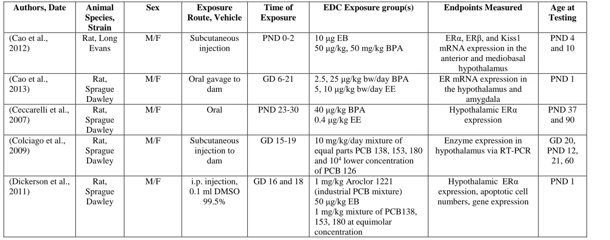
Table 1.1: EDC Effects on the Hypothalamus
Authors, Date Animal Species, Strain
Sex Exposure
Route, Vehicle
Time of Exposure
EDC Exposure group(s) Endpoints Measured Age at Testing (Cao et al.,
2012)
Rat, Long Evans
M/F Subcutaneous injection
PND 0-2 10 μg EB
50 μg/kg, 50 mg/kg BPA
ERα, ERβ, and Kiss1 mRNA expression in the
anterior and mediobasal hypothalamus
PND 4 and 10
(Cao et al., 2013)
Rat, Sprague Dawley
M/F Oral gavage to dam
GD 6-21 2.5, 25 μg/kg bw/day BPA 5, 10 μg/kg bw/day EE
ER mRNA expression in the hypothalamus and
amygdala
PND 1
(Ceccarelli et al., 2007)
Rat, Sprague Dawley
M/F Oral PND 23-30 40 μg/kg BPA 0.4 μg/kg EE
Hypothalamic ERα expression
PND 37 and 90
(Colciago et al., 2009)
Rat, Sprague Dawley
M/F Subcutaneous injection to
dam
GD 15-19 10 mg/kg/day mixture of equal parts PCB 138, 153, 180 and 104 lower concentration of PCB 126
Enzyme expression in hypothalamus via RT-PCR
GD 20, PND 12,
21, 60
(Dickerson et al., 2011)
Rat, Sprague Dawley
M/F i.p. injection, 0.1 ml DMSO
99.5%
GD 16 and 18 1 mg/kg Aroclor 1221 (industrial PCB mixture) 50 μg/kg EB
1 mg/kg mixture of PCB138, 153, 180 at equimolar concentration
Hypothalamic ERα expression, apoptotic cell numbers, gene expression
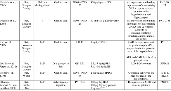
Table 1.1 continued
(Facciolo et al., 2002) Rat, Sprague Dawley M/F, not distinguished
Oral, to dam GD 0 – PND 23
400 μg/kg/day BPA stt2 expression and binding in presence of α containing GABA type A receptor
agonists in the hypothalamus and
hippocampus
PND 10, 23
(Facciolo et al., 2005)
Rat, Sprague Dawley
F Oral, to dam GD 0 – PND 23
40 and 400 μg/kg/day BPA stt3 expression and binding in presence of α containing GABA type A receptor
agonists in extrahypothalamic structures, hippocampus,
and cortex
PND 7, 55
(Hays et al., 2002)
Rat, Holtzman
Sprague Dawley
M/F Oral, to dam GD 15 1 μg/kg TCDD GAD 67 expression and progestin receptor (PR) expression in the preoptic area of the hypothalamus
AhR and GAD dual label in preoptic area
PND 3
(He, Paule, & Ferguson, 2012)
Rat, Sprague Dawley
M/F Oral gavage, to dam
GD 6-21 2.5, 25 μg/kg BPA 5.0, 10.0 μg/kg EE
SDN-POA volume PND 21
(Hobler et al., 2010)
Rat, Wistar
M/F Oral, to dam GD 6 – PND 58
2 mg/kg/day TPTCl Aromatase activity in the preoptic area of the
hypothalamus
PND 1, 21, 58
(Khurana, Ranmal, & Ben-Jonathan, 2000)
Rat, Fischer
344
M/F Subcutaneous injection
PND 1-5 100 μg day BPA 100 μg day octylphenol 5 μg day DES
ER expression in MBH and anterior pituitary
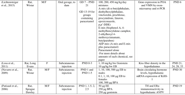
Table 1.1 continued
(Lichtensteiger et al., 2015)
Rat, Wistar
M/F Oral gavage, to dam
GD 7 – PND 21
GD 13-19 for groups containing paracetamol
100, 200, 450 mg/kg/day mixtures A-mix (di-n-butylphthalate, diethylhexylphthalate, vinclozolin, prochloraz, procymidone, linuron, epoxiconazole, p,p’-DDE)
E-mix (bisphenol A, 4-methylbenzylidene camphor, 2-ethylhexyl
4-methoxycinnamate, butylparaben)
AEP-mix (A-mix and E-mix plus paracetamol)
Paracetamol alone -For more details about amounts of each chemical, see paper
Gene expression in POA and VMN by exon-microarray and rt-PCR
PND 6
(Losa et al., 2011)
Rat, Long Evans
F Subcutaneous injection
PND 0-3 1, 10 mg/kg bw Genistein 10 μg/kg bw EB
Kiss fiber density in the hypothalamus
PND 21, 24, 28, 33 (Navarro et al.,
2009)
Rat, Wistar
M/F Subcutaneous injection
PND 1 and PND 1-5
1, 10, 100, 500 μg EB to males
0.1, 1, 10, 100 μg EB to females
100, 500 μg BPA
Brain circulating hormone levels, hypothalamic mRNA expression of
KiSS-1
PND 30
(Patisaul et al., 2006)
Rat, Sprague Dawley
M/F Subcutaneous injection
PND 1, 1.5, 2, 2.5
50 μg EE 250 μg BPA 250 μg genistein
TH and ERα immunoreactivity in hypothalamic AVPV
Table 1.1 continued
(Patisaul et al., 2007)
Rat, Sprague Dawley
M/F Subcutaneous injection
PND 1, 1.5, 2, 2.5
PND 85
50 μg EE 250 μg BPA 250 μg genistein
Gonadectomized adults with 10 μg EB and 48 hours later 500 μg progesterone
AVPV and SDN volume, calbindin labeled neuron numbers, GnRH neuronal
activation
PND 19 and
PND 98
(Rebuli et al., 2014)
Rat, Sprague Dawley
F Oral gavage, to the dam
GD 6 – PND 90
2.5, 25, 260, or 2700 μg /kg bw/day BPA
0.5, 5.0 μg/kg bw/day EE
Hypothalamic estrogen receptor expression
PND 21, 90
(Rubin et al., 2006)
Mouse, CD-1
M/F Osmotic pump to dam
GD 8 – PND 16
25, 250 ng/kg bw per day BPA
TH neuron number in preoptic area
PND 22-24, 27-29,
post natal week 6-9 (Walker et al.,
2014)
Rat, Sprague Dawley
M/F Subcutaneous injection to
dam
GD 16, 18 1 mg/kg A1221 50 μg/kg EB
Gene expression and DNA methylation in the AVPV
and ARC
Table 1.2: EDC Effects on the Hippocampus
Authors, Date Animal Species, Strain
Sex Exposure
Route, Vehicle
Time of Exposure
EDC Exposure group(s) Endpoints Measured Age at Testing (Facciolo et al.,
2002) Rat, Sprague Dawley M/F, not distinguished
Oral, to dam GD 0 – PND 23
400 μg/kg/day BPA stt2 expression and binding in presence of α containing GABA type A receptor
agonists in the hypothalamus and
hippocampus
PND 10, 23
(Facciolo et al., 2005)
Rat, Sprague Dawley
F Oral, to dam GD 0 – PND 23
40 and 400 μg/kg/day BPA stt3 expression and binding in presence of α containing GABA type A receptor
agonists in extrahypothalamic structures, hippocampus,
and cortex
PND 7, 55
(K. Kim et al., 2009) Mouse, ICR M/F, not distinguished Subcutaneous injection GD 14.5-18.5 and Postnatal week 8
5, 10, 20 mg/kg BPA neonatal hippocampal development and adult hippocampal neurogenesis PND 1, 22, Postnatal week 8 (Kunz et al.,
2011) Rat, Sprague Dawley M/F, not distinguished
Oral, to dam GD 6 – PND 20
1 mg/L BPA Hippocampal histology, growth, morphology (hippocampus, motor and
somatosensory cortices), glial and neuronal development (cingulum),
myelination (cortex and corpus callosum)
PND 20
(Takahashi et al., 2009)
Rat, F344/N
M Subcutaneously injected osmotic pump
to dam
GD 7 – PND 1
1 mg/kg PCB 106 Gene expression in cerebral cortex, hippocampus, and
striatum
Table 1.2 continued
(X. B. Xu et al., 2014)
Rat, Sprague Dawley
M Oral, to dam GD 21 – PND 21
0.1 mg/L BPA
500 μg/kg ER antagonist ICI 182,780
Hippocampal mRNA, phosphorylation, and total protein expression for ERα
PND 7, 11, 21
(X. H. Xu et al., 2010)
Rat, Sprague Dawley
M Oral gavage, to dam
GD 7 – PND 21
0.05, 0.5, 5, 50, 200 mg/kg/day BPA
Hippocampal NMDAR subunit, ERβ and aromatase
cytrochrome P450 protein expression
PND 4, 7, 14, 21, 56
(X. Xu, Xie, et al., 2013)
Mouse, ICR
M Oral, to dam GD 7 – PND 21
4, 0.4 or 0.04 mg/kg/day BPA Pyramidal cell synaptic density in the CA1 region
of the hippocampus, Synaptic structural modification in pyramidal cells of the CA1 region of
the hippocampus, hippocampal western blot
PND 14, 21 and 56
(X. Xu, Xie, et al., 2013)
Mouse, ICR
M Oral, to dam GD 7 – PND 21
0.04, 0.4, 40 mg/kg/day BPA Synaptic density and structural modification of
pyramidal cells in CA1 region of hippocampus, synaptic protein and
receptor expression
PND 14, 21, 56
(Zoeller, Bansal, & Parris, 2005)
Rat, Sprague Dawley
M Oral, to dam GD 6 – PND 15
1, 10, 50 mg/kg BPA Hippocampal mRNA expression of TH responsive genes
PND 15
(Zoeller et al., 2000) Rat, Sprague Dawley M/F, not distinguished
Oral, to dam GD 6 – PND 21
1, 4, 8 mg/kg A1254 Hippocampal and cerebellar mRNA expression of TH
responsive genes
Table 1.3: EDC Effects on Cortex, Cerebellum, and Mid-brain
Authors, Date Animal Species, Strain
Sex Exposure
Route, Vehicle
Time of Exposure
EDC Exposure group(s) Endpoints Measured Age at Testing (Ahmed, 2011) Rat,
Wistar M/F, not distinguished Gastric intubation, to dam
GD 1 – PND 30
0.2 or 0.4 μg/kg bw/ day TCDD
Cerebellar monoamine and GABA concentrations and
acetylcholinesterase activity (AchE)
GD 16, 19, PND 10, 20, 30
(Chang et al., 2005)
Rat, Sprague Dawley
M/F Oral, to dam GD 15 2 μg/kg TCDD Expression of cytochrome P450 1A1 (CYP1A1), Bax, Bcl-2, Bcl-xl using western blot and RT-PCR in cortex
and cerebellum
PND 0 and
PND115-125
(Facciolo et al., 2005)
Rat, Sprague Dawley
F Oral, to dam GD 0 – PND 23
40 and 400 μg/kg/day BPA stt3 expression and binding in presence of α containing GABA type A receptor
agonists in extrahypothalamic structures, hippocampus,
and cortex
PND 7, 55
(Fernandez et al., 2010)
Mouse, Dark-Agouti
M Oral, to dam GD 18 0.7 μg/kg TCDD Myelin protein expression, morphological analysis of myelination, expression of myelination related genes
in medulla oblongata, cerebellum, telencephalon,
diencephalon
PND 2/3, 14, 30,
Table 1.3 continued
(Itoh et al., 2012)
Mouse, ICR/Jcl
M/F Subcutaneous injection, to the
dam
GD 0.5-postnatal week 3
20 μg/kg bw/day BPA neuronal migration in the ventricular zone, gene expression changes in embryonic telencephalon, DiI labeled cortical neurons
in somatosensory cortex at postnatal week 3 and 12, gene expression in cortex,
thalamus and pons
GD 10.5-18.5, Postnatal weeks 3 and 12 and PND
1, 4, 8
(Kawai et al., 2007)
Mouse, ICR
M Oral, to dam GD 11-17 2 ng/g bw/day BPA ERα, ERβ, and serotonin immunoreactivity in the
dorsal raphe nucleus
PND 35, 63, 91
(Morse et al., 1996)
Rat, Wistar
M/F Oral, to dam GD 10-16 5, 25 mg/kg Aroclor 1254 Forebrain and cerebellum thyroid hormone concentrations and thyroid
hormone metabolism
GD 20, PND 21 and 90
(Nayyar, Zawia, & Hood, 2002)
Rat, Sprague Dawley
M/F, not distinguished
Oral, to dam GD 15 0.25, 0.5, 1.0 μg/kg bw dioxin Electrophoretic mobility shift assay of nuclear extract from cortex and
cerebellum
PND 3, 5, 10, 15, 20,
30
(Nishizawa, Imanishi, et al., 2005)
Mouse, ICR
M/F Oral, to dam GD 6.5-17.5 0.02, 2, 200, 20,000 μg/kg day BPA
5 μg/kg EE
AhR, AhRR, Arnt expression in cortex and cerebellum, regulation of
CYP1A1 and GST
GD 14.5 and 18.5 (Nishizawa et al., 2003) Mouse, ICR
M/F Oral, to dam GD 6.5-17.5 2 μg/kg day BPA RARα, and RXRα expression in cortex and
cerebellum
GD 12.5, 14.5, 16.5,
and 18.5 (Nishizawa,
Morita, et al., 2005)
Mouse, ICR
M/F Oral, to dam GD 6.5-17.5 0.02, 2, 200, 20,000 μg/kg day BPA
AhR, RARα, and RXRα expression in cortex and
cerebellum
GD 14.5 and 18.5
(Takahashi et al., 2009)
Rat, F344/N
M Subcutaneously injection, dam
GD 7 – PND 1
1 mg/kg PCB 106 Gene expression in cerebral cortex, hippocampus, and
striatum
Table 1.3 continued
(Zoeller et al., 2000)
Rat, Sprague Dawley
M/F, not distinguished
Oral, to dam GD 6 – PND 21
1, 4, 8 mg/kg A1254 Hippocampal and cerebellar mRNA expression of TH
responsive genes
PND 5, 15, 30
(Zsarnovszky et al., 2005)
Rat, Sprague Dawley
M/F Intracerebellar injection
PND 4-12 10-12 - 10-10 M and 10-7 – 10-6 M BPA
10-10 M EE
Activated ERK1/2 signaling in cerebellum
Table 1.4: EDC Effects on Whole Brain and Embryonic Brain Regions
Authors, Date Animal Species, Strain
Sex Exposure
Route, Vehicle
Time of Exposure
EDC Exposure group(s) Endpoints Measured Age at Testing (Fernandez et al.,
2010)
Mouse, Dark-Agouti
M Oral, to dam GD 18 0.7 μg/kg TCDD Myelin protein expression, morphological analysis of myelination, expression of myelination related genes
in medulla oblongata, cerebellum, telencephalon,
diencephalon
PND 2/3, 14, 30,
135
(Fujita et al., 2006)
Mouse, C57BL/6
M/F, not distinguished
Oral, to dam GD 7 20 μg/kg TCDD Genes expression in whole fetal brain
GD 12
(Gauger et al., 2004) Rat, Sprague Dawley M/F, not distinguished
Oral, to dam GD 6-16 1, 4 mg/kg A1254 TH responsive gene expression in fetal cortex
GD 16
(Itoh et al., 2012)
Mouse, ICR/Jcl
M/F Subcutaneous injection, to the
dam
GD 0.5-postnatal week 3
20 μg/kg bw/day BPA neuronal migration in the ventricular zone, gene expression changes in embryonic telencephalon, DiI labeled cortical neurons
in somatosensory cortex at postnatal week 3 and 12, gene expression in cortex,
thalamus and pons
GD 10.5-18.5, Postnatal weeks 3 and 12 and PND
1, 4, 8
(Komada et al., 2012) Mouse, C57BL/6J F and M/F-not distinguished
Oral GD 8.5-13.5 20 and 200 μg/kg BPA fetal neurogenesis of the cortical plate of the dorsal
telencephalon
GD 14.5
(Morse et al., 1993)
Rat, Wistar
M/F, not distinguished
Oral, to dam GD 1 or GD 1-18
0.2, 0.6, 1.8 mg/kg bw hexachlorobiphenyl (HCB) 0.6 mg/kg bw
Tetrachlorobiphenyl (TCB)
Activity of type II thyroxine – 5’ deiodinase in
the whole brain
Table 1.4 continued
(Nakamura et al., 2006) Mouse, ICR/Jcl M/F, not distinguished Subcutaneous injection to dam GD 0-10.5, 12.5, 14.5, or
16.5
20 μg/kg BPA cell proliferation, neuronal differentiation and migration in the ventricular zone and cortical plate, gene expression in embryonic telencephalon
GD 10.5, 12.5, 14.5 and 16.5
(Naveau et al., 2014)
Rat, Wistar
M/F, not distinguished
Oral to dam GD 6-17, 19, or 20
6 mg/kg/day Aroclor 1254 cell proliferation, S phase and cell cycle exit in the cortex, ventricular zone, and cortical plate, cell death and neuronal differentiation rate, neuronal migration
GD 17, 19, 20
(Wolstenholme et al., 2012)
Mouse, C57BL/6J
M/F Oral, to the dam
GD 0 – 18.5 5 mg BPA/kg diet whole brain gene expression by microarray
and rtPCR
Figure
Figure 1.1: The Percentage of studies including sex in their analyses. Studies including males only, females only, males and females, and males and/or females, but were not distinguished in analysis were counted and
CHAPTER 2—Prenatal Bisphenol A Exposure Alters Sex-Specific Estrogen Receptor Expression in the Neonatal Rat Hypothalamus and Amygdala
Authors: Jinyan Cao1, Meghan E. Rebuli1, James Rogers1, Karina L. Todd1, Stephanie M. Leyrer1, Sherry A. Ferguson2, Heather B. Patisaul1,3
Affiliations: 1Department of Biology, NCSU, Raleigh, North Carolina 27695; 2Division of Neurotoxicology, NCTR/FDA, Jefferson, Arkansas 72079; 3Keck Center for Behavioral
Biology, NCSU, Raleigh, North Carolina 27695
Published: 2013, Toxicological Sciences 133(1), 157-173
doi: 10.1093/toxsci/kft035 Abstract
Bisphenol A (BPA) exposure is ubiquitous, and in laboratory animals, early-life BPA
exposure has been shown to alter sex specific neural organization, neuroendocrine
physiology, and behavior. The specific mechanisms underlying these brain-related outcomes,
however, remain largely unknown, constraining the capacity to ascertain the potential human
relevance of neural effects observed in animal models. In the perinatal rat brain, estrogen is
masculinizing, suggesting that BPA-induced perturbation of estrogen receptor (ESR)
expression may underpin later in-life neuroendocrine effects. We hypothesized that prenatal
BPA exposure alters sex-specific ESR1 (ERα) and ESR2 (ERβ) expression in postnatal
limbic nuclei. Sprague Dawley rats were mated and gavaged on gestational days (GDs) 6–21
with vehicle, 2.5 or 25 μg/kg bw/day BPA, or 5 or 10 μg/kg bw/day ethinyl estradiol.
An additional group was restrained but not gavaged (naïve control). Offspring were
and amygdala by in situ hybridization. Relative to the vehicle group, significant effects of
BPA were observed on ESR1 and ESR2 expression throughout the mediobasal hypothalamus
and amygdala in both sexes. Significant differences in ESR expression were also observed in
the mediobasal hypothalamus and amygdala of the naïve control group compared with the
vehicle group, highlighting the potential for gavage to influence gene expression in the
developing brain. These results indicate that ESR expression in the neonatal brain of both
sexes can be altered by low-dose prenatal BPA exposure, with the caveat of the stress of
gavage.
Key Words
brain; endocrine disruptor; endocrine disruption; hypothalamus; development; sexually
dimorphic; ethinyl estradiol.
Disclaimer
This document has been reviewed in accordance with the U.S. Food and Drug
Administration (FDA) policy and approved for publication. Approval does not signify that
the contents necessarily reflect the position or opinions of the FDA, nor does mention of
trade names or commercial products constitute endorsement or recommendation for use. The
findings and conclusions in this report are those of the authors and do not necessarily
represent the views of the FDA.
Introduction
Human exposure to bisphenol A (BPA), a component of polycarbonate plastics,
epoxy resins, dental sealants, thermal receipts, and other products, is indisputably low but