Copyright2000 by the Genetics Society of America
Toward a Physical Map of
Drosophila buzzatii
: Use of Randomly Amplified
Polymorphic DNA Polymorphisms and Sequence-Tagged Site Landmarks
Hafid Laayouni, Mauro Santos and Antonio Fontdevila
Grup de Biologia Evolutiva (GBE), Departament de Gene`tica i de Microbiologia, Universitat Auto`noma de Barcelona, 08193 Bellaterra (Barcelona), Spain
Manuscript received December 7, 1999 Accepted for publication August 21, 2000
ABSTRACT
We present a physical map based on RAPD polymorphic fragments and sequence-tagged sites (STSs) for therepletagroup speciesDrosophila buzzatii.One hundred forty-four RAPD markers have been used as probes forin situhybridization to the polytene chromosomes, and positive results allowing the precise localization of 108 RAPDs were obtained. Of these, 73 behave as effectively unique markers for physical map construction, and in 9 additional cases the probes gave two hybridization signals, each on a different chromosome. Most markers (68%) are located on chromosomes 2 and 4, which partially agree with previous estimates on the distribution of genetic variation over chromosomes. One RAPD maps close to the proximal breakpoint of inversion2z3but is not included within the inverted fragment. However, it
was possible to conclude from this RAPD that the distal breakpoint of2z3had previously been wrongly
assigned. A total of 39 cytologically mapped RAPDs were converted to STSs and yielded an aggregate sequence of 28,431 bp. Thirty-six RAPDs (25%) did not produce any detectable hybridization signal, and we obtained the DNA sequence from three of them. Further prospects toward obtaining a more developed genetic map than the one currently available forD. buzzatiiare discussed.
A
common tenet in evolutionary biology is that an distribution (Carson and Wasserman 1965; Barker1977;Fontdevilaet al.1981, 1982;Haouaset al.1984).
ultimate understanding of evolution by natural
selection requires an integrated approach from genetics A substantial number of articles in ecological genetics
(e.g.,Barker andEast1980;Barker 1982;Santoset
and ecology. Unfortunately, there seems to be an
in-creasing gap between our current knowledge from very al.1989;ThomasandBarker1990;Quezada-Dı´azet
al. 1992; Santos 1994), life-history evolution (Ruiz
well-studied genomes and the ecological scenarios
where these genomes have evolved. As a noteworthy et al.1986;Hassonet al.1991;Santoset al.1992;
Barba-example, compare the massive amount of information dillaet al.1994;Betra´net al.1998), quantitative
genet-in recent releases of the FlyBase (FlyBase Consortium ics (ProutandBarker1989;Ruizet al.1991;Thomas
1999)—the comprehensive database for the fruitfly— andBarker1993;Leibowitzet al.1995;Santos1996),
with the number of entries for Drosophila inEndler’s thermal adaptation (KrebsandLoeschcke1996, 1997,
(1986, pp. 129–153) broad review of direct demonstra- 1999;Imashevaet al.1997), colonization (Fontdevila
tions of selection on naturally occurring genetic varia- et al.1981, 1982;HalliburtonandBarker1993;Rossi
tion: just one forDrosophila buzzatiiand two forD. melano- et al. 1996), and speciation (NaveiraandFontdevila
gaster! Because of this empirical restriction, we need a 1986; 1991a,b) have focused on D. buzzatii.Conversely
reasonable model where both approaches to under- toD. melanogaster, this wealthy state of affairs markedly
standing evolution can be successfully combined. contrasts with a paucity of molecular markers inD.
buz-Perhaps the best-characterized ecology of any Dro- zatii, still restricted to a few allozymes (Schaferet al.
sophila group is for therepleta group species, and we 1993; Betra´net al.1995). [A molecular marker is
de-agree withPowell(1997, p. 149) in that “anyone look- fined here as “any genetic variant that allows scoring of
ing for a system to connect ecology with genetics would conspecific individuals at the molecular level.” This is
do well to consider therepleta group.” Particularly, D. a somewhat narrower definition than that provided by
buzzatiiprovides a valuable model system for studies in KingandStansfield(1997) for a genetic marker, but is
natural populations and evolutionary genetics. Thus, operationally and implicitly used in evolutionary biology
this species is restricted to the cactus niche, feeding (Avise 1994) and quantitative genetics (Lynch and
and breeding in rotting tissues, but has a worldwide Walsh1998).]
To overcome this deficiency, here we present the first
extensive effort to map by in situ hybridization to the
Corresponding author:Mauro Santos, Departament de Gene`tica i polytene chromosomes of D. buzzatii a large number de Microbiologia, Universitat Auto`noma de Barcelona, Facultat de
(144) of reproducible randomly amplified polymorphic Cie`ncies, Edifici Cn, 08193 Bellaterra (Barcelona), Spain.
E-mail: [email protected] DNA (RAPD;WelshandMcClelland1990;Williams
1798 H. Laayouni, M. Santos and A. Fontdevila
for 20 min. After centrifugation for 15 min in an Eppendorf et al.1990) markers. RAPDs have been successfully
ap-centrifuge, the supernatant was added to 1 volume of 2-propa-plied to the construction of linkage maps in a variety
nol and left standing at room temperature for 5 min, which
of organisms (e.g.,Reiteret al.1992;Postlethwaitet was followed by a 10-min Eppendorf centrifugation. The pellet
al.1994;HuntandPage1995;Dimopouloset al.1996) was washed with 70% ethanol. Residual ethanol was removed
by drying the precipitate in a desiccator for 5 min, after which and are becoming a frequently used tool in population
the DNA was resuspended in 100l of sterile distilled water.
and evolutionary genetics (Smithet al.1994;de Zande
DNA amplifications:A set of 78 random decamer oligonu-andBijlsma1995; Apostolet al.1996; Espinasaand
cleotides purchased from Genosys Biotechnologies Inc.
(Cam-Borowsky1998). bridge, UK) and 5 from Operon Technologies Inc. (Alameda,
In addition to convenience for recombination map- CA) were used as single primers for the amplification of RAPD
ping, RAPDs can provide sequence-tagged sites (STSs; sequences. Primers are listed in Table 1 as designated by the
suppliers. Olsonet al.1989) that serve as physical entry points to
The conditions reported byWilliamset al.(1990) for creat-the genome. STSs can also be a rich source for detecting
ing RAPD markers by PCR were optimized for use with D. previously undescribed potential genes even in very
well-buzzatiitemplate DNA. All reaction volumes were 25l,
over-studied genomes (Louiset al.1997). We therefore have layered with 50l of light mineral oil (Sigma Chemical Co.,
determined 39 STS landmarks from cloned RAPD se- St. Louis). Each reaction consisted of 1⫻ activity buffer
(GIBCO BRL, Gaithersburg, MD), 1.6 mmMgCl2, 200mof quences, and all sequences were checked against both
each dNTP (Boehringer Mannheim, Indianapolis), 400 nm nucleic acid and protein databases for potential
primer, template DNA (ⵑ30–40 ng), and 0.8 units of Taq matches. These STSs also allow us to roughly estimate
polymerase (GIBCO BRL). Only one primer and one genomic
the overall base composition of theD. buzzatiigenome. DNA sample were added to any single reaction. Amplification
The physical map obtained comprises a total of 73 effec- was achieved in an MJ Research Inc. (Watertown, MA)
ther-mocycler programmed as follows: a preliminary 5-min dena-tively unique RAPD markers (39 of these are STSs),
turation at 94⬚; 45 cycles of 30 sec at 94⬚(denaturation), 1 min together with 9 RAPDs that gave two hybridization
sig-at 35⬚ (anneal), and 1 min at 72⬚ (extension); and a final nals, each on a different chromosome. The results
ob-extension at 72⬚for 5 min followed by storage at 4⬚.
Electropho-tained from the combined use of different techniques resis was performed in 1.4% agarose gels (SeaKem) with
Tris-allow the first comprehensive approach to the genome HCl acetate/EDTA (TAE) buffer for 5 hr at 70 V, constant
ofD. buzzatii. We hope the information provided here voltage. Reaction products were analyzed alongside small mo-lecular weight marker VI (Boehringer Mannheim). Ethidium will be an important tool for further development of a
bromide-stained gels (0.5 g/ml) were visualized on a UV reasonably saturated genetic map in this species.
transilluminator and photographed with a Polaroid camera or digitalized with a Bio-Print image management system. After testing for polymorphism and reproducibility (see below), the MATERIALS AND METHODS
RAPD bands chosen as probes were gel purified, reamplified using the same decameric primer that identified the RAPD Fly material:D. buzzatiiflies were collected from a natural
polymorphism, and labeled forin situhybridization. population in an abandonedOpuntia ficus-indicaplantation at
Polytene chromosome preparation andin situhybridization: Carboneras on the Mediterranean coast of Spain (Almerı´a;
Probes (300 ng–1g DNA) were labeled with digoxygenin-37⬚N, 1⬚9⬘W; seeRuizet al.1986 for details). Between the
11-dUTP by the random primer method using the Boehringer 10th and 12th of September 1993, 36 rotting Opuntia cladodes
Mannheim labeling kit, and the total yield from the labeling were collected, placed individually in transparent plastic
con-reaction (500 ng–2g) was quantified according to the in-tainers on a bed of sand, closed with a fine-meshed fabric, and
structions supplied by the manufacturer. Third instar larvae kept at room temperature (22–27⬚) in the makeshift laboratory
were grown at low densities at 18⬚ in a modified version of near the field site. From the adult flies that emerged from 28
David’s killed-yeast culture medium (David 1962). Salivary rots, a high number of isofemale strains were established by
gland chromosomes suitable for in situ hybridization were pairwise mating in vials (2⫻8 cm, with 5 ml of food) of virgin
prepared according toLabradoret al.(1990). Prehybridiza-females and males. The isofemale strains were maintained
tion, hybridization, and detection were carried out as de-at 23⬚ by one single brother-sister mating for the firstⵑ18
scribed byde Frutoset al.(1989). Hybridization temperature generations and full-sib matings (4–8 mating pairs per vial)
was 37⬚. Chromosomes were observed by phase contrast with thereafter and passed through a minimum ofⵑ36 generations
a Zeiss Axioscope photomicroscope at ⫻400 magnification before RAPD screening. Therefore, the probability that a
neu-and digitalized with a Bio-Print image management system. tral allele was still segregating in any particular isofemale strain
Chromosome mapping:The karyotype of most repleta spe-is practically negligible (see,e.g.,Gale1990). The population
cies, includingD. buzzatii, consists of five telocentric chromo-at Carboneras is polymorphic for the two common
cosmopoli-somes (1⫽X,2,3,4,5) and a dot (6) chromosome ( Wasser-tan 2st and 2j and for the two rare cosmopolitan 2jz3 and
man 1992). Hybridization signals were localized on the 2jq7, second-chromosome arrangements, as well as for the rare
polytene chromosomes using theD. repleta(Wharton1942) cosmopolitan4stand4s(Fontdevilaet al.1981; for a
descrip-andD. buzzatii(Ruizet al.1982;RuizandWasserman1993) tion seeRuizet al.1984).
cytological maps. The maps of D. buzzatii are cut-and-paste DNA isolation:DNA was isolated from individual males from
reconstructions of theD. repletamap according to the sequence each isofemale strain. The following protocol is a modification
of inversions proposed for their respective phylogenies. The of that described inLatorreet al.(1986). Each fly was
homog-molecular organizations of Mueller/Sturtevant/Novitski chro-enized in a 1.5-ml microcentrifuge tube containing 160l of
mosomal elements D (⫽ 4) and E (⫽ 2) in D. repleta and 10 mmTris/60 mmNaCl/5% (wt/vol) sucrose/10 mmEDTA,
D. buzzatii (see Powell 1997, p. 307—but note that exact pH 7.8. One hundred microliters of 1.25% SDS/300 mmTris/
correspondence of chromosomal arms inD. hydeiis misplaced 5% sucrose/10 mmEDTA, pH 9, were then added. The
mix-and readers should refer toLoukasand Kafatos 1986 for ture was incubated at 65⬚for 30 min, after which 60l of 5m
1800 H. Laayouni, M. Santos and A. Fontdevila
bridization (Ranzet al.1997). Within the limits of potential resolution, Ranz et al. (1997) concluded that the formerly proposed cytogenetic relationships between both species seem to be consistent with the new results.
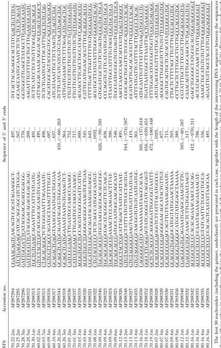
DNA sequencing:Thirty-nine single-signal RAPD markers (see below) were converted to STSs (Olsonet al.1989). Gel-purified RAPD fragments (10–100 ng) were directly cloned into pGEM-T Vector (Promega, Madison, WI). DNA mini-preparations were made from positive clones of transformed JM109 Escherichia colicells. The DNA sequences were deter-mined by the dideoxy method (Sangeret al.1977)using an ALF sequencer (Pharmacia Biotech, Piscataway, NJ). Nucleo-tide sequences were determined on both DNA strands and includedⵑ80 nucleotides of the T vector flanking the clon-ing site.
Nucleic acid searches were performed using the BLAST program (Altschulet al.1997). BLASTN was used to search the nucleic acid database, BLASTX to search the protein data-base with the putative translations of the STSs in all six frames, and ORF Finder program to look for potential open reading frames (ORFs). Alignments were also obtained using the de-fault option of the program CLUSTAL W (version 1.6)
(Thompsonet al.1994).
Figure1.—RAPD profile for the decameric primer G-80.16.
Lanes 2–15 are the PCR amplification products from individ-ual template DNA samples coming from 14 independent iso-RESULTS AND DISCUSSION
female strains ofD. buzzatii.Lane 1 indicates the molecular weight standards, and their sizes are given on the left-hand RAPD products and RAPD product profiles:
Forty-side (in base pairs). Lane 16 is the negative control. Polymor-four random decamer oligonucleotides (Table 1)
phic and reproducible RAPD bands used as probes forin situ yielded reproducible and polymorphic DNA fragments.
hybridization are indicated by arrows. A fragment was considered polymorphic when absent
in at least 1 individual out of 14 from different (i.e.,
independent) isofemale strains,i.e., when the recessive
In situhybridizations were routinely carried out using (absence) allele was at an average frequency of at least
a D. buzzatii strain fixed for 2st and 4st gene arrange-7% in the natural population (a more restrictive
crite-ments. A total of 108 RAPDs produced a single or multi-rion than the standard 5 or 1% used in population
ple signals (up to 15), and Table 2 gives the
hybridiza-genetics; see,e.g.,Hedrick1985). Repeating the
ampli-tion sites on the chromosomes from the salivary glands. fication using a set of five or more individuals that had
Sixty-three RAPDs gave a single and consistently detect-rendered polymorphic bands tested the reproducibility
able hybridization signal that must correspond to the of the different profiles. A particular band was
consid-site of the polymorphic locus, and in 10 additional cases ered as reproducible when the profiles from the two
there were one or two extra secondary signals on the independent amplifications were consistent in all
indi-same or different chromosomes that were absent in viduals tested.
some preparations. No variation in signal localization Those 44 primers generated 547 scorable marker
was ever detected among the several nuclei examined bands (an average of 12.4 bands per primer), of which
for a given probe. Hence, a total of 73 RAPDs with an 257 (47%) were polymorphic. RAPD reproducibility
average length of 942 bp (aggregate map length ofⵑ69
(see above) was obtained for 144 (56%) fragments,
kb) were considered to behave as effectively unique and
which were used as probes for in situ hybridization.
to be valuable as markers for physical map construction. RAPDs were named according to the decameric primer
Fourteen RAPDs gave two primary signals, and in nine that identified the RAPD polymorphism, followed by a
cases these signals were located on different chromo-digit that increases as the relative mobility of the band
somes, thus potentially increasing the number of useful increases. Figure 1 shows a typical example of RAPD
markers for further recombinational maps. Figure 2, products. A negative but nonsignificant correlation
be-a–e, shows a picture ofD. buzzatiipolytene chromosomes
tween the G⫹ C content of the decameric primer (as
indicating the cytological positions of the 73 primary-given by the first number in the primers from Genosys
singled signal RAPDs, together with the 9 primary-dou-Biotechnologies, Cambridge, UK) and the number of
bled signal RAPDs on different chromosomes (boldface
polymorphic bands scored was observed (SpearmanrS⫽
type).
⫺0.302;P ⫽ 0.055). On the other hand, there was a
Two RAPDs map close to known inversion break-positive and statistically significant correlation between
points, and they were converted to STSs for further
the G ⫹ C content of the primer and the fraction of
analyses (see below). RAPD 70.18.1 maps on 2(F1d)
polymorphic bands that were reproducible (rS⫽0.453;
(Figure 2b), close to the proximal breakpoint of
inver-P⫽ 0.003).
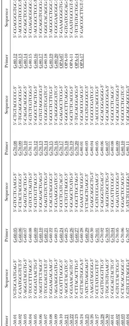
TABLE 2
Localization byin situhybridization on the salivary gland chromosomes ofD. buzzatiiof the 108 RAPDs used as probes
Hybridization signal
Primer RAPD Size (bp) Primary Secondary
G-60.09 60.09.4 640 X(C3c)
G-70.01 70.01.4 610 X(E3c)⫹4(F3a)
G-70.10 70.10.3 735 X(centromere)⫹4(B2c)
G-70.12 70.12.2 900 X(B4c)⫹4(C3d)
G-70.13 70.13.2 1365 X(E4b)
G-80.13 80.13.3 940 X(B1a) 2(D4c)
G-50.22 50.22.1 595 2(B2d-f )⫹3(F2e)
G-50.30 50.30.1 551 2(G3a-b)
G-60.03 60.03.2 1630 2(B2d-f )
G-60.03 60.03.5 1000 2(B2d-f )
G-60.03 60.03.7 556 2(E2a)
G-60.03 60.03.8 450 2(E2a)⫹4(A3d)
G-60.05 60.05.3 1605 2(B2d-f )
G-60.10 60.10.4 697 2(D5b)
G-60.21 60.21.2 1290 2(D5b)
G-60.24 60.24.3 1200 2(D4e-f )
G-60.26 60.26.1 840 2(Bli) 3(C3c)⫹5(D1f )
G-60.26 60.26.2 793 2(G3a-b) 5(G1a)
G-60.29 60.29.1 1500 2(D3g)
G-70.03 70.03.4 405 2(F5d)
G-70.09 70.09.1 1320 2(C7e)
G-70.09 70.09.5 450 2(G1b)
G-70.10 70.10.1 1200 2(A1a)
G-70.10 70.10.5 550 2(A2c)⫹4(G4e)
G-70.14 70.14.3 1005 2(B2a)⫹2(G5h)
G-70.16 70.16.2 1320 2(G5b)
G-70.18 70.18.1 2000 2(F1d)
G-70.19 70.19.1 1165 2(B3f )
G-70.20 70.20.2 1150 2(B2c)
G-70.20 70.20.4 650 2(C3e)
G-80.07 80.07.1 2000 2(G3a-b) 2(D5a)
G-80.07 80.07.2 1085 2(E5e)
G-80.07 80.07.3 937 2(E5a)
G-80.09 80.09.1 560 2(A4a)
G-80.13 80.13.2 1035 2(B2c)
G-80.17 80.17.1 1450 2(A4e) 2(B2c)
G-80.20 80.20.2 500 2(A4d)⫹3(B4d)
OPA-14 OPA-14.1 450 2(D2d)
OPA-14 OPA-14.2 400 2(D2d)
G-50.28 50.28.5 269 3(G4e)
G-60.02 60.02.2 555 3(D5d)
G-60.03 60.03.3 1530 3(C2c)
G-60.03 60.03.4 1200 3(G5d)⫹3(centromere)
G-60.05 60.05.5 685 3(G2d) 4(A4c)
G-60.09 60.09.3 757 3(A1e)
G-60.10 60.10.5 355 3(E4c)
G-60.24 60.24.2 1400 3(C5c)⫹3(D4b)
G-60.26 60.26.3 424 3(C3c)
G-70.03 70.03.1 575 3(F4a)
G-80.10 80.10.3 925 3(A1b) 3(G2a)
G-80.12 80.12.1 1200 3(E1f-g)
G-80.16 80.16.1 1760 3(A2d)
G-50.22 50.22.2 557 4(E1c)
G-50.25 50.25.2 627 4(A1g)
G-50.28 50.28.3 885 4(A2e)
G-60.10 60.10.1 1535 4(C1b)
G-60.29 60.29.2 1115 4(A2g-h) 5(G1a)
G-70.01 70.01.2 812 4(G1f )
1802 H. Laayouni, M. Santos and A. Fontdevila
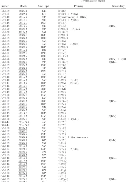
TABLE 2
(Continued)
Hybridization signal
Primer RAPD Size (bp) Primary Secondary
G-70.01 70.01.3 777 4(G1f )
G-70.04 70.04.1 703 4(E2d)
G-70.08 70.08.1 1092 4(G1g)
G-70.09 70.09.2 1200 4(G1e)⫹5(G3e)
G-70.09 70.09.3 1005 4(E4g)⫹5(C2a)
G-70.09 70.09.4 898 4(E4g)
G-70.09 70.09.6 400 4(C3g)
G-70.12 70.12.1 925 4(C2d)⫹4(D2b)
G-70.12 70.12.3 590 4(E4b)
G-70.12 70.12.4 555 4(E4b)
G-70.16 70.16.3 1130 4(G1d)
G-80.09 80.09.4 453 4(D4a)
G-80.09 80.09.5 449 4(D2a)
G-80.10 80.10.1 2000 4(E1b)
G-80.10 80.10.2 1100 4(E1d)
G-80.14 80.14.2 1030 4(C2a)⫹4(G1b-c)
G-80.16 80.16.3 848 4(F3c)
G-80.16 80.16.4 616 4(D2a) 2(E6e)
G-80.16 80.16.5 543 4(E2g)
G-80.19 80.19.1 2500 4(G4a)
G-50.28 50.28.1 1590 5(G2f ) 3(B4d)
G-60.03 60.03.6 905 5(D1f )
G-60.27 60.27.1 590 5(G4a)
G-60.29 60.29.3 1100 5(G1b)
G-70.13 70.13.3 479 5(B1a)
G-70.13 70.13.4 421 5(B1a)
G-80.07 80.07.4 775 5(G4b)
G-80.12 80.12.3 610 5(G2c)
G-80.19 80.19.3 870 5(B3c)
G-50.21 50.21.1 650 2(E3b)⫹2(G5)⫹4(A4f )⫹5(F1d)⫹
3(B5d)⫹3(G4)
G-50.25 50.25.1 860 X(A1f )⫹3(C4c)⫹2(D3a-g)⫹
2(E2a)⫹3(G2)4(C3c)⫹5(A3f )
G-50.28 50.28.2 905 4(G3)⫹X,2,3,4,5(centromeres)
G-60.05 60.05.4 1530 2(G1e)⫹2(B3e)⫹2(centromere)
G-60.05 60.05.6 600 3(F2b-c)⫹3(F2c-d)⫹4(A2)
G-60.06 60.06.1 1365 X,2,3,4,5(centromeres)
G-70.03 70.03.3 495 X(B2g)⫹2(E4c)⫹2(G1e)
G-70.10 70.10.2 840 2(G3b)⫹4(B2d)⫹2,4,
5(centromeres)
G-70.14 70.14.1 1150 2(G3b)⫹4(B1d)⫹5(G3c-d)⫹
2(centromere)
G-70.14 70.14.2 1130 2(G2i-j)⫹3(D4b)⫹4(A5a)
G-70.14 70.14.4 950 2(B1k)⫹2(D3a-b)⫹2(centromere)
G-70.16 70.16.1 1630 X(F3b)⫹X(G2f )X,3,
5(centromeres)
G-80.12 80.12.2 650 4(B1b)⫹5(E3d)⫹5(G2a)
G-80.14 80.14.3 785 2(D5g-h)⫹3(A2b)⫹4(G1f )
G-80.17 80.17.2 800 2(B2b)⫹2(G1a-f )⫹5(E4a-e)
G-80.20 80.20.1 755 X(centromere)⫹2(B3)⫹4(D1)
G-80.20 80.20.3 450 2(A4a)⫹3(A1c)⫹3(B1)⫹5(A3d)
OPA-7 OPA-7.1 1200 3(D/E)⫹4(F3d)⫹5(C2d)
OPA-7 OPA-7.2 800 2(E4/5)⫹4(A5b)⫹4(F1e-f )
OPA-17 OPA-17.2 1400 ⬎5 positions
G-70.03 70.03.2 585 ⵑ15 positions
Figure 2.—Blueprints of the standard chromosome arrangements ofD. buzzatii in-dicating the cytological localizations of the 73 RAPDs with a single primary signal, to-gether with the 9 RAPDs that produced two primary signals, each on different chromo-somes (indicated in boldface type), as inferred from the in situ hybridizations. (a) Chromosome X, (b) chromosome 2, (c) chromosome3, (d) chromosome4, (e) chromosome 5. The standard arrange-ments are cut-and-paste reconstructions of the D. repleta map (Wharton 1942) ac-cording to the sequence of inversions pro-posed for their respective phylogenies
(RuizandWasserman1993). The relative
order of those markers that hybridized on the same band is not known for certain. On the basis of information in Table 4, 50.25.2sts on4(A1g) likely marks the ho-mologous to gene kls, and 80.12.3sts on 5(G2c) the homologous to geneshot, both inD. melanogaster.The breakpoints of the polymorphic inversions on the second (2j, 2jz3,2jq7) and fourth (4s) chromosomes are
also shown. To recover the chromosomal segments included in inversions 2z3 and 2q7, segment2jfirst must be inverted. The
question mark indicates that the distal breakpoint of inversion2z3is not the same
Figure2.—Continued.
(Ruiz et al. 1984). This RAPD was also used as probe the inverted fragment. However, some discrepancies
were apparent when comparing the position of the hy-forin situhybridization on aD. buzzatiistrain fixed for
2jz3gene arrangement. Figure 3 shows the hybridization bridization signal with the putative distal chromosome
structures that should be observed. Thus, if we assume
1806 H. Laayouni, M. Santos and A. Fontdevila
Figure2.—Continued.
that the distal breakpoints of2jand2z3were exactly the 2z3is indeed more proximal than that for2j, somewhere
around2(E4b-c).
same on 2(C6c) (see Ruiz et al. 1984), the segment
2(E4) should lie just after (proximal→distal direction) A conspicuous feature from Figure 2, a–e, is that RAPDs are unevenly distributed among chromosomes. the hybridization signal and this was not the case. The
Mueller/Sturtevant/Novitski chromosomal elements, sequences. They could be unambiguously aligned but there is a big indel of 74 nucleotides and a significant and the percentage of total euchromatin assigned to
each of these elements (X-A, 18%; 2-E, 22.6%; 3-B, number of mismatches. This suggests that they could
represent two closely related loci, but for the time being
21.4%; 4-D, 20.3%; 5-C, 17.7%; see Wasserman 1982;
Schaferet al. 1993), a higher-than-expected number we cannot discard the possibility of a length polymor-phism. To summarize, it is not clear whether or not of single-signal RAPDs were located on chromosomes
2and 4(2
(4)⫽ 22.0;P ⬍ 0.001. The conclusion does all the RAPDs that were obtained from an identical
decameric primer and happen to hybridize on the same not qualitatively change after correcting for the
differ-ent number ofXchromosomes in males). chromosome band necessarily characterize the same
locus. On the other hand, dissimilar RAPDs (i.e., those Twenty-one RAPDs gave more than two primary
sig-nals on the salivary gland chromosomes, and in a num- obtained with different decameric primers) that map to
the same location likely mark different loci (cf. 60.26.2sts ber of cases they were located on the centromeres
(Ta-ble 2). The presence of other copies of the same gene and 50.30.1sts on chromosome 2 and 80.16.4sts and
80.09.5stson chromosome4). family, pseudogenes or DNA segments sharing a
se-quence homology, and/or transposable elements of di- Sequence analyses of RAPD markers: A total of 39
cytologically mapped RAPD markers were gel purified, verse types are probably the reason to observe multiple
signals. reamplified by PCR, and cloned using T vectors. The
clones were subjected to partial DNA sequence analysis
Clustering of RAPDs: The extent of clustering of
RAPD markers on chromosomes 2 and 4 (i.e., those from both ends and thus were converted to STSs, which
are valuable markers for physical map construction and with a higher number of RAPDs) was investigated by
means of a goodness-of-fit test (SokalandRohlf1995) can also reveal previously undescribed potential genes
(Louis et al. 1997). In most cases the total base pair
of the observed number of hybridization signals per
chromosome section to that expected from a Poisson length of the clones was sequenced, representing an
aggregate sequence of 28,431 bp (27,654 bp after
ex-distribution. Chromosome2is divided into 38 sections
(Wharton1942) of ⵑ600 kb each (assuming that D. cluding70.01.3sts; see above). STS landmarks were
des-ignated by adding the suffix “sts” to the name of the buzzatiihasⵑ2000 bands in the polytene chromosomes
asD. hydeiandⵑ50 kb per band; seeLaird1973;Hartl original RAPD marker. Table 3 lists these STSs and presents the terminal 30 bp from each end. In all cases et al.1994), and the distribution of signals (Figure 2b)
was as follows: 12 sections with one signal, 4 with two, but three (see the slight variation in primer sequences
reported as a footnote in Table 3), the decamer
oligonu-2 with three, and 1 with six (G(Williams’ correction) ⫽ 9.41;
P⫽0.094). The previous values could overestimate the cleotide that was used to generate the RAPD was present
at each terminus as expected.
degree of clustering because RAPDs60.03.2and60.03.5
[section2(B2)], RAPDsOPA14.1andOPA14.2[section STSs allow us a rough approximation of the variation
in nucleotide composition over the different chromo-2(D2)], and RAPDs60.03.7and60.03.8[section2(E2)]
might represent the same loci. Chromosome4is divided somes of D. buzzatii. Thus, overall G ⫹ C content is
41.18% (compared to 42.86% for D. melanogaster and
into 32 sections (Wharton1942), and the distribution
of signals (Figure 2d) was the following: 4 sections with 40.82% for the distant relative D. virilis; both values
estimated from the buoyant densities reported inGall
one signal, 5 with two, 2 with three, 1 with four, and 1
with five (G(Williams’ correction) ⫽ 13.18; P ⫽ 0.010). As for et al.1971), and the corresponding figures for the
au-tosomes are the following: 36.05% for chromosome5
chromosome2, this distribution of signals could
overes-timate the degree of clustering because RAPDs70.12.3 (aggregate sequence of 2252 bp), 38.87% for
chromo-some3 (aggregate sequence of 4809 bp), 40.54% for
and 70.12.4 [section 4(E4)] and RAPDs 70.01.2 and
70.01.3[section4(G1)] might represent the same loci. chromosome 4 (aggregate sequence of 9383 bp), and
43.52% for chromosome 2 (aggregate sequence of
To see whether or not that was indeed the case, we
derived STSs for RAPDs70.01.2and70.01.3and com- 10,219 bp). Assuming that STSs are representative of
the whole genome, these figures would tentatively sug-pared their sequences. They could be unambiguously
aligned and matched almost perfectly with a big gap gest that chromosome2is relatively rich in coding
re-gions (see Li1997).
from nucleotides 494 to 511 due to a higher number
of GT repeats in70.01.2sts(see Table 3 for their partial All STS sequences were checked against both nucleic
acid and protein databases for potential matches. We sequence information). After counting those two sets
of RAPDs as a single marker,G(Williams’ correction)⫽10.63 (P⫽ were particularly interested in those STSs (80.07.3sts
and70.18.1sts) derived from the two RAPDs that map 0.014). Therefore, RAPDs are not randomly distributed
along chromosome4, and there seems to be a higher- near second chromosome paracentric inversion
break-points (Figure 2b). Thus, the proximal breakpoint of than-expected number of hybridization signals in the
central part. inversion2jlies between thenAcR-96AandPp1␣-96A
genes, has been recently cloned and sequenced, and
Similarly, we derived STSs for RAPDs 70.13.3 and
able element named Galileo (Ca´ceres et al. 1999). An extensive reorganization within Mueller/Sturte-vant/Novitski chromosome elements has occurred in 80.07.3sts was checked against both nucleic acid and
protein databases and “hits” with an apparently un- Drosophila evolution, but chromosomal homologies
have been generally conserved (SegarraandAguade´
known gene in Drosophila (see below).
As in the distant relativeD. virilis(von Allmenet al. 1992; Kress 1993; Hartl and Lozovskaya 1994;
Segarraet al.1995, 1996; Ranz et al.1997; Vieiraet
1996), the genesAntennapedia(Antp) andUltrabithorax
(Ubx) in D. buzzatii seem to be together because they al.1997). This allowed us to check our hybridization
signals with those reported for D. melanogaster, and in
map on the same 2(F1c-d) band (Ranz et al. 1997),
close to the putative proximal breakpoint 2(F1c) of general there was a good agreement. Thus,klsmaps in
D. melanogaster on chromosome 3L, very close to the
inversion 2z3 (Ruiz et al. 1984). 70.18.1sts maps on
2(F1d) (Figure 2b) and, as described above, is not in- telomere, and this agrees quite well with the position
of RAPD50.25.2on chromosome4(Table 2 and Figure
cluded within the inversion fragment (Figure 3). No
significant hits with known genes were found in BLAST 2d). shot maps on chromosome 2R and, accordingly,
RAPD 80.12.3 maps on chromosome 5 in D. buzzatii
searches for 70.18.1sts, and we do not know whether
the relative positions ofAntp-Ubx-70.18.1stsare still con- (Table 2 and Figure 2e). In one case (80.16.4sts) the
correspondence was with the secondary signal
(chromo-served in the2jz3gene arrangement.
Table 4 lists the 22 STSs that rendered significant some 3R in D. melanogaster and chromosome 2 in D.
buzzatii; Table 2 and Figure 2b), and in three additional “hits” in BLAST searches of the GenBank databases and
also shows the protein alignments between conceptually cases (50.22.2sts, 70.03.4sts, and 80.07.2sts) there was
no correspondence with the cytological location re-translated STSs and the respective hits representing
known genes. As expected, the significant hits were in ported for the genomic scaffolds inD. melanogaster.
Simi-larities in sequences between different proteins are most cases with protein sequences or genomic scaffolds
fromD. melanogaster, but in three instances (70.09.4sts, likely the cause for the lack of correspondence, which
is clearly suggested by the two hits of50.22.2sts(Table 4).
80.07.3sts, and 70.19.1sts) the hits were with protein
sequences from other taxa that have not yet been de- Negative results:In spite of up to three attempts, 36
RAPDs (25%) did not produce any detectable
hybridiza-scribed in Drosophila. Interestingly, 70.09.4sts and
80.07.3stsshow reasonably good alignments with their tion signal on the polytene chromosomes (Table 5). We have obtained the DNA sequences from a sample of corresponding matches (see Table 4) and might have
identified novel Drosophila putative genes. three of those RAPDs (70.08.2, 70.09.7, and 70.14.5,
with an aggregate sequence of 1606 bp) to further inves-Thirteen STSs hit with Drosophila sequences of
known chromosomal location. From the alignments ob- tigate whether or not they present special features to
preventin situhybridization. The three sequences have
served in Table 4 and the corresponding chromosomal
homologies (see below), we conclude that 50.25.2sts an overall G⫹C content (41.10%) very similar to the
STSs, and no repetitive regions were detected. No
sig-likely marks the homologous to geneklarsicht(kls) and
80.12.3ststhe homologous to geneshort stop(shot); both nificant hits were found when these sequences were checked against both nucleic acid and protein
data-genes were previously known and mapped inD.
melano-gaster(FlyBase Consortium1999). An intriguing case bases. However, the sequence70.14.05presents an ORF of 207 amino acids (data not shown), and several
addi-is the hit of80.12.1stswith alcohol dehydrogenase (Adh)
genes ofD. buzzatiiand the Tc1-like DNA transposable tional clues to suggest that this sequence is part of a
coding region (compositional differences among codon
element ofD. virilis.In many species of therepletagroup
(includingD. buzzatii) theAdh region contains a pseu- positions relatively large and similar to the functional
genes inD. buzzatii;i.e., G⫹C highest in third position
dogene (Adh-⌿) and twoAdh functional genes (Adh-2
andAdh-1), arranged 5⬘to 3⬘, that have arisen by two and lowest in second position).
A likely cause for the lack of hybridization signal can
independent duplication events (Menotti-Raymondet
al.1991; Yumet al.1991;Sullivan et al.1994). Align- be an underreplication of those sequences during the
formation of polytene chromosomes. This will be the
ment of80.12.1stswith theD. buzzatii Adhregion
(Gen-Bank accession no. U65746) shows substantial matches case for all sequences within the␣-heterochromatin and
some sequences within the-heterochromatin (Gallet
between the intervening sequence of genesAdh-2and
Adh-1(from nucleotides 5475 to 5580) and nucleotides al.1971;Gall1973;Glaseret al.1997). However, no firm conclusion can be made on the available data and
455...560 of80.12.1sts, and alignment with Tc1-like from
D. virilis(GenBank accession no. U26938) shows sub- further work is in progress.
Conclusions and prospects:The present results help
stantial matches for nucleotides 276...312 of80.12.1sts
with the inverted repeats of the element.Adhmaps at understand the observed differences in the distribution
of genetic variation over chromosomes in species of the3(G1a) band inD. buzzatii(Labradoret al.1990),
while RAPD80.12.1maps at the3(E1f-g) band (Table the repletagroup of Drosophila (Zouros 1976). Thus,
enzyme heterozygosity is highest for chromosome2, but
2, Figure 2c) and could reflect a transposon-mediated
sepa-1812 H. Laayouni, M. Santos and A. Fontdevila
TABLE 4
Sequences producing significant alignments with theD. buzzatiiSTSs in searches using the BLAST program
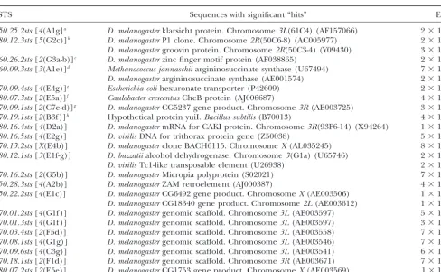
STS Sequences with significant “hits” Ei
50.25.2sts[4(A1g]a D. melanogasterklarsicht protein. Chromosome3L(61C4) (AF157066) 2⫻10⫺61 80.12.3sts[5(G2c)]b D. melanogasterP1 clone. Chromosome2R(50C6-8) (AC005977) 2⫻10⫺31 D. melanogastergroovin protein. Chromosome2R(50C3-4) (Y09430) 3⫻10⫺29 60.26.2sts[2(G3a-b)]c D. melanogasterzinc finger motif protein (AF038865) 2⫻10⫺19 60.09.3sts[3(A1e)]d Methanococcus jannaschiiargininosuccinate synthase (U67494) 7⫻10⫺15 D. melanogasterargininosuccinate synthase (AE001574) 2⫻10⫺13 70.09.4sts[4(E4g)]e Escherichia colihexuronate transporter (P42609) 2⫻10⫺41
80.07.3sts[2(E5a)]f Caulobacter crescentusCheB protein (AJ006687) 4⫻10⫺24
70.09.1sts[2(C7e-d)]g D. melanogasterCG5237 gene product. Chromosome3R(AE003725) 3⫻10⫺34 70.19.1sts[2(B3f )]h Hypothetical protein yuiI.Bacillus subtilis(B70013) 4⫻10⫺10 80.16.4sts[4(D2a)] D. melanogastermRNA for CAKI protein. Chromosome3R(93F6-14) (X94264) 1⫻10⫺19
80.16.5sts[4(E2g)] D. virilisDNA for trithorax protein gene (Z50038) 5⫻10⫺8
70.13.2sts[X(E4b)] D. melanogasterclone BACH6115. ChromosomeX(AL035245) 8⫻10⫺7 80.12.1sts[3(E1f-g)] D. buzzatiialcohol dehydrogenase. Chromosome3(G1a) (U65746) 2⫻10⫺8
D. virilisTc1-like transposable element (U26938) 2⫻10⫺8
70.16.2sts[2(G5b)] D. melanogasterMicropia polyprotein (S02021) 7⫻10⫺32
50.28.3sts[4(A2b)] D. melanogasterZAM retroelement (AJ000387) 4⫻10⫺41
50.22.2sts[4(E1c)] D. melanogasterCG6492 gene product. ChromosomeX(AE003506) 1⫻10⫺18 D. melanogasterCG18340 gene product. Chromosome2L(AE003612) 1⫻10⫺14 70.01.2sts[4(G1f )] D. melanogastergenomic scaffold. Chromosome3L(AE003597) 5⫻10⫺7 70.01.3sts[4(G1f )] D. melanogastergenomic scaffold. Chromosome3L(AE003597) 3⫻10⫺11 70.03.4sts[2(F5d)] D. melanogastergenomic scaffold. Chromosome3L(AE003558) 7⫻10⫺25 70.08.1sts[4(G1g)] D. melanogastergenomic scaffold. Chromosome3L(AE003546) 7⫻10⫺16 70.09.6sts[4(C3g)] D. melanogastergenomic scaffold. Chromosome3L(AE003541) 6⫻10⫺11 70.18.1sts[2(F1d)] D. melanogastergenomic scaffold. Chromosome3R(AE003671) 7⫻10⫺12 80.07.2sts[2(E5e)] D. melanogasterCG1753 gene product. ChromosomeX(AE003569) 1⫻10⫺104
Access number is given in parentheses. Cytological locations of STSs are from Table 2, and those forD. melanogasterhits were obtained from FlyBase (http://astorg.u.strasbg.fr:7081). 70.01.2sts and 70.01.3sts are not independent (see text for details). Protein alignments between conceptually translated STSs and hits representing knownD. melanogastergenes and/or presumably undescribed genes in Drosophila are given as footnotes from a to h. The numbers flanking the top lines indicate nucleotide positions of the amino acid residues in the STSs; numbers flanking the bottom lines indicate amino acid positions in the known protein. Percent similarity and identity are also indicated.
a50.25.2sts⫻klarsicht protein (D. melanogaster) (sim: 92%; iden: 89%)
b80.12.3sts⫻groovin protein (D. melanogaster) (sim: 97%; iden: 91%)
c60.26.2sts⫻zinc-finger motif protein (D. melanogaster) (sim: 51%; iden: 33%)
TABLE 4
(Continued)
d60.09.3sts⫻argininosuccinate synthase-like (D. melanogaster) (sim: 54%; iden: 38%)
e70.09.4sts⫻hexuronate transporter (E. coli) (sim: 66%; iden: 49%)
f80.07.3sts⫻cheB protein (C. crescentus) (sim: 73%; iden: 64%)
g70.09.1sts⫻CG5237 gene product (D. melanogaster) (sim: 52%; iden: 46%)
1814 H. Laayouni, M. Santos and A. Fontdevila
TABLE 4
(Continued)
h70.19.1sts⫻hypothetical protein yuiI (B. subtilis) (sim: 64%; iden: 52%)
iIndicates the number of hits one can “expect” by chance when searching a database of a particular size.
rated and were treated as a unity. The apportioning of any signal) will be used as genetic markers to provide
a link between the physical and more extensive linkage RAPDs observed here certainly suggests that average
variability levels on the autosomes ofD. buzzatiiare2ⱖ maps, also covering chromosomes3and4.In addition,
they will help to increase the density of markers (includ-4 ⬎ 3 ⬎ 5, contrary to the observed distribution of
spontaneous visible markers that placed chromosome ing microsatellites) around specific genomic regions to
search for quantitative trait loci of fitness-related traits
4as the least variable (Schaferet al.1993).
The physical map of D. buzzatii now comprises 73 such as body size (see Betra´net al. 1998). [A caveat:
effectively unique RAPD markers (39 of these are STSs) because the cytological maps ofD. buzzatiiare
cut-and-and 53 genes whose cytological position is already paste reconstructions of theD. repletamap (see above),
known (Figure 2, a–e). On the other hand, the current exact correspondence between the physical and the
ge-genetic map is poorly developed and consists of three netic maps for the relative positions of markers is
ex-linkage groups (chromosomesX,2, and5) that include pected, provided the proposed cytogenetic
relation-visible mutants and enzyme loci (Schaferet al.1993). ships betweenD. repletaandD. buzzatiiare fully correct.]
The RAPDs obtained here (along with those that gave M. Labrador and J. E. Quezada-Dı´az were of great help during the
secondary signals, those that gave hybridization signals initial steps of this work. We thank A. Leibowitz and J. E.
Quezada-Dı´az for their assistance in collecting the thousands of flies raised on different chromosomes, and the 36 that did not give
TABLE 5
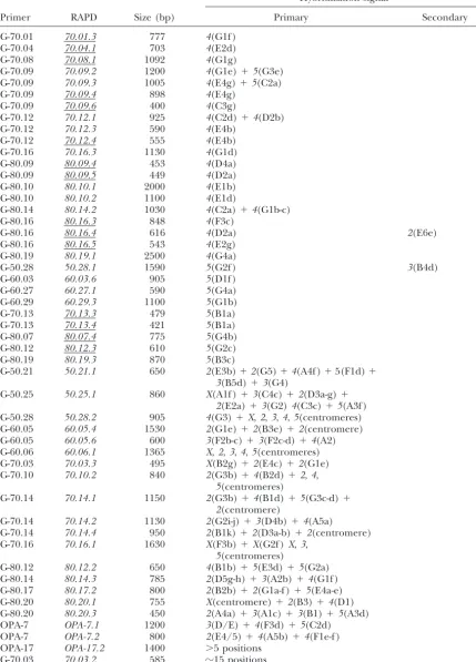
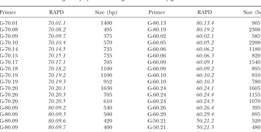
RAPDs that did not give any hybridization signal on the salivary gland chromosomes ofD. buzzatii
Primer RAPD Size (bp) Primer RAPD Size (bp)
G-70.01 70.01.1 1400 G-80.13 80.13.4 905
G-70.08 70.08.2 495 G-80.19 80.19.2 2300
G-70.09 70.09.7 375 G-60.02 60.02.1 585
G-70.10 70.10.4 570 G-60.05 60.05.2 2200
G-70.14 70.14.5 735 G-60.06 60.06.2 1180
G-70.15 70.15.1 735 G-60.06 60.06.3 820
G-70.17 70.17.1 705 G-60.09 60.09.1 1540
G-70.18 70.18.2 1100 G-60.09 60.09.2 895
G-70.19 70.19.2 1100 G-60.10 60.10.2 810
G-70.19 70.19.3 952 G-60.10 60.10.3 780
G-70.20 70.20.1 1630 G-60.24 60.24.1 1605
G-70.20 70.20.3 705 G.60.24 60.24.4 1155
G-70.20 70.20.5 610 G-60.24 60.24.5 1070
G-80.09 80.09.2 540 G-60.26 60.26.4 395
G-80.09 80.09.3 500 G-60.29 60.29.4 895
G-80.09 80.09.6 420 G-50.21 50.21.2 520
G-80.09 80.09.7 400 G-50.21 50.21.3 480
Genome Projects and community literature. Nucleic Acids Res. from Opuntia rots, L. Alarco´n and F. Rodrı´guez-Trelles for providing
27:85–88. information on the sequence ofXdhinD. buzzatiibefore publishing,
Fontdevila, A., A. Ruiz, G. AlonsoandJ. Ocan˜ a,1981 The evolu-M. P. Garcı´a-Guerreiro for helpful advice within situhybridizations,
tionary history ofDrosophila buzzatii.I. Natural chromosomal poly-F. Rodrı´guez-Trelles for helpful discussion and careful reading of
morphism in colonized populations of the Old World. Evolution earlier drafts, and M. Peiro´ for technical assistance. One of us (H.L.) 35:148–157.
is very grateful to M. R. Goldsmith for providing a stimulating intellec- Fontdevila, A., A. Ruiz, J. Ocan˜ aandG. Alonso,1982 The evolu-tual environment during his stay in the Department of Zoology, Uni- tionary history ofDrosophila buzzatii.II. How much has chromo-versity of Rhode Island. Two anonymous referees and the communicat- somal polymorphism changed in colonization? Evolution 36:
843–851. ing editor provided very helpful comments on the manuscript. H.L.
Gale, J. S., 1990 Theoretical Population Genetics. Unwin Hyman, was supported by a FP94-00215104 fellowship from the Ministerio de
London. Educacio´n y Ciencia (Spain). This work was supported by grants
Gall, J. G.,1973 Repetitive DNA in Drosophila, pp. 59–74 in
Sympo-PB93/0843 and PB96-1136 from the Direccio´n General de Ensen˜anza
sium on Molecular Cytology, edited byB. HamkaloandJ.
Papacon-Superior e Investigacio´n Cientı´fica (DGESIC, Spain) to A.F. and grant stantinou.Plenum Press, New York.
SGR98 from the Direccio´ General de Recerca (Generalitat de Cata- Gall, J. G., E. H. CohenandM. L. Polan,1971 Repetitive DNA lunya) to the GBE. sequences in Drosophila. Chromosoma33:319–344.
Glaser, R. L., T. J. LeachandS. E. Ostrowski,1997 The structure of heterochromatic DNA is altered in polyploid cells ofDrosophila melanogaster.Mol. Cell. Biol.17:1254–1263.
Halliburton, R.,andJ. S. F. Barker,1993 Lack of mitochondrial
LITERATURE CITED
DNA variation in AustralianDrosophila buzzatii.Mol. Biol. Evol.
Altschul, S. F., T. L. Madden, A. A. Scha¨ffer, J. Zhang, Z. Zhang 10:484–487.
et al., 1997 Gapped BLAST and PSI-BLAST: a new generation Haouas, S., Y. Carton, M. MarrakchiandJ. David,1984 Repro-of protein database search programs. Nucleic Acids Res.25:3389– ductive strategy of Drosophila species (D. buzzatiiandD. melanogas-3402. ter) associated with the prickly pear of Opuntia in Tunisia. Acta
Apostol, B. L., W. C. Black, P. ReiterandB. R. Miller,1996 Popu- Œcologica5:175–179.
lation genetics with RAPD-PCR markers: the breeding structure Hartl, D. L.,andE. R. Lozovskaya,1994 Genome evolution: be-ofAedes aegyptiin Puerto Rico. Heredity76:325–334. tween the nucleosome and the chromosome, pp. 579–592 in
Avise, J. C.,1994 Molecular Markers: Natural History and Evolution. Molecular Ecology and Evolution: Approaches and Applications, edited
Chapman & Hall, New York. byB. Schierwater, B. Streit, G. P. WagnerandR. DeSalle. Barbadilla, A., A. Ruiz, M. SantosandA. Fontdevila,1994 Mat- Springer, New York.
ing pattern and fitness-component analysis associated with inver- Hartl, D. L., D. I. Nurminsky, R. W. JonesandE. R. Lozovskaya,
sion polymorphism in a natural population ofDrosophila buzzatii. 1994 Genome structure and evolution in Drosophila: applica-Evolution48:767–780. tions of the framework P1 map. Proc. Natl. Acad. Sci. USA91:
Barker, J. S. F.,1977 Cactus-breeding Drosophila: a system for the 6824–6829.
measurement of natural selection, pp. 403–430 inMeasuring Selec- Hasson, E., J. C. Vilardi, H. Naveira, J. J. Fanara, C. Rodrı´guez tion in Natural Populations, Vol. 19 of Lecture Notes in Biomathematics, et al., 1991 The evolutionary history ofDrosophila buzzatii.XVI. edited byF. B. ChristiansenandT. Fenchel.Springer, Berlin. Fitness component analysis in an original natural population
Barker, J. S. F.,1982 Population genetics of Opuntia breeding Dro- from Argentina. J. Evol. Biol.4:209–225.
sophila in Australia, pp. 209–224 inEcological Genetics and Evolu- Hedrick, P. W., 1985 Genetics of Populations. Jones and Barlett,
tion: The Cactus-Yeast Drosophila Model System, edited by J. S. F. Boston.
BarkerandW. T. Starmer.Academic Press, New York. Hunt, G. J.,andR. E. Page, Jr.,1995 Linkage map of the honey bee,
Barker, J. S. F.,andP. D. East,1980 Evidence for selection follow- Apis mellifera, based on RAPD markers. Genetics139:1371–1382. ing perturbation of allozyme frequencies in a natural population Imasheva, A. G., V. Loeschcke, L. A. Zhivotovsky and O. E.
of Drosophila. Nature284:166–168. Lazebny,1997 Effects of extreme temperatures on phenotypic variation and developmental stability in Drosophila melanogaster Betra´n, E., J. E. Quezada-Dı´az, A. Ruiz, M. SantosandA.
Fontde-vila, 1995 The evolutionary history of Drosophila buzzatii. andDrosophila buzzatii.Biol. J. Linn. Soc.61:117–126.
King, R. C.,andW. D. Stansfield,1997 A Dictionary of Genetics, Ed. XXXII. Linkage disequilibrium between allozymes and
chromo-some inversions in two colonising populations. Heredity74:188– 5. Oxford University Press, New York.
Krebs, R. A.,andV. Loeschcke,1996 Acclimation and selection 199.
Betra´n, E., M. SantosandA. Ruiz,1998 Antagonistic pleiotropic for increased resistance to thermal stress inDrosophila buzzatii.
Genetics142:471–479. effect of second-chromosome inversions on body size and early
life-history traits inDrosophila buzzatii.Evolution52:144–154. Krebs, R. A.,andV. Loeschcke,1997 Estimating heritability in a threshold trait: heat-shock tolerance inDrosophila buzzatii.
Hered-Ca´ceres, M., J. M. Ranz, A. Barbadilla, M. LongandA. Ruiz,1999
Generation of a widespread Drosophila inversion by a transpos- ity79:252–259.
Krebs, R. A.,andV. Loeschcke, 1999 A genetic analysis of the able element. Science285:415–418.
Carson, H. L.,andM. Wasserman,1965 A widespread chromo- relationship between life-history variation and heat-shock toler-ance inDrosophila buzzatii.Heredity83:46–53.
somal polymorphism in a widespread species,Drosophila buzzatii.
Am. Nat.99:111–115. Kress, H.,1993 The salivary gland chromosomes ofDrosophila virilis: a cytological map, pattern of transcription and aspects of
chromo-David, J.,1962 A new medium for rearing Drosophila in axenic
conditions. Dros. Inf. Serv.36:128. some evolution. Chromosoma102:734–742.
Labrador, M., H. NaveiraandA. Fontdevila,1990 Genetic
map-de Frutos, R., K. KimuraandK. R. Peterson,1989 In situ
hybridiza-tion of Drosophila polytene chromosomes with digoxigenin- ping of theAdhlocus in therepletagroup of Drosophila byin situhybridization. J. Hered.81:83–86.
dUTP labeled probes. Trends Genet.5:366.
de Zande, L. V.,andR. Bijlsma,1995 Limitation of RAPD technique Laird, C. D.,1973 DNA of Drosophila chromosomes. Annu. Rev. Genet.7:177–204.
in phylogeny reconstruction in Drosophila. J. Evol. Biol.8:645–
656. Latorre, A., A. MoyaandF. J. Ayala,1986 Evolution of mitochon-drial DNA inDrosophila subobscura.Proc. Natl. Acad. Sci. USA.
Dimopoulos, G., L. Zheng, V. Kumar, A. della Torre, F. C. Kafatos
et al., 1996 Integrated genetic map ofAnopheles gambiae: use of 83:8649–8653.
Leibowitz, A., M. SantosandA. Fontdevila,1995 Heritability RAPD polymorphisms for genetic, cytogenetic and STS
land-marks. Genetics143:953–960. and selection on body size in a natural population ofDrosophila buzzatii.Genetics141:181–189.
Endler, J. A.,1986 Natural Selection in the Wild.Princeton University
Press, Princeton, NJ. Li, W.-H.,1997 Molecular Evolution.Sinauer, Sunderland, MA.
Louis, C., E. Maduen˜ o, J. Modolell, M. M. Omar, G. Papagiannakis Espinasa, L.,andR. Borowsky,1998 Evolutionary divergence of
AP-PCR (RAPD) patterns. Mol. Biol. Evol.15:408–414. et al., 1997 One-hundred and five new potentialDrosophila mela-nogastergenes revealed through STS analysis. Gene195:187–193.
1816 H. Laayouni, M. Santos and A. Fontdevila
Loukas, M.,andF. C. Kafatos,1986 The actin loci in the genus Drosophila buzzatii: larval crowding and male mating success. Evo-Drosophila: establishment of chromosomal homologies among lution50:2530–2535.
distantly related species byin situhibridization. Chromosoma94: Santos, M., A. Ruiz andA. Fontdevila,1989 The evolutionary 297–308. history ofDrosophila buzzatii.XIII. Random differentiation as a
Lynch, M.,andB. Walsh,1998 Genetics and Analysis of Quantitative partial explanation of the observed chromosomal variation in a
Traits.Sinauer, Sunderland, MA. structured natural population. Am. Nat.133:183–197.
Menotti-Raymond, M., W. T. StarmerandD. T. Sullivan,1991 Santos, M., A. Ruiz, J. E. Quezada-Dı´az, A. Barbadilla andA.
Characterization of the structure and evolution of theAdhregion Fontdevila,1992 The evolutionary history ofDrosophila
buz-ofDrosophila hydei.Genetics127:355–366. zatii.XX. Positive phenotypic covariance between field adult
fit-Naveira, H.,andA. Fontdevila,1986 The evolutionary history of ness components and body size. J. Evol. Biol.5:403–422.
Drosophila buzzatii.XII. The genetic basis of sterility in hybrids Schafer, D. J., D. K. Fredline, W. R. Knibb, M. M. Greenand betweenD. buzzatiiand its siblingD. seridofrom Argentina. Genet- J. S. F. Barker,1993 Genetics and linkage mapping ofDrosophila
ics114:841–857. buzzatii.J. Hered.84:188–194.
Naveira, H.,andA. Fontdevila,1991a The evolutionary history Segarra, C.,andM. Aguade´,1992 Molecular organization of the ofDrosophila buzzatii.XXI. Cumulative action of multiple sterility Xchromosome in different species of the obscura group of Dro-factors on spermatogenesis in hybrids ofD. buzzatiiandD. koep- sophila. Genetics130:513–521.
ferae.Heredity67:57–72. Segarra, C., E. R. Lozovskaya, G. Ribo´ , M. Aguade´andD. L. Hartl,
Naveira, H.,andA. Fontdevila,1991b The evolutionary history 1995 P1 clones fromDrosophila melanogasteras markers to study ofDrosophila buzzatii.XXII. Chromosomal and genic sterility in the chromosomal evolution of Muller’sAelement in two species male hybrids ofD. buzzatiiandD. koepferae.Heredity66:233–239. of the obscura group of Drosophila. Chromosoma104:129–136.
Naveira, H., C. Pla andA. Fontdevila,1986 The evolutionary Segarra, C., G. Ribo´andM. Aguade´,1996 Differentiation of Mull-history ofDrosophila buzzatii.XI. A new method for cytogenetic er’s chromosomal elements DandEin the obscura group of localization based on asynapsis of polytene chromosomes in inter- Drosophila. Genetics144:139–146.
especific hybrids of Drosophila. Genetica71:199–212. Smith, J. J., J. S. Scott-Craig, J. R. Leadbetter, G. L. Bush,
Olson, M., L. Hood, C. CantorandD. Botstein,1989 A common D. L. Robertset al., 1994 Characterization of random amplified language for physical mapping of the human genome. Science polymorphic DNA (RAPD) products fromXanthomonas campestris 245:1434–1435. and some comments on the use of RAPD products in phylogenetic
Postlethwait, J. H., S. L. Johnson, C. N. Midson, W. S. Talbot, analysis. Mol. Phylogenet. Evol.3:135–145.
M. Gateset al., 1994 A genetic linkage map for the Zebrafish. Sokal, R. R.andF. J. Rohlf,1995 Biometry, Ed. 3. Freeman, New
Science264:699–703. York.
Powell, J. R.,1997 Progress and Prospects in Evolutionary Biology: The
Sullivan, D. T., W. T. Starmer, S. W. Curtiss, M. Menotti-Ray-Drosophila Model.Oxford University Press, New York.
mondandJ. Yum,1994 Unusual molecular evolution of anAdh Prout, T.,andJ. S. F. Barker,1989 Ecological aspects of the
herita-pseudogene in Drosophila. Mol. Biol. Evol.11:443–458. bility of body size inDrosophila buzzatii.Genetics123:803–813.
Thomas, R. H. andJ. S. F. Barker, 1990 Breeding structure of
Quezada-Dı´az, J. E., M. Santos, A. RuizandA. Fontdevila,1992
natural populations ofDrosophila buzzatii: effects of the distribu-The evolutionary history of Drosophila buzzatii.XXV. Random
tion of larval substrates. Heredity64:355–365. mating in nature. Heredity63:373–379.
Thomas, R. H.,andJ. S. F. Barker,1993 Quantitative genetic
analy-Ranz, J. M., C. SegarraandA. Ruiz,1997 Chromosomal homology
sis of the body size and shape ofDrosophila buzzatii.Theor. Appl. and molecular organization of Muller’s elementsDandEin the
Genet.85:598–608.
Drosophila repletaspecies group. Genetics145:281–295.
Thompson, J. D., D. G. HigginsandT. J. Gibson,1994 CLUSTAL
Ranz, J. M., M. Ca´ceresandA. Ruiz,1999 Comparative mapping
W: improving the sensitivity of progressive multiple sequence of cosmids and gene clones from a 1.6 Mb chromosomal region
alignment through sequence weighting, positions-specific gap ofDrosophila melanogasterin three species of the distantly related
penalties and weight matrix choice. Nucleic Acids Res.22:4673– subgenus Drosophila. Chromosoma108:32–43.
4680.
Reiter, R. S., J. G. K. Williams, K. A. Feldmann, J. A. Rafalski,
Vieira, J., C. P. Vieira, D. L. HartlandE. R. Lozovskaya,1997
S. V. Tingeyet al., 1992 Global and local genome mapping in
Discordant rates of chromosome evolution in theDrosophila virilis Arabidopsis thalianaby using recombinant inbred lines and
ran-species group. Genetics147:223–230. dom amplified polymorphic DNAs. Proc. Natl. Acad. Sci. USA
von Allmen, G., I. Hogga, A. Spierer, F. Karch, W. Benderet al., 89:1477–1481.
1996 Splits in fruitflyHoxgene complexes. Nature380:116.
Rossi, M. S., E. Barrio, A. Latorre, J. E. Quezada-Dı´az, E. Hasson
Wasserman, M.,1982 Evolution of the repletagroup, pp. 61–139
et al., 1996 The evolutionary history ofDrosophila buzzatii.XXX.
inThe Genetics and Biology of Drosophila, Vol. 3b, edited by M.
Mitochondrial DNA polymorphism in original and colonizing
Ashburner, H. L. CarsonandJ. N. Thompson, Jr.Academic populations. Mol. Biol. Evol.13:314–323.
Press, New York.
Ruiz, A.,andM. Wasserman,1993 Evolutionary cytogenetics of the
Wasserman, M.,1992 Cytological evolution of theDrosophila repleta Drosophila buzzatiispecies complex. Heredity70:582–596.
Ruiz, A., A. FontdevilaandM. Wasserman,1982 The evolutionary species group, pp. 455–552 inDrosophila Inversion Polymorphism, history of Drosophila buzzatii. III. Cytogenetic relationships be- edited by C. B. KrimbasandJ. R. Powell.CRC Press, Boca tween two sibling species of the buzzatii cluster. Genetics101: Raton, FL.
503–518. Welsh, J.,andM. McClelland,1990 Fingerprinting genomes using
Ruiz, A., H. NaveiraandA. Fontdevila,1984 La historia evolutiva PCR with arbitrary primers. Nucleic Acids Res.18:7213–7218. deDrosophila buzzatii.IV. Aspectos citogene´ticos de su polimor- Wharton, L. T.,1942 Analysis of the repleta group of Drosophila. fismo cromoso´mico. Gene´t. Ibe´r.36:13–35. Univ. Texas Publ.4228:23–52.
Ruiz, A., A. Fontdevila, M. Santos, M. SeoaneandE. Torroja, Williams, J. G. K., A. R. Kubelik, K. J. Livak, J. A. Rafalskiand 1986 The evolutionary history ofDrosophila buzzatii.VIII. Evi- S. V. Tingey,1990 DNA polymorphism amplified by arbitrary dence for endocyclic selection acting on the inversion polymor- primers are useful as genetic markers. Nucleic Acids Res.18: phism in a natural population. Evolution40:740–755. 6531–6535.
Ruiz, A., M. Santos, A. Barbadilla, J. E. Quezada-Dı´az, E. Hasson Yum, J. S., W. T. StarmerandD. T. Sullivan,1991 The structure
et al., 1991 Genetic variance for body size in a natural population of theAdh locus ofDrosophila mettleri: an intermediate in the ofDrosophila buzzatii.Genetics128:739–750. evolution of theAdhlocus in the repletagroup of Drosophila.
Sanger, F., S. NicklenandA. R. Coulson,1977 DNA sequencing Mol. Biol. Evol.8:857–867.
with chain-terminating inhibitors. Proc. Natl. Acad. Sci. USA74: Zouros, E.,1976 The distribution of enzyme and inversion polymor-5463–5467. phism over the genome of Drosophila: evidence against balancing
Santos, M.,1994 Heterozygote deficiencies under Levene’s popula- selection. Genetics83:169–179. tion subdivision structure. Evolution48:63–78.