A Monthly Journal of Computer Science and Information Technology
ISSN 2320–088X
IJCSMC, Vol. 2, Issue. 4, April 2013, pg.532 – 539
RESEARCH ARTICLE
© 2013, IJCSMC All Rights Reserved
532
A Robust DR Classification from Blood
Vessel Features
Alka Vijayan1, Antony Judice.A2 1
P.G Scholar, Arunachala College of Engineering for Women, Kanyakumari, Tamilnadu, India
2
Assistant Professor, Arunachala College of Engineering for Women, Kanyakumari, Tamilnadu, India
1
vijayanalka@gmail.com; 2 pravinhireling@gmail.com
Abstract— A Diabetic Retinopathy (DR) is a condition occurring in persons with diabetes, which causes progressive damage to the retina that leads to blindness. Early detection of retinopathy in individuals with diabetes is critical in preventing visual loss. In this project prior Diabetic retinopathy diagnosis implement based on SVM classifier. To improve the classifier accuracy depends upon the retinal blood vessel features. For blood vessel extraction proposes a function based on the evaluation of measurable features describing retinal blood vasculature. Specifically, this proposal enables vascular structure assessment through its characterization as connected segments with measurable area and length. Thus, this function is sensitive to vasculature features such as connectivity, area and length, and supplements widely-used metrics based on contingency tables. The classification scheme proposed appears useful for characterizing overall retinopathy severity of patients on the basis of gradings of fundus photographs. The data presented may be of help in planning trials of treatment aimed at slowing the development or progression of retinopathy.
Key Terms: - Diabetic Retinopathy; SVM Classifier; quality evaluation function
I. INTRODUCTION Diabetic Retinopathy
The retina is the light-sensitive layer of cells at the back of the eye as shown in Fig 1. It converts light into electrical signals. The signals are sent to the brain through the optic nerve and the brain interprets them to produce the images that you see. To work effectively, the retina needs a constant supply of blood, which it receives through a network of tiny blood vessels. Over time, a continuously high blood sugar level can cause the blood vessels to become blocked or to leak. This damages the retina and stops it from working.
© 2013, IJCSMC All Rights Reserved
533
Diabetic retinopathy [2] is a condition occurring in persons with diabetes, which causes progressive damage to the retina, the light sensitive lining at the back of the eye. It is a serious sight-threatening complication of diabetes.
Diabetic retinopathy is the result of damage to the tiny blood vessels that nourish the retina as shown in Fig 2. They leak blood and other fluids that cause swelling of retinal tissue and clouding of vision. The condition usually affects both eyes. The longer a person has diabetes, the more likely they will develop diabetic retinopathy. If left untreated, diabetic retinopathy can cause blindness.
Fig 2: A retina showing signs of diabetic retinopathy Symptoms of diabetic retinopathy include:
• Seeing spots or floaters in your field of vision • Blurred vision
• Having a dark or empty spot in the center of your vision • Difficulty seeing well at night
In patients with diabetes, prolonged periods of high blood sugar can lead to the accumulation of fluid in the lens inside the eye that controls eye focusing. This changes the curvature of the lens and results in the development of symptoms of blurred vision. The blurring of distance vision as a result of lens swelling will subside once the blood sugar levels are brought under control. Better control of blood sugar levels in patients with diabetes also slows the onset and progression of diabetic retinopathy.
Often there are no visual symptoms in the early stages of diabetic retinopathy. Early detection and treatment can limit the potential for significant vision loss from diabetic retinopathy.
Treatment of diabetic retinopathy varies depending on the extent of the disease. It may require laser surgery to seal leaking blood vessels or to discourage new leaky blood vessels from forming. Injections of medications into the eye may be needed to decrease inflammation or stop the formation of new blood vessels. In more advanced cases, a surgical procedure to remove and replace the gel-like fluid in the back of the eye, called the vitreous, may be needed. A retinal detachment, defined as a separation of the light-receiving lining in the back of the eye, resulting from diabetic retinopathy, may also require surgical repair.
If you are a diabetic, you can help prevent or slow the development of diabetic retinopathy by taking your prescribed medication, sticking to your diet, exercising regularly, controlling high blood pressure and avoiding alcohol and smoking.
Diabetic Eye Problems
Diabetic retinopathy is classified into two types:
1. Non-proliferative diabetic retinopathy (NPDR) is the early state of the disease in which symptoms
will be mild or non-existent. In NPDR, the blood vessels in the retina are weakened causing tiny bulges called microanuerysms to protrude from their walls. The microanuerysms may leak fluid into the retina, which may lead to swelling of the macula.
Fig 3: Non-proliferative diabetic retinopathy (NPDR)
2. Proliferative diabetic retinopathy (PDR) is the more advanced form of the disease. At this stage,
© 2013, IJCSMC All Rights Reserved
534
new blood vessel may leak blood into the vitreous, clouding vision. Other complications of PDR include detachment of the retina due to scar tissue formation and the development of glaucoma. Glaucoma is an eye disease defined as progressive damage to the optic nerve. In cases of proliferative diabetic retinopathy, the cause of this nerve damage is due to extremely high pressure in the eye. If left untreated, proliferative diabetic retinopathy can cause severe vision loss and even blindness.
Fig 4: Proliferative diabetic retinopathy (PDR)
Objective
A Diabetic retinopathy is a potentially blinding complication of diabetes. Early stage of vision loss diagnosis is one of the challenge processes. The main objective of this project is to improve the quality of colour fundus image vessel segmentation and to identify the diabetic retinopathy by using SVM algorithm and evaluating the performances of this segmentation. To improve the classifier accuracy depends upon the retinal blood vessel features.
II. METHODOLOGY Block Diagram
Fig 5: Block Diagram
A. Read Colour Fundus Image (CFI)
© 2013, IJCSMC All Rights Reserved
535
addition to this, the values of the QEFs [7] under study were also calculated for the same images. Thus, human- and functions-provided evaluations are compared to measure the correspondence degree between them through some statistical approach.
B. Preprocessing
In this process we resize the image as per the requirements. Also we enhance the brightness of the image. The importance of image resizing scheme is greatly felt in the fields of medical image processing, computer graphics, image database etc. Image resizing is an operation that enlarges or reduces the size of the displayed image. The two main purposes of resizing images are to increase the size of the object within the image for ease of visualisation and to decrease the size of the image so that the entire image can be visualised on a monitor or display device with a lower spatial resolution than the image itself. During the process of resizing, image pixels are mapped onto a smaller or larger image matrix. Various methods to achieve the remapping of pixels are used. The common methods are nearest neighbour interpolation and bi-linear interpolation. Another more time-consuming method is bi-cubic or cubic convolution.
The enhancement is widely used for medical image processing and as a preprocessing step in speech recognition, texture synthesis, and many other image/video processing applications. The final preprocessing step consists on generating a new vessel enhanced image, which proves more suitable for further extraction of moment invariants based features while bright retinal structures are removed (i.e., optic disc, possible presence of exudates or reflection artifacts), the darker structures remaining after the opening operation become enhanced (i.e., blood vessels, fovea, possible presence of microaneurysms or haemorrhages).
C. Vascular Tree Segmentation
The segmentation [1], [7] and analysis of retinal vasculature form an essential part of several practical applications such as detection of hypertension, diabetes, stroke and cardiovascular diseases. In case of ophthalmologic conditions, the segmentation and measurement of the retinal vessels is of primary interest in the diagnosis and treatment of a diabetic retinopathy that directly affect the morphology of the retinal vessel tree. Also the accurate segmentation of the retinal blood vessels is often an essential prerequisite step in the automated analysis of retina for characterizing the detected lesions and in identifying false positives. The retinal vasculature is mainly comprised of arteries and veins [5] that are visible within the retinal image. These two spread out from the optic disc [4], [6] and branch successively to occupy different regions of the fundus. Compared to the other anatomical structures vessels have a lower reflectance. Thus, they appear darker relative to the background in the colour retinal image. When the retinal vessels have been enhanced compared to the background, the next step is to extract the vessels from the image.
Vessels [1] appear darker than the background, their width is always smaller than a certain value, they are piecewise linear and they are connected in a tree like way. However these properties hold only approximately: Due to the presence of noise, the vessels are often disconnected, and not each pixel on a vessel appears darker than the background. The vessel borders appear often unsharp.
In the following, we work on the green channel fg of the RGB color space, because blood containing features appear most contrasted in this channel. In order to eliminate the noise and small “walls”, that may disconnect the vascular tree, we apply a simple Gaussian filter, followed by an opening of size 2.
Texture Analysis Types
The images used in this example are shown in Fig. 6. Three synthetic segmentations [7] were generated from manual vessel segmentation in image (a) according to the following criteria.
• Fig. 6(b): Firstly, N true vessel pixels from wider vessels are labeled as background; secondly, N true background pixels located at the edges of narrower vessels are labelled as vessel.
• Fig. 6(c): Firstly, N true vessel pixels from thinner vessels are labeled as background; secondly, N true background pixels located at the edges of wider vessels are labeled as vessel.
• Fig. 6(d): Firstly, N true vessel pixels from thinner vessels are labeled as background; secondly, N true random background pixels are labeled as vessel.
© 2013, IJCSMC All Rights Reserved
536
Fig 6: Images used to show the dependence of CAL on vascularity features. (a) Reference-standard. (b)-(d) Images created by distorting (a).
In this project we will extract the features such as brightness, colour, shape, texture. This QEF is based on three functions that evaluate Connectivity (C) [7], Area (A) [7] and Length (L) [7] in vessel segmentations with
respect to their corresponding reference-standard images. Denoting as the segmentation to be evaluated and
as the reference image, these functions are defined within the [0,1] interval as follows.
Connectivity (C): This factor assesses the fragmentation degree between and . Since the vascular tree is a connected structure, proper vascular segmentation is expected to have only a few connected components (ideally one). This factor penalizes fragmented segmentations by comparing the number of connected
components in and with regard to the total number of vessel pixels in .
Mathematically
(1)
where min is the minimum function, and stand for the number of connected components
in and , respectively, and denotes the cardinality of . Note that, for the sake of simplicity, segmentation is referred to the set of vessel pixels exclusively, thus excluding the set of background pixels.
Area (A): This factor, based on the Jaccard coefficient, evaluates the degree of overlapping areas between S
and SG and is defined as
(2)
Function is a morphological dilation using a disc of pixels in radius. The introduction of this operator
provides tolerance to slight differences in vessel width. The magnitude of this tolerance is controlled through .
Length (L): This factor measures the degree of coincidence between and in terms of total length and is
formally expressed as
(3)
where is an homotopic skeletonization and is a morphological dilation with a disc of pixels in radius
to reduce the impact of slight differences in vessel tracing. The value of controls sensitivity degree to these differences.
According to these features, a function is defined to be monotonically increasing as
(4) where
(5)
© 2013, IJCSMC All Rights Reserved
537
In this work, the product of , , and is proposed as a QEF for global quality assessment in retinal vessel segmentations. This function will be referred to as hereafter
(6)
The product of , , and was selected because it tends to preserve equal quality in all features. On the other hand, this choice also allows the interpretation of segmentation results from the evaluation of important vascularity features.
D. SVM Classifier
Support vector machine (SVM) [8] is a widely used technique for pattern recognition and classification in a variety of applications for its ability for detecting patterns in experimental databases. SVM has become an essential machine-learning method for the detection and classification of particular patterns in medical images. In the literature, it can be found several fields in which SVM are applied: cancer, tumor, or nodule detection, vascular analysis, dementia detection etc. Regarding image modalities, SVM has been applied to a variety of image types: magnetic resonance images (MRI), SPECT or PET, ultrasound images, etc.
SVM techniques consist of two separate steps: first of all a given set of binary labeled training data is used for training; then new unlabeled data can be classified according to the learned behavior. SVM separates a given set of binary labeled training data by means of a hyperplane that is maximally distant from the two possible classes. The objective is to build a function F with the training data, as expressed in (1), able to properly classify new unclassified data.
(7)
The training data are formed by p different profiles (set of images of each subject), each one containing N variables, together with their proper label. Thus, the training database can be expressed as
(8) where xn are the variables of the profile p and y the corresponding label.
Linear discriminant functions define decision hyperplanes in the N-dimensional feature space
(9)
where is w the weight vector that is orthogonal to the decision hyperplane and w0 is the threshold. The
optimization task consists of finding the unknown parameters wn, and w0 that define the decision hyperplane.
The hyperplane is not unique and the selection process focuses on maximizing the generalization performance of the classifier, that is, the ability of the classifier, designed using the training set, to operate satisfactorily with new data. The vectors that define the separation hyperplane are called support vectors (SVs). Among the different design criteria, the maximal margin hyperplane is usually selected since it leaves the maximum margin of separation between the two classes.
When no linear separation of the training data is possible, SVM can work in combination with kernel techniques so that the hyperplane defining the SVM corresponds to a nonlinear decision boundary. However, in this work, only the linear case is treated.
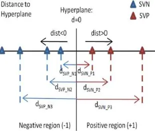
Fig. 7. Example of SVP_N and SVN_P distribution in terms of distance to hyperplane
Fig. 7 shows an example of SVP_N and SVN_P distribution in terms of distance to the hyperplane. Notice that the classifier places them in the wrong subspace. According to these wrongly classified SVs, it can be defined the security region where decisions are risky and no classification is performed. In this case, new values of PerrNeg and PerrPos are obtained. For instance, imagine that SVP_N1 is extracted from the classifier and no
decision is made for observations whose classifier output is between dSVP_N1 and the separation plane: in this
© 2013, IJCSMC All Rights Reserved
538
classifier will decrease the error rate, with the drawback of not classifying some observations whose SVM output is inside the security region and the decision is considered as too risky. In general, the new SVM classifier with bounds of confidence can be defined as follows.
1) Select the desired value of PerrNeg and PerrPos: PerrNegDesired, and PerrPosDesired, respectively.
2) Take all the SVs and obtain {SVP_N1,...., SVP_Nk1} and {SVN_P1,...., SVN_Pk2}.
3) Define ia for indexing the SVP_N and ib for indexing SVN_P. Set them initially to 1.
4) Compute the values of PerrNeg and PerrPos with the desired ones in step 1.
a) If PerrPos ≥PerrPosDesired , extract the SVP_N(ia): [dSVP_N(ia),0] is the security region of the negative region
(-1). Increase ia and compute again step 4a.
b) If PerrNeg ≥PerrNegDesired, extract SVN_P (ib): [0,dSVN_P(ib)] is the security region of positive region (+1).
Increase and compute again step 4b.
c) The SVM-based classifier is ready, with{+1,-1} for properly classified data and {-0.5,+0.5}for risky decisions. The use of bounds of confidence and the so-called “security zone” provokes that the new classifier turns the old binary output of the classifier (which was {+1,-1}) into in a new four-cases output {+1, +0.5, -0.5, -1}, where {+1,-1} have the same meaning than in the old classifier and {+0.5,-0.5} are related to observations remaining in the +1 or -1 side of the hyperplane, respectively, but too risky to be defined as class +1 or -1. It is clear that, as the values of PerrPos and PerrNeg are defined when considering the SVs as the evaluation set, these
values are guaranteed a priori when considering an evaluation set of data different from the one in the training step. However, although the values of PerrPos and PerrNeg are not guaranteed, the existence of the security zone
increases the success rate of the classifier, at the expense of having some unclassified outputs.
III. RESULTS
Fig 8: Segmentation Accuracy
Fig 9: Performance evaluation of Diabetic Retinopathy
IV.CONCLUSION
© 2013, IJCSMC All Rights Reserved
539
process in a smooth way. The accuracy obtained is about 89%. It has been found that classification accuracy is high when compared to the existing method.
V. SCOPE FOR FURTHER STUDIES
SVM have shown promising results in object detection and recognition, content-based image retrieval, text recognition, biometrics, speech recognition and also used for regression. SVM became famous when using images as input, it gave accuracy comparable to neural-networks with hand-designed features in a handwriting recognition tasks.
REFERENCES
[1] Thomas Walter and Jean-Claude Klein, “Segmentation of Color Fundus Images of the Human Retina: Detection of the Optic Disc and the Vascular Tree Using Morphological Techniques.” ISMDA 2001, LNCS 2199, pp. 282-287, 2001
[2] C. Sinthanayothin, J. F. Boyce, T. H. Williamson, H.L. Cook, E. Mensah, S. Lal and D. Usher, “Automated Detection of Diabetic Retinopathy on Digital Fundus Images”, 2002 Diabetic UK. Diabetic Medicine, 19, 105-112.
[3] M. Niemeijer, J. Staal, B. van Ginneken, M. Loog and M.D. Abr`amoff. “Comparative Study of Retinal Vessel Segmentation Methods” on a New Publicly Available Database, SPIE Medical Imaging, 5370:648-656, 2004
[4] M. Foracchia, E. Grisan, and A. Ruggeri, “Detection of optic disc in retinal images by means of a geometrical model of vessel structure,” IEEE Trans. Med. Imag., vol. 23, no. 10, pp. 1189–1195, Oct. 2004.
[5] Claudia Kondermann, Daniel Kondermann and Michelle Yan “Blood Vessel Classification into Ateries and Veins in Retinal Images,” Proc. SPIE 6512, Medical Imaging 2007: Image Processing, 651247 March 02, 2007.
[6] A. H. A. R. Youssif, A. Z. Ghalwash, and A. R. Ghoneim, “Optic disc detection from normalized digital fundus images by means of a vessels’ direction matched filter,” IEEE Trans. Med. Imag., vol. 27, no. 1, pp. 11–18, Jan. 2008.
[7] Manuel Emilio Gengundez-Arias, Arturo Aquino, Jose Manuel Bravo, and Diego Marin “A Function for Quality Evaluation of Retinal Vessel Segmentations,” IEEE Trans. Med. Imag., vol. 31, no. 2, Feb 2012.