_____________________________________________________________________________________________________ *Corresponding author: E-mail: [email protected], [email protected];
www.sciencedomain.org
Material Selection for Constructing an Intraoral
Stent Used in Radiotherapy: Analysis of Density and
Structure
Lázara Joyce Oliveira Martins
1*, Ana Flávia Sanches Borges
2,
Gustavo Zanna Ferreira
1, Simone Zuquerato Sansavino
3,
Ana Tarsila Fonseca Siosaki
4, Americo Tabata
5and Paulo Sérgio da Silva Santos
11
Department ofStomatology, School of Dentistry of Bauru, University of São Paulo, Brazil.
2
Department of Operative Dentistry, Endodontics and Dental Materials, School of Dentistry of Bauru, University of São Paulo, Brazil.
3
Department of Medical Physics, Prof. Nair Araújo Antunes Oncologic Center, Brazil.
4
Department of Radiologist, Prof. Nair Araújo Antunes Oncologic Center, Brazil.
5
Department of Physics, School of Sciences of Bauru, São Paulo State University, Brazil.
Authors’ contributions
This work was carried out in collaboration between all authors. Authors LJOM and PSSS designed the study, wrote the protocol and wrote the first draft of the manuscript. Authors AFSB and GZF managed the literature searches. Authors SZS, ATFS and AT analyses of the study performed the radiotherapy and spectroscopy analysis. All authors read and approved the final manuscript.
Article Information
DOI: 10.9734/BJMMR/2016/26308 Editor(s): (1) Jingli Xu, College of Pharmacy, University of New Mexico, USA. (2)Masahiro Hasegawa, Department of Orthopaedic Surgery, Mie University Graduate School of Medicine, 2-174 Edobashi, Tsu City, Mie, 514-8507, Japan. Reviewers: (1) Vaishnavi Vedam, Asian Institute of Medicine, Science and Technology (AIMST) University, Malaysia. (2)Muhammad Kashif, University of Health Sciences, Lahore, Pakistan. (3)Anonymous, Greek Anticancer Institute, Saint Savvas Hospital, Athens, Greece. (4)Vidya Ajila, Nitte University, India. Complete Peer review History:http://sciencedomain.org/review-history/15279
Received 10th April 2016 Accepted 13th June 2016 Published 4th July 2016
ABSTRACT
Neck and head cancer is a very common disease. Radiotherapy is one of the treatments, and its side effects affect the patient’s quality of life because the radiation targets both neoplastic and healthy tissues. This study aimed to select material that exhibited the greatest number of
characteristics that are biocompatible with human tissue and that are strong enough to construct an intraoral stent. The stent would be used to mechanically isolate the palate, tongue, and mouth floor to prevent radiation in head and neck cancer cases requiring radiotherapy, thereby aiming to decrease the treatment’s side effects. The following materials were selected to be submitted to a first analysis by computed tomography: polyacetal (white and black), polymethylmethacrylate (PMMA), polyurethane, and polyvinyl chloride. By observing the density through Hounsfield unit (HU) analysis, the materials with HU values closest to that of water (HU = 0) were selected for the structural analysis after the radiotherapeutic protocol through micro Raman spectroscopy. After undergoing radiation, PMMA with HUs of 177 and 179 without structural modification had the best density results; this was verified by micro Raman spectroscopy. PMMA seems to be a promising material due to its density and structural integrity after the radiotherapeutic protocol.
Clinical Significance: The material with the HU value that is most compatible with the oral tissues
does not interfere with the action of x-ray beams or with the main function of mechanically isolating the oral tissues, thus reducing the side effects from radiotherapy, improving the patient’s quality of life after the treatment.
Keywords: Cancer; polymethylmethacrylate; radiotherapy; spectrometry.
1. INTRODUCTION
Head and neck cancer, particularly mouth cancer, is significantly prevalent worldwide. According to data from the National Cancer Institute of Brazil, in 2014, 11,280 new cases of mouth cancer were diagnosed in Brazil alone. The people most affected by mouth cancer are alcoholics, smokers, those infected by human papillomavirus, and those who have been exposed to excessive ultraviolet A [1].
The main treatments for head and neck cancer have been surgery, chemotherapy, and radiotherapy (with local effect). Although radiotherapy has the fundamental function of containing the tumour’s development and decreasing its extent, this therapy causes many side effects for the patient, such as radiodermatitis, oral mucositis, candidiasis, xerostomia, radiation caries, osteoradionecrosis, trismus, and loss of taste [2]. These side effects compromise the patient’s quality of life during and after the cancer treatment.
The most common side effect of radiotherapy for head and neck cancer is oral mucositis— ulcerations on the oral mucosa—which causes severe discomfort during feeding, directly and markedly affecting the patient’s quality of life [3].
Radiotherapy affects both neoplastic and healthy tissue because the tumour is very close to the surrounding (sound) tissue. Attempting to reduce the treatment’s side effects on healthy tissue, researchers have conducted studies to develop an intraoral stent [4-6]. The main characteristic of an intraoral stent is the mechanical separation of
the intraoral tissues: palate, tongue, and mouth floor [2,4,5].
Use of an intraoral stent eases treatment planning through the use of computerized tomography, decreases the extent of oral mucositis acquired during the treatment, and decreases the damage to the surrounding muscle tissues, taste buds, and salivary glands. During radiotherapy appointments, the oral tissue is immobilized, enabling more accurate dosimetry of the neoplastic tissue [4].
The material for intraoral stent construction should neither block the radiation nor irritate the oral mucosa. It should also be nontoxic, inert, thermomechanically resistant (because it requires sterilization by moist heat), and easy to handle (since the patient will use it many times) [4]. Moreover, cost is an important factor in making the intraoral stent affordable for most patients who require radiotherapy.
To prevent radiation blockage, the material should exhibit a density as close as possible to that of water. The radiation interacts differently depending on the characteristics of each medium; ideally, the material should interfere significantly with neither the absorption nor the scattering. Hounsfield units (HUs) are used in measuring the electronic density of the medium, which is related to the medium’s atomic number. Depending on the atomic number and the radiation energy, the type of interaction varies [7].
which were submitted to a protocol similar to that of the oral radiotherapy used to treat head and neck cancer.
2. MATERIALS AND METHODS
Four materials were selected: polyacetal (white and black), polymethylmethacrylate (PMMA), polyurethane, and polyvinyl chloride (PVC). Firstly, the materials were submitted to computerized tomography to verify the HUs and compare them with that of water (HU = 0). The tomographic phases were performed at a centre for cancer treatment. The statistical analysis used in this research is qualitative and descriptive of the results obtained. These four materials were chosen due to their common use in dentistry, their resistance, and their biological compatibility with patients’ mouths. Even today, no studies in the area have described the most suitable material for making intraoral stents.
2.1 Preanalysis of Density
Cylinders measuring 2 cm in diameter and 1 cm in thickness of each material polyacetal (white and black), PMMA, polyurethane, and PVC (n=2) were used. The specimens were placed over a 1-cm wax lamina to obtain a sufficient height for the radiation reach its maximum peak at the centre of the samples during the computerized tomography (Activion, Toshiba, Japan), which oscillated with a mean build-up of 1.5 cm (the build-up is the region prior to the point of maximum dose absorbed by the medium). When the ionizing radiation enters any medium, it interacts such that its point of maximum absorbed dose varies with the radiation energy; thus, a higher energy results in a deeper maximum dose [7]. The specimens were individually irradiated at 6 MV power inside a standard field of 10 × 10 cm with a dose rate of 500 UM/min (monitor unit) (Activion, Toshiba, Japan). The software (Eclipse, Varian, California,
USA) read the images to obtain the density measurements and to plan the radiotherapy.
2.2 Radiotherapeutic Protocol
The radiotherapeutic protocol was applied at the maximum dose for the head and neck areas (7000 cGy) with a dose rate of 300 cGy/min for 23.3 minutes. Next, the samples were stored in an incubator (Marconi, Piracicaba, SP, Brazil) at relative humidity and at 37ºC until the spectroscopy was performed.
2.3 Micro Raman Spectroscopy
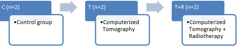
The studied material, PMMA (n=6) was divided according to the groups assigned in Figs. 1 and 2.
Five (micrometre-scale) measurements were executed using a Raman spectroscope consisting of a triple monochromator (Jobin Yvon model T64000) with 64 cm of focal distance. The first two monochromators were used as the laser line filter, and the third monochromator was used as the spectrographic stage. The Raman sign excitation was carried out using a laser beam with a wavelength of 488.0 nm and a power of about 10 mW (Laser Spectra-Physic model Stabilite 2070). To eliminate the laser plasma lines, an interferometer filter (Newport) was used. A configuration known as retro-scattering, in which the incident and scattered laser beams are in the same direction (with the sample surface forming a 90º angle), was used. To focus the laser beam, a microscope (Olympus model BX40) was used with ×50 lenses, enabling a beam of diameter of approximately 10 µm. The detection of the Raman sign was executed using a CCD camera (Jobin Yvon model SpectraOne) refrigerated with liquid nitrogen. The entrance slit 500 µm, and the device resolution was approximately 5 cm–1.
Fig. 1. Experimental conditions for each material
Fig. 2. Sample PMMA specimens for three groups
C indicates the control group (no treatment); T, computerized tomography; T+R, computerized
tomography + radiotherapy
The micro Raman spectroscopy created a light beam, which passed through a semitransparent mirror, scattering part of the light to a microscope, where the beam was focused on the sample (horizontal axis). The scattered light beam (Raman sign) came back through the microscope and was reflected by the semitransparent mirror and scattered towards the monochromator (vertical axis). Because part of the incident laser beam was also reflected to the monochromator through the semitransparent mirror, this was mixed with the Raman sign. Accordingly, monochromators 1 and 2 were used to filter the laser beam to produce the Raman sign at the spectrographic stage. Next, the Raman sign was registered by the CCD camera.
This research was conducted in 2014 at three research centres: University of São Paulo, São Paulo State University and the Prof.Nair Araújo Antunes Oncologic Center - all in São Paulo, Brazil.
3. RESULTS
The results obtained at the first phase of the study through the measurement of the samples’ HU values indicated that the highest values were found for PMMA, which had values closest to that of water (HU = 0).
Table 1. The density of each material, as indicated by HU (n=2)
Material Material density
(HU value) sample 1 and 2
Polymethylmethacrylate 177 and 179 Polyacetal (white) 385 and 487 Polyacetal (black) 441 and 444 Polyurethane 765 and 771
PVC 1167 and 1215
3.1 Micro Raman Spectroscopy
The Raman spectroscopy exhibited a peak series corresponding to the number of the waves
in the characteristic bands of the PMMA polymer, with main values of 366, 497, 606, 818, 978, 1127, 1195, 1464, and 1733 cm–1 (landmarks). No statistical differences were seen among groups C (control without treatment), T (tomography), and T+R (tomography followed by radiotherapy). By analysing the quality of the bands in relation to the bandwidth and the appearance of new bands, no statistically significant differences were found, suggesting that the radiotherapeutic treatment after computed tomography did not provoke alterations in the polymer’s molecular arrangement (Fig. 3).
Fig. 3. Spectra of polymethylmethacrylate according to the treatments. The x-axis shows the waves with 366, 497, 606, 818, 978,
1127, 1195, 1464, and 1733 cm–1, which are related to the band characteristics of the polymer identified by the Raman sign. The
y-axis shows the intensity (an arbitrary number)
4. DISCUSSION
The density of the materials should be as close as possible to that of water because the intraoral stent should interfere with neither radiation absorption nor scattering. This feature was only verified for one of the four materials tested: PMMA.
lead to insignificant density variation over the treatment period [8].
PMMA presents as a powder (containing acrylic polymers or copolymers, benzoyl peroxide, pigments [TiO2], colour opacifiers, plasticizers,
and coloured organic fibres) or as a liquid (containing methyl methacrylate, hydroquinone, accelerators, plasticizers, and ethylene glycol dimethacrylate). The material has few residual contaminants and is of low cost [8]. Structurally, it is a semicrystalline material, which characterizes a certain periodicity of its molecular arrangement, as identified by Raman sign (Willis et al., 1968; Smit et al. [9]). The materials can be identified by examining the peaks, resulting in the corresponding number of waves. The number of waves for each peak band was attributed to the PMMA spectrum by Willis et al. (1968) using a comparison with infrared spectroscopy:
602aν(C–COO), νs (C–C–O); 853a ν(CH2); 925b
ν(CH2); 999a O–CH3; 1081a ν(C–C); 1264a
ν(C–O), ν(C–COO); 1460a δa(C–H) ofα-CH3,
δa(C–H) of O–CH3. There are 1,648
combinations of bands involving ν(C=C) and
ν(C–COO); 1736a ν(C=O) of (C–COO). Also in that study, the authors classified the identified bands as strong, medium, weak, polarized, or nonpolarized signs. The number of waves for the peaks identified in the present study demonstrated similar values to those of the peaks reported in the literature: 366, 497, 606, 818, 978, 1127, 1195, 1464, and 1733 cm–1. In Raman spectroscopy, the number of waves does not need to be exact, as small variations can occur without mischaracterizing the signs.
The qualitative characteristics of the bands can be analyzed by the superposition, intensity, shape, and standard inside each spectrum [9]. In the analysis of the band characteristics of PMMA, identified by the number of waves and peaks, there were no qualitative differences among the spectra of each group, suggesting that the radiotherapeutic treatment did not provoke modifications in the polymer’s molecular arrangement polymer. The semicrystalline structure means that the atoms are arranged at certain periodicities inside the molecules (that is, the monomers). On the other hand, the monomers are linked by the so-called polymerization process, which results in rigidity and enables the periodicity to be detected by the number of waves of the peaks, as was previously
identified by Willis et al. (1968) and Smit et al. [9]. Conversely, a modification strong enough to change the PMMA band characteristics occurs, as in the present study (radiotherapeutic protocol: 7000 cGy with a dose rate of 300 cGy/min for 23.3 minutes), the differences can be detected [9,10].
To obtain good quality in PMMA polymerization, it is necessary that the polymer and monomer be mixed at the specific ratio described by the manufacturer to avoid internal and external porosity in the stent, which will lead to a less mechanically resistant device. Avoiding temperatures above the boiling point of the liquid during the polymerization process decreases the material’s porosity, which suggests further challenges when PMMA stents require sterilization.
PMMA material has enough features to be used in intraoral stents that patients will use in planning and in all radiotherapy treatment sessions. PMMA is inert and tough, and it neither blocks radiation nor causes harm to patients during radiotherapy. Thus, healthy tissue from around the tumour region will not be affected, reducing the side effects of radiotherapy.
5. CONCLUSIONS
PMMA showed the ideal density for use in intraoral stents because, after the radiotherapeutic protocol, it did not exhibit structural alterations.
CONSENT
It is not applicable.
ETHICAL APPROVAL
It is not applicable.
ACKNOWLEDGEMENTS
The authors would like to thank Prof. Nair Araújo Antunes Oncologic Center, Bauru, SP, Brazil, at where the radiation protocol of this study took place.
COMPETING INTERESTS
REFERENCES
1. Instituto Nacional Do Câncer José Alencar Gomes Da Silva. Estimativa: Incidência de câncer no Brasil. Rio de Janeiro: Inca. 2014;124. Brazilian.
2. Verrone JR, Alves FA, Prado JD, Boccaletti KW, Sereno MP, Silva MLG, Jaguar GC. Impact of intraoral stent on the side effects of radiotherapy for oral cancer. Head Neck. 2013;35(7):E213-7.
DOI: 10.1002/hed.23028 Epub 2012 Jun 19
3. Huber MA, Terezhalmy GT. The head and neck radiation oncology patient. Quintessence International. 2003;34(9): 693-717.
4. Johnson B, Sales L, Winston A, Liao J, Laramore G, Parvathaneni U. Fabrication of customized tongue-displacing stents: Considerations for use in patients receiving head and neck radiotherapy. J Am Dent Assoc. 2013;144(6):594-600. 5. Kaanders JHAM, Fleming TJ. Devices
valuable in head and neck radiotherapy.
Int J Radiat Oncol Biol Phys. 1992;23(3):639-45.
6. Wang RR, Levona WO. A direct method for fabricating tongue- shielding stent. J Prosthet Dent. 1995;74(2):171-3.
7. Khan FM. The physics of radiation therapy. 5a ed. Lippincott Williams & Wilkins. 2014;624.
8. Jha KC, Zhu H, Dhinojwala A, Tsige M. Molecular structure of poly(methyl methacrylate) surface II: Effect of stereoregularity examined through all-atom molecular dynamics. Langmuir. 2014; 30(43):12775-85.
DOI: 10.1021/la5023328 Epub 2014 Oct 24
9. Smit EE, Erckens RJ, Hendrikse F, Motamedi M, Wicksted JP, March WF. Identification of intraocular lens materials using confocal Raman spectroscopy. J Cataract Refract Surg. 1999;25(11):1498-504.
10. Willis HA, Zichy VJI and Hendra PJ. The laser-Raman and infra-red spectra of polymethyl methacrylate. Polymer. 1969; 10:737–46.
_________________________________________________________________________________ © 2016 Martins et al.; This is an Open Access article distributed under the terms of the Creative Commons Attribution License (http://creativecommons.org/licenses/by/4.0), which permits unrestricted use, distribution, and reproduction in any medium, provided the original work is properly cited.
Peer-review history: