1071-412X/05/$08.00⫹0 doi:10.1128/CDLI.12.6.727–735.2005
Copyright © 2005, American Society for Microbiology. All Rights Reserved.
Antibody Responses in Reindeer (
Rangifer tarandus
) Infected with
Mycobacterium bovis
W. R. Waters,
1* M. V. Palmer,
1J. P. Bannantine,
1R. Greenwald,
2J. Esfandiari,
2P. Andersen,
4J. McNair,
3J. M. Pollock,
3and K. P. Lyashchenko
2United States Department of Agriculture, Agricultural Research Service, National Animal Disease Center, Bacterial
Diseases of Livestock Research Unit, Ames, Iowa1; Chembio Diagnostic Systems, Inc., Medford, New York2;
Bacteriology Department, Veterinary Sciences Division, Stormont, Belfast, United Kingdom3; and
Statens Serum Institut, Copenhagen, Denmark4
Received 9 March 2005/Returned for modification 19 March 2005/Accepted 5 April 2005
Despite having a very low incidence of disease, reindeer (Rangifer tarandus) are subject to tuberculosis (TB) testing requirements for interstate shipment and herd accreditation in the United States. Improved TB tests are desperately needed, as many reindeer are falsely classified as reactors by current testing procedures. Sera collected sequentially from 11 (experimentally)Mycobacterium bovis-infected reindeer and 4 noninfected rein-deer were evaluated by enzyme-linked immunosorbent assay (ELISA), immunoblotting, and multiantigen print immunoassay (MAPIA) for antibody specific toM. bovisantigens. Specific antibody was detected as early as 4 weeks after challenge withM. bovis. By MAPIA, sera were tested with 12 native and recombinant antigens, which were used to coat nitrocellulose. AllM. bovis-infected reindeer developed responses to MPB83 and a fusion protein, Acr1/MPB83, and 9/11 had responses to MPB70. Other antigens less commonly recognized included MPB59, ESAT-6, and CFP10. Administration of purified protein derivatives for skin testing boosted serum antibody responses, as detected by each of the assays. Of the noninfected reindeer, 2/4 had responses that were detectable immediately following skin testing, which correlated with pathological findings (i.e., presence of granulomatous lesions yet the absence of acid-fast bacteria). The levels of specific antibody produced by infected reindeer appeared to be associated with disease progression but not with cell-mediated immunity. These findings indicate thatM. bovisinfection of reindeer elicits an antibody response to multiple antigens that can be boosted by skin testing. Serological tests using carefully selected specific antigens have potential for early detection of infections in reindeer.
Mycobacterium bovis infection of reindeer (Rangifer
taran-dus) is rare, especially in North America, where there are no published reports of the occurrence of tuberculosis (TB) in this species. Despite the low incidence of disease, reindeer are subject to regulations in the United States Department of Agriculture (USDA)Bovine Tuberculosis Eradication Uniform
Methods and Rules(29), requiring testing for interstate
move-ment and herd accreditation. For TB surveillance of reindeer within the United States, a single cervical test (a measure of delayed-type hypersensitivity) is the primary test and the com-parative cervical skin test (CCT) is used to confirm infection. Although exact numbers are difficult to ascertain, many rein-deer have tested positive by skin testing for TB surveillance. Strains ofMycobacterium bovis or other Mycobacterium spp. within theMycobacterium tuberculosiscomplex, however, have not been isolated from these reindeer upon necropsy. Reasons for the high rate of false-positive reactions elicited by skin testing are unclear, although it may be due to unusual exposure to nontuberculousMycobacteriumspp. or an exaggerated cel-lular immune response to mycobacterial antigens. Therefore, improved TB tests are urgently needed to avoid the unneces-sary slaughter of reindeer falsely identified as TB reactors.
For TB surveillance of captive wildlife (e.g., zoos or game
farms) and nontraditional livestock, blood-based TB assays are appealing, as they require a single handling event, thereby minimizing capture-associated injuries. Blood-based assays are also more readily used in capture surveillance programs with free-ranging wildlife (e.g., white-tailed deer [Odocoileus
virgin-ianus] in Michigan or Cape buffalo in Africa). While in vitro
cell-based assays appear promising, they require processing of the blood sample within 24 h and are subject to complications inherent with overnight delivery (e.g., temperature fluctuations and delays in setup). For these reasons, serological tests are particularly attractive for use in TB surveillance of nontradi-tional livestock and wildlife. A major limiting factor for the development of serological TB assays, particularly for rein-deer, is the lack of specific information about antigens recog-nized by antibodies that are produced during disease. Thus, experimentalM. bovisinfection studies are necessary to char-acterize the humoral immune response and to identify the most reactive antigens that could be employed in serodiagnos-tic tests.
Previous studies with cattle and white-tailed deer have vealed both similarities and differences in the antibody re-sponses againstM. bovisinfection of these two species (17, 18, 34). In particular, antigen recognition patterns appear to differ from animal to animal in both species, and antibody levels are significantly elevated shortly after the intradermal tuberculin injection(s) for skin testing. Little, if anything, is known con-cerning antibody responses of reindeer toM. bovisinfection.
The present study describes the humoral response of
rein-* Corresponding author. Mailing address: United States Depart-ment of Agriculture, Agricultural Research Service, National Animal Disease Center, P.O. Box 70, Ames, IA 50010-0070. Phone: (515) 663-7756. Fax: (515) 663-7458. E-mail: rwaters@nadc.ars.usda.gov.
727
on August 17, 2020 by guest
http://cvi.asm.org/
deer to experimental infection withM. bovis. Specific objec-tives were to determine (i) antigen recognition patterns by serum antibodies, (ii) relationships of the humoral responses with cell-mediated immunity and disease progression, (iii) the effect of tuberculin skin testing on the antibody response, and (iv) the potential for serological tests in TB surveillance pro-grams for reindeer.
MATERIALS AND METHODS
Animals, challenge, and necropsy.Thirty-four castrated male reindeer ( Ran-gifer tarandus) of approximately 9 months of age were obtained from a TB-free herd in Michigan and housed at the National Animal Disease Center, Ames, Iowa, according to institutional guidelines and approved animal care and use protocols. Eleven animals were experimentally infected withM. bovis, and four animals were kept noninoculated as a control group. ForM. bovisinfection, the challenge inoculum (105
CFU in 0.2 ml of phosphate buffered saline [PBS], pH 7.2) was instilled directly into the tonsillar crypts of anesthetized reindeer (n⫽
11) as described for the inoculation of white-tailed deer (21). The strain ofM. bovisused for the challenge inoculum (95-1315 [USDA, Animal Plant and Health Inspection Service {APHIS} designation]) was originally isolated from a white-tailed deer in Michigan (24). Inoculum consisted of mid-log-phaseM. bovis
cells grown in Middlebrook’s 7H9 medium supplemented with 10% oleic acid-albumin-dextrose complex (Difco, Detroit, Michigan) plus 0.05% Tween 80 (Sigma Chemical Co., St. Louis, Missouri). At the time of inoculation, reindeer were moved from an outdoor pen into climate-controlled rooms (two to three animals/room) within a biosafety level 3 confinement facility. Negative airflow exited the building through HEPA filters, ensuring that air from animal pens was pulled towards a central corridor and through HEPA filters before exiting the building. The airflow velocity was 10.4 air changes/h. Four noninoculated rein-deer were housed in a climate-controlled room in a building (biosafety level 2 facility) separate from the building in which the infected reindeer were housed. Additionally, serum samples from 19 reindeer housed outdoors at the National Animal Disease Center were obtained.
Thirteen months after inoculation, reindeer in the infected (n⫽11) and noninoculated (n⫽4) groups were euthanized by an intravenous injection of sodium pentobarbital (Fort Dodge Animal Health, Fort Dodge, Iowa) and ex-amined. Various tissues were collected for bacteriologic culture and microscopic examination. Detailed descriptions of cellular immune responses (35), bacterio-logic culture, histopathology, and gross necropsy results are presented elsewhere (23).
CCT.Ninety days afterM. bovisinoculation, reindeer were tested for in vivo responsiveness to mycobacterial antigens by a modified CCT technique enabling the collection of biopsy specimens for which the dermal reactions to purified protein derivatives (PPDs) at 24, 48, and 72 h postinjection could be analyzed (23, 33). Briefly, the cervical region was clipped and animals injected intrader-mally in three separate locations withM. bovisPPD and a single location with
Mycobacterium aviumPPD (PPDs obtained from National Veterinary Services Laboratory, Ames, Iowa). A standard CCT (i.e., single intradermal injection each ofM. aviumPPD andM. bovisPPD) was performed 8 months afterM. bovis
inoculation (22, 29). For each of the skin tests, 100g ofM. bovisPPD and 40
g ofM. aviumPPD were administered at each respective location according to guidelines described in USDA, APHIS circular 91-45-011 (29). Skin thickness at each injection site was measured prior to injection of PPDs and 72 h after administration. Changes in skin thickness were calculated and responses plotted on a scattergram developed by USDA for the interpretation of the CCT for bison, cattle, and cervidae (Form VS-6-22D). Responses by individual reindeer within both infected and noninfected groups are presented elsewhere (23).
Enzyme-linked immunosorbent assay (ELISA).Lipoarabinomannan (LAM)-enriched mycobacterial antigen was prepared fromM. bovisstrain 95-1315 as described previously (31). Briefly, bacilli harvested from 4-week cultures were sonicated in PBS, further disrupted with 0.1- to 0.15-mm glass beads (Biospec Products, Bartlesville, Oklahoma) in a bead beater (Biospec Products), centri-fuged, filtered (0.22-m-pore-size filter), and digested in a 1-mg/ml proteinase K (Roche Molecular Biochemicals, Indianapolis, Indiana) solution (50 mM Tris, 1 mM CaCl2buffer, pH 8.0) for 1 h at 50°C. Protein concentrations were deter-mined (Bio-Rad, Hercules, California) and antigen stored at⫺20°C.
Immulon II 96-well microtiter plates (Dynatech, Chantilly, Virginia) were coated with 100l/well (4g) antigen diluted in carbonate/bicarbonate coating buffer (pH 9.6). Antigen-coated plates, including control wells containing coating buffer alone, were incubated for 15 h at 4°C. Plates were washed three times with
200l/well PBS containing 0.05% Tween 20 (PBST) (Sigma), and blocked with 200l/well of a commercial milk diluent/blocking solution (Kirkegaard and Perry Laboratories, Gaithersburg, Maryland). After incubation for 1 h at 37°C in the blocking solution, wells were washed nine times with 200l/well PBST and test sera added to wells (100l/well). Test and control sera were diluted 1:100 in PBS containing 0.1% gelatin. After incubation for 20 h at 4°C with diluted test sera, wells were washed nine times with 200l/well PBST and incubated for 1 h at 37°C with 100l/well of biotin-protein G (Sigma) diluted 1:5,000 in PBS plus 0.1% gelatin. Wells were washed nine times with 200l/well PBST and incubated for 1 h at 37°C with 100 l/well of peroxidase-streptavidin (Sigma) diluted 1:2,000 in PBS plus 0.1% gelatin. Wells were washed nine times with 200l/well PBST and incubated for 4 min at room temperature with 100l/well of SureBlue 3,3⬘,5,5⬘-tetramethyl benzidine nsrsid6847181\delrsid6847181\charrsid6847181
microwell peroxidase substrate (Kirkegaard and Perry Laboratories). The reac-tion was stopped by the addireac-tion of 100l/well of 0.18 M sulfuric acid and the absorbances (450 nm) of individual wells measured using an automated ELISA plate reader (Molecular Devices, Menlo Park, California). Data are presented as the changes in optical density readings calculated by subtracting the mean optical density readings for wells receiving coating buffer alone (two replicates) from the mean optical density readings for antigen-coated wells (two replicates) receiving the same serum sample.
Immunoblot assay.Electrophoresis and immunoblot assays were performed using previously described procedures (2) with the following modifications. The antigen for immunoblotting was a whole-cell sonicate (WCS) ofM. bovisstrain 95-1315. Following standard culture, mycobacteria were pelleted (10,000⫻gfor 20 min) and washed twice with cold PBS. Pellets were resuspended in PBS and sonicated on ice with a probe sonicator. Sonication consisted of three 10-min bursts at 18 W on ice, with 10-min chilling periods between sonications. Debris was removed by centrifugation (12,000⫻gfor 5 min) and supernatants harvested and stored at⫺20°C. Protein concentrations were determined by using a protein assay (Bio-Rad, Richmond, CA). Comparisons of the reactivities of serial serum samples against WCS antigen were conducted using a slot-blotting device (Bio-Rad, Richmond, CA). Antigen (200g) was electrophoresed through prepara-tive 12% (wt/vol) polyacrylamide gels and transferred to nitrocellulose filters. These filters were placed in a blocking solution consisting of PBST and 2% (wt/vol) bovine serum albumin (PBST-BSA). After the blocking, the filters were placed into the slot blot device, and individual sera, diluted 1:200 in PBST-BSA, were added to independent slots. After a 2-h incubation with gentle rocking, blots were washed three times with PBST and incubated with horseradish per-oxidase-conjugated protein G (Sigma) diluted 1:40,000 in PBST-BSA for 1.5 h. Blots were again washed three times with PBST and developed for chemilumi-nescence in SuperSignal detection reagent (Pierce Chemical Co.).
Multiantigen print immunoassay (MAPIA).The following recombinant anti-gens ofM. boviswere purified to near homogeneity as polyhistidine-tagged proteins (Rv numbers according to the classification of Cole et al. [3] in paren-theses): ESAT-6 (Rv3875) and CFP10 (Rv3874) produced at the Statens Serum Institut, Copenhagen, Denmark; MPB59 (Rv1886c), MPB64 (Rv1980c), MPB70 (Rv2875), and MPB83 (Rv2873) (produced at the Veterinary Sciences Division, Stormont, Belfast [15]). Alpha-crystallin (Acr1) (Rv3391) and the 38-kDa pro-tein PstS1 (Rv0934) were purchased from Standard Diagnostics, Seoul, South Korea. Polyprotein fusions CFP10/ESAT-6 and Acr1/MPB83 were constructed by overlapping PCR using gene-specific oligonucleotides to amplify the genes fromM. tuberculosisH37Rv chromosomal DNA. The fused polygene PCR prod-ucts were cloned into the pMCT6Escherichia coliexpression vector by using SmaI/BamHI restriction enzymes. The polyproteins were purified to near homo-geneity by exploiting the polyhistidine tag encoded by the vector.M. bovisculture filtrate (MBCF) was obtained from a field strain ofM. bovis(T/91/1378; Veter-inary Sciences Division, Belfast, United Kingdom) cultured in synthetic Sauton’s medium. MAPIA was performed as described previously (19). Briefly, antigens were immobilized on nitrocellulose membrane (Schleicher & Schuell, Keene, NH) at a protein concentration of 0.05 mg/ml by using a semiautomated air-brush-printing device (Linomat IV; Camag Scientific Inc., Wilmington, DE). The membrane was cut, perpendicular to the antigen bands, into 4-mm-wide strips. Strips were blocked for 1 h with 1% nonfat skim milk in PBS with 0.05% Tween 20 and then incubated for 1 h with serum samples diluted 1:40 in blocking solution. After being washed, strips were incubated overnight with horseradish peroxidase-conjugated protein G (Sigma, St. Louis, MO) diluted 1:1,000, fol-lowed by another washing step. Deer antibodies bound to printed antigens were visualized with 3,3⬘,5,5⬘-tetramethyl benzidine (Kirkegaard and Perry Laborato-ries, Gaithersburg, MD).
Statistics.Data were analyzed by repeated measures (one-way analysis of variance followed by a Tukey-Kramer multiple comparisons test, using a com-mercially available statistics program [InStat 2.00; GraphPAD Software, San
on August 17, 2020 by guest
http://cvi.asm.org/
Diego, CA]). Pearson’s product-moment correlations between MAPIA and pa-thology scores were computed.
RESULTS
Infection status.Inoculation ofM. bovisinto tonsillar crypts of reindeer resulted in gross and microscopic lesions in all inoculated animals (23). Tuberculous lesions were most prom-inent in medial retropharyngeal lymph nodes and were also detected in tonsils, mesenteric lymph nodes, lungs, and lung-associated lymph nodes. Infected reindeer did not have clinical signs of TB or disseminated disease after 1 year of coloniza-tion, suggesting low-grade chronic infection.M. boviscultures were isolated from tissues of 6/11 infected animals but not
from control reindeer; however, granulomatous lesions with no acid-fast bacteria were detected within the tracheobronchial lymph nodes of 2/4 noninoculated reindeer (Table 1).
Serological response to LAM-enriched antigens and WCS.
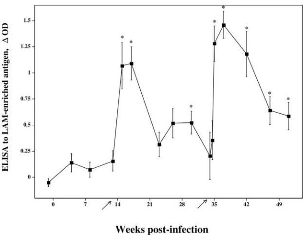
Antibodies toM. bovisLAM were detected by ELISA in sera taken 15 weeks after challenge withM. bovis, 2 weeks after the injection of PPDs for skin testing (Fig. 1). By 22 weeks post-challenge, responses to LAM had waned and were again boosted upon the injection of PPDs for the second skin test. The two noninoculated reindeer that had granulomatous le-sions also had responses toM. bovisLAM that were detectable after the skin test (Table 1).
By the immunoblotting ofM. bovisWCS, responses by in-fected reindeer were detected as early as 8 weeks postchal-lenge (Fig. 2). Specific bands of reactivity at⬍15,⬃25,⬃32,
⬃42, ⬃75, and ⬃100 kDa were detected. Responses were boosted by the injection of PPDs for the skin test. As was the case with the LAM ELISA, injection of PPDs for the skin test elicited a response detectable by the immunoblotting of sam-ples from two of the four noninoculated reindeer (animals 123 and 129), although the response was detectable only after the second skin test for animal 123. Immunoblot responses (i.e., intensity and number of bands) by 10/11 infected reindeer greatly exceeded the detected responses in samples from the two responding noninoculated reindeer. The two noninocu-lated reindeer had only a single, light band of reactivity at⬃25 kDa, whereas sera from infected reindeer elicited multiple, heavy bands of reactivity upon immunoblotting.
Serological response to purified proteins.To further char-acterize the humoral immune response, the MAPIA was used to determine responses to a panel of recombinantM.
tubercu-losis/M. bovisproteins. Antigens recognized by sera from M.
FIG. 1. Response kinetics of serum antibody specific for LAM-enriched antigen. Sera fromM. bovis-infected reindeer (■; n⫽ 11) were analyzed for reactivity toM. bovis-derived LAM by ELISA. Data are presented as mean (⫾standard errors of the mean) changes in optical density (⌬OD). Arrows on thexaxis indicate time points when purified protein derivatives were injected for skin testing (CCT). Asterisks (*) indicate responses that exceed (P⬍0.05) preinfection responses (i.e., week⫺1).
TABLE 1. Comparison of pathologies and serologies from noninoculated reindeer
Animal
ELISA antibody response (⌬OD)a
Pathologyb
Day⫺7 Post-CCT
123 ⫺0.021 0.891 g, m
124 ⫺0.626 0.023 —
129 ⫺0.066 0.478 g, m
132 0.023 ⫺0.013 —
aChange in optical density (⌬OD) (response toM. bovis-derived
lipoarabino-mannan-enriched antigen minus the response to no antigen) by sera from non-inoculated reindeer at the initiation of the study (day⫺7) and 2 weeks after injection of purified protein derivative for the initial comparative cervical test (post-CCT) 16 weeks later.
bg, gross lesions consistent with tuberculosis; m, caseonecrotic granuloma(s)
seen upon microscopic examination (Mycobacterium boviswas not identified by culture or in situ PCR and no acid-fast bacteria present in lesions); —, gross or microscopic lesions not detected.
on August 17, 2020 by guest
http://cvi.asm.org/
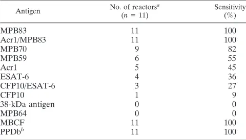
bovis-infected reindeer included MPB83, MPB70, MPB59, Acr1, ESAT-6, CFP10, and fusion proteins ESAT-6/CFP10 and Acr1/MPB83, although not all antigens were recognized at each time point and some antigens were not recognized by sera from each animal (Table 2; Fig. 3). For comparison, two com-plex antigens (M. bovisPPD and MBCF) were included in the assay and were recognized, at least at some time point, by each
M. bovis-infected animal. Responses by infected reindeer were
detected as early as 4 weeks postchallenge by MAPIA (Fig. 3B). With animal 126, MPB83 and ESAT-6 were both early antigenic targets with reactivities to each of the single proteins
and their respective fusion proteins (Fig. 3B). As with the ELISA and immunoblotting, the intradermal tuberculin injec-tion for skin testing boosted responses of infected reindeer to several antigens (Fig. 4). Serum antibodies against a limited number of antigens (MPB83 and/or MBCF) were elicited by skin testing in 2/4 noninoculated reindeer (animals 123 and 129) (Fig. 4A). However, the detected antibody levels in sam-ples from the experimentally infected reindeer consistently exceeded the detected antibody levels in samples from the two responding noninoculated reindeer (Fig. 4).
Specificity of MAPIA. As indicated, responses in samples from 2/4 noninoculated reindeer after the skin test were de-tected, indicating prior sensitivity to cross-reactive antigens. To evaluate the specificity of MAPIA, samples from noninfected reindeer were evaluated. These samples included sera ob-tained from reindeer in the infected group (n⫽11) and the noninoculated group (n⫽4) at the preinoculation time point as well as samples collected from 19 reindeer that were part of another study. Serum antibodies against a limited number of antigens (5/34 responded to MPB83, 2/34 responded to ESAT-6, and 1/34 responded to MBCF) were detected and indicative of relative specificities of 85%, 91%, and 97% for MPB83, ESAT-6, and MBCF, respectively. Further studies to evaluate a larger sample size (i.e.,⬎200 reindeer serum sam-ples) from a wide range of locations (i.e., Alaska to Tennessee) within the United States to better estimate the specificity of MAPIA are under way.
Association of antibody responses with pathology and bac-teriology findings.Individual serological responses character-ized by MAPIA appeared to correlate with pathological and bacteriologic results. To provide this comparison, infected re-indeer were divided into three subgroups, strong, moderate, and weak antibody reactors, based on magnitude of the hu-moral responses and number of antigens recognized in MAPIA (Table 3). Additionally, MAPIA and pathology scores were calculated based upon MAPIA band intensities/numbers and necropsy findings, respectively. By use of linear correlation analysis, a positive correlation between MAPIA and pathology scores was detected (r⫽0.68;P⫽0.02) (Table 3). All infected animals had positive histopathology results (i.e., presence of granulomatous lesions with acid-fast organisms present), and most of them had gross lesions consistent with TB found in at least one tissue specimen. Each subgroup of antibody respond-ers had one animal without gross lesions. Among the strong antibody responders, animal 128 did not have gross lesions; however, this animal was euthanized 7 months postinfection, or 6 months earlier than the other infected reindeer. Further,
M. bovisculture was recovered from tissues of 3/3 strong
an-tibody responders, 0/5 moderate anan-tibody responders, and 1/3 weak antibody responders. Intriguingly, there were only two animals withM. bovisstrains isolated from the lungs, and both were found among the group of strong antibody responders (i.e., animals 121 and 128). Also, the subgroups clearly differed by extent of lesions andM. bovisreplication in various tissues (Table 3) so that the strong antibody responders had a greater number of tissues with lesions and/orM. bovis than did the other two subgroups.
Antibody responses and cell-mediated immunity.The sero-logical findings obtained for each animal in the present study were compared with skin test results and gamma interferon
FIG. 2. Preparative immunoblots ofM. bovisstrain 95-1315 WCS antigen probed with sera from a reindeer experimentally infected with M. bovis. Molecular mass markers are indicated in the left margin (in kDa) and weeks postinfection in the lower margin. Arrows in the lower margin indicate time points when purified protein derivatives were injected for skin testing (CCT). Symbols in the lower left margin refer to sera from known noninfected (⫺) and infected (⫹) white-tailed deer used as controls for the assay. Numbers in the lower margin refer to weeks relative to infection. Immunoblots were performed on sam-ples from each reindeer at all time points indicated. Responses pre-sented in this figure are representative of 10/11 of the infected rein-deer. One infected reindeer (animal 125) had only minimal responses detectable by immunoblot analysis.
TABLE 2. Rates of antibody responses to protein antigens detected by MAPIA in reindeer infected withMycobacterium bovis
Antigen No. of reactors
a
(n⫽11)
Sensitivity (%)
MPB83 11 100
Acr1/MPB83 11 100
MPB70 9 82
MPB59 6 55
Acr1 5 45
ESAT-6 4 36
CFP10/ESAT-6 3 27
CFP10 1 9
38-kDa antigen 0 0
MPB64 0 0
MBCF 11 100
PPDbb 11 100
aResponses are indicative of rates of antigen recognition for each animal
measured at various time points postinfection.
bPPDb,M. bovispurified protein derivative.
on August 17, 2020 by guest
http://cvi.asm.org/
(IFN-␥) responses that are described in detail elsewhere (23, 35). All 11 infected reindeer tested positive by CCT (APHIS Form VS-6-22D) for the interpretation of CCT for bison, cat-tle, and cervidae, using either possible interpretation (23), per-formed 3 and 8 months afterM. bovisinoculation. No
corre-lation between the skin test reactions obtained withM. bovis
PPD and the levels of serum immunoglobulin responses de-tected by ELISA or MAPIA was found. In particular, at 3 months postinfection, the strongest MAPIA responders (ani-mals 121, 126, and 128) produced skin test reactions (changes
FIG. 3. Antibody responses to recombinant antigens detected by MAPIA in reindeer experimentally infected withM. bovis. Arrows in the upper margin indicate time points when purified protein derivatives were injected for skin testing (CCT). Antigens printed are shown in the right margin. Strips represent different time points during infection when serum samples were collected (shown in weeks in the lower margin). Representative responses by two differentM. bovis-infected animals, animal 120 (A) and animal 126 (B), are provided to demonstrate the variability in antigen recognition patterns.
on August 17, 2020 by guest
http://cvi.asm.org/
in skin thickness) ranging from 17.5 mm to 24.9 mm, while the weakest MAPIA responders (animals 125, 127, and 136) pro-duced skin test reactions ranging from 12.9 mm to 19.3 mm, demonstrating no significant difference between the extreme subgroups. Further, the weakest CCT reactor, animal 135 (3.0 mm), and the strongest CCT reactor, animal 131 (25.0 mm),
had essentially the same magnitude of serological response (Fig. 4). Similarly, there was no correlation between humoral responses and in vitro IFN-␥ production to M. bovis PPD stimulation (data not shown). Interestingly, animal 126, the only strong and early ESAT-6 antibody responder (Fig. 3), did not have a prominent in vitro IFN-␥response to this antigen.
FIG. 4. Effects of injection of PPDs for skin testing on antibody responses (as measured by MAPIA) by noninfected (A) and infected (B) reindeer toM. bovisantigens. Reindeer were injected with PPDs for skin testing 3 months after challenge withM. bovisand sera collected at indicated time points for analysis of antibody. Numbers in the upper margins indicate animal identification numbers. Numbers in lower margins indicate weeks relative to skin test. Antigens are indicated in the right margin.
on August 17, 2020 by guest
http://cvi.asm.org/
Instead, the highest levels of IFN-␥under ESAT-6 stimulation were produced by animal 120, which showed no antibody re-sponse against this antigen throughout the course of infection (Fig. 3).
DISCUSSION
The present study demonstrates that experimental infection of reindeer withM. boviselicits antibody responses detectable by several immunoassays using various mycobacterial antigens. It is the first study, as far as we know, to evaluate antibody responses and antigen recognition patterns by reindeer toM.
bovis infection. In MAPIA, the most frequently recognized
proteins were MPB83, MPB70, and MPB59. Other antigens, including ESAT-6 and CFP10, had moderate individual sero-reactivity (36% and 9%, respectively) and potential for detec-tion of early infecdetec-tion (reindeer 126) (Fig. 3B). Sera from two of four noninoculated reindeer exhibited weak reactivities to mycobacterial antigens, and these responses correlated with pathological findings. Lesions found in lung-associated lymph nodes from these two reindeer were granulomatous with no isolation of M. bovis and no detection of acid-fast bacteria. Infection withM. tuberculosiscomplex mycobacteria was fur-ther ruled out by in situ PCR for IS6110. In a concurrent study evaluating cellular immune responses, each of these two rein-deer had intermittent IFN-␥responses toM. bovis PPD con-sidered positive, yet these two reindeer did not exhibit positive IFN-␥responses to rESAT6/CFP10 (35). Interestingly, IFN-␥ responses to M. bovis PPD by these two reindeer exceeded concurrent responses toM. aviumPPD. One of the two non-infected reindeer exhibiting positive antibody responses also tested positive by skin test (second CCT only) (23). It should be noted, however, that both IFN-␥and skin test responses to
M. bovisPPD by infected reindeer greatly exceeded those of
noninfected reindeer (23, 35). Together, these findings indi-cate that lymphocytes (both B and T cells) from nontubercu-lous reindeer react to certain antigens within M. bovisPPD, likely due to exposure to cross-reactive epitopes of other non-tuberculous species of bacteria, highlighting the unique re-sponse of this host to mycobacterial antigens.
Reindeer are susceptible to infection with other mycobacte-rial agents, such asM. aviumsubsp.paratuberculosisand
My-cobacterium kansasii(13). Lesions inM. kansasii-infected
re-indeer closely resemble those seen from M. bovis-inoculated reindeer (13). Antigens traditionally considered specific for the
M. tuberculosis complex (e.g., ESAT-6, CFP10, and MPB83)
are also produced byM. kansasiiand have been shown to elicit an immune response inM. kansasii-sensitized/infected cattle, humans, nonhuman primates, and guinea pigs (6, 7, 8, 9, 30; W. R. Waters and K. P. Lyashchenko, unpublished observa-tions). In the present study, the only other mycobacterium isolated wasMycobacterium duvaliifrom a sample of kidney.
Mycobacterium duvaliiis commonly associated with soil and is
not known to be a pathogen of animals. While this is specula-tion, it is likely that responses detected by noninoculated re-indeer resulted from infection/sensitization with nontubercu-lous mycobacteria or closely related bacteria, resulting in a cross-reactive response.
In earlier studies, LAM has proven useful in antibody-based tests of mycobacterial infections in cattle and white-tailed deer and provides a tool to objectively evaluate antibody response kinetics upon experimental inoculation (5, 12, 14, 25–28, 31, 32, 34). However, this antigen is broadly cross-reactive with nontuberculous mycobacteria. The use of purified proteins could improve the specificities of antibody-based TB tests, but the sensitivities might be insufficient. Previous serological stud-ies on tuberculous cattle, Eurasian badgers (Meles meles), and
TABLE 3. Association of the tuberculin-boosted antibody responses toM. bovisantigens with pathology/bacteriology findings in experimentally infected reindeer
Animal
Results for antibodies in MAPIA Pathology and bacteriology results
Band
intensitya No. of
bands
MAPIA
scoreb Gross lesions Culture from
tissues
No. of positive
tissuesc Pathology
scored
121 ⫹⫹⫹ 4 7 Positive Positive 3 5
126 ⫹⫹⫹ 6 9 Positive Positive 5 7
128e ⫹⫹⫹ 4 7 Positive Positive 4 6
120 ⫹⫹ 4 6 Negative Negative 1 1
131 ⫹⫹ 4 6 Negative Negative 1 1
133 ⫹⫹ 4 6 Negative Negative 1 1
134 ⫹⫹ 3 5 Negative Negative 3 3
135 ⫹⫹ 3 5 Negative Negative 0 0
125 ⫹/⫺ 2 2.5 Negative Negative 2 2
127 ⫹ 4 5 Positive Positive 1 3
136 ⫹ 3 4 Negative Negative 0 0
a
Magnitude of antibody response to most seroreactive antigen(s) detected by MAPIA 2 to 4 weeks after skin testing performed 3 months postinfection was scored visually as strong (⫹⫹⫹), moderate (⫹⫹), weak (⫹), or very weak (⫹/⫺).
b
MAPIA score is the band intensity (e.g.,⫹⫹⫹represents 3 and⫹/⫺represents 0.5) plus number of bands.
c
At necropsy (13 months postinfection), 15 tissue specimens, including tonsil, lung, spleen, liver, kidney, and 10 different lymph nodes, were examined for gross lesions consistent with tuberculosis and histopathology (caseonecrotic granuloma with acid-fast bacilli); the tissues were also used to isolateM. bovisby bacteriologic culture; disease status was confirmed for all 11 infected reindeer by positive histopathology findings in one or more tissue specimens (not shown). Numbers of tissues with positive gross pathology and/or bacteriology results are shown.
d
The pathology score is the value for gross lesions plus the value for culture from tissues plus the number of positive tissues. Positive and negative findings were assigned values of 1 and 0, respectively. Pearson’s correlation coefficient was 0.68 (P⫽0.02) for the comparison of MAPIA scores to pathology scores.
e
Euthanized 7 months postinfection due to complications unrelated toM. bovisinoculation.
on August 17, 2020 by guest
http://cvi.asm.org/
white-tailed deer have demonstrated that antibody responses of these species toM. bovisare characterized by recognition of variable patterns of multiple proteins (1, 10, 17, 18, 20, 34). MPB70 and MPB83 proteins are predominantly reactive in cattle, whereas MPB83 is the most serodominant antigen in white-tailed deer and, particularly, in European badgers (4, 10, 11, 20, 34). Sera from M. bovis-infected cattle, white-tailed deer, and badgers also react with other specific proteins (e.g., ESAT-6 and CFP10), but the set of most frequently recognized antigens may vary among the host species (10, 17, 18, 20, 34). Thus, it is crucial to determine antigen recognition patterns over the course ofM. bovisinfection for each of the various hosts. Utilizing several of the most immunodominant antigens as defined for reindeer in the present study, a simple serolog-ical test can be developed using advanced immunoassay tech-nologies, such as the lateral-flow format suitable for rapid field testing (10).
The present study also demonstrated that humoral re-sponses did not correlate with T-cell immunity of infected reindeer. However, the extent of pathology developed in indi-viduals correlated well with the magnitude of their antibody responses. These findings are in agreement with recent obser-vations on antibody responses ofM. bovis-infected cattle and white-tailed deer (19, 34, 36), thus indicating that the serolog-ical approach may have not only diagnostic value but also prognostic potential. As is the case with cattle (36), it appears that antibody levels are positively correlated with disease pro-gression and may be indicative of host susceptibility among cervid species. Disease progression in white-tailed deer is more rapid than that in reindeer (23); likewise, antibody responses
byM. bovis-infected white-tailed deer (34) are more vigorous
and develop more rapidly than those of reindeer (present study). Specific IFN-␥responses by inoculated reindeer were robust, detected early, and maintained throughout the dura-tion of the study (35), indicative of a prominent cellular re-sponse. The results indicate that antibody-based tests provide a convenient and inexpensive antemortem tool to evaluate disease progression (positive correlation with antibody) and/or protective immunity (negative correlation with antibody) in animals used in trials for new vaccines. To serve this purpose, the serological assays must differentiate antibody responses produced against vaccine antigens from those induced byM.
bovisinfection.
Studies with cattle and white-tailed deer have also demon-strated that antibody responses of infected animals can be boosted significantly by the intradermal tuberculin injection administered for routine skin testing (11, 15). These anamnes-tic responses are mediated mostly by IgG1 in cattle and may involve multiple antigens (16, 17). In the present study, we found a similar boosting effect of tuberculin skin testing on humoral responses toM. bovisin experimentally infected re-indeer. The sharply elevated antibody levels appeared 2 weeks after skin testing and gradually declined a few weeks later, suggesting that B-cell memory primed byM. bovisinoculation played a role. This phenomenon requires further investigation and, if proven diagnostically specific, may be a powerful ap-proach to enhance the potential of TB serodiagnosis. For in-stance, a serological TB test could be used in conjunction with standard skin testing procedures (i.e., as a complementary or confirmatory test).
Ultimately, it would be advantageous to develop and imple-ment a stand-alone blood-based assay for TB surveillance of reindeer and other species. Another option is for antibody-based tests to be used as confirmatory tests performed several weeks after skin testing procedures. This approach could prove useful in decreasing the number of reindeer slaughtered due to false-positive skin test reactions to the complex cocktail of antigens contained withinM. bovis PPD. Future studies will determine the specificity of such a test, particularly given the likelihood of confounding cross-reactive responses probably elicited by exposure to nontuberculous mycobacteria.
ACKNOWLEDGMENTS
USDA, APHIS, in part, provided funds for these studies. We thank Peter Lasley, Rebecca Lyon, Jessica Pollock, and Shelly Zimmerman for excellent technical support. We also thank Richard Auwerda, Gary Buck, Doug Ewing, Andrew Gibson, Don Hackbarth, Todd Holtz, Terry Krausman, David Panthen, Brian Pottebaum, Don Robinson, Jay Steffen, Johann Thiel, Wayne Varland, and Larry Wright for excellent animal care.
REFERENCES
1.Amadori, M., K. P. Lyashchenko, M. L. Gennaro, J. M. Pollock, and I. Zerbini.2002. Use of recombinant proteins in antibody tests for bovine tuberculosis. Vet. Microbiol.85:379–389.
2.Bannantine, J., and J. R. Stabel.2000. HspX is present within Mycobacte-rium paratuberculosis-infected macrophages and is recognized by sera from some infected cattle. Vet. Microbiol.76:343–358.
3.Cole, S. T., R. Brosch, J. Parkhill, T. Garnier, C. Churcher, D. Harris, S. V. Gordon, et al.1998. Deciphering the biology ofMycobacterium tuberculosis
from the complete genome sequence. Nature393:537–544.
4.Fifis, T., C. Costopoulos, L. A. Corner, and P. R. Wood.1992. Serological reactivity toMycobacterium bovisprotein antigens in cattle. Vet. Microbiol.
30:343–354.
5.Gaborick, C., M. D. Salman, R. P. Ellis, and J. Triantis.1996. Evaluation of a five-antigen ELISA for diagnosis of tuberculosis in cattle and cervidae. J. Am. Vet. Med. Assoc.209:962–966.
6.Geluk, A., K. E. Van Meijgaarden, K. L. Franken, B. Wieles, S. M. Arend, W. R. Faber, B. Naafs, and T. H. Ottenhoff.2004. Immunological crossre-activity of theMycobacterium lepraeCFP-10 with its homologue in Mycobac-terium tuberculosis. Scand. J. Immunol.59:66–70.
7.Geluk, A., K. E. van Meijgaarden, K. L. Franken, Y. W. Subronto, B. Wieles, S. M. Arend, E. P. Sampaio, T. de Boer, W. R. Faber, B. Naafs, and T. H. Ottenhoff.2002. Identification and characterization of the ESAT-6 homo-logue ofMycobacterium lepraeand T-cell cross-reactivity withMycobacterium tuberculosis. Infect. Immun.70:2544–2548.
8.Gey van Pittius, N. C., R. M. Warren, and P. D. van Helden.2002. ESAT-6 and CFP-10: what is the diagnosis? Infect. Immun.70:6509–6511. 9.Gey Van Pittius, N. C., J. Gamieldien, W. Hide, G. D. Brown, R. J. Siezen,
and A. D. Beyers.2001. The ESAT-6 gene cluster ofMycobacterium tuber-culosisand other high G⫹C Gram-positive bacteria. Genome Biol.2:0044– 0048.
10.Greenwald, R., J. Esfandiari, S. Lesellier, R. Houghton, J. Pollock, C. Aa-gaard, P. Andersen, R. G. Hewinson, M. Chambers, and K. Lyashchenko.
2003. Improved serodetection ofMycobacterium bovisinfection in badgers (Meles meles) using multiantigen test formats. Diagn. Microbiol. Infect. Dis.
46:197–203.
11.Harboe, M., H. G. Wiker, J. R. Duncan, M. M. Garcia, T. W. Dukes, B. W. Brooks, C. Turcotte, and S. Nagai.1990. Protein G-based enzyme-linked immunosorbent assay for anti-MPB70 antibodies in bovine tuberculosis. J. Clin. Microbiol.28:913–921.
12.Jark, U., I. Ringena, B. Franz, G. F. Gerlach, M. Beyerbach, and B. Franz.
1997. Development of an ELISA technique for serodiagnosis of bovine paratuberculosis. Vet. Microbiol.51:189–198.
13.Kiupel, M., H. Simmon, D. Berry, and C. Bolin.2003.Mycobacterium kan-sasiiin reindeer (Rangifer tarandus), p. 120.InProceedings of the American Association of Veterinary Laboratory Diagnosticians.
14.Koets, A. P., V. P. Rutten, M. de Boer, D. Bakker, P. Valentin-Weigand, and W. van Eden.2001. Differential changes in heat shock protein-, lipoarabi-nomannan-, and purified protein derivative-specific immunoglobulin G1and G2isotype responses during bovineMycobacterium aviumsubsp.
paratuber-culosisinfection. Infect. Immun.69:1492–1498.
15.Lightbody, K. A., R. A. Skuce, S. D. Neill, and J. M. Pollock.1998. Myco-bacterial antigen-specific antibody responses in bovine tuberculosis: an ELISA with potential to confirm disease status. Vet. Rec.142:295–300.
on August 17, 2020 by guest
http://cvi.asm.org/
16.Lightbody, K. A., J. McNair, S. D. Neill, and J. M. Pollock.2000. IgG isotype antibody responses to epitopes of theMycobacterium bovisprotein MPB70 in immunised and in tuberculin skin test-reactor cattle. Vet. Microbiol.75:177– 188.
17.Lyashchenko, K., A. O. Whelan, R. Greenwald, J. M. Pollock, P. Andersen, R. G. Hewinson, and H. M. Vordermeier.2004. Association of tuberculin-boosted antibody responses with pathology and cell-mediated immunity in cattle vaccinated withMycobacterium bovisBCG and infected withM. bovis. Infect. Immun.72:2462–2467.
18.Lyashchenko, K. P., J. M. Pollock, R. Colangeli, and M. L. Gennaro.1998. Diversity of antigen recognition by serum antibodies in experimental bovine tuberculosis. Infect. Immun.66:5344–5349.
19.Lyashchenko, K. P., M. Singh, R. Colangeli, and M. L. Gennaro.2000. A multi-antigen print immunoassay for the development of serological diag-nosis of infectious diseases. J. Immunol. Methods242:91–100.
20.McNair, J., D. M. Corbett, R. M. Girvin, D. P. Mackie, and J. M. Pollock.
2001. Characterization of the early antibody response in bovine tuberculosis: MPB83 is an early target with diagnostic potential. Scand. J. Immunol.
53:365–371.
21.Palmer, M. V., D. L. Whipple, and S. C. Olsen.1999. Development of a model of natural infection withMycobacterium bovisin white-tailed deer. J. Wildl. Dis.35:450–457.
22.Palmer, M. V., D. L. Whipple, and W. R. Waters.2001. Tuberculin skin testing in white-tailed deer (Odocoileus virginianus). J. Vet. Diagn. Investig.
13:530–533.
23.Palmer, M. V., W. R. Waters, T. C. Thacker, and B. V. Thomsen. Experi-mental infection of reindeer (Rangifer tarandus) withMycobacterium bovis. Submitted for publication.
24.Schmitt, S. M., S. D. Fitzgerald, T. M. Cooley, C. S. Bruning-Fann, L. Sullivan, D. Berry, T. Carlson, R. B. Minnis, J. B. Payeur, and J. Sikarskie.
1997. Bovine tuberculosis in free-ranging white-tailed deer from Michigan. J. Wildl. Dis.33:749–758.
25.Sugden, E. A., B. S. Samagh, D. R. Bundle, and J. R. Duncan.1987. Li-poarabinomannan and lipid-free arabinomannan antigens ofMycobacterium paratuberculosis. Infect. Immun.55:762–770.
26.Sugden, E. A., A. H. Corner, B. S. Samagh, B. W. Brooks, C. Turcotte, K. H. Nielsen, R. B. Stewart, and J. R. Duncan.1989. Serodiagnosis of ovine paratuberculosis, using lipoarabinomannan in an enzyme-linked immunosor-bent assay. Am. J. Vet. Res.6:850–854.
27.Sugden, E. A., K. Stilwell, E. B. Rohonczy, and P. Martineau.1997.
Com-petitive and indirect enzyme-linked immunosorbent assays for Mycobacte-rium bovisinfections based on MPB70 and lipoarabinomannan antigens. Can. J. Vet. Res.61:8–14.
28.Sugden, E. A., K. Stilwell, and A. Michaelides.1997. A comparison of lipoarabinomannan with other antigens used in the absorbed enzyme immu-noassays for the serological detection of cattle infected withMycobacterium paratuberculosis. J. Vet. Diagn. Investig.9:413–417.
29.United States Department of Agriculture.1999. Bovine tuberculosis eradi-cation uniform methods and rules. APHIS 91-45-011. U.S. Government Printing Office, Washington, D.C.
30.Vosloo, W., P. Tippoo, J. E. Hughes, N. Harriman, M. Emms, D. W. Beatty, H. Zappe, and L. M. Steyn.1997. Characterisation of a lipoprotein in My-cobacterium bovis(BCG) with sequence similarity to the secreted protein MPB70. Gene188:123–128.
31.Waters, W. R., M. V. Palmer, and D. L. Whipple.2002.Mycobacterium bovis-infected white-tailed deer (Odocoileus virginianus): detection of immu-noglobulin specific to crude mycobacterial antigens by ELISA. J. Vet. Diag. Investig.14:470–475.
32.Waters, W. R., J. M. Miller, M. V. Palmer, J. R. Stabel, D. E. Jones, K. A. Koistinen, E. M. Steadham, M. J. Hamilton, W. C. Davis, and J. P. Ban-nantine.2003. Early induction of a humoral and cellular immune response during experimentalMycobacterium aviumsubsp.paratuberculosisinfection of calves. Infect. Immun.71:5130–5138.
33.Waters, W. R., M. V. Palmer, S. C. Olsen, R. E. Sacco, and D. L. Whipple.
2003. Immune responses of elk toMycobacterium bovisbacille Calmette Guerin vaccination. Vaccine21:1518–1526.
34.Waters, W. R., M. V. Palmer, J. P. Bannantine, D. L. Whipple, R. Greenwald, J. Esfandiari, P. Andersen, J. McNair, J. M. Pollock, and K. P. Lyashchenko.
2004. Antigen recognition by serum antibodies in white-tailed deer (Odocoileus virginianus) experimentally infected withMycobacterium bovis. Clin. Diagn. Lab. Immunol.11:849–855.
35.Waters, W. R., M. V. Palmer, R. E. Slaughter, S. L. Jones, J. E. Pitzer, and F. C. Minion.A blood-based gamma interferon assay for the detection of
Mycobacterium bovis-infected reindeer (Rangifer tarandus). Submitted for publication.
36.Welsh, M. D., R. T. Cunningham, D. M. Corbett, R. M. Girvin, J. McNair, R. A. Skuce, D. G. Bryson, and J. M. Pollock.2005. Influence of pathological progression on the balance between cellular and humoral immune responses in bovine tuberculosis. Immunology114:101–111.