Western University Western University
Scholarship@Western
Scholarship@Western
Electronic Thesis and Dissertation Repository
7-22-2015 12:00 AM
Bioactive Glass-Ceramic Coating of Titanium Substrates by
Bioactive Glass-Ceramic Coating of Titanium Substrates by
Alkaline Hydrothermal Process
Alkaline Hydrothermal Process
Mohamed GebrilThe University of Western Ontario
Supervisor Dr. Amin Rizkalla
The University of Western Ontario
Graduate Program in Biomedical Engineering
A thesis submitted in partial fulfillment of the requirements for the degree in Master of Engineering Science
© Mohamed Gebril 2015
Follow this and additional works at: https://ir.lib.uwo.ca/etd
Part of the Biomaterials Commons
Recommended Citation Recommended Citation
Gebril, Mohamed, "Bioactive Glass-Ceramic Coating of Titanium Substrates by Alkaline Hydrothermal Process" (2015). Electronic Thesis and Dissertation Repository. 2965.
https://ir.lib.uwo.ca/etd/2965
This Dissertation/Thesis is brought to you for free and open access by Scholarship@Western. It has been accepted for inclusion in Electronic Thesis and Dissertation Repository by an authorized administrator of
BIOACTIVE GLASS-CERAMIC COATING OF TITANIUM
SUBSTRATES BY ALKALINE HYDROTHERMAL PROCESS
(Thesis Format: Integrated Article)
by
Mohamed Ibrahim Osman Gebril
Graduate Program in Biomedical Engineering
A thesis submitted in partial fulfillment of the requirements for the degree of
Master of Engineering Science
The school of Graduate and Postdoctoral Studies The University of Western Ontario
London, Ontario, Canada
© Mohamed Gebril 2015
ABSTRACT
Surface modification is a well-known approach to enhance the osseointegration of
titanium dental implants. In this study, a novel hydrothermal method for coating titanium
surfaces with bioactive glass was developed. Our method included sol-gel synthesis of
bioactive glass, followed by hydrothermal coating of titanium under different NaOH
concentrations. The surface properties of coated substrates were evaluated by scanning
electron microscopy, X-ray diffraction, energy dispersive X-ray spectroscopy, and
surface profilometry. By varying the alkalinity of the hydrothermal process, different
surface topographies, crystalline phases and chemistries could be obtained. Soaking the
hydrothermally coated titanium substrates in simulated body fluid resulted in
hydroxyapatite deposition, demonstrating bioactivity. All titanium surfaces were
biocompatible and the topography of the coated titanium surfaces played a major role in
determining the attachment of MC3T3-E1 osteoblastic cells. Our studies suggest that this
novel coating method has the potential to improve the osseointegration of dental
implants.
KEYWORDS
Dental implants, Titanium, Glass-ceramic, Osseointegration, Nanostructure, Sodium
titanate, Sol-gel, Hydrothermal synthesis, Topography, Bioactivity, Cell attachment,
Focal adhesion.
CO-AUTHORSHIP STATEMENT
Chapter 1 “ Introduction” and chapter 2 “Literature Review and Background” were
written by Mohamed Gebril. Drs. Amin Rizkalla, S.J. Dixon, D.W. Hamilton and Noelle
Ochotny did revisions and editing.
Chapter 3 entitled “Sol-gel Hydrothermal Bioactive Glass Coating of Titanium
Substrates” was written by Mohamed Gebril with suggestions from Drs. Amin Rizkalla,
S.J. Dixon, D.W. Hamilton, Roberta Flemming and Noelle Ochotny.
Experiments were designed by Dr. Amin Rizkalla. Mohamed Gebril performed all
experiments. Bioactive glass synthesis, hydrothermal coating and most characterization
work were conducted in Dr. Amin Rizkalla’s Lab. Cell work was performed in Dr. S.J.
Dixon’s Lab.
Chapter 4 entitled “Summary and Conclusions” was written by Mohamed Gebril and
edited by Drs. Amin Rizkalla and Noelle Ochotny.
ACKNOWLEDGMENTS
“If anyone travels on a road in search of knowledge, Allah will cause him to travel on one of the roads of Paradise” (Prophet Muhammad peace be upon him)
First, I thank ALLAH who gave me all the blessings to complete my Master’s degree.
May Allah forgive my sins and reward me the Paradise for the sake of this degree.
I would like to thank my supervisor, Dr. Amin Rizkalla, for his valuable guidance and
support. I appreciate his time and effort for helping me to learn new laboratory
techniques and improving my research skills. I would like to extend my appreciation to
my advisory committee, Drs. Jeff Dixon, Douglas Hamilton and Jin Zhang for their
continuous advice, help and encouragement. I would also like to thank Dr. Roberta
Flemming for her help with the XRD data analysis and providing me with the Eva
Software. Many thanks to Dr. Noelle Ochotny for her help with the cell work. I am
grateful to Dr. Paul Charpentier for providing the Bruker D2 PHASER desktop
diffractometer to collect the XRD data.
Special thanks go to Zakir for his technical assistance with XRD and Tim Goldhawk for
his assistance with SEM. I would like to thank Patrick and John from Dr. Flemming’s'
lab.
I would like to extend my thanks to all the staff at the Biomedical Engineering Program
at Western University especially Christine Ellwood and to my lab-mates including
Christopher Chen, Dibakar Mondal and Greg Luvison for their help and support. I am
grateful to my friends in the Dixon’s lab for their continuous help. I would also like to
I want to thank my father who would be very proud to see me succeed in my life and
achieve this degree. I really wish he was here to share this moment with me. Although he
is no longer in our world, I believe that he can see me. My endless thanks go to my
mother, my sisters, my brother and my friends for their unconditional love, support and
care.
I would like to thank the Natural Sciences and Engineering Research Council of Canada
(NSERC) for providing the financial support for our research.
TABLE OF CONTENT
ABSTRACT ... ii
CO-‐AUTHORSHIP STATEMENT ... iii
ACKNOWLEDGMENTS ... iv
TABLE OF CONTENT ... vi
LIST OF TABLES ... ix
LIST OF FIGURES ... x
LIST OF APPENDICES ... xii
LIST OF ABBREVIATIONS ... xiii
CHAPTER 1: INTRODUCTION ... 1
1.1 Overview ... 1
1.2 Objectives of the Thesis ... 2
1.3 Hypothesis ... 3
1.4 Thesis Outline ... 3
1.5 References ... 4
CHAPTER 2: LITERATURE REVIEW AND BACKGROUND ... 5
2.1 Bone Anatomy and Physiology ... 5
2.1.1 Structure and Function of Bone ... 5
2.1.2 Functions of Bone[5] ... 6
2.1.3 Bone Cells and Remodeling ... 6
2.2 Dental Implants ... 10
2.2.1 Overview ... 10
2.2.2. Indications and Contraindications for Dental Implants ... 10
2.3 Osseointegration ... 11
2.3.1 Factors affecting osseointegration ... 11
2.3.2 Peri-‐implant Healing ... 11
2.4 Bioactivity of Dental Implants ... 12
2.5 Dental Implant Materials ... 14
2.5.1 Metals ... 14
2.6 Surface Modifications of Titanium ... 15
2.6.2 Chemical Methods ... 16
2.6.3 Passivation Treatments ... 18
2.6.4 Other Chemical Surface Treatments ... 19
2.7 Bio-‐ceramics and Bioactive Glass Coatings ... 19
2.7.1 Calcium Phosphates ... 19
2.7.2 Bioactive Glass ... 21
2.8 Production of Coatings on Titanium surface ... 23
2.8.1 Sol-‐Gel ... 23
2.8.2 Plasma Spraying ... 24
2.8.3 Ion-‐Beam Methods ... 25
2.8.4 Laser Methods ... 26
2.8.5 RF Sputtering ... 26
2.8.6 Hydrothermal Treatment ... 26
2.8.7 Electrochemical Cathodic Deposition ... 27
2.8.8 Thermal Substrate Method ... 27
2.9 Rational of the Study ... 28
2.10 References ... 29
CHAPTER 3: SOL-‐GEL HYDROTHERMAL COATING OF TITANIUM SUBSTRATES WITH BIOACTIVE GLASS ... 35
3.1 Introduction ... 35
3.2 Materials and Methods ... 37
3.2.1 Bioactive Glass Synthesis ... 37
3.2.2 Aqueous Solution Preparation ... 38
3.2.3 Surface Preparation of Titanium ... 38
3.2.4 Hydrothermal Coating ... 39
3.2.5 Physical, Chemical and Biological Characterization ... 39
3.3 Results ... 42
3.3.1 Scanning Electron Microscopy (SEM) and Energy Dispersive X-‐ray Spectroscopy (EDX) of Sol-‐Gel Hydrothermal Coatings ... 42
3.3.2 X-‐ray Diffraction ... 47
3.3.3 Surface Roughness ... 49
3.3.4 Bioactivity ... 50
3.3.5 Cell interaction to Substrate ... 52
3.5 Conclusions ... 63
3.6 References ... 64
CHAPTER 4: CONCLUSIONS ... 68
4.1 Summary and Conclusions ... 68
4.2 Contributions to the Current State of Knowledge ... 70
4.2.1 General Significance ... 70
4.3 Limitations ... 70
4.4 Future Directions ... 71
4.5 References ... 73
APPENDIX AND CURRICULUM VITAE ... 74
APPENDIX A: COPYRIGHT PERMISSIONS ... 75
CURRICULUM VITAE ... 77
LIST OF TABLES
LIST OF FIGURES
Figure 2-1 : The diaphysis of a long bone, from spongy bone tissue and the medullary
cavity on the left.. Compact bone tissue in the middle. The periosteum is on the right. Osteocyte is shown at the top right. Reproduced from Anatomy and Physiology: From Science to Life, 2nd edition with permission of John Wiley & Sons, Inc. Appendix A [10]. ... 9
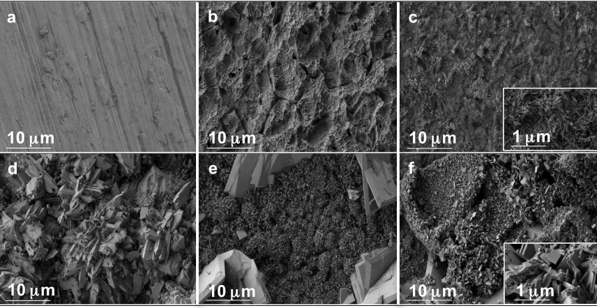
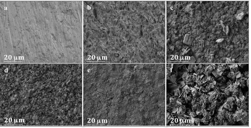
Figure 3-1 a) Polished Ti surface as control. b) Sand-blasted and acid-etched Ti surface. c) Titanium substrate coated with bioactive glass dispersed in 0.25 M NaOH under hydrothermal condition at 170ºC for 24 h. d) Titanium substrate coated with bioactive glass dispersed in 0.5 M NaOH under hydrothermal condition at 170ºC for 24 h. e) Titanium substrate coated with bioactive glass dispersed in 1 M NaOH under hydrothermal condition at 170ºC for 24 h. f) Titanium substrate coated with bioactive
glass dispersed in 2 M NaOH under hydrothermal condition at 170ºC for 24 h. (n=3) .... 43
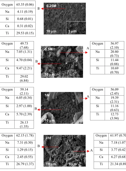
Figure 3-2 Elemental analysis of the hydrothermally coated titanium surfaces under various NaOH concentrations. The mean values are triplicate measurements of the atomic
percent of each element from random fields of view. The numbers in brackets are SD. .. 44
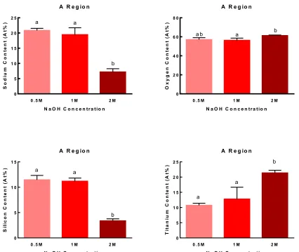
Figure 3-3 Histogram comparing the sodium, oxygen, silicon and titanium contents in the “a” region as a function of NaOH concentration. Data are means ± SD of triplicate measurements from random fields of view (n=3). Different lower case letters indicate significant differences at p < 0.05. ... 45
Figure 3-4 Histogram comparing the calcium, sodium, oxygen, silicon and titanium
contents in the “b” region as a function of NaOH concentration. Data are means ± SD of
triplicate measurements from random fields of view (n=3). Different lower case letters indicate significant differences at p < 0.05. ... 46
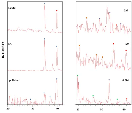
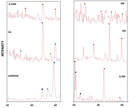
Figure 3-5 XRD analysis of Polished Ti and SA-Ti surfaces reveal 00-050-0787 (N)- Titanium oxide ( ) on its surface. Ti surface treated under 0.25 M NaOH showed -00-002-1359 (D)- Titanium Oxide ( ) and 01-089-0799 (C)- sodium titanate ( ). Ti surfaces treated under 0.5 M NaOH showed 011-0039 (l)-beta calcium phosphate ( ) and 00-010-0179-(*) sodium phosphate ( ). Ti surfaces treated under 1M NaOH showed
01-089-0800 (C)-sodium titanate ( ) and 00-051-1380 (*)- SiO2 ( ). Ti surfaces treated under 2
M NaOH showed 00-018-1243 (D)- sodium silicate ( ), 00-031-1329 (*)-sodium titanate ( ) and 00-046-0570 (C)- SiO2 ( ). Data was analyzed using EVA software. ... 48
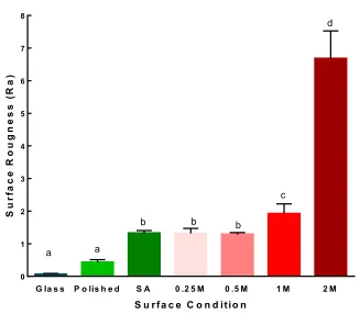
Figure 3-6 Surface roughness values of different Ti surfaces. Data are means ± SD of
triplicate specimens. Different lower case letters indicate significance differences at p <
Figure 3-7a) Polished Ti (control) and b) SA Ti did not exhibit hydroxyapatite deposition on their surfaces after 7 days immersion in SBF at 37 ºC. c, d, e, f) Ti surfaces coated with BG under different alkaline conditions (0.25, 0.5, 1 and 2 M NaOH) showed
hydroxyapatite-like crystals on their surfaces after soaking in SBF for 1 week. (n=3) ... 50
Figure 3-8 XRD analysis after immersion of substrata in SBF for 7 days. Polished Ti and SA surfaces showed 00-011-0029 (N)- calcium titanium oxide ( ) and 00-011-0177 (D)-beta calcium phosphate ( ). Ti surfaces treated under 0.25 M NaOH showed 01-086-0740 (C)- hydroxyapatite ( ). Ti surfaces treated under 0.5 M NaOH showed 00-044-0763 (D)-calcium phosphate hydrate ( ) and 01-086-0740 (C) -hydroxyapatite ( ). Ti surfaces treated under 1 M and 2 M NaOH showed 01-086-0740 (C)-hydroxyapatite ( ) and
00-032-1369 (N)- titanium phosphate ( ). Data was analyzed using EVA software. ... 51
Figure 3-9 MC3T3-E1 osteoblastic cells were cultured on the substrata for 24 hours. Cells were then fixed and labeled for nuclei (blue), filamentous actin (red) and vinculin (green). Cells were imaged by fluorescence microscopy. a) Fluorescence image of MC3T3-E1 cells on control glass. b) Fluorescence image of MC3T3-E1 cells on Polished Ti. c) Fluorescence image of MC3T3-E1 cells on sand-blasted and acid-etched Ti. d) Fluorescence image of MC3T3-E1 cells on glass-ceramic coating prepared with 0.25 M NaOH. e) Fluorescence image of MC3T3-E1 cells on glass-ceramic coating prepared with 0.5 M NaOH. f) Fluorescence image of MC3T3-E1 cells on glass-ceramic coating prepared with 1 M NaOH. g) Fluorescence image of MC3T3-E1 cells on glass-ceramic
coating prepared with 2 M NaOH. Scale bars represent 50µm. Images are representative
of triplicate independent experiments. ... 53
Figure 3-10 Histogram comparing the number of cells on glass and different Ti substrates. Data are means ± SD of triplicate independent experiments (n=3). Different lower case letters indicate significant differences at p < 0.05. ... 54
LIST OF APPENDICES
APPENDIX A: COPYRIGHT PERMISSIONS
LIST OF ABBREVIATIONS
Al2O3 Aluminum oxide
BCP Biphasic calcium phosphates
beta-TCP Beta tricaclcium phosphate
BG Bioactive glass
BPS Buffered phosphate solution
BSA Bovine serum albumin
Ca(NO3)2 Calcium nitrate
Ca2+
calcium ion
CaP Calcium phosphate
Cl- Chlorine ion
CPTi Commercial pure titanium
DAPI 4', 6-diamidino-2-phenylindole
ECM Extracellular matrix
EDX Energy dispersive x-ray spectroscopy
FBS Fetal bovine serum
HA Hydroxyapatite
HCA Hydroxycarbonate apatite
HCl Hydrochloric acid
HCO3
-Bicarbonate ion
HPO4
HVSFS High-velocity suspension flame spraying
ICDD International centre for diffraction data
K+ Potassium ion
MC3T3-E1 Osteoblast-like cells
Mg++ Magnesium ion
Na+ Sodium ion
NaOH Sodium hydroxide
P Polished
PLD Pulsed laser deposition
PSA Polysialic acid
Ra Surface roughness
RF Radio frequency
RFGD Radiofrequency glow discharge
SA Sandblasted acid etched.
SBF Simulated body fluid
SEM Scanning electron microscopy
SiO2 Silicon oxide
SLA Sand-blasted large grit acid etched
TEOS Tetraethyl orthosilicate
TEP Triethyl phosphate
Ti Titanium
TiO2 Titanium oxide
TMOS Tetramethoxysilane
XRD X-ray diffraction
CHAPTER 1: INTRODUCTION
1 . 1 O V E R V I E W
Dental implants are medical devices placed in maxillary and mandibular bone to provide
mechanical support for the replacement of lost teeth. Fabricated from titanium alloy,
dental implants can be used to replace single teeth, or as anchors to support fixed bridges,
removable partial or complete dentures [1]. Brånemark first presented the concept of
osseointegration, in which titanium implants bond to bone without the formation of an
interfacial layer [2]. Since Brånemark’s initial discovery, the design and surface
modifications of implants have been modified in attempts to optimize their longevity and
clinical success [3] .
Brånemark and colleagues published their landmark paper on the replacement of teeth
with titanium in a fully edentulous patient. Titanium and its alloys are the most widely
used materials for dental implants owing to their superior properties, including
mechanical strength, biocompatibility and corrosion resistance [4]. The ability of titanium
to avoid corrosion is due to the formation of a titanium oxide surface layer. Titanium is a
bioinert material and crucially it does not develop any chemical bond with bone. Several
researchers have modified titanium implants in attempts to enhance the process of new
bone formation in apposition to the implant to increase implant success rates and
longevity. A wide range of surface modifications including mechanical and chemical
approaches have been developed that are reviewed in Chapter 2. Increased rates of
osseointegration were reported when titanium was coated with either hydroxyapatite or
stimulates the recruitment of osteogenic cells as well as their subsequent differentiation
and production of mineralized extracellular matrix [5]. On the other hand, calcium
phosphates are biocompatible, osteoconductive material that strongly bond to bone [6, 7].
Different methods of surface coating are described in the literature including plasma
spraying and dip coating. However, such techniques lead to unfavourable properties in
the formed layers due to an imbalance of coating on the surface, as well as the
requirement of high temperature that result in nonhomogeneous properties. Indeed, in
certain cases, bioactive glass and hydroxyapatite added to implants using these
techniques failed, largely due to delamination [3]. Alternate methods for forming and
coating these layers are needed which is the focus of this thesis.
1 . 2 O B J E C T I V E S O F T H E T H E S I S
Taking into account the limitations described in formation of homogenous layers of
hydroxyapatite and bioactive glass on titanium the overall focus of this study is to
synthesize a bioactive and osteoconductive nanowire coating onto titanium substrates by
a two stage sol-gel-hydrothermal process, and to develop a novel sol-gel hydrothermal
coating method onto titanium (Ti) implants. We also examine the effects of reaction pH
and time under hydrothermal conditions on the surface topography and chemistry of the
1 . 3 H Y P O T H E S I S
We hypothesize that the hydrothermal conditions and pH levels of the reaction will
modulate the physical, chemical and biological properties of the coatings as well as
enhance cell attachment on the surface of the coated titanium.
1 . 4 T H E S I S O U T L I N E
As highlighted above, the objectives and hypothesis of this study focuses on the
development of the bioactive glass coating on the surface of titanium through a two-step
sol-gel hydrothermal process. The literature review is presented in chapter 2. Chapter 3
includes sol-gel synthesis of the bioactive glass and hydrothermal treatment process
along with the different characterization techniques to validate the success of the
developed approach. Chapter 4 provides a conclusion of this study and suggestions for
future work.
1 . 5 R E F E R E N C E S
1. Spiekermann, H., Implantology. 1995: Thieme.
2. Branemark, P.-I., Osseointegration and its experimental background. The Journal
of prosthetic dentistry, 1983. 50(3): p. 399-410.
3. Le Guéhennec, L., et al., Surface treatments of titanium dental implants for
rapid osseointegration. Dental materials, 2007. 23(7): p. 844-854.
4. Leyens, C. and M. Peters, Titanium and titanium alloys. 2003: Wiley Online
Library.
5. Salinas, A.J., A.I. Martin, and M. Vallet Reg, Bioactivity of three CaO P2O5
SiO2 sol gel glasses. Journal of biomedical materials research, 2002. 61(4): p.
524-532.
6. Wilson, J. and S.B. Low, Bioactive ceramics for periodontal treatment:
comparative studies in the Patus monkey. Journal of Applied Biomaterials, 1992.
3(2): p. 123-129.
7. El-Ghannam, A., Bone reconstruction: from bioceramics to tissue engineering.
Expert review of medical devices, 2005. 2(1): p. 87-101.
CHAPTER 2: LITERATURE REVIEW AND BACKGROUND
2 . 1 B O N E A N A T O M Y A N D P H Y S I O L O G Y
2.1.1 STRUCTURE AND FUNCTION OF BONE
Bone is a well-organized tissue that is composed of cells and extracellular matrix (ECM).
The ECM contains organic and inorganic components. The organic and inorganic
components provide bone’s strength and flexibility. The organic component represents
40% of bone and is mainly composed of collagen type I and also includes glycoproteins,
peptides, lipid materials, and adsorbed serum proteins [1]. Inorganic ECM is mainly
composed of hydroxyapatite (HA), calcium phosphate, calcium carbonate, calcium
fluoride and magnesium fluoride. Bone compressive and shear strength as well as
hardness are enhanced by the carbonated HA complex [2]. Organic-inorganic structure of
bone is formed as a result of nucleation of HA along the collagen fibers [3] The unique
structure of bone explains its mechanical properties as well as its resistance towards
different compressive and tensile forces applied during daily activities [2, 3].
Regarding the macroscopic structure of bone, there are two different types of bone that
can be differentiated by the degree of macroporosity (Figure 2.1) [3, 4]. Cancellous also
known as spongy or trabecular bone is the less dense type of bone. It has more than
double the porosity of compact bone, and is rich in blood vessels, bone marrow and
connective tissues. Cancellous bone plays a major role in hematopoiesis [4]. On the other
hand, compact bone also known as cortical bone is the main structural element of the
bone has a superior ability to withstand the stress upon bone 20 times more than
cancellous bone [2].
Bone cells are supplied with oxygen and nutrition as well as removal of cellular
byproducts through a network of blood vessels. In cortical bone, nerves and blood vessels
run through Haversian and Volkmann’s canals.
2.1.2 FUNCTIONS OF BONE[5]
1. Protection of internal organs and the central nervous system
2. Reservoir of inorganic ions
3. Hematopoiesis
4. Mechanical support of soft tissues
5. Enable motion by providing articulations and attachments for muscles
2.1.3 BONE CELLS AND REMODELING
Bone is a living tissue that undergoes continuous cycles of deposition and resorption [6].
Understanding the unique properties of bone as well as the action of bone cells is
essential for the development of new biologically relevant biomaterials. The process of
bone remodeling involves various types of bone cells in order to carry out bone
resorption and formation. Osteoclasts resorb old or damaged bone and osteoblasts form
new bone. In a healthy adult, there is a balance between resorption and deposition to
maintain the skeletal system. New bone is originated in form of lamellae or concentric
by osteoblasts forming new lamellae, blood and nutrients are supplied through the
Haversian canal to older lamellae.
2.5.1.1 OSTEOBLASTS
Osteoblasts are differentiated cells that originate from mesenchymal stem cells, followed
by proliferation and differentiation into pre-osteoblasts and then maturation. Osteoblast
development occurs in three main stages [7, 8]:
1. Cell proliferation
2. Matrix production and maturation
3. Mineralization
Osteoblasts synthesize and secrete different proteins including type I collagen. This is
followed by mineralization of the matrix [2]. Some osteoblasts become encased within
the bone matrix they have formed and terminally differentiate into osteocytes
2.5.1.2 OSTEOCYTES
Osteocytes have less metabolic activity than osteoprogenitor or osteoblastic cells.
Osteocytes respond to mechanical stimulation and can send signals to osteoblasts and
osteoclasts to promote bone formation and bone resorption.They play a role in the
process of maintaining local bone The processes that connect adjacent osteocytes permit
communication among cells as well as delivery of nutrients [1].
2.5.1.3 OSTEOCLASTS
Osteoclasts are bone resorbing cells. Hydrochloric acid and proteolytic enzymes are
of the extracellular bone matrix. Thereafter, the organic matrix is degraded by the action
of hydrolytic enzymes. Degradation products are then removed and released into the
Figure 2-1 : The diaphysis of a long bone, from spongy bone tissue and the medullary
cavity on the left. Compact bone tissue in the middle. The periosteum is on the right. Osteocyte is shown at the top right. Reproduced from Anatomy and Physiology:
From Science to Life, 2nd edition with permission of John Wiley & Sons, Inc.
2 . 2 D E N T A L I M P L A N T S
2.2.1 OVERVIEW
Dental implants are medical devices that are placed within maxilla and/or mandible in
order to provide mechanical support to different prosthetic appliances used such as a
single crown or bridge, or a partial or complete denture [11]. Different materials, designs
and surface modifications of dental implants are developed because of their increased
clinical importance as a solution for patients with either esthetic or functioning issues.
Historical studies reported that missing teeth replacement was introduced early in the
history by using either homologous or alloplastic materials. These materials might
provoke systemic responses [11]. In the 19th century, different studies demonstrated the
use of rubber, gold and porcelain for teeth replacements. In 1939, Stock developed a
threaded vitallium dental implant [11]. While studying bone healing in a rabbit model, a
great turning point occurred in 1952, when a Swedish orthopedic surgeon Branemark.
discovered a titanium cylinder placed in the tibia of a rabbit could not be removed [12].
The retentive titanium presented the concept of osseointegration.In 1962, Brånemark and
his team treated the first edentulous patient [13].
2.2.2. INDICATIONS AND CONTRAINDICATIONS FOR DENTAL IMPLANTS
Dental implants are used for replacement of single, partial and complete loss of teeth.
They are contraindicated in case of unfavorable occlusal function, macroglossia, which is
known as enlarged tongue in completely edentulous patient, systemic disorders such as
uncontrolled diabetes and patients receiving radiotherapy due to the inhibition of bone
2 . 3 O S S E O I N T E G R A T I O N
It is known as direct functional and structural bond formation between bone tissue and
artificial implant surface [15].
Osseointegration refers to the irreversible biological stability of implant in bone tissue. It
is reported to be as a result to the absence of negative local or systemic reactions towards
the implant. Studies exploring the mechanism of osseointegration from have been
conducted [16].A 5-year clinical study using screw-shaped titanium implants showed
that proper healing, operator skills as well as stress distribution are very important for the
successful bone formation [17].
2.3.1 FACTORS AFFECTING OSSEOINTEGRATION
Implant material, design and surface topography are responsible for enhancing
osseointegration [18]. Moreover, healthy bone and pharmacological agents such as
bisphosphonates enhance osseointegration. The effect of bisphosphonates is debatable.
Other studies reported that its usage inhibits bone formation since it alters bone formation
cycle by activation of osteoblasts and inhibition of osteoclastic activity [19, 20]. In
contrast, there are several factors that inhibit osseointegration such as inadequate implant
stability, excessive occlusal stresses, radiation therapy, pharmacological agents such as
NSAIDs, osteoporosis, rheumatoid arthritis, smoking and alcoholism [19, 21-24].
2.3.2 PERI-IMPLANT HEALING
After surgical placement of the implant within the maxilla or the mandible within less
The blood clot forms a fibrin matrix, induces foreign body reaction and increases
phospholipid hydrolysis and intracellular calcium. Then, osteoconduction occurs in
which osteogenic cells proliferate at the implant surface. De novo bone formation takes
place as osteogenic cells differentiate into osteoblasts. Woven bone is produced then
remodeled and replaced by mature lamellar bone [16, 25, 26].
Osteoconduction is defined as stimulation of undifferentiated osteogenic cells
proliferation and their migration to the implant surface. Next is osteoinduction, which is
defined as osteogenic cells differentiation into osteoblasts that form new bone. Finally,
peri-implant osteogenesis is the process that ultimately results in the biological fixation
required for implant anchorage.
Primary stability of a dental implant is very important for osseointegration; it depends on
the implant design, accuracy of surgical placement as well as implant location.
Inadequate stability enhances fibrous tissue formation, that inhibit osseointegration,
resulting in loosening of the implant and its eventual failure [16, 26].
Peri-implant osteogenesis may fail for a number of reasons, including: 1. impaired
vascularization; 2. inadequate source of osteogenic cells; 3. hyperactive osteoclasts; 4.
abnormal cell proliferation; or 5. abnormal local or systemic inflammatory responses
[27].
2 . 4 B I O A C T I V I T Y O F D E N T A L I M P L A N T S
Bioactive materials are used to replace, as well as to reconstruct bony defects. Bioactive
glass bonds directly to the surface of bone through production of an apatite layer. To
HA layer. First, immersion of samples in simulated body fluid (SBF) is done followed by
evaluation. SBF was developed by Kokubo et al. [28] and Hench et al. [29]. It resembles
human blood plasma composition, except for less HCO3- and more Cl-.
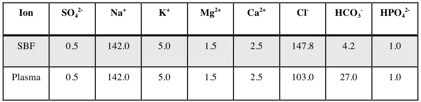
Table 2-1. Ionic composition of SBF and blood plasma
Ion SO42- Na+ K+ Mg2+ Ca2+ Cl- HCO3- HPO4
2-SBF 0.5 142.0 5.0 1.5 2.5 147.8 4.2 1.0
Plasma 0.5 142.0 5.0 1.5 2.5 103.0 27.0 1.0
The production of an apatite layer indicates the bonding ability of the material to bone.
The main stages of apatite formation on a bioactive glass include ion exchange of Na+
and K+ with H+ or H
3O- from the solution. SiOH4 is released to the solution. The
silicon-to-oxygen bonds break down, forming silanol on the materials surface. Thereafter,
condensation and re-polymerization of the SiO2-rich layer on the surface occur. Ca and Pi
ions deposit on the surface forming a layer of amorphous calcium phosphate. At the end,
amorphous calcium phosphate undergoes crystallization to form hydroxycarbonate
apatite HCA [30, 31] .
The mechanism of bioactivity is very important, especially the last two steps. Knowing
that the bioactivity test is time-dependent, the longer the time needed for the material to
2 . 5 D E N T A L I M P L A N T M A T E R I A L S
2.5.1 METALS
2.5.1.1 STAINLESS STEEL
Temporary orthopedic devices such as screws and plates are formed of 316L Stainless
steel is used for. However it is strong, easy to machine and inexpensive, it is not used as
dental implant material because of high corrosion as well as inability to osseointegrate
compared to titanium [18, 32].
2.5.1.2 COBALT-BASE ALLOYS
These alloys are produced of chromium and cobalt. They are characterized by high
corrosion resistance due to formation of chromium oxide layer, but these alloys are
inferior to titanium. They are mostly used in total joint replacements [18, 33].
2.5.1.3 TITANIUM AND TITANIUM ALLOYS
Titanium and its alloys have excellent biocompatibility and superior mechanical
properties. Titanium is characterized by forming a passive protective oxide layer that
enhances its corrosion resistance in physiological environment. Titanium is used in either
99.75% pure form known as commercial pure titanium or as Ti-6Al-4V alloy with 6%
aluminum and 4% vanadium. Owing to superior mechanical properties as well as
corrosion resistance, commercial pure titanium (CPTi) has been widely used. Different
composition of carbon, hydrogen, iron, nitrogen and oxygen results in four grades of
CPTi. The different composition of each grade of titanium has a significant influence on
their mechanical properties. Currently grade 4 CPTi as well as titanium alloy is widely
Once titanium is in contact with body fluids, a thin passive oxide layer is formed on the
surface of the implant. Different oxides are formed including TiO, TiO2 and Ti2O3 but
TiO2 is the most stable oxide layer[36]. Biocompatibility, high corrosion resistance and
excellent osseointegration are greatly influenced by the oxide layer [36].
Different surface treatments were introduced to increase osseointegration by stimulating
proliferation of osteoblasts on the implant surface. These include: acid etching [37], sand
blasting [37], alkaline heat treatment [37], calcium phosphate coating, plasma spraying,
dip coating [38], and bioactive glass coating.
2 . 6 S U R F A C E M O D I F I C A T I O N S O F T I T A N I U M
2.6.1 MECHANICAL METHODS
Mechanical approaches for surface modifications include removal, shaping, or treatment
of titanium surfaces through the application of physical forces or removal of surface
material by cutting or abrasive action.
2.6.1.1 SURFACE POLISHING
In polishing, a smooth surface finish is formed using fine abrasive grades accompanied
by lubrication [39]. Alumina, diamond and SiC are the most common used polishing
media. The finest polishing grades can produce surfaces with roughness value Ra of 0.1
µm or less [40]. However, polishing is usually used as an intermediate step prior to other
surface treatments; polishing as a final surface modification develops mechanical surface
stresses and alters the chemical composition of the titanium surface [39]. The main
impact of polishing is removal of the native surface layer, descaling and smoothening of
2.6.1.2 SURFACE BLASTING
Surface blasting is the application of particles under high velocity to surfaces. It is mainly
used for cleaning, descaling, removal of the native surface layer, increasing surface
roughness and enhancing adhesion of bonded materials [41-43]. Various ceramics
including alumina, silica, titania of different particle sizes are used for surface blasting of
titanium. Particle size is the most critical parameter controlling the process of blasting
and its impact on the surface topography. For example, a surface roughness of 0.5-1.5
µm Ra value is produced using alumina particles of 25-75 µm. On the other hand, the use
of alumina particles of 200-600 µm resulted in Ra of 2-6 µm [43, 44]. Moreover, smooth
rounded surfaces are formed by gentle shot peening. Chemical treatment of the blasted
surface is recommended to remove the particles embedded during blasting. The ability of
the alumina and silica particles to alter the chemical composition of titanium surfaces
encouraged the use of blasting prior to the application of hydroxyapatite on the surface of
titanium [45]. A thin oxide layer of less than 10 nm of TiO2 is expected on the blasted
surfaces accompanied by traces of blasting medium [39].
2.6.2 CHEMICAL METHODS
Chemical methods reported for surface modification of titanium include the chemical
reaction taking place between chemicals and the surface of titanium.
2.6.2.1 SOLVENT CLEANING
Solvent cleaning is mainly used to remove oils, fatty surface and greased contaminates
that remain after manufacturing. Alcohols, ketones and chlorinated carbons are common
remain as residues on the surface [46, 47]. This is due to the reaction occurring between
the organic solvents and titanium oxide layer.
2.6.2.2 WET CHEMICAL ETCHING
Wet chemical etching is a process that depends mainly on the reaction between certain
chemicals and the titanium oxide layer. Removal of the native oxide layer and increasing
surface roughness are the ultimate goals of the etching process
2.6.2.2.1 Acid Etching
Acid etching removes oxide scales and forms clean surfaces. The surface topography of
acid etched titanium is affected mainly by the pretreatment condition of titanium. Mild
treatments did not have any effect on the surface condition [39].In contrast, significant
differences in the surface topography are observed when titanium surfaces are
extensively etched. In case of alloyed titanium, differences in etching alpha and beta
phases result in surface topography in which beta phase is protruding from alpha [41].
Blasted surfaces prior to etching show higher surface roughness [41, 48]. In general
etching produces a surface roughness varying from 0.1 µm or more according to the
pretreatment topography [49, 50]. A thin oxide layer of less than 10 nm is formed on the
surface of titanium as a result of etching. The oxide layer grows slowly over a year from
3 nm to 6 nm [51].
There are two main methods of acid etching. First is by using a mixture of nitric acid and
hydrofluoric acid in a ratio of 10:1. In one method, hydrofluoric acid reacts with the
surface titanium oxide forming titanium fluorides and free hydrogen. The 10:1 ratio must
of titanium [52]. In the other method, a mixture of 1:1 hydrochloric acid and sulphuric
acid is used. The degree of pickling or etching is mainly affected by the temperature, acid
concentration and treatment time that vary from one minute to one hour [41, 48, 53] .
2.6.2.2.2 Alkaline Etching
Alkaline treatment of titanium is performed using 5M NaOH for 24 hours at 60 °C [54,
55]. It produces a sodium titanate gel layer of 1 µm thickness as well as surface
porosities. Alkaline etching is used mainly as a pretreatment to gel-derived apatite
coating [53]. In some cases, the use of alkaline etching after acid etching produces a high
surface roughness [53].
2.6.3 PASSIVATION TREATMENTS
The main purpose of passivation treatments of titanium is the formation of a stable oxide
layer to prevent ion release [39]. There are two main approaches regarding passivation,
heat treatment in air or immersion in a strong oxidizing agent.
2.6.3.1 NITRIC ACID PASSIVATION
It is mainly formed using HNO3 solution in which titanium is immersed for 30 minutes at
room temperature followed by rinsing and drying in order to neutralize the surface [56].
Usually passivation is the last step in surface modification of titanium. Although nitric
acid passivation was reported to have no effect on the titanium oxide layer, higher ion
2.6.3.2 HEAT TREATMENT
It is used as an alternative treatment method to passivation. It has no direct effect on the
surface topography but it has an influence on the oxide scale layer. Heat treatment at 400
°C produces an oxide layer of 30 nm [59].
2.6.4 OTHER CHEMICAL SURFACE TREATMENTS
In addition to the previously explained methods, there are other approaches that are
reported in the literature regarding the surface modification of titanium. These methods
include apatite-coating techniques that will be explained in the following section.
2.6.4.1 HYDROGEN PEROXIDE TREATMENT
The reaction between the hydrogen peroxide and the titanium oxide produces Ti gels. The
biocompatibility of the titanium upon using hydrogen peroxide is explained by the
reaction between the hydrogen peroxide and titanium. The hydrogen peroxide is formed
during inflammatory reaction [60, 61] .
2 . 7 B I O - C E R A M I C S A N D B I O A C T I V E G L A S S C O A T I N G S
2.7.1 CALCIUM PHOSPHATES
Calcium phosphates are a biocompatible, osteoconductive material that bonds strongly to
bone [62, 63]. HA is widely used for dental owing to its similar composition to that of the
mineral phase of bone and teeth. Different methods are being used for the production of
HA such as wet chemical methods [64], solid-state reactions [65], hydrolysis methods
[65] and sol-gel chemistry. Considering hydrolysis, acid calcium phosphates such as
anhydrous dicalcium phosphate, octacalcium and dicalcium phosphate dihydrate
deficient apatite precipitate is formed under alkaline conditions through either reaction
between calcium nitrate and ammonium phosphate or drop-wise precipitation of
phosphoric acid to a suspension of calcium hydroxide [66, 67]. Sol-gel chemistry
involves hydrolysis of metal alkoxides and calcium salts followed by polycondensation.
Homogenous as well as controlled composition of the final product are the main
characteristics of the sol-gel process. However, calcination at high temperature is
required in order to produce crystalline HA. Calcination results in production of
secondary phases of HA such as granular particle shapes and beta-tricalcium phosphate
(TCP) [68, 69].
In contrast, exposing calcium and phosphorus precursors to high pressure through
hydrothermal process produces crystalline HA [70]. A recent study reports the synthesis
of HA nanowire through a combination of solvothermal processes and sol-gel chemistry
[71].
Synthesis of HA bone scaffold is challenging owing to various degradability levels
between different forms of HA. For example crystalline HA is poorly degradable while
the fragility of the amorphous HA limits its use [72].
Crystalline HA has a limited osteoconductivity and bioactivity owing to its chemical
stability in body fluids. In contrast, amorphous HA has a high dissolution rate that
initiates an immune system response [72]. Biphasic calcium phosphates (BCP), formed of
a combination of beta-TCP and HA, was developed to assure proper functioning of the
HA [73]. Different studies reported the excellent effect of HA and its ability to bond
application of the calcium phosphate coatings for total joint arthroplasty due to the
enhanced osseointegration levels [75].
2.7.2 BIOACTIVE GLASS
In 1971, Hench and his colleges developed a specific glass formulation, known as
bioactive glass in system Na2O- Cao-SiO2-P2O5, which does not initiate formation of
fibrotic tissue [76]. Bioactive glasses are silicate-based glasses that are amorphous and
biologically active. They are able to produce a strong bond when they are in contact with
body fluids through formation of a bone-like HA layer [77, 78]. A series of reactions take
place as a bonding mechanism of silicate glasses to bone [30]. Ions released from the
bioactive glass produce a silica gel layer that stimulates favorable intracellular and
extracellular responses to enhance bone synthesis. An amorphous calcium phosphate
layer is formed due to ion exchange between body fluids and bioactive glass followed by
crystallization to form carbonated HA.
Melt-derived bioactive glass was first introduced, in which melts of SiO2 and P2O5
network formers and CaO and Na2O network modifiers are quenched [76]. In the
beginning of 1900s, sol-gel produced bioactive glass replaced the melt-derived. The main
advantages of sol-gel bioactive glass include mixing at the molecular level that provides
better control over chemical homogeneity and composition of the produced glass, low
temperature process and high surface area of the bioactive glass that enhances the
In sol-gel chemistry a series of steps take place in order to form bioactive glass including
hydrolysis and polycondensation of metal alkoxide followed by gelation, aging, drying
and densification [79]. A metal alkoxide M-ORx in which an oxygen linkage O bonds a
metallic ion M to a functional group R. Tetraethoxysilane (TEOS) as well as
tetramethoxysilane (TMOS) are the most commonly used metal precursor owing to their
high reactivity to water.
Understanding the mechanism of hydrolysis and condensation reactions is essential to
investigate the reaction parameters. There are many factors affecting it including
temperature, nature and concentration of the electrolyte, nature of solvent and type of
alkoxide precursor. Hydrolysis reaction depends specifically on the electrolyte
concentration, the form of alkoxide group, (the bulkier the alkoxide group the lower rate
of hydrolysis), and R- ratio of water to TEOS [30, 79].
Regarding gelation, a 3-D network of polycondensed particles produces the gel. The
gelation process is influenced by the extent of cross-linking of particles and the particle
size. In aging, viscosity of the solution increases and pore size decreases owing to further
polycondensation [79, 80]. Sufficient strength of gel is essential in order to avoid
cracking during drying. Considering drying, cracking is observed owing to an increase in
the capillary pressure especially in small pores less than 20 nm due to removal of liquid
from the gel [81]. Controlling the rate of hydrolysis and condensation to form
monodispersed pore sizes prevents cracking of gels during drying [80-82]. Knowing that
glass is normally an amorphous material, it needs to be sintered or heated to get rid of
Due to the inferior mechanical properties of bioactive glass, it has limited applications. In
the apatite containing glass, it was reported that the bonding of the glass to the bone is
achieved through the bond between bone apatite and glass-ceramic. In the processing of
bioactive glass, two main approaches were described by researchers including
melt-derived and sol-gel melt-derived bioactive glass. Melt-melt-derived bioactive glass is characterized
by lower dissolution rate as well as lower rate of apatite layer formation on the surface of
the glass. In contrast, sol-gel derived glass showed higher dissolution rate, higher rate of
HA formation and higher crystallinity compared to melt-derived bioactive glass [80]. The
sol-gel bioactive glass shows continuous release of silica ions and break down of the
glass network giving calcium and phosphorus that enhances the process of hydroxyl
carbonate apatite formation.
2 . 8 P R O D U C T I O N O F C O A T I N G S O N T I T A N I U M S U R F A C E
The production of surface coating on the surface of titanium, mostly hydroxyapatite or
calcium phosphate coating, encouraged a lot of researchers to attempt to enhance the
biological properties of titanium, combining the superior mechanical properties of
titanium with the excellent bioactivity of calcium phosphate or bioactive glass. These
coatings would enhance the process of new bone formation accordingly increase the
success rate of the implant.
2.8.1 SOL-GEL
Using sol-gel method, HA coatings are developed on the surface of titanium[68].
Titanium substrates are dipped in calcium, mostly nitrate salts and phosphorus gels, for a
certain period of time. The coatings produced are characterized by high porosity, low
influence since it increases the surface area in contact with tissue fluids. In order to
enhance adhesion as well as density, coatings are sintered. Various forms of calcium
phosphates are formed as a result of different sintering temperatures [83, 84].
Dip coating method is used to apply two layers of coating[85]. After primarily deposition
of calcium and nitrate solution, the film is dried at 200 °C. Thereafter, second deposition
is performed followed by drying at 750 °C. Higher corrosion resistance as well as higher
adhesion ability of the coating is observed due to the formation of an intermediate layer
of TiO2 between HA and titanium. The optimal oxide layer is 200 nm. If its thickness is
less than 200 nm, lower corrosion resistance is observed. In addition, an oxide layer of
more than 200 nm reduces the adhesion ability between the coating and titanium due to
the thermal mismatch.
Different developments of the mentioned technique were investigated [85]. For example
a CaTiO2 was applied as an intermediate layer to enhance adhesion of HA to the titanium
surface. Altering the precursors, such as using HA/ethanol solution, was investigated in
order to develop a coating with higher roughness, porosity, homogeneity and bonding to
the surface [86].
Owing to the long processing time as well as post-sintering limitations, the use of sol-gel
coatings in the industry is limited.
2.8.2 PLASMA SPRAYING
Plasma spraying is a commonly used technique for surface modification of implants by
complex of thermal changes involving powder particles, the plasma zone as well as the
substrate are observed.
Regarding the mechanism of plasma spraying technique, particles are exposed to very
high heating temperature for few seconds in the 10,000 °C jet. However particles undergo
different melting rates, some particles do not melt due to the limited time in the plasma
zone. Thereafter HA droplets are impacted on the surface of titanium [87-90].
Limitations of the plasma spraying technique include, nonhomogeneous coating due to
the formation of a mixture of crystalline and amorphous HA as a result of quick exposure
to high temperature, low Ca/P ratio as a result of the reaction due to the high temperature
applied and formation of rough surfaces [87, 91]. Although surface roughness is
considered an enhancing factor of the process of implant to bone bonding, the roughness
should be in a certain range considering that extremely rough surfaces are not favorable.
Moreover, it is reported that plasma sprayed coatings exhibit different bond strengths to
the implant surface. This results in the formation of microcracks, nonuniform coating and
limited delamination resistance [92, 93].
2.8.3 ION-BEAM METHODS
In ion beam method, calcium and phosphorus ions are embedded on the surface of the
substrate. Thereafter, the substrates are exposed to SBF in order to form a titanium
hydroxide layer that acts as bonding sites for HA. It is characterized by strong adhesive
bonding of the coating to the surface of the substrate. The main limitations of this
technique include formation of amorphous coatings on the surface of the substrate as well
2.8.4 LASER METHODS
It is known that plasma sprayed HA showed nonhomogeneous coating with high levels of
thickness. Moreover the coating is mechanically bonded to the surface of the substrate
that is poorly accepted for clinical application. In contrast, pulsed laser deposition PLD is
able to form a thin crystalline coating with acceptable adhesive bonding to the substrate
surface. Regardless the advantages of the PLD method, the expensive cost of the process
as well as the machine itself may limit its use [95, 96].
2.8.5 RF SPUTTERING
In this technique, RF-magnetron sputtering from calcium phosphate glass targets,
followed by post-annealing, is used to produce a thin apatite layer on the substrate. The
production of calcium phosphate occurs as a result of sputtering in which phosphorous
oxide is lost. The higher Ca/P ratio of the target glass as well as the higher post-annealing
temperature, the more crystalline phases formed. Lately, sputtered HA showed excellent
results regarding bone formation. The length of the process as well as the expensive cost
of RF sputtering limit its application [87, 97].
2.8.6 HYDROTHERMAL TREATMENT
Hydrothermal treatment is performed on already existing coating in order to form a
targeted phase [38, 98-100]. Plasma spraying, anodic oxidation or any reported
techniques can be used. Thereafter the substrates are hydrothermally treated in water or
SBF aqueous medium in order to produce HA. A thin layer of HA is formed in case of
hydrothermally treated anodic oxide film that has calcium and phosphorus. Compared to
hydrothermal treatment. Although hydrothermal treatment enhances the HA formation, it
alters the stability of the oxide layer bonding to the surfaces [98, 99, 101]. The
hydrothermal condition including reaction pH, temperature, pressure and reaction time
are the main factors affecting the process of HA formation [93]. Increase in temperature
and pH accelerates the release of calcium and phosphorus ions from the original coating
and eventually increases the HA production.
2.8.7 ELECTROCHEMICAL CATHODIC DEPOSITION
CaP is deposited on the surface of the substrate using cathodic deposition under ambient
temperature [102, 103]. Good shape conformity, room temperature process, uniform
coating thickness as well as short processing times are the main advantages of this
technique. The major disadvantages of this method include the development of stresses in
the coatings and inadequate bonding to the surfaces[104, 105].
2.8.8 THERMAL SUBSTRATE METHOD
In this method, an aqueous solution of calcium and phosphorus is used to produce CaP
coating on titanium surfaces. The immersed substrates are exposed to high temperature of
1008 °C known as Joule heating. Thereafter CaP coating is produced. HA, which is the
major component of the coating, increases as a result of elevated reaction time and
temperature. Heating the aqueous solution containing calcium and phosphorous ions
under accurate conditions of pH and temperature has a critical effect on the production of
HA because it is less soluble under higher temperature. The diversity of crystallinities
produced is considered as the main disadvantage of this technique that limits its use
2 . 9 R A T I O N A L O F T H E S T U D Y
Considering the drawbacks of methods of surface coating applications reported in the
literature, the target of this study is to synthesize a bioactive and osteoconductive
nanowire coating onto titanium substrates by a two stage sol-gel-hydrothermal process to
develop a novel sol-gel hydrothermal coating method onto Ti implants and to study the
effect of reaction pH and time under hydrothermal conditions on surface topography,
chemistry and osteoblast like-cell attachment to the bioactive glass coating. We
developed bioactive glass coating on the surface of titanium through various
hydrothermal conditions. We hypothesize that pH levels of the reactions will modulate
physical, chemical and biological properties of the coatings as well as enhancement of
2 . 1 0 R E F E R E N C E S
1. Bilezikian, J.P., L.G. Raisz, and T.J. Martin, Principles of Bone Biology:
Two-Volume Set. 2008: Academic Press.
2. Clarke, B., Normal bone anatomy and physiology. Clinical journal of the
American Society of Nephrology, 2008. 3(Supplement 3): p. S131-S139.
3. Rho, J.-Y., L. Kuhn-Spearing, and P. Zioupos, Mechanical properties and the
hierarchical structure of bone. Medical engineering & physics, 1998. 20(2): p.
92-102.
4. Seeley, R.R. and T.D. Stephens, Tate (2000). Anatomy and physiology,
McGraw-Hill.
5. Hadjidakis, D.J. and I.I. Androulakis, Bone remodeling. Annals of the New York
Academy of Sciences, 2006. 1092(1): p. 385-396.
6. Dimitriou, R., et al., Bone regeneration: current concepts and future directions.
BMC medicine. 9(1): p. 66.
7. Siggelkow, H., et al., Development of the osteoblast phenotype in primary human
osteoblasts in culture: comparison with rat calvarial cells in osteoblast
differentiation. Journal of cellular biochemistry, 1999. 75(1): p. 22-35.
8. Barrère, F., C.A. van Blitterswijk, and K. de Groot, Bone regeneration:
molecular and cellular interactions with calcium phosphate ceramics.
international Journal of Nanomedicine, 2006. 1(3): p. 317.
9. Mulari, M.T.K., et al., Osteoclast ruffled border has distinct subdomains for
secretion and degraded matrix uptake. Traffic, 2003. 4(2): p. 113-125.
10. Jenkins GW, K.C., Tortora GJ, Anatomy and physiology. 2007: Wiley
11. Anusavice, K.J., C. Shen, and H.R. Rawls, Phillips' science of dental materials:
Elsevier Health Sciences.
12. Spiekermann, H., Implantology. 1995: Thieme.
13. Ratner, B.D., et al., Biomaterials science: an introduction to materials in
medicine. 2004: Academic press.
14. Misch, C.E., Contemporary implant dentistry. 2007: Elsevier Health Sciences.
15. Hobkirk, J.A., R.M. Watson, and L.J.J. Searson, Introducing dental implants.
2003: Churchill Livingstone.
16. Mavrogenis, A.F., et al., Biology of implant osseointegration. J Musculoskelet
Neuronal Interact, 2009. 9(2): p. 61-71.
17. Adell, R., et al., A 15-year study of osseointegrated implants in the treatment of
the edentulous jaw. International journal of oral surgery, 1981. 10(6): p. 387-416.
18. Sykaras, N., et al., Implant materials, designs, and surface topographies: their
effect on osseointegration. A literature review. The International journal of oral &
maxillofacial implants, 1999. 15(5): p. 675-690.
19. Marco, F., et al., Peri-implant osteogenesis in health and osteoporosis. Micron,
2005. 36(7): p. 630-644.
20. Javed, F. and K. Almas, Osseointegration of dental implants in patients
undergoing bisphosphonate treatment: a literature review. Journal of
21. Dahners, L.E. and B.H. Mullis, Effects of nonsteroidal anti-inflammatory drugs
on bone formation and soft-tissue healing. Journal of the American Academy of
Orthopaedic Surgeons, 2004. 12(3): p. 139-143.
22. Weiss, C.M., Principles and practice of implant dentistry. 2001.
23. Isidor, F., Loss of osseointegration caused by occlusal load of oral implants. A
clinical and radiographic study in monkeys. Clinical oral implants research, 1996.
7(2): p. 143-152.
24. Ihde, S., et al., Effects of radiation therapy on craniofacial and dental implants: a
review of the literature. Oral Surgery, Oral Medicine, Oral Pathology, Oral
Radiology, and Endodontology, 2009. 107(1): p. 56-65.
25. Davies, J.E., Understanding peri-implant endosseous healing. Journal of dental
education, 2003. 67(8): p. 932-949.
26. Nedir, R., et al., Predicting osseointegration by means of implant primary
stability. Clinical Oral Implants Research, 2004. 15(5): p. 520-528.
27. Franchi, M., et al., Early detachment of titanium particles from various different
surfaces of endosseous dental implants. Biomaterials, 2004. 25(12): p. 2239-2246.
28. Kokubo, T., et al., Solutions able to reproduce in vivo surface†structure
changes in bioactive glass†ceramic A†W3. Journal of biomedical materials
research, 1990. 24(6): p. 721-734.
29. Filgueiras, M.R., G. La Torre, and L.L. Hench, Solution effects on the surface
reactions of a bioactive glass. Journal of biomedical materials research, 1993.
27(4): p. 445-453.
30. Hench, L.L., Bioceramics: from concept to clinic. Journal of the American
Ceramic Society, 1991. 74(7): p. 1487-1510.
31. Han, Y.J., et al., Investigation of the bioactivity and biocompatibility of different
glass interfaces with hydroxyapatite, fluorohydroxyapatite and 58S bioactive
glass. Biofactors, 2007. 30(4): p. 205-216.
32. Planell, J.A., et al., Bone repair biomaterials. 2009: Elsevier.
33. Varano, R., et al., The effect of microstructure on the wear of cobalt-based alloys
used in metal-on-metal hip implants. Proceedings of the Institution of Mechanical
Engineers, Part H: Journal of Engineering in Medicine, 2006. 220(2): p. 145-159.
34. Natali, A.N. and P.G. Pavan, 11 Numerical approach to dental biomechanics.
Dental biomechanics, 2003: p. 211.
35. Leyens, C. and M. Peters, Titanium and titanium alloys. 2003: Wiley Online
Library.
36. Powers, J.M. and J.C. Wataha, Dental Materials-: Properties and Manipulation:
Elsevier Health Sciences.
37. Le Guéhennec, L., et al., Surface treatments of titanium dental implants for
rapid osseointegration. Dental materials, 2007. 23(7): p. 844-854.
38. Frauchiger, V.M., et al., Anodic plasma-chemical treatment of CP titanium
surfaces for biomedical applications. Biomaterials, 2004. 25(4): p. 593-606.
39. Brunette, D.M., Titanium in medicine: material science, surface science,
engineering, biological responses, and medical applications. 2001: Springer