1556-6811/09/$08.00
⫹
0
doi:10.1128/CVI.00455-08
Copyright © 2009, American Society for Microbiology. All Rights Reserved.
Effect of Porcine Circovirus Type 2 (PCV2) Vaccination of the Dam
on PCV2 Replication In Utero
䌤
D. M. Madson,
1A. R. Patterson,
1S. Ramamoorthy,
1N. Pal,
1X. J. Meng,
2and T. Opriessnig
1*
Department of Veterinary Diagnostic and Production Animal Medicine, College of Veterinary Medicine, Iowa State University,
Ames, Iowa,
1and Department of Biomedical Sciences and Pathobiology, Center for Molecular Medicine and Infectious Diseases,
College of Veterinary Medicine, Virginia Polytechnic Institute and State University, Blacksburg, Virginia
2Received 4 December 2008/Returned for modification 24 February 2009/Accepted 31 March 2009
The aims of this study were to determine if porcine circovirus type 2 (PCV2) vaccination of the dam is
effective in preventing fetal PCV2 infection and reproductive failure. Twelve pregnant, PCV2-naïve sows were
randomly divided into four groups, with three sows in each group. Group 1 sows served as noninoculated,
nonvaccinated negative controls, group 2 sows were vaccinated with a commercially available PCV2 vaccine at
28 days of gestation and were not inoculated, group 3 sows were vaccinated at 28 days of gestation and
inoculated with PCV2b at 56 days of gestation, and group 4 sows were inoculated with PCV2b but were not
vaccinated. Serum samples from all sows were collected weekly throughout the gestation period, and sows were
allowed to farrow naturally. At parturition, sow colostrum samples, presuckle serum samples, and tissues from
the piglets were collected. Reproductive failure was not observed under the study conditions. PCV2 vaccination
induced PCV2-specific immunoglobulin G and serum neutralizing antibodies in sows from groups 2 and 3 and
prevented detectable PCV2 viremia in the dams after challenge. In group 3, PCV2 DNA was detected in
colostrum samples, fetuses, and live-born pigs; however, microscopic lesions and PCV2-specific antigen were
not present in any of the fetuses in this group. The results from this study indicate that vertical transmission
of PCV2 can occur in PCV2-vaccinated dams.
Porcine circovirus type 2 (PCV2) is a nonenveloped,
single-stranded, circular DNA virus of approximately 1.7 kb and is
classified in the
Circoviridae
family, in the genus
Circovirus
(38). PCV2 has three currently recognized genotypes, namely,
PCV2a, PCV2b, and PCV2c (4, 26). Multiple disease entities
are recognized with PCV2 infection in swine and include
pneu-monia, diarrhea, wasting, and reproductive failure (26).
PCV-associated disease (PCVAD) is used to describe the
multisys-temic disease manifestations related to PCV2 infection.
PCV2 was first described for growing high-health-status pigs
in Canada (9) and was later found to be associated with
re-productive disease in mature animals (24, 39).
PCV2-associ-ated reproductive failure was first reported in Canada.
Clini-cally, the cases were characterized by late-term abortions,
decreased numbers of viable piglets, and increased numbers of
stillborns and mummified fetuses (24, 39). Gross lesions of
PCV2-associated fetal infection included dilated
cardiomyop-athy, pulmonary edema, hepatomegaly, and ascites. The most
consistent microscopic changes associated with PCV2 infection
of fetuses include myocardial degeneration, necrosis, fibrosis,
and nonsuppurative myocarditis (39). These changes are due
to an apparent PCV2 tropism for fetal myocardiocytes (34).
However, this tropism diminishes in late gestation, and
in-creased levels of PCV2 DNA can be detected in lymphoid
tissues (33). In addition, PCV2 was found to be capable of
crossing the placenta and causing fetal infection in
PCV2-negative sows during viremia after intranasal inoculation (29).
It has been demonstrated that PCV2 inoculation of sows 3
weeks before parturition can result in lethargy, abortion, and
delivery of stillborn piglets as early as 7 days postinoculation
(29).
During 2004 and 2005, PCVAD in growing pigs spread
rap-idly throughout North America, resulting in severe disease
characterized by high morbidity, high mortality, and decreased
growth efficiencies. Molecular characterization of the PCV2
strains involved in these outbreaks identified PCV2b, which
had not been reported previously in North America (2).
There-after, multiple PCV2 vaccines became available for disease
prevention. However, an approved sow vaccine to protect
against PCV2-associated reproductive failure is not currently
available in the United States. The objective of this study was
to determine if PCV2 vaccination of the dam is effective in
preventing fetal PCV2 infection and reproductive losses.
MATERIALS AND METHODS
Animals and breeding.Twelve specific-pathogen-free, crossbred sows of uni-form genetics were purchased from a single herd serologically negative for PCV2, porcine reproductive and respiratory syndrome virus (PRRSV), porcine parvovirus (PPV), swine influenza virus (SIV), and encephalomyocarditis virus (EMCV). All sows were synchronized for estrus detection by use of a commercial product (Matrix; Intervet Inc., Millsboro, DE), using the manufacturer’s recom-mended dose (15 mg/sow/day) and duration (15 days). Twenty hours after re-moval, each sow received 5 ml of gonadotropin (P.G. 600; Intervet Inc., Mills-boro, DE) intramuscularly and was then artificially inseminated with PCV2 DNA-free extended semen (28) for three consecutive days upon estrus detection. Sow pregnancy was confirmed at 28 days of gestation by ultrasonography.
Experimental design and sample collection.The experimental protocol was approved by the Iowa State University Institutional Animal Care and Use Com-mittee. Sows were randomly divided into four groups of three sows each. All groups were housed separately in a biosafety level 2 facility for the duration of the study. Group 1 sows served as noninoculated and nonvaccinated controls. Group 2 and 3 sows were vaccinated with a licensed, commercially available
* Corresponding author. Mailing address: Department of
nary Diagnostic and Production Animal Medicine, College of
Veteri-nary Medicine, Iowa State University, Ames, IA 50011. Phone: (515)
294-1950. Fax: (515) 294-3564. E-mail: tanjaopr@iastate.edu.
䌤
Published ahead of print on 8 April 2009.
830
on August 17, 2020 by guest
http://cvi.asm.org/
PCV2 vaccine at 28 days of gestation. At 56 days of gestation, all sows in groups 3 and 4 were inoculated oronasally with an infectious PCV2b stock. All sows were allowed to farrow naturally.
Serum samples were collected from all sows prior to breeding and weekly thereafter until parturition. At parturition, sow colostrum samples and live-born-piglet presuckle serum samples were collected. All live-born-piglets and fetuses (stillborn and mummified) from these sows were necropsied on the day of parturition.
Vaccination. Group 2 and 3 sows received 1 ml of a killed, baculovirus-expressed, commercially available PCV2 vaccine (Ingelvac CircoFLEX; Boehr-inger Ingelheim Vetmedica Inc.) intramuscularly in the neck at 28 days of gestation. The PCV2 vaccine is labeled for administration to healthy pigs of more than 3 weeks of age for the reduction of inflammation and lymphoid depletion associated with PCV2 infection. The vaccine was stored according to the man-ufacturer’s instructions until use.
Inoculation.Sows from groups 3 and 4 were inoculated oronasally with 5 ml (2.5 ml per nostril) of PCV2b stock (strain NC-16485) (18) with an infectious
titer of approximately 104.250% tissue culture infective doses per ml at 56 days
of gestation. The PCV2b stock was produced by transfecting PK-15 cells with an infectious clone as previously described (7), and the titer of the PCV2b stock was determined by using an immunofluorescence assay with a PCV2-specific
anti-body (7). The PCV2 inoculum was stored at⫺80°C until inoculation.
Clinical assessment.All sows were visually monitored weekly for clinical signs of PCV2-associated disease, including weight loss, diarrhea, and dyspnea. In addition, sows from groups 2 and 3 were visually monitored for 2 h following vaccination for adverse reactions. Sows from groups 3 and 4 were monitored daily for pyrexia, lethargy, and vaginal discharge for 2 weeks following inocula-tion at 56 days of gestainocula-tion.
Serology.Live-born-piglet presuckle serum samples, sow colostrum samples, and sow serum samples, collected prior to insemination and at 28, 56, 84, and 112 days of gestation, were tested for anti-PCV2 antibodies, using a recombinant PCV2 capsid protein-based enzyme-linked immunosorbent assay (ELISA) as described previously (23). Samples having a sample-to-positive-control (S/P)
ratio ofⱖ0.2 were considered to be positive. The S/P ratio for each sample was
calculated by dividing the sample optical density at 450 nm by the positive control optical density. The sensitivity and specificity of the PCV2 ELISA used in this study were previously determined to be 79.9% and 99.6%, respectively, for a cutoff value of 0.2 (23).
The presence of anti-PCV2 neutralizing antibodies was assessed in all sow serum samples collected from all groups at 56 and 112 days of gestation, using a fluorescence focus neutralization assay performed according to the standard operating procedure at the Iowa State University Veterinary Diagnostic Labo-ratory (32). PCV2 isolate ISU-98-15237 was used in the assay.
In addition, sow serum samples collected on day 112 of gestation were tested for the presence of anti-PRRSV antibodies by ELISA (IDEXX Laboratories, Inc., Westbrook, ME), anti-SIV H1N1 and H3N2 antibodies by ELISA (IDEXX Laboratories, Inc., Westbrook, ME), anti-PPV antibodies by hemagglutination inhibition (21), and anti-EMCV antibodies by virus neutralization (protocol number BPPRL2109; National Veterinary Service Laboratory, Ames, IA).
PCR.Sow serum samples, colostrum samples, and live-born-piglet presuckle
sera were tested by quantitative real-time PCR for the presence and amount of PCV2 DNA (27). DNA extraction was carried out using a commercially available DNA extraction isolation kit (QIAamp DNA Mini kit; Qiagen, Valencia, CA) according to the manufacturer’s recommendations. The quantitative real-time PCR parameters, primers, and probes were as described previously (27).
Necropsy and microscopic examination.Hearts, lungs, livers, kidneys, brains, spleens, tonsils, thymuses, and pinnae (ear notches) were collected from all live-born and stillborn piglets. A similar set of tissues was collected from mum-mified fetuses if postmortem tissue identification was possible at necropsy. Pla-cental samples from the dams were also obtained at parturition. Tissue sections were placed in 10% neutral buffered formalin, allowed to fix completely,
em-bedded in paraffin wax, cut into 4-m-thick sections, and stained with
hematox-ylin and eosin. Stained tissues were evaluated for the presence of microscopic lesions, such as inflammation and lymphoid depletion, in a blinded fashion by a veterinary pathologist.
IHC.Immunohistochemical (IHC) staining was performed on selected tissue
sections (myocardium, lungs, pinnae, tonsils, and placenta), using a polyclonal rabbit anti-PCV2 serum for the detection of PCV2 antigen as described previ-ously (37).
Statistical analysis.Summary statistics for all groups were calculated to assess the overall quality of the data. Repeated data measurements (sow PCR and serology) were log transformed and analyzed using multivariate analysis of vari-ance (multivariate ANOVA) to detect significant differences over time. If
dif-ferences were significant (P ⬍ 0.05), a nonparametric one-way ANOVA
(Kruskal-Wallis test) was used to analyze the data at each time point, followed by pairwise comparisons using the Wilcoxon rank sum test. For nonrepeated measures (piglet viremia, piglet anti-PCV2 antibodies, colostral antibodies, and colostral PCV2 DNA), a nonparametric Kruskal-Wallis one-way ANOVA fol-lowed by pairwise comparisons using the Wilcoxon rank sum test was used. Statistical analysis was performed using JMP 7.0.2 (SAS Institute, Cary, NC).
RESULTS
Clinical assessment and farrowing.
Adverse vaccine
reac-tions were not observed in group 2 and 3 sows following
vac-cination. Clinical disease (pyrexia, lethargy, wasting, diarrhea,
dyspnea, vaginal discharge, or abortion) associated with PCV2
infection was not observed during gestation in group 3 and 4
sows. All sows maintained pregnancy and farrowed at 114 to
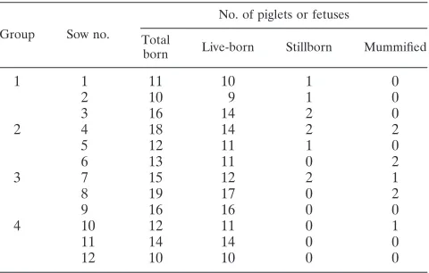
116 days postinsemination. Table 1 summarizes litter
charac-teristics by group.
Serology.
All sows were negative for anti-PCV2 antibodies
prior to insemination and seronegative for PRRSV, SIV
(H1N1 and H3N2), PPV, and EMCV at parturition. Group 1
sows (negative controls) remained negative for anti-PCV2
an-tibodies for the duration of the study. Table 2 summarizes sow
anti-PCV2 antibody development by group at 28, 56, 84, and
112 days of gestation. All sows in groups 2, 3, and 4 had
detectable anti-PCV2 antibodies at 112 days of gestation. A
significant effect of time (
P
⫽
0.027) was observed for the
production of anti-PCV2 antibodies; however, differences
be-tween groups were not observed.
PCV2 neutralizing antibodies were identified in all group 2,
3, and 4 sows during gestation, whereas group 1 sows remained
seronegative for PCV2 (Table 2). A significant effect of time
(
P
⬍
0.001) was observed for serum neutralizing antibody
production; however, significant group differences were not
observed at 56 and 112 days of gestation.
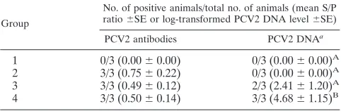
Colostrum-associated anti-PCV2 antibodies were present in
all group 2, 3, and 4 sows but were not detected in group 1 sows
(Table 3). A significant statistical difference was not observed
between groups (
P
⬎
0.05).
Anti-PCV2 immunoglobulin G (IgG) antibodies were not
detected in presuckle sera collected from group 1, 2, and 4
TABLE 1. Summary of expelled piglets and fetuses by litter and
group at parturition for nonvaccinated and noninoculated sows
(group 1), sows vaccinated at 28 days of gestation (groups 2
and 3), and sows oronasally inoculated with PCV2b
(groups 3 and 4) at 56 days of gestation
Group Sow no.
No. of piglets or fetuses Total
born Live-born Stillborn Mummified
1
1
11
10
1
0
2
10
9
1
0
3
16
14
2
0
2
4
18
14
2
2
5
12
11
1
0
6
13
11
0
2
3
7
15
12
2
1
8
19
17
0
2
9
16
16
0
0
4
10
12
11
0
1
11
14
14
0
0
12
10
10
0
0
on August 17, 2020 by guest
http://cvi.asm.org/
live-born piglets. Conversely, two live-born piglets in group 3
(from the same litter) had detectable anti-PCV2 antibodies,
with S/P ratios of 0.26 and 0.47 (Table 4).
PCV2 DNA detection by PCR.
PCV2 DNA was not detected
by quantitative real-time PCR amplification in weekly serum
samples collected from group 1, 2, or 3 sows prior to artificial
insemination or during gestation. In contrast, two of three
group 4 sows developed detectable PCV2 viremia during
ges-tation. PCV2 viremia in group 4 sows first appeared at 70 days
of gestation. Sows remained PCV2 viremic for four to seven
consecutive weeks, and one of three sows had detectable PCV2
DNA in serum at parturition. Significant differences in sow
viremia were not observed between groups (
P
⬎
0.05) (data
not shown).
Colostrum samples collected at parturition were negative for
PCV2 DNA for groups 1 and 2. In contrast, PCV2 DNA was
detected in two of three group 3 sows and three of three group
4 sows (Table 3). The amount of PCV2 DNA present in
co-lostrum of group 4 sows (mean log PCV2 genomic copies/ml
colostrum) was significantly (
P
⫽
0.042) higher than that
ob-served for group 3 (Table 3).
Presuckle sera collected from group 1 and 2 live-born piglets
did not contain PCV2 DNA. PCV2 DNA was detected in sera
from group 3 and group 4 live-born piglets (Table 4). Group 3
live-born viremic piglets originated from two different litters
(one and two piglets).
Macroscopic lesions, microscopic examination, and IHC.
At
necropsy, no gross lesions were observed in live-born piglets or
stillborns from group 1, 2, 3, or 4 sows. Furthermore, no
mi-croscopic tissue changes associated with PCV2 infection or
PCV2 antigen were detected in live-born piglets, stillborns, or
mummified fetuses from group 1, 2, or 3 sows or in examined
placental sections. However, one live-born piglet from a group
4 sow had mild multifocal myocardial degeneration and
necro-sis (Table 4), with lymphoid depletion in the tonsils and spleen.
In this piglet, moderate to abundant amounts of PCV2 antigen
were detected in the myocardium and tonsils, as determined by
IHC stains (Table 4).
DISCUSSION
PCV2-associated viremia in the dam is the most likely source
of fetal infection (29, 30), though semen transmission appears
to be a possible route (13, 14, 17, 18, 19, 35). Previous
exper-imental studies proved that PCV2 is capable of crossing the
placenta in naïve sows after intranasal inoculation during late
gestation (29). In the current study, oronasal PCV2 inoculation
resulted in detectable viremia, development of anti-PCV2
an-tibodies, and the presence of PCV2 DNA in colostrum samples
of group 4 (unvaccinated) sows. PCV2 viremia of the dam
resulted in transplacental infection of fetuses in utero,
con-firming the potential of vertical transmission of PCV2
infec-tion. In contrast to other experimental and field observations
of PCV2-associated reproductive failure (11, 24, 29, 31, 39),
abortion, dam illness, and increased numbers of nonviable
piglets were not observed under the conditions of this study.
Differences between studies may be related to differences in
the amount or virulence of the PCV2 isolate used for
inocu-lation. However, our findings suggest that vertical PCV2
trans-mission in the field could occur at a higher incidence than what
has been reported.
In recent years, multiple PCV2 vaccines have become
avail-able to combat PCVAD. Currently, three vaccines are licensed
for growing pigs of 3 to 4 weeks of age or older, and another
vaccine is labeled for prefarrowing usage on dams. In a PCV2
vaccination study involving 72 growing animals, vaccination
prevented detectable PCV2-associated viremia and reduced
PCV2 fecal and oral shedding of viral DNA (8). In another
experimental study, vaccination induced neutralizing
antibod-ies and reduced PCV2 viremia, fecal shedding, and
PCV2-associated microscopic lesions (25). Similar observations were
made during field investigations involving PCV2-vaccinated
and nonvaccinated pigs (6, 10, 15).
To date, the performance of only one PCV2 vaccine has
been studied experimentally with breeding animals. Sow
vac-cination reduced the number of stillborn piglets at parturition,
TABLE 2. Prevalence and sow-associated mean group levels of anti-PCV2 IgG and serum neutralizing antibodies for nonvaccinated and
noninoculated sows (group 1), sows vaccinated at 28 days of gestation (groups 2 and 3), and sows oronasally inoculated with
PCV2b (groups 3 and 4) at 56 days of gestation
Group
No. of sows with IgG antibody/no. of sows in group (mean S/P ratio⫾SE) on day of gestation
No. of sows with neutralizing antibody/ no. of sows in group (mean antibody
levela⫾SE) on day of gestation
28 56 84 112 56 112
1
0/3 (0.00
⫾
0.00)
0/3 (0.00
⫾
0.00)
0/3 (0.00
⫾
0.00)
0/3 (0.00
⫾
0.00)
0/3 (1.81
⫾
0.00)
0/3 (1.81
⫾
0.00)
2
0/3 (0.00
⫾
0.00)
2/3 (0.29
⫾
0.15)
2/3 (0.28
⫾
0.11)
3/3 (0.56
⫾
0.05)
3/3 (2.31
⫾
0.20)
3/3 (2.41
⫾
0.00)
3
0/3 (0.00
⫾
0.00)
2/3 (0.08
⫾
0.15)
2/3 (0.29
⫾
0.15)
3/3 (0.43
⫾
0.11)
2/3 (2.11
⫾
0.30)
3/3 (2.51
⫾
0.10)
4
0/3 (0.00
⫾
0.00)
0/3 (0.00
⫾
0.00)
1/3 (0.23
⫾
0.21)
3/3 (0.40
⫾
0.12)
0/3 (1.81
⫾
0.00)
3/3 (2.81
⫾
0.44)
a
Mean log-transformed serum neutralizing antibody level.
TABLE 3. Prevalence of anti-PCV2 IgG antibodies and PCV2
DNA in colostrum of nonvaccinated and noninoculated sows
(group 1), sows vaccinated at 28 days of gestation (groups 2
and 3), and sows oronasally inoculated with PCV2b
(groups 3 and 4) at 56 days of gestation
Group
No. of positive animals/total no. of animals (mean S/P
ratio⫾SE or log-transformed PCV2 DNA level⫾SE)
PCV2 antibodies PCV2 DNAa
1
0/3 (0.00
⫾
0.00)
0/3 (0.00
⫾
0.00)
A2
3/3 (0.75
⫾
0.22)
0/3 (0.00
⫾
0.00)
A3
3/3 (0.49
⫾
0.12)
2/3 (2.41
⫾
1.20)
A4
3/3 (0.50
⫾
0.14)
3/3 (4.68
⫾
1.15)
BaDifferent superscripts within the column indicate significant (P⬍0.05)
dif-ferences in group mean PCV2 DNA amounts.
on August 17, 2020 by guest
http://cvi.asm.org/
decreased prewean piglet mortality, improved sow mortality,
and improved farrowing rates (1, 3, 5, 12, 16, 36). Vaccination
was found to be successful in reducing PCV2-associated
repro-ductive failure and improving sow performance under field
conditions. However, to our knowledge, the effect of sow
vac-cination on fetal PCV2 replication in utero has not been
in-vestigated to date.
Detectable differences in the immune response following
vaccination of pregnant animals and differences in immune
regulation in the uterus during pregnancy have been reported
(22, 40). In the current study, sows were vaccinated after
con-firmation of pregnancy due to the limited availability of
PCV2-naïve sows. PCV2 vaccination of pregnant animals had no
detectable adverse effects when the vaccine was administered
at 28 days of gestation, eliminated detectable PCV2 viremia,
and induced both serum neutralizing antibodies and colostral
anti-PCV2 antibodies. However, vaccination did not prevent
the presence of PCV2 DNA in colostral samples, fetal PCV2
viremia, or the development of anti-PCV2 IgG antibodies in
piglets following PCV2 challenge at 56 days of gestation.
Pos-sible considerations for the lack of PCV2 viremia in group 3
sows include a limited detection rate due to the timing of
serum collection (weekly) and the sensitivity of the PCR assay.
Interestingly, live-born group 3 piglets that were viremic or had
anti-PCV2 antibodies at birth did not have detectable PCV2
antigen in tissues (myocardium, lungs, tonsils, or pinnae). This
suggests that either fetal infection occurred later in gestation
than that in group 4 piglets or dam vaccination reduced fetal
PCV2 replication.
Although PCV2 vaccination in this report did not prevent
fetal infection, two group 3 piglets developed specific
anti-PCV2 antibodies in utero without having detectable presuckle
viremia, microscopic lesions associated with fetal infection, or
detectable PCV2 antigen in tissue. This implies that
immuno-competent fetuses (
⬎
70 days of gestation) are able to clear
infection prior to parturition without associated microscopic
lesions or detectable PCV2 antigen. This observation has not
been reported previously and is similar to the case for PPV
infection in fetal swine (20).
In summary, PCV2 infection of naïve pregnant sows may not
result in reproductive failure but can be associated with fetal
infection. PCV2 vaccination of pregnant sows induced
neutral-izing and anti-PCV2 antibodies in serum and colostrum but did
not prevent vertical transmission. Sow vaccination also did not
prevent colostral shedding of PCV2, which can be another
route of vertical transmission. Furthermore, a proportion of
fetuses infected in utero were able to clear PCV2 infection
prior to parturition.
ACKNOWLEDGMENTS
This project was supported by Boehringer Ingelheim Vetmedica Inc.
No financial interest exists between the authors and Boehringer
Ingelheim Vetmedica Inc.
We thank Joseph Bender, Jeremy Johnson, Paul Thomas, and Troy
Worth for their assistance with collection of diagnostic samples
throughout the study and the Iowa State University Laboratory Animal
Resources staff for daily animal care.
REFERENCES
1.Bech, A. B., and L. Kunstmann.2008. Effect of sow vaccination with
Circo-vac on the performances of 3 Danish herds in Northern Jutland, p. 109.In
Proceedings of the 20th International Pig Veterinary Society Congress, Durban, South Africa, vol. 2. Hein Jonker Media Management, Durban, South Africa.
2.Cheung, A. K., K. M. Lager, O. I. Kohutyuk, A. L. Vincent, S. C. Henry, R. B. Baker, R. R. Rowland, and A. G. Dunham.2007. Detection of two porcine circovirus type 2 genotypic groups in United States swine herds. Arch. Virol.
152:1035–1044.
3.Delisle, C., G. Delisle, N. Bridoux, J. C. Thibault, S. Longo, and F. Joisel.
2008. Results of sow vaccination against PCV2 with Circovac in France:
improvement of reproduction parameters, p. 47.InProceedings of the 20th
International Pig Veterinary Society Congress, Durban, South Africa, vol. 1. Hein Jonker Media Management, Durban, South Africa.
4.Dupont, K., E. O. Nielsen, P. Baekbo, and L. E. Larsen.2008. Genomic analysis of PCV2 isolates from Danish archives and a current PMWS
case-control study supports a shift in genotypes with time. Vet. Microbiol.128:
56–64.
5.Ebbesen, T., and L. Kunstmann.2008. Effect of sow vaccination with
Cir-covac on stillborn piglets, p. 81.InProceedings of the 20th International Pig
Veterinary Society Congress, Durban, South Africa, vol. 2. Hein Jonker Media Management, Durban, South Africa.
6.Fachinger, V., R. Bischoff, S. B. Jedidia, A. Saalmuller, and K. Elbers.2008. The effect of vaccination against porcine circovirus type 2 in pigs suffering
from porcine respiratory disease complex. Vaccine26:1488–1499.
7.Fenaux, M., P. G. Halbur, G. Haqshenas, R. Royer, P. Thomas, P. Nawagit-gul, M. Gill, T. E. Toth, and X. J. Meng.2002. Cloned genomic DNA of type 2 porcine circovirus is infectious when injected directly into the liver and lymph nodes of pigs: characterization of clinical disease, virus distribution,
and pathologic lesions. J. Virol.76:541–551.
8.Fort, M., M. Sibila, A. Allepuz, E. Mateu, F. Roerink, and J. Segales.2008. Porcine circovirus type 2 (PCV2) vaccination of conventional pigs prevents viremia against PCV2 isolates of different genotypes and geographic origins.
Vaccine26:1063–1071.
9.Harding, J., and E. Clark.1997. Recognizing and diagnosing postweaning
multisystemic wasting syndrome (PMWS). J. Swine Health Prod.5:201–203.
10.Horlen, K. P.2008. A field evaluation of mortality rate and growth perfor-mance in pigs vaccinated against porcine circovirus type 2. J. Am. Vet. Med.
Assoc.232:906–912.
11.Johnson, C. S., H. S. Joo, K. Direksin, K. J. Yoon, and Y. K. Choi.2002. Experimental in utero inoculation of late-term swine fetuses with porcine
circovirus type 2. J. Vet. Diagn. Investig.14:507–512.
TABLE 4. Incidence of PCV2 DNA and anti-PCV2 IgG antibodies in presuckle serum samples, light microscope-associated lesions
(myocardial necrosis, myocarditis, or fibrosis), and IHC staining for PCV2 antigen in tissues (heart, lung, tonsil, and pinnae) by
group for piglets from nonvaccinated and noninoculated sows (group 1), sows vaccinated at 28 days of gestation (groups 2
and 3), and sows oronasally inoculated with PCV2b (groups 3 and 4) at 56 days of gestation
Group na
No. of animals with characteristic/total no. of animals
Presuckle serum Microscopic
lesions in heart
PCV2 antigen PCV2
DNA
PCV2
antibodies Heart Lung Tonsil Pinnae
1
37
0/33
0/33
0/37
0/37
0/37
0/37
0/37
2
43
0/36
0/36
0/43
0/43
0/43
0/43
0/43
3
50
3/45
2/45
0/50
0/50
0/50
0/50
0/50
4
36
1/35
0/35
1/36
1/36
0/36
1/36
0/36
aTotal number of live-born piglets, stillborns, and mummified fetuses.
on August 17, 2020 by guest
http://cvi.asm.org/
12.Joisel, F., A. Brune, A. Schade, S. Longo, and C. Charreyre.2008. Improve-ment of reproduction performance induced by PCV2 vaccination of sows and
gilts with Circovac in 277 German sow farms, p. 72.InProceedings of the
20th International Pig Veterinary Society Congress, Durban, South Africa, vol. 2. Hein Jonker Media Management, Durban, South Africa.
13.Kim, J., D. U. Han, C. Choi, and C. Chae.2001. Differentiation of porcine circovirus (PCV)-1 and PCV-2 in boar semen using a multiplex nested
polymerase chain reaction. J. Virol. Methods98:25–31.
14.Kim, J., D. U. Han, C. Choi, and C. Chae.2003. Simultaneous detection and differentiation between porcine circovirus and porcine parvovirus in boar semen by multiplex seminested polymerase chain reaction. J. Vet. Med. Sci.
65:741–744.
15.Kixmoller, M., M. Ritzmann, M. Eddicks, A. Saalmuller, K. Elbers, and V. Fachinger.2008. Reduction of PMWS-associated clinical signs and
co-infec-tions by vaccination against PCV2. Vaccine26:3443–3451.
16.Kunstmann, L., and L. Lau.2008. Effect of sow vaccination with Circovac on
the performance of 34 Danish herds, p. 75.InProceedings of the 20th
International Pig Veterinary Society Congress, Durban, South Africa, vol. 2. Hein Jonker Media Management, Durban, South Africa.
17.Larochelle, R., A. Bielanski, P. Muller, and R. Magar.2000. PCR detection and evidence of shedding of porcine circovirus type 2 in boar semen. J. Clin.
Microbiol.38:4629–4632.
18.Madson, D. M., S. Ramamoorthy, C. Kuster, N. Pal, X. J. Meng, P. G. Halbur, and T. Opriessnig.2008. Characterization of shedding patterns of porcine circovirus type 2a and 2b in experimentally inoculated mature boars.
J. Vet. Diagn. Investig.20:725–734.
19.McIntosh, K. A., J. C. Harding, S. Parker, J. A. Ellis, and G. D. Appleyard.
2006. Nested polymerase chain reaction detection and duration of porcine circovirus type 2 in semen with sperm morphological analysis from naturally
infected boars. J. Vet. Diagn. Investig.18:380–384.
20.Mengeling, W. L.2006. Porcine parvovirus, p. 373–386.InB. E. Straw, J. J. Zimmerman, S. D’Allaire, and D. J. Taylor (ed.), Diseases of swine. Black-well Publishing, Ames, IA.
21.Mengeling, W. L., J. F. Ridpath, and A. C. Vorwald.1988. Size and antigenic comparisons among the structural proteins of selected autonomous
parvo-viruses. J. Gen. Virol.69:825–837.
22.Morein, B., G. Blomqvist, and K. Hu.2007. Immune responsiveness in the
neonatal period. J. Comp. Pathol.137(Suppl. 1):S27–S31.
23.Nawagitgul, P., P. A. Harms, I. Morozov, B. J. Thacker, S. D. Sorden, C. Lekcharoensuk, and P. S. Paul.2002. Modified indirect porcine circovirus (PCV) type 2-based and recombinant capsid protein (ORF2)-based enzyme-linked immunosorbent assays for detection of antibodies to PCV. Clin.
Diagn. Lab. Immunol.9:33–40.
24.O’Connor, B., H. Gauvreau, K. West, J. Bogdan, M. Ayroud, E. G. Clark, C. Konoby, G. Allan, and J. A. Ellis.2001. Multiple porcine circovirus 2-asso-ciated abortions and reproductive failure in a multisite swine production
unit. Can. Vet. J.42:551–553.
25.Opriessnig, T., D. M. Madson, J. R. Prickett, D. Kuhar, J. K. Lunney, J. Elsener, and P. G. Halbur.2008. Effect of porcine circovirus type 2 (PCV2) vaccination on porcine reproductive and respiratory syndrome virus
(PRRSV) and PCV2 coinfection. Vet. Microbiol.131:103–114.
26.Opriessnig, T., X. J. Meng, and P. G. Halbur.2007. Porcine circovirus type 2 associated disease: update on current terminology, clinical manifestations,
pathogenesis, diagnosis, and intervention strategies. J. Vet. Diagn. Investig.
19:591–615.
27.Opriessnig, T., S. Yu, J. M. Gallup, R. B. Evans, M. Fenaux, F. Pallares, E. L. Thacker, C. W. Brockus, M. R. Ackermann, P. Thomas, X. J. Meng, and P. G. Halbur.2003. Effect of vaccination with selective bacterins on
conven-tional pigs infected with type 2 porcine circovirus. Vet. Pathol.40:521–529.
28.Pal, N., Y. W. Huang, D. M. Madson, C. Kuster, X. J. Meng, P. G. Halbur, and T. Opriessnig.2008. Development and validation of a duplex real-time PCR assay for the simultaneous detection and quantification of porcine circovirus type 2 and an internal control on porcine semen samples. J. Virol.
Methods149:217–225.
29.Park, J.-S., J. Kim, Y. Ha, K. Jung, C. Choi, J.-K. Lim, S.-H. Kim, and C. Chae.2005. Birth abnormalities in pregnant sows infected intranasally with
porcine circovirus 2. J. Comp. Pathol.132:139–144.
30.Pensaert, M. B., R. E. Sanchez, Jr., A. S. Ladekjær-Mikkelsen, G. M. Allan, and H. J. Nauwynck.2004. Viremia and effect of fetal infection with porcine viruses with special reference to porcine circovirus 2 infection. Vet.
Micro-biol.98:175–183.
31.Pittman, J. S.2008. Reproductive failure associated with porcine circovirus
type 2 in gilts. J. Swine Health Prod.16:144–148.
32.Pogranichniy, R. M., K. J. Yoon, P. A. Harms, S. L. Swenson, J. Zimmerman, and S. Sorden.2000. Characterization of immune response of young pigs to
porcine circovirus type 2 infection. Viral Immunol.13:143–153.
33.Sanchez, R. E., Jr., P. Meerts, H. J. Nauwynck, and M. B. Pensaert.2003. Change of porcine circovirus 2 target cells in pigs during development from
fetal to early postnatal life. Vet. Microbiol.95:15–25.
34.Sanchez, R. E., Jr., H. J. Nauwynck, F. McNeilly, G. M. Allan, and M. B. Pensaert.2001. Porcine circovirus 2 infection in swine foetuses inoculated at
different stages of gestation. Vet. Microbiol.83:169–176.
35.Schmoll, F.2008. Prevalence of PCV2 in Austrian and German boars and
semen used for artificial insemination. Theriogenology69:814–821.
36.Schøning, T., P. Nielsen, and L. Lau.2008. Effect of Circovac (Merial) on porcine circovirus type 2 (PCV2) sow reproductive failure and mortality: a
case report, p. 108.InProceedings of the 20th International Pig Veterinary
Society Congress, Durban, South Africa, vol. 2. Hein Jonker Media Man-agement, Durban, South Africa.
37.Sorden, S. D., P. A. Harms, P. Nawagitgul, D. Cavanaugh, and P. S. Paul.
1999. Development of a polyclonal-antibody-based immunohistochemical method for the detection of type 2 porcine circovirus in formalin-fixed,
paraffin-embedded tissue. J. Vet. Diagn. Investig.11:528–530.
38.Todd, D., M. Bendinelli, P. Biagini, S. Hino, A. Mankertz, S. Mishiro, C. Niel, H. Okamoto, S. Raidal, B. W. Ritchie, and G. Teo.2005. Circoviridae, p. 327–
334.InM. A. Fauquet, J. Mayo, U. Maniloff, and L. A. B. E. Desselberger (ed.),
Virus taxonomy. Elsevier Academic Press, San Diego, CA.
39.West, K. H., J. M. Bystrom, C. Wojnarowicz, N. Shantz, M. Jacobson, G. M. Allan, D. M. Haines, E. G. Clark, S. Krakowka, F. McNeilly, C. Konoby, K. Martin, and J. A. Ellis.1999. Myocarditis and abortion associated with intrauterine infection of sows with porcine circovirus 2. J. Vet. Diagn.
In-vestig.11:530–532.
40.Winter, A. J., C. E. Hall, R. H. Jacobson, D. R. Verstreate, M. P. Meredith, and W. L. Castleman.1986. Effect of pregnancy on the immune response of
cattle to a Brucella vaccine. J. Reprod. Immunol.9:313–325.