C
LINICAL ANDV
ACCINEI
MMUNOLOGY, June 2008, p. 925–931
Vol. 15, No. 6
1556-6811/08/$08.00
⫹
0
doi:10.1128/CVI.00500-07
Copyright © 2008, American Society for Microbiology. All Rights Reserved.
Expression of a Functional Single-Chain Variable-Fragment Antibody
against Complement Receptor 1 in
Streptococcus gordonii
䌤
Jennifer B. Knight,
1Scott A. Halperin,
1,3Kenneth A. West,
1and Song F. Lee
1,2,3*
Department of Microbiology and Immunology, Faculty of Medicine,
1and Department of Applied Oral Sciences, Faculty of
Dentistry,
2Dalhousie University, and Department of Pediatrics, Dalhousie University, and the IWK Health Centre,
3Halifax, Nova Scotia, Canada
Received 17 December 2007/Returned for modification 21 January 2008/Accepted 24 March 2008
Streptococcus gordonii
, an oral commensal organism, is a candidate vector for oral-vaccine development.
Previous studies have shown that recombinant
S. gordonii
expressing heterologous antigens was weakly
immunogenic when delivered intranasally. In this study, antigen was specifically targeted to antigen-presenting
cells (APC) in order to potentiate antigen-APC interactions and increase the humoral immune response to the
antigen. To achieve this goal, a single-chain variable-fragment (scFv) antibody against complement receptor 1
(CR1) was constructed. Anti-CR1 scFv purified from
Escherichia coli
was able to bind to mouse mixed
lymphocytes and bone marrow-derived dendritic cells. The in vivo function of the anti-CR1 scFv protein was
assessed by immunizing mice intranasally with soluble scFv and determining the immune response against the
hemagglutinin (HA) peptide located on the carboxy terminus of the scFv. The serum anti-HA immunoglobulin
G (IgG) immune response was dose dependent; as little as 100 ng of anti-CR1 scFv induced a significant IgG
immune response, while such a response was minimal when the animals were given an unrelated scFv. The
anti-CR1 scFv was expressed in
S. gordonii
as a secreted protein, which was functional, as it bound to dendritic
cells. Mice orally colonized by the anti-CR1-secreting
S. gordonii
produced an anti-HA IgG immune response,
indicating that such an approach can be used to increase the immune response to antigens produced by this
bacterium.
Streptococcus gordonii
is a commensal bacterium found in
the oral cavities of humans. The organism is considered to be
an attractive vector as a live-oral-vaccine vehicle (14, 23). A
number of heterologous antigens have been expressed in this
organism as either secreted proteins (15, 27) or cell
wall-an-chored proteins (16, 17, 19, 20, 25, 26). In the murine
oral-colonization model, the recombinant
S. gordonii
was able to
establish long-term colonization (16, 20). However, there are
difficulties in stimulating a strong protective immune response
against recombinant antigens following oral colonization.
Antigen targeting to immune cells has the potential to
ma-nipulate the immune system and elicit an immune response
more efficiently. Monoclonal immunoglobulin G (IgG)
anti-bodies have long been used as specific targeting vehicles. A
number of reports have indicated success in achieving
en-hanced immune responses using antibodies to complement
receptor 1 (CR1) and CR2 (3, 8, 30), Fc receptors (1, 2), and
dendritic cell DEC205 receptor (5, 6). However, there are
limitations in using intact IgG as a targeting vehicle; these
limitations include a weak extravascular transport ability for
IgG and difficulties with expressing whole IgG by bacteria.
Single-chain variable-fragment (scFv) antibodies, however,
of-fer a number of advantages, e.g., they can be readily produced
by bacteria and can be easily engineered genetically as fusion
proteins carrying polypeptide antigens. In the context of
anti-gen targeting, scFvs against CR1 and -2 (21, 24), DEC205 (9),
CD3 (31), and natural killer NKG2D receptor (29) have been
reported with some degree of success.
In this study, we have taken the approach of expressing an
scFv antibody against CR1 in
S. gordonii
to target immune
cells. CR1 is a phagocytic receptor expressed by a number of
immune cells, including dendritic cells, macrophages,
neutro-phils, eosinoneutro-phils, and B cells, as well as erythrocytes. The
anti-CR1 scFv was tested for binding to target cells in vitro and
used in intranasal immunization in mice.
MATERIALS AND METHODS
Bacteria and growth conditions.S. gordoniiwas cultivated in Todd-Hewitt broth containing 0.5% yeast extract at 37°C aerobically without shaking.
Kana-mycin and tetracycline, when needed, were included in the medium at 250g/ml
and 10g/ml, respectively. RecombinantEscherichia coliwas grown aerobically
with vigorous shaking at 37°C in Luria Bertani broth (1% tryptone, 0.5% yeast extract, and 1% NaCl [wt/vol]) or Super Broth (1% MOPS [morpholinepropane-sulfonic acid], 3% tryptone, 2% yeast extract [wt/vol]) containing either 100
g/ml of ampicillin or 50g/ml of kanamycin. All antibiotics were purchased
from Sigma-Aldrich Chemical Co. (Oakville, ON, Canada).
Construction of the anti-CR1 scFv.The anti-CR1 antibody gene was obtained from the anti-CR1 monoclonal antibody-producing hybridoma HB8592 (Amer-ican Type Culture Collection, Manassas, VA). The cells were grown in modified
Dulbecco’s medium supplemented with 5 mM-mercaptoethanol and 10% fetal
calf serum (Sigma-Aldrich). Total RNA was isolated from 1⫻106hybridoma
cells by extraction with the Trizol reagent (Invitrogen Life Technologies,
Burl-ington, ON, Canada). The RNA obtained was dissolved in 40l of
diethylpyro-carbonate-treated water. cDNA was synthesized from the RNA by reverse tran-scription using oligo(dT) as the primer and murine leukemia virus reverse transcriptase (Invitrogen) according to the manufacturer’s instructions. The
vari-able light-chain (VL) and heavy-chain (VH) antibody fragments were amplified
by PCR using mixed primers as described by Barbas et al. (4). The resulting
0.4-kb VLor VHDNA fragments were gel purified and used in overlapping PCR
to produce the scFv antibody DNA. The resulting 0.8-kb scFv fragment was
* Corresponding author. Mailing address: Department of Applied
Oral Sciences, Faculty of Dentistry, Dalhousie University, Halifax,
Nova Scotia, Canada B3H 3J5. Phone: (902) 470-7522/494-8799. Fax:
(902) 494-6621. E-mail: [email protected].
䌤
Published ahead of print on 2 April 2008.
925
on August 17, 2020 by guest
http://cvi.asm.org/
ligated into the SfiI sites of the phagemid pComb3X (4). The ligated DNA was
transformed intoE. coliXL1-Blue. The resulting construct (pCR1) was verified
by restriction analysis and DNA sequencing (The John P. Robarts Research Institute DNA Sequencing Facility, London, ON, Canada).
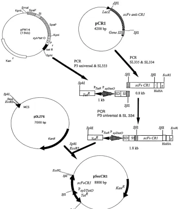
Cloning of the anti-CR1 scFv gene inS. gordonii.To express the anti-CR1 scFv inS. gordonii, pSecCR1 was constructed. The construction of pSecCR1 is out-lined in Fig. 1. Briefly, the DNA coding for the anti-CR1 scFv and the C-terminal histidine and hemagglutinin (HA) tags was obtained by PCR using the primer pair SL334 (CGGAATTCCGTTAAGAAGCGTAGTCCGGAACGTC; the EcoRI site is underlined) and SL335 (GAGGCCCAGGCGGCCGAGCTC). A 1-kb DNA fragment carrying the TetR repressor gene, the tetracycline-inducible
xyl/tetOpromoter, and the ribosomal binding site and signal sequence ofspaP
originating fromStreptococcus mutans, was amplified from pPM14 (18) using
primers P3 Universal (ACGCCAAGCTTGCATGCCTGC; the SphI site is un-derlined) and SL333 (GAGCTCGGCCGCCTGGGCCTCATCGGCAAAAAC CTTTTG). The two fragments were joined together via overlapping PCR, and the resulting 1.8-kb PCR product was cloned into the SphI and EcoRI sites on theE. coli-Streptococcusshuttle vector pDL276, creating pSecCR1. pSecCR1 was
transformed intoS. gordonii(hppG::Tetr
) via natural transformation (13).
Isolation of scFvs fromE. coli.pCR1 was transformed intoE. coliTB1 (New England Biolabs, Mississauga, ON, Canada) for the production of scFv. As a
control, pK8 (pComb3X carrying scFv recognizing pertussis toxin [S. F. Lee,
K. G. Chan, and S. A. Halperin, unpublished data]) was also introduced intoE.
coliTB1. The recombinantE. coliwas grown in 500 ml of Super Broth for 16 h.
Cells were harvested by centrifugation (10,000⫻g) and resuspended in 5 ml
NiCAM wash buffer (50 mM NaH2PO4, 300 mM NaCl, 20 mM imidazole) with
1 mM phenylmethylsulfonyl fluoride and sonicated (15 10-s bursts at amplitude 35 separated by 10-s cooling periods; Vibra cell; Sonics and Materials Inc.,
Danbury, CT). The sonicate was centrifuged at 20,000⫻gfor 20 min at 4°C. The
clear supernatant was passed through a 1.5-ml NiCAM column (Sigma-Aldrich) equilibrated with the wash buffer. Unwanted proteins were removed with 20 ml of wash buffer. The bound scFv proteins were eluted with 5 ml of elution buffer
(50 mM NaH2PO4, 300 mM NaCl, 250 mM imidazole). The protein was
con-centrated by ultrafiltration using Amicon Ultra-Free-CL centrifugal filter devices (5,000-Da molecular mass cutoff).
scFv isolation fromS. gordonii.Anti-CR1 scFv was isolated from the culture
supernatant of an overnightS. gordoniiculture (100 ml) using a NiCAM affinity
column essentially as described above. Briefly, the culture supernatant was passed through the NiCAM column twice, unwanted proteins were removed by
a 20-ml wash, and scFv was eluted with four 500-l elution buffers. The eluates
were used in Western blotting and enzyme-linked immunosorbent assay (ELISA) as described below.
FIG. 1. Construction of pSecCR1. See Materials and Methods for details. SS, signal sequence; SD, ribosomal-binding site; HIS, hexahistidine tag.
on August 17, 2020 by guest
http://cvi.asm.org/
SDS-PAGE and Western immunoblotting.Protein samples were analyzed by sodium dodecyl sulfate-polyacrylamide gel electrophoresis (SDS-PAGE) on 12% polyacrylamide gels using the buffer system of Laemmli (12). Proteins were stained with Coomassie blue. The scFv protein concentration was determined by comparison with bovine serum albumin (BSA) standards on the same SDS-PAGE gel using Image J software (National Institutes of Health, Bethesda, MD). For Western immunoblotting, proteins were transferred to nitrocellulose membranes (28) and antigens were detected using an anti-HA monoclonal an-tibody (1/20,000; Sigma-Aldrich) and goat anti-mouse IgG alkaline phosphatase-conjugated antibody (1/20,000; Sigma-Aldrich).
Isolation of mixed lymphocytes.Mixed lymphocytes were isolated from blood obtained from BALB/c mice using Ficoll Paque Plus (Amersham Biosciences, Baie d’Urfe, PQ, Canada) according to the manufacturer’s instructions. The mixed lymphocytes were used to coat 96-well polystyrene microtiter plates.
Bone marrow-derived dendritic cells.Dendritic cells were isolated and cul-tured from the femurs and tibias of BALB/c mice as described previously (7). The cells were harvested on day 6 and used in ELISA and immunofluorescence assays as described below.
Immunization.The anti-CR1 and -K8 scFvs isolated as described above were dialyzed against phosphate-buffered saline (PBS) and used to immunize BALB/c
mice (female; 5 weeks old;n⫽4; Charles River Laboratory, St. Constant,
Quebec, Canada) using lipopolysaccharide (LPS) (10g) fromE. coli6127:B8
(Sigma-Aldrich) as an adjuvant. The mice were immunized intranasally with 0.1
or 5g of scFv. One group of mice received LPS as a control. Intranasal
immunization was achieved by pipetting 30l of prepared antigen dropwise into
the nostrils of sedated animals. The animals were immunized on days 1, 23, 33, and 95. Sera and saliva were collected on day 7 prior to immunization and on day 107 using methods described previously (11).
Oral colonization.The oral colonization of 4-week-old BALB/c mice (female;
n⫽3) withS. gordonii SecCR1 andS. gordoniiSL3 was carried out using
methods described previously (16).S. gordoniiSL3 is a kanamycin-resistant strain
but lacks the anti-CR1 gene (14). To induce the expression of the anti-CR1 scFv,
the animals were given intraperitoneal injections of 100g tetracycline in 100l
of PBS on days 1, 8, 16, and 22 (18). The animals were euthanized on day 29, at which time microbiological swabs were obtained from the nasal cavity, oral cavity, and pharynx (14, 16). The swabs were inoculated onto brain heart infusion agar plates containing kanamycin. The colonies obtained were observed to have
colony morphology similar to that ofS. gordoniiand were gram-positive cocci in
short chains. Western immunoblot analysis of culture supernatants of selected colonies confirmed the presence of the 33-kDa scFv protein. Saliva was collected 2 days prior to colonization and on day 28. Sera were obtained 1 day prior to colonization and at euthanasia.
ELISA.ELISAs were used to test the function of scFv in binding to mixed lymphocytes and dendritic cells and detection of HA-specific antibodies in saliva and sera. For the detection of scFv binding, 96-well flat-bottom polystyrene microtiter plates (Corning Inc., Corning, NY) were coated with mixed lympho-cytes and dendritic cells (8,000 cells/well), and the cells were lightly fixed with 0.125% gluteraldehyde at 4°C overnight. After overnight incubation, the coated
plates were used immediately or stored at⫺80°C until they were used. The plates
were blocked with 1% (wt/vol) BSA in PBS containing 0.1% (wt/vol) Tween 20 (PBST) for 1 h at room temperature, and scFv (200 ng/well) was added. The plates were incubated at 4°C for 16 h and washed with PBST, and the bound scFv was detected with commercial HA monoclonal antibody and goat anti-mouse alkaline phosphatase conjugates.
For the detection of HA-specific antibodies in sera and saliva, microtiter plates were coated with 100 ng/well recombinant cyclophilin 18 (rC18) in ELISA
coat-ing buffer at 4°C overnight. The C18 gene originatcoat-ing fromToxoplasma gondii
was PCR amplified from pKJ97 (10) using the primer pair SL330 (CCTGGCC CAGGCGGCCGAAAATGCCGGAGTCAGAAAG; the SfiI site is underlined) and SL331 (CCTGGCCGGCCTGGCCCTCCAACAAACCAATGTCCGT; the SfiI site is underlined). The amplified 514-bp PCR fragment encoding the mature C18 was inserted into the SfiI sites on pComb3X. The rC18 protein was isolated
as a single 18-kDa protein fromE. colilysate using NiCAM chromatography as
described above. rC18 had no homology to the anti-CR1 or K8 scFv but con-tained the same HA tag, and the protein was large enough to coat the wells easily. Preliminary results showed that consistent ELISA results were obtained compared to the inconsistent results when HA peptide was used. The plates were blocked with 1% BSA in PBST, and serially diluted sera or saliva was added (11). IgG antibodies were detected by the alkaline phosphatase-conjugated goat anti-mouse IgG. IgA antibodies were detected by a biotinylated goat anti-anti-mouse IgA
(␣-chain specific; 1/20,000; Sigma-Aldrich), followed by an avidin-alkaline
phos-phatase conjugate (1/20,000; Sigma-Aldrich). The titers of antibodies were
ex-pressed as the reciprocal of the dilution that produced anA405reading 0.05
higher than that of the pooled preimmune samples.
Immunofluorescence.Bone marrow-derived dendritic cells obtained as de-scribed above were used as the CR1-expresssing cells. For a non-CR1-producing control, mouse TC-1 lung epithelial cells (ATCC) were used. TC-1 cells were cultured in RPMI 1640 medium to 90% confluence according to the supplier’s instructions. The cells were detached with a 0.25% (wt/vol) trypsin–0.53 mM EDTA solution and centrifuged. One million dendritic cells or TC-1 cells were
centrifuged (350⫻g; 5 min; 4°C) and washed once in PBS. The cells were
resuspended in 0.5 ml of PBS-BSA buffer (PBS plus 1% BSA), and 1g of a rat
anti-mouse CD16/CD32 (Fc␥III/II receptors) monoclonal IgG (BD Biosciences,
Mississauga, ON, Canada) was added. The cells were incubated on ice for 5 min
to allow the antibody to bind to the Fc␥III/II receptors to eliminate background
binding in subsequent steps. Following incubation, the cells were centrifuged and resuspended in 0.8 ml PBS-BSA buffer. The cells were divided into four equal aliquots and incubated at 4°C with gentle mixing as follows: aliquot 1 with 200 ng anti-CR1, aliquot 2 with 200 ng K8 scFv, and aliquos 3 and 4 with PBS-BSA buffer alone. After 1 h, the cells were washed in PBS-BSA, anti-HA (1/200) antibody was added to the first three aliquots, and PBS-BSA buffer was added to the fourth aliquot. The cells were further incubated for 1 h. The cells were then
washed, and 0.5g of a rat anti-mouse IgG-specific fluorescein
isothiocyanate-conjugated monoclonal antibody (clone R35-95; BD Biosciences) was added to
the first three aliquots. To the fourth aliquot of cells, 0.25g of a rat anti-mouse
CR1/2-specific fluorescein isothiocyanate-conjugated monoclonal antibody (clone eBIo8D9; eBiosciences, San Diego, CA) was added. The cells were
incu-bated for 30 min at room temperature and washed, and a 10-l droplet was
allowed to dry as a smear on a glass slide in the dark. A drop of emulsion containing 90% glycerol and 10% PBS was applied to prevent rapid quenching of the fluorescence, and a coverslip was placed on top of the emulsion. The cells were viewed with a Leica DM2500 microscope (495-nm excitation; 525-nm emis-sion), and images were captured with a digital camera. The images were con-verted to black and white and pixel recon-verted to allow easier viewing using the PhotoShop program.
Statistical analysis.The results were analyzed with the paired Student’sttest,
and aPvalue of⬍0.05 was considered significant.
Nucleotide sequence accession number.The GenBank accession number for the anti-CR1 scFv nucleotide sequence is EF694984.
RESULTS
Construction and expression of the anti-CR1 scFv.
The
anti-CR1 scFv was constructed from RNA isolated from anti-anti-CR1
monoclonal-antibody-producing hybridoma cells. The
con-struct was verified by restriction analysis and DNA sequencing.
The amino acid sequence deduced from the DNA sequence
revealed the expected framework and
complement-determin-ing regions with the expected cysteine residues in both the
V
Kand V
Hdomains. In addition, the glycine-serine linker and
the histidine and HA tags introduced in the cloning were
identifiable (Fig. 2).
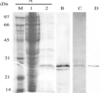
E. coli
carrying pCR1 was shown to express a 27-kDa protein
recognized by the commercial anti-HA antibody, indicating
that scFv was correctly constructed. The 27-kDa protein was
subsequently isolated from the
E. coli
lysate via affinity
chro-matography on a NiCAM column (Fig. 3). The isolated 27-kDa
protein, in addition to a smaller protein, presumably a
degra-dation product, was recognized by the commercial anti-HA
antibody. The control K8 scFv was similarly isolated and
rec-ognized by the anti-HA antibody (Fig. 3). The amount of
isolated anti-CR1 and K8 scFvs was approximately 130
g/liter
of culture.
Binding of anti-CR1 scFv to lymphocytes and dendritic
cells.
The ability of the isolated anti-CR1 scFv to bind to
murine mixed lymphocytes and bone marrow-derived dendritic
cells was examined. As shown in Fig. 4, the isolated anti-CR1
scFv bound to the two cell types while the control K8 scFv
V
OL. 15, 2008
ANTI-CR1 scFv
927
on August 17, 2020 by guest
http://cvi.asm.org/
generated against pertussis toxin did not. The results indicated
that the anti-CR1 scFv was functional. Binding was also
ob-served using immunofluorescence microscopy (Fig. 5). The
anti-CR1 scFv clearly bound to bone marrow-derived dendritic
cells (Fig. 5B), while the K8 scFv did not (Fig. 5C). A
com-mercial anti-mouse CR1 antibody also bound to the dendritic
cells (Fig. 5A). As expected, none of the antibodies bound to
the control epithelial cells (Fig. 5D to F).
Induction of immune response to HA.
The ability of the
anti-CR1 scFv to induce an immune response was tested in a
murine immunization experiment. The HA tag present on the
carboxy terminus of anti-CR1 served as the antigen. The
un-related K8 scFv, which also contained the same HA tag, was
used as a control. The animals were given scFv intranasally
with LPS as the adjuvant. As shown in Fig. 6, all groups that
received the anti-CR1 scFv showed a strong IgG response, with
the strongest response in the group that received 5
g of
protein. The increase in immune response between animals
immunized intranasally with 0.1
g and 5
g anti-CR1 was
statistically significant (
P
⫽
0.03). Although an IgG response
was also observed in the control group receiving the K8 scFv,
the response was weaker. The responses from immunization
FIG. 2. Deduced amino acid sequence of the anti-CR1 scFv. The framework regions (FR), complement-determining regions (CDR; boldface
letters), cysteine residues for disulfide bond formation, glycine-serine linker, and hexahistidine and HA tags are indicated.
FIG. 3. SDS-PAGE and Western immunoblotting of anti-CR1
scFv protein produced by
E. coli
. (A) Coomassie blue-stained
SDS-PAGE gel. Lane M, low-molecular-mass protein markers; lane 1,
crude cell lysate; lane 2, anti-CR1 scFv eluted from the NiCAM
col-umn. (B) Immunoblot of the isolated anti-CR1 scFv probed with the
anti-HA antibody. (C) Coomassie blue-stained K8 scFv isolated by
NiCAM chromatography. (D) Immunoblot of the isolated K8 scFv
probed with the anti-HA antibody.
FIG. 4. Binding of anti-CR1 scFv to mouse mixed lymphocytes
(A) and bone marrow-derived dendritic cells (B). The function of the
purified scFv protein was determined by its ability to bind to cells
expressing the ligand. The bound anti-CR1 scFv protein was detected
by the anti-HA antibody. The results shown are means
⫾
standard
deviations of triplicate wells.
on August 17, 2020 by guest
http://cvi.asm.org/
with 0.1
g anti-CR1 and 0.1
g K8 were significantly different
(
P
⫽
0.008). The response by mice that received 5
g of
anti-CR1 was also higher than in mice that received 5
g of K8
(
P
⫽
0.048). A response was not observed in the group that
received only LPS. A salivary IgA response was not observed in
any of the groups.
Expression and function of anti-CR1 in
S. gordonii
.
The
anti-CR1 scFv gene was subcloned into pDL276 as outlined in
Fig. 1. The expression of scFv was under the control of a
tetracycline-inducible
xyl-tetO
promoter. The promoter was
shown to be functional in
E. coli
and
S. gordonii
(18).
E. coli
carrying pSecCR1 was shown to express a 33-kDa protein
recognized by the commercial anti-HA antibody, indicating the
gene had been successfully subcloned (data not shown).
pSecCR1 was transformed into a tetracycline-resistant strain
of
S. gordonii
DL-1 (
hppG
::
tet
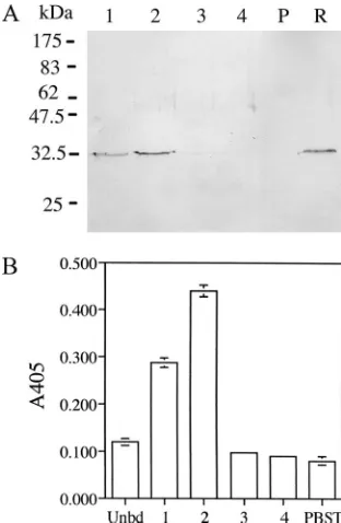
). The transformant secreted a
33-kDa protein recognized by the commercial HA
anti-body, while such a band was absent in the culture supernatant
from the parent strain (Fig. 7A). The 33-kDa protein bound to
the NiCAm column and could be eluted with imidazole. The
eluted protein was able to bind to mouse dendritic cells in the
ELISA (Fig. 7B), indicating that it was functional.
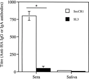
Induction of an immune response following oral
coloniza-tion with
S. gordonii
.
The ability of the anti-CR1-producing
S.
gordonii
to induce an immune response was tested in a mouse
oral-colonization study. The results showed that the two
groups of mice were colonized to the same degree. In mice
given
S. gordonii
SecCR1, the oral cavity, pharynx, and nasal
cavity contained 1,013
⫾
200 (mean
⫾
standard error), 115
⫾
56, and 20
⫾
10 CFU of
S. gordonii
, respectively. In mice given
S. gordonii
SL3, there were 950
⫾
370, 130
⫾
77, and 25
⫾
7
CFU of
S
.
gordonii
in these sites. Sera from mice colonized
with the anti-CR1-producing
S. gordonii
showed an immune
response, while that from mice colonized with the control
S.
gordonii
SL3 did not (Fig. 8). No difference in IgA response
was observed in saliva obtained from both groups.
DISCUSSION
In the present study, the single-chain recombinant antibody
against CR1 was constructed from the cDNA obtained from a
hybridoma. The construct was verified by DNA sequencing.
The scFv was successfully expressed in
E. coli
and
S. gordonii
.
The binding assay showed that the scFv was able to bind to
dendritic cells and mixed lymphocytes, indicating that the scFv
retained its function. The expression of a functional anti-CR1
scFv derived from a different hybridoma, 7G6, was previously
reported by Prechl et al. (24), although that scFv remained in
the insoluble fraction of
E. coli
while ours was mostly soluble.
To the best of our knowledge, this is the first report of the
FIG. 5. Detection of anti-CR1 scFv binding to mouse bone
mar-row-derived dendritic cells by immunofluorescence labeling. (A to C)
Dendritic cells reacted with a commercial anti-CR1/2 monoclonal
an-tibody, anti-CR1 scFv, and K8 scFv, respectively. (D to F) Mouse TC-1
epithelial cells reacted with a commercial anti-CR1/2 monoclonal
an-tibody, anti-CR1 scFv, and K8 scFv, respectively. See Materials and
Methods for details.
FIG. 6. Antigen-specific serum IgG response in BALB/c mice
fol-lowing intranasal immunization with the CR1 scFv. CR1,
anti-CR1-scFv; K8, antipertussis toxin scFv; LPS, LPS alone. The results
shown are means
⫾
standard errors of three or four individual sera.
FIG. 7. Expression and function of anti-CR1 scFv in
S. gordonii
.
(A) Immunoblot of culture supernatant from
S. gordonii
SecCR1 (lane
R) or parent
S. gordonii
(lane P) concentrated 20-fold by
trichloroace-tic acid precipitation and anti-CR1 scFv isolated by NiCAM
chroma-tography. Lanes 1 to 4 are fractions from the column. (B) Eluted (bars
1, 2, 3, and 4) and unbound (Unbd) fractions from the NiCAM column
were tested for binding to dendritic cells in ELISA. PBST, wells in
which PBST was added in place of fractions. The results shown are
means
⫾
standard deviations of triplicate wells.
V
OL. 15, 2008
ANTI-CR1 scFv
929
on August 17, 2020 by guest
http://cvi.asm.org/
expression of anti-CR1 scFv in
S. gordonii.
Oggioni et al. (22)
previously described the surface expression of an scFv against
the
Streptococcus mutans
major surface protein antigen P1 in
S.
gordonii
and demonstrated the ability of such an scFv to reduce
dental caries in an animal model. These results collectively
indicate that functional scFvs can be expressed in
E. coli
and
S.
gordonii
.
The anti-CR1 scFv isolated from
E. coli
was able to elicit a
very robust antibody response when given intranasally. The
antibody response was significantly higher than that from mice
immunized with the control K8 scFv, indicating that the
anti-CR1 scFv works as postulated and efficiently targets antigen to
phagocytic cells, resulting in an increased immune response.
These data are in agreement with those observed for the
anti-CR1/2 whole IgG molecule coupled to ovalbumin (3) or
an-thrax lethal toxin (30). However, our result is superior to that
reported for the anti-CR1/2 scFv derived from hybridoma 7G6,
which failed to elicit a significant antibody response (24) even
though the scFv was taken up and the influenza virus peptide
carried on the scFv was efficiently presented by
antigen-pre-senting cells in vitro (21). The difference may be due to the use
of an LPS adjuvant in our experiment, whereas no adjuvant
was used for the 7G6-derived scFv. LPS is known to
up-regu-late the expression of major histocompatibility complex
mole-cules, CD40 ligand, and cytokines by antigen-presenting cells,
leading to full activation of T cells and ultimately a strong
immune response. Therefore, we believe that LPS played a
role as an adjuvant in facilitating the robust immune response
to the HA peptide in our experiment. The adjuvant effect of
LPS is likely responsible for the observed immune response to
the control K8 scFv, particularly at 5-
g doses.
It is noteworthy that the superior immune response
ob-served in this study was obtained via intranasal immunization.
All the studies reported in the literature were performed using
the parenteral route. In view of the actual amount of HA
administered (0.1
g of anti-CR1 contains approximately 0.003
g of HA peptide) and the fact that a large quantity of antigen
is usually needed for mucosal immunization, the observed
im-mune response is quite remarkable and further indicates the
efficiency of the targeting ability of the anti-CR1 scFv.
The results of the oral-colonization experiment are in
agree-ment with those observed using purified scFv delivered
intra-nasally. We estimated that the amount of anti-CR1 scFv
pro-duced by
S. gordonii
was 17 ng/10
9CFU (unpublished data),
and in previous colonization studies, we reported that
S.
gor-donii
was present to the order of 10
3CFU on the oral-nasal
pharynx (14). Thus, the amount of scFv produced during
colonization at a given time is small (but production is
contin-uous), and yet an immune response was observed, further
indicating the efficiency of the anti-CR1 scFv.
It is also interesting that in both scFv protein
intranasal-immunization and bacterial-colonization experiments, only a
systemic IgG response was observed. Typically, intranasal
im-munization elicits both a systemic and a mucosal antibody
response, and this was certainly the case with other protein
antigens (11, 14). On the other hand, our previous results from
oral colonization with the recombinant
S. gordonii
elicited only
a weak mucosal IgA response (16). The deviation from these
past results suggests that the anti-CR1 scFv may have a unique
feature in that it selectively promotes a systemic response,
although the mechanism remains unclear.
In summary, an scFv against CR1 was constructed and
ex-pressed as a functional protein by both
E. coli
and
S. gordonii
.
The scFv was able to elicit an enhanced immune response to
the HA peptide linked to its C terminus, presumably due to its
antigen-targeting ability. An immune response was also
ob-served in a mouse
S. gordonii
oral-colonization experiment,
indicating that the scFv may be used as an antigen-targeting
tool in the bacterium.
ACKNOWLEDGMENTS
We thank Yi-Jing Li and Annette Morris for their valuable technical
assistance. We also thank Carlos Barbas III for providing pComb3X
and Keith Joiner for providing pKJ97.
J. B. Knight was an IWK graduate studentship recipient. This study
was supported by the Canadian Institutes of Health Research (CIHR).
REFERENCES
1.Adamova, E., M. C. Walsh, R. R. Gosselin, K. Hale, M. T. Preissler, R. F. Graziano, and E. J. Gosselin.2005. Enhanced antigen-specific antibody and cytokine responses when targeting antigen to human FcGamma receptor
type I using an anti-human Fc␥receptor type I-streptavidine fusion protein
in an adjuvant-free system. Immunol. Investig.34:417–429.
2.Anderson, C. F., and D. M. Mosser.2002. Cutting edge: biasing immune
responses by directing antigen to macrophage Fc␥receptors. J. Immunol.
198:3697–3701.
3.Baiu, D. C., J. Prechl, A. Tchorbanov, H. D. Molina. A. Erdei, A. Sulica, P. J. A. Capel, and W. L. W. Hazenbos.1999. Modulation of the humoral immune response by antibody-mediated antigen targeting to complement
receptors and Fc␥receptors. J. Immun.162:3125–3130.
4.Barbas, C. L., D. R. Barton, and J. K. Scott.2001. Phage display: a laboratory manual. Cold Spring Harbor Laboratory Press, Plainview, NY.
5.Bonifaz, L., D. Bonnyay, K. Mahnke, M. Rivera, M. C. Nussenzwei, and R. M. Steinman.2002. Efficient targeting of protein antigen to the dendritic cell receptor DEC 205 in the steady state leads to antigen presentation on
major histocompatibility complex class I products and peripheral CD8⫹T
cell tolerance. J. Exp. Med.196:1627–1638.
6.Bonifaz, L. C., D. P. Bonnyay, A. Charalambous, D. I. Darguste, S. Fuji, H. Soares, M. K. Brimnes, B. Moltedo, T. M. Moran, and R. M. Steinman.2004. In vivo targeting of antigens to maturing dendritic cells via the DEC 205
receptor improves T cell vaccination. J. Exp. Med.199:815–824.
7.Chan, K. G., M. Mayer, E. M. Davis, S. A. Halperin, T. J. Lin, and S. F. Lee.
2007. The roles ofD-alanylation ofStreptopcoccus gordoniilipoteichoic acid
in innate and adaptive immunity. Infect. Immun.75:3033–3042.
8.Craig, M. L., M. L. Reinagel, E. N. Marti, R. Schlimgen, A. Nardin, and R. P. Taylor. 1999. Infusion of bispecific monoclonal antibody complexes into monkeys provides immunologic protection against later challenge with a
model pathogen. Clin. Immunol.92:170–180.
9.Demangel, C., J. Zhou, A. B. H. Choo, G. Shoebridge, G. M. Halliday, and W. J. Britton.2005. Single chain antibody fragments for the selective
tar-geting of antigens to dendritic cells. Mol. Immunol.42:979–985.
10.High, K. P., K. A. Joiner, and R. E. Handschumacher.1994. Isolation, cDNA
FIG. 8. Immune response from oral colonization with
S. gordonii
.
Antigen-specific IgG antibody in sera and IgA antibody in saliva from
mice colonized with
S. gordonii
SecCR1 or SL3. The results shown are
means
⫾
standard errors of three individual sera or salivas.
*
,
P
⬍
0.05.
on August 17, 2020 by guest
http://cvi.asm.org/
sequences, and biochemical characterization of the major
cyclosporine-bind-ing proteins ofToxoplasma gondii. J. Biol. Chem.269:9105–9112.
11.Knight, J. B., Y. Y. Huang, S. A. Halperin, R. Anderson, A. Morris, A. MacMillan, T. Jones, D. S. Burt, G. Van Nest, and S. F. Lee.2006. Immu-nogenicity and protective efficacy of a recombinant filamentous
hemagglu-tinin fromBordetella pertussis. Clin. Exp. Immunol.144:5543–5551.
12.Laemmli, U. K.1970. Cleavage of structural proteins during the assembly of
the head of bacteriophage T4. Nature227:680–685.
13.Lee C. W., S. F. Lee., and S. A. Halperin.2004. Expression and
immunoge-nicity of a recombinant diphtheria toxin fragment A inStreptococcus
gordo-nii. Appl. Environ. Microbiol.70:4569–4574.
14.Lee, S. F.2003. Oral colonization and immune responses toStreptococcus gordonii: potential use as a vector to induce antibody against respiratory
pathogens. Curr. Opin. Infect. Dis.16:231–235.
15.Lee, S. F., S. A. Halperin, J. B. Knight, and A. Tait.2002. Purification and
immunogenicity of a recombinantBordetella pertussisS1S3FHA fusion
pro-tein expressed byStreptococcus gordonii. Appl. Environ. Microbiol.68:4253–
4258.
16.Lee, S. F., S. A. Halperin, H. Wang, and A. MacArthur.2002. Oral
coloni-zation and immune responses toStreptococcus gordoniiexpressing a pertussis
toxin S1 fragment in mice. FEMS Microbiol. Lett.208:175–178.
17.Lee, S. F., R. J. March, S. A. Halperin, G. Faulkner, and L. Gao.1999. Surface expression of a protective recombinant pertussis toxin S1 subunit
fragment inStreptococcus gordonii. Infect. Immun.67:1511–1516.
18.Mallaley, P. P., S. A. Halperin, A. Morris, A. MacMillian, and S. F. Lee.
2006. Expression of pertussis toxin S1 fragment by inducible promoters in oral streptococci and immune responses elicited during oral colonization in
mice. Can. J. Microbiol.52:436–444.
19.Medaglini, D., A. Ciabattini, M. R. Spinosa, T. Maggi, H. Marcotte, M. R. Oggioni, and G. Pozzi.2001. Immunization with recombinantStreptococcus gordoniiexpressing tetanus toxin fragment C confers protection from lethal
challenge in mice. Vaccine19:1931–1939.
20.Medaglini, D., G. Pozzi, T. P. King, and V. A. Fischetti.1995. Mucosal and systemic immune responses to a recombinant protein expressed on the
sur-face of the oral commensal bacteriumStreptococcus gordoniiafter oral
col-onization. Proc. Natl. Acad. Sci. USA92:6868–6872.
21.Molna´r, E., J. Prechi, A. Isaa´k, and A. Erdei.2003. Targeting with scFv: immunomodulation by complement receptor specific constructs. J. Mol. Rec.
16:318–323.
22.Oggioni, M. R., C. Beninati, M. Boccanera, D. Medaglini, M. R. Spinosa, T.
Maggi, S. Conti, W. Magliani, F. De Bernardis, G. Teti, A. Cassone, G. Pozzi, and L. Polonelli.2001. Recombinant Streptococcus gordoniifor mucosal
delivery of a scFv microbicidal antibody. Int. Rev. Immunol.20:275–287.
23.Oggioni, M. R., D. Medaglini, T. Maggi, and G. Pozzi.1999. Engineering the gram positive cell surface for construction of bacterial vaccine vectors.
Meth-ods19:163–173.
24.Prechl, J., A. Tchorbanov, A. Horvath, D. C. Baiu, W. Hazenbos, E. Rajnavolgyi, I. Kurucz, P. J. Capel, and A. Erdei.1999. Targeting of influ-enza epitopes to murine CR1/CR2 using single-chain antibodies.
Immuno-pharmacology42:159–165.
25.Ricci, S., D. Medaglini, C. M. Rush, A. Marcello, S. Peppoloni, R. Manga-nelli, G. Palu, and G. Pozzi.2000. Immunogenicity of the B monomer of
Escherichia coliheat-labile toxin expressed on the surface ofStreptococcus gordonii. Infect. Immun.68:760–766.
26.Sharma, A., H. Nagata, N. Hamada, H. T. Sojar, D. E. Hrudy, H. K. Kuramitsu, and R. J. Genco.1996. Expression of functionalPorphyromonas gingivalisfimbrillin polypeptide domains on the surface ofStreptococcus gordonii. Infect. Immun.62:3933–3938.
27.Sharma, A., H. T. Sojar, D. E. Hruby, H. K. Kuramitsu, and R. J. Genco.
1997. Secretion ofPorphyromonas gingivalisfimbrillin polypeptides by
re-combinantStreptococcus gordonii. Biochem. Biophys. Res. Commun.238:
313–316.
28.Towbin, H., T. Staehelin, and J. Gordon.1979. Electrophoretic transfer of proteins from polyacrylamide gels to nitrocellulose sheets: procedure and
some applications. Proc. Natl. Acad. Sci. USA76:4350–4354.
29.Von Strandmann, E. P., H. P. Hansen, K. S. Reiners, R. Schnell, P. Borch-mann, S. Merkert, V. R. Simhadri, A. Draube, M. Reiser, I. Purr, M. Hallek, and A. Engret.2006. A novel bispecific protein (ULBP2-BB4) targeting the NKG2D receptor on natural killer (NK) cells and CD138 activates NK cells and has potent antitumor activity against human multiple myeloma in vitro
and in vivo. Blood107:1955–1962.
30.Whipple, E. C., A. H. Ditto, R. S. Shanahan, J. J. Gatesman, S. F. Little, R. P. Taylor, and M. A. Lindorfer.2007. Low doses of antigen coupled to anti-CR2 mAbs induce rapid and enduring IgG immune responses in mice and
cynomolgus monkeys. Mol. Immunol.44:377–388.
31.Yoshida, S., T. Kobayashi, H. Matsuoka, C. Seki, W. L. Gosnell, S. P. Chang, and A. Ishii.2002. T cell activation and cytokine production via a bispecific single-chain antibody fragment targeted to blood-stage malaria parasites.
Blood101:2300–2306.